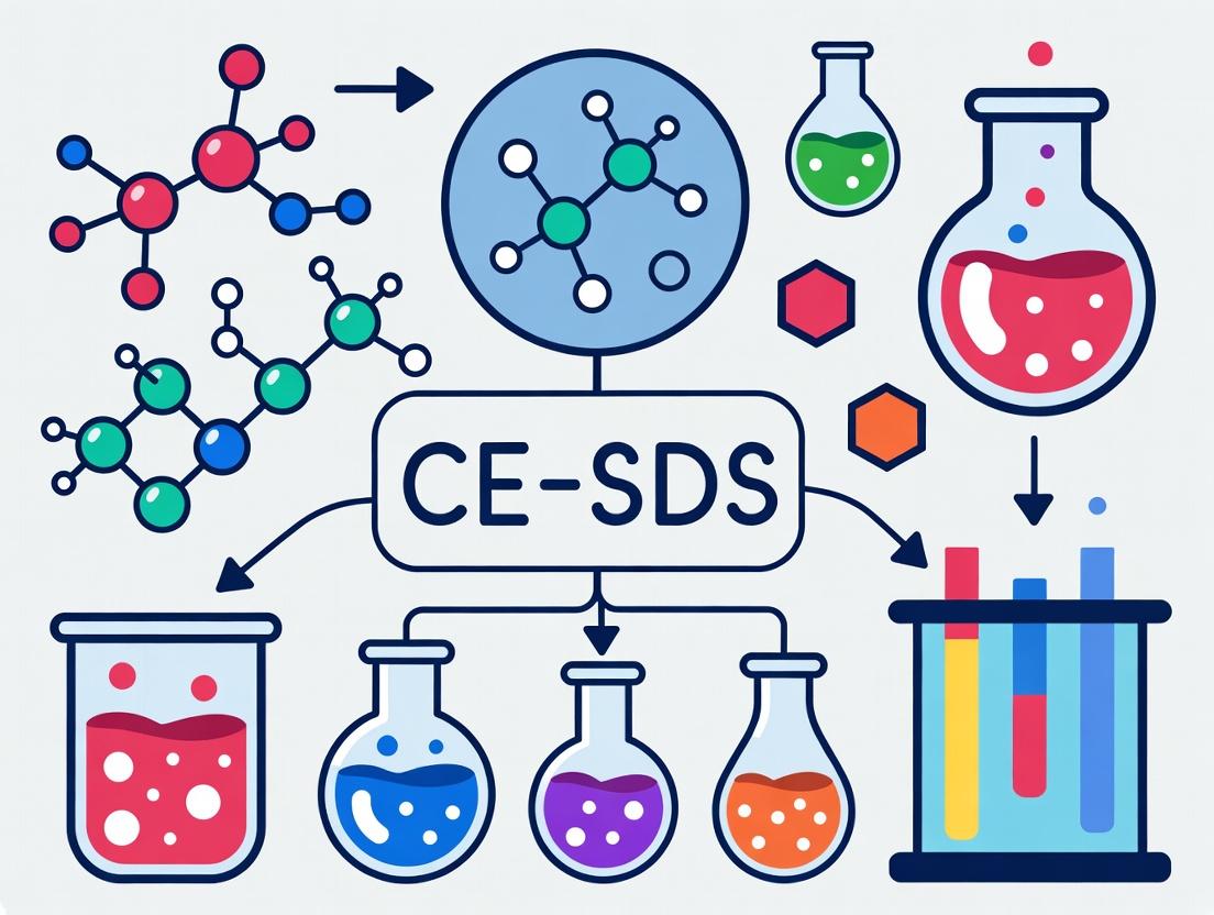
Ultimate Guide to CE-SDS: Mastering Protein Purity Analysis for Biopharma Development
This comprehensive guide explores Capillary Electrophoresis with Sodium Dodecyl Sulfate (CE-SDS) as a critical analytical tool for assessing protein therapeutic purity.
Ultimate Guide to CE-SDS: Mastering Protein Purity Analysis for Biopharma Development
Abstract
This comprehensive guide explores Capillary Electrophoresis with Sodium Dodecyl Sulfate (CE-SDS) as a critical analytical tool for assessing protein therapeutic purity. Tailored for researchers, scientists, and drug development professionals, the article covers foundational principles from how CE-SDS separates and quantifies protein variants like fragments and aggregates. It provides detailed, step-by-step methodological protocols for both reduced and non-reduced analysis, real-world applications in QC and stability studies, and advanced troubleshooting for common issues like poor resolution, sensitivity, and reproducibility. Finally, it validates CE-SDS against orthogonal techniques like SDS-PAGE and SEC, highlighting its regulatory compliance, superior quantification, and role in method comparability studies for robust purity assessment throughout the drug development lifecycle.
CE-SDS Fundamentals: The Science of Protein Separation and Purity Assessment
What is CE-SDS? Principles of Capillary Electrophoresis with SDS.
Capillary Electrophoresis with Sodium Dodecyl Sulfate (CE-SDS) is a high-resolution, automatable analytical technique used primarily for the size-based separation and purity analysis of proteins under denaturing conditions. It is a critical tool in biopharmaceutical development for assessing the purity and integrity of protein therapeutics, such as monoclonal antibodies (mAbs), and for detecting and quantifying product-related impurities like fragments and aggregates.
The core principle involves the covalent modification of proteins with a fluorescent dye (typically for laser-induced fluorescence, LIF, detection) or their detection via UV absorbance, followed by separation in a capillary filled with a sieving polymer matrix. Prior to analysis, the protein sample is denatured and uniformly coated with the anionic surfactant SDS. This SDS coating imparts a consistent, negative charge-to-mass ratio to all proteins. When an electric field is applied, proteins migrate through the polymer sieving matrix based primarily on their hydrodynamic size (molecular weight), with smaller molecules migrating faster than larger ones. This allows for highly reproducible molecular weight estimation and quantitative impurity profiling.
Key Principles and Methodologies
CE-SDS Modalities: UV vs. LIF Detection
Two primary detection modes are employed, each with distinct advantages.
Table 1: Comparison of CE-SDS Detection Methods
| Parameter | UV Detection | LIF Detection |
|---|---|---|
| Labeling | Non-covalent (inherent UV absorbance) | Covalent (fluorescent dye, e.g., 5- or 6-carboxyfluorescein succinimidyl ester) |
| Sensitivity | ~ 0.1 mg/mL (μg range) | ~ 0.01 mg/mL (ng range) |
| Dynamic Range | ~ 2 orders of magnitude | ~ 3-4 orders of magnitude |
| Primary Use | Purity, aggregates, fragments | High-sensitivity impurity analysis, low-abundance species |
| Sample Prep | Simpler (mix with SDS buffer) | Requires labeling, quenching, and cleanup steps |
Quantitative Performance Metrics
CE-SDS methods are rigorously validated for use in regulated environments.
Table 2: Typical CE-SDS Method Performance Characteristics
| Performance Attribute | Typical Result |
|---|---|
| Precision (Repeatability) | %RSD for migration time: < 1.0%; %RSD for peak area: < 5.0% |
| Linearity Range | 0.1 - 2.0 mg/mL (UV); 0.01 - 1.0 mg/mL (LIF) (R² > 0.98) |
| Limit of Quantitation (LOQ) | ~0.1% (LIF mode for impurity peaks) |
| Accuracy (Spike Recovery) | 80-120% for known impurities |
| Size Resolution | Capable of resolving fragments differing by ~5-10 kDa |
Experimental Protocols
Protocol 1: CE-SDS Analysis of a Monoclonal Antibody (Reduced, UV Detection)
This protocol is for assessing the purity and fragment content of a reduced mAb, separating light chain (LC) and heavy chain (HC).
Materials: CE instrument with UV detector, bare fused silica capillary (50 μm i.d., total length 30-40 cm), CE-SDS run buffer (commercial sieving matrix with SDS), sample buffer (containing SDS and a reducing agent like β-mercaptoethanol or DTT), 0.1N HCl, 0.1N NaOH, deionized water.
Procedure:
- Capillary Conditioning: Flush capillary with 0.1N NaOH for 5 min, deionized water for 5 min, and run buffer for 10 min.
- Sample Preparation: Dilute protein to 1 mg/mL in sample buffer. Add reducing agent to a final concentration of 50 mM (e.g., DTT). Heat at 70°C for 5-10 minutes. Centrifuge briefly.
- Instrument Setup: Set detection wavelength to 220 nm. Set sample tray temperature to 5-10°C.
- Injection: Hydrodynamic or electrokinetic injection (e.g., 5-10 kV for 20-40 sec).
- Separation: Apply constant voltage of 15-20 kV (negative polarity, cathode at detector side) for 20-40 minutes. Temperature: 20-25°C.
- Post-run Analysis: Flush capillary with 0.1N HCl for 2 min, water for 2 min, and run buffer for 5 min between runs.
- Data Analysis: Integrate peaks. Identify main LC and HC peaks. Calculate % purity and % fragments/impurities relative to total peak area.
Protocol 2: High-Sensitivity Impurity Analysis (Non-Reduced, LIF Detection)
This protocol is optimized for detecting low-level aggregates and fragments without reduction.
Materials: LIF-CE instrument (excitation ~488 nm, emission ~520 nm), derivatization kit (containing fluorescent dye, reaction buffer, quench solution), purification spin columns.
Procedure:
- Protein Labeling:
- Dilute protein to 1-2 mg/mL in labeling buffer.
- Add fluorophore reagent at a molar dye-to-protein ratio of 10:1 to 20:1.
- Incubate at 35°C for 30 minutes in the dark.
- Quenching & Cleanup:
- Add quenching reagent to stop the reaction. Incubate for 10 minutes.
- Desalt the labeled sample using a spin column to remove excess dye.
- Sample Denaturation: Mix the purified, labeled sample with non-reducing SDS sample buffer. Heat at 70°C for 5 minutes. Centrifuge.
- CE Analysis: Use an LIF-equipped CE system. Follow a separation protocol similar to Protocol 1, but with LIF detection parameters. Injection times may be shorter due to higher sensitivity.
- Data Analysis: Identify the main monomer peak. Quantify pre-monomer (fragments) and post-monomer (aggregates) peaks as a percentage of total detected signal.
Visualization: CE-SDS Workflow and Principles
Title: CE-SDS Experimental Workflow
Title: CE-SDS Separation Principle
The Scientist's Toolkit: Essential Research Reagents & Materials
Table 3: Key Reagent Solutions for CE-SDS Analysis
| Item | Function & Importance |
|---|---|
| SDS Sample Buffer | Contains SDS for protein denaturation/coating, and often a reducing agent (DTT) or alkylating agent (iodoacetamide). Critical for consistent charge masking. |
| Sieving Polymer Run Buffer | A dynamic sieving matrix (e.g., dextran, PEG, or commercial polymers) in a conductive buffer. Enables size-based separation within the capillary. |
| Fluorescent Derivatization Kit | For LIF-CE-SDS. Contains a succinimidyl ester dye (e.g., 5-FAM), reaction buffer, and quench solution. Enables high-sensitivity detection. |
| Capillary Conditioning Solutions | 0.1-1.0 M NaOH (for activating silica), 0.1-1.0 M HCl, and deionized water. Essential for maintaining capillary performance and reproducibility. |
| Internal Standard (ISS) | A low molecular weight, stable protein (e.g., insulin) labeled for LIF. Used to normalize migration times and correct for run-to-run variability. |
| Size Markers | A set of pre-stained proteins covering a known molecular weight range. Used to generate a calibration curve for accurate molecular weight estimation. |
| Protein Purification Spin Columns | Used post-labeling in LIF protocols to remove excess fluorescent dye, which can cause high background noise. |
1. Introduction and Application Notes Within the development of biotherapeutics, the purity and integrity of protein reagents (e.g., antibodies, recombinant proteins) are critical for research reproducibility and therapeutic efficacy. Capillary Electrophoresis-Sodium Dodecyl Sulfate (CE-SDS) has emerged as a principal, quantitative technique for assessing these attributes under denaturing conditions. This protocol details the application of CE-SDS for characterizing key quality metrics—purity, fragmentation, and aggregation—framed within a broader thesis on developing robust analytical control methods for protein reagent characterization in drug development.
2. Key Quantitative Metrics: Data Summary The primary output of a CE-SDS analysis is an electropherogram from which quantitative percentages are derived for each species. The following table summarizes the target metrics and their typical impact.
Table 1: Key CE-SDS Metrics for Protein Reagent Purity Testing
| Metric | Description | Typical Acceptance Criterion | Impact on Reagent Quality |
|---|---|---|---|
| Main Peak Purity | Percentage of intact, full-length protein. | >90% (research); >95% (therapeutic) | Direct measure of desired product. |
| Fragmentation | Sum percentage of lower molecular weight (LMW) species (e.g., light/heavy chain, clip variants). | <10% total | Indicates chemical/ enzymatic degradation, affects potency. |
| Aggregation | Sum percentage of high molecular weight (HMW) species (covalent or non-covalent under denaturing conditions). | <5% total | Can impact immunogenicity and pharmacokinetics. |
| Pre-peaks | Small, early-eluting species (e.g., free dye, small peptides). | <2% | Often related to sample preparation artifacts. |
3. Detailed CE-SDS Protocol for Purity and Heterogeneity Assessment Protocol: CE-SDS Analysis of a Monoclonal Antibody Under Reducing and Non-Reducing Conditions
I. Research Reagent Solutions & Materials (The Scientist's Toolkit) Table 2: Essential Materials for CE-SDS Analysis
| Item | Function |
|---|---|
| CE-SDS Analyzer (e.g., PA 800 Plus, Maurice) | Instrument platform for automated capillary electrophoresis with UV and/or laser-induced fluorescence (LIF) detection. |
| Bare Fused Silica Capillary (50 µm i.d., 30.2 cm length) | Separation pathway for SDS-protein complexes. |
| CE-SDS Running Buffer (10x, proprietary) | Provides consistent ionic strength and SDS milieu for separation. Diluted to 1x with deionized water. |
| Acidic Wash Solution (e.g., 0.1 M HCl) | Cleans capillary and prepares inner silica surface. |
| Basic Wash Solution (e.g., 0.1 M NaOH) | Critical for removing adsorbed material and conditioning the capillary. |
| SDS-MW Sample Buffer (with internal standard) | Denatures proteins, imparts uniform negative charge via SDS binding, and includes a lower MW marker for migration time normalization. |
| Fluorescent Dye (5-Dye, MW Standard) | Optional dye for non-covalent, pre-separation labeling of proteins for highly sensitive LIF detection. |
| Iodoacetamide (IAM) | Alkylating agent used in sample preparation to prevent reformation of disulfide bonds after reduction, locking fragments in reduced state. |
II. Sample Preparation Protocol
- Protein Denaturation: Dilute protein reagent to 1-2 mg/mL in PBS.
- Reduced Analysis: For a 50 µL aliquot, add 2.5 µL of 0.5 M DTT (final 25 mM). Vortex and incubate at 70°C for 10 minutes. Immediately add 2.5 µL of 1.0 M IAM (final 50 mM). Vortex and incubate at 70°C for 5 minutes in the dark.
- Non-Reduced Analysis: For a 50 µL aliquot, add 2.5 µL of 1.0 M IAM (optional, for cysteine blocking). Vortex and incubate at 70°C for 5 minutes in the dark.
- Final Preparation: To both reduced and non-reduced samples, add 95 µL of SDS-MW sample buffer. Vortex and centrifuge briefly. Heat at 70°C for 10 minutes. Cool to room temperature before injection.
III. Instrumental Method & Analysis
- Capillary Conditioning: Rinse capillary with 0.1 M NaOH (5 min), deionized water (3 min), 0.1 M HCl (5 min), deionized water (3 min), and 1x CE-SDS running buffer (10 min). Apply pressure (e.g., 50 psi).
- Sample Injection: Hydrodynamic injection (e.g., 5.0 psi for 40 seconds).
- Separation: Apply constant voltage of +15-20 kV (reversed polarity) for 30-40 minutes. Monitor at 220 nm (UV) or with LIF detection.
- Data Integration: Use instrument software to integrate peak areas. Identify peaks by relative migration time (RMT) compared to internal standard. Calculate percentages: (Peak Area / Total Integrated Area) x 100%.
4. Visualization of CE-SDS Workflow and Data Interpretation
Title: CE-SDS Analytical Workflow from Sample to Data
Title: Interpreting CE-SDS Electropherogram Profiles
Application Notes
The analysis of protein therapeutic purity, particularly for monoclonal antibodies (mAbs) and other biologics, is a critical quality attribute in biopharmaceutical development. Within the context of a broader thesis on CE-SDS method development for protein reagent purity testing, this document details the superior performance of Capillary Electrophoresis with Sodium Dodecyl Sulfate (CE-SDS) over traditional slab gel SDS-Polyacrylamide Gel Electrophoresis (SDS-PAGE).
Key Advantages of CE-SDS
- Superior Resolution & Peak Capacity: CE-SDS separates proteins in a capillary based on their hydrodynamic size, offering higher resolving power. This allows for clear distinction between the main product (e.g., intact mAb light and heavy chains) and critical impurities like fragments (e.g., heavy chain, light chain, non-glycosylated heavy chain) and aggregates.
- Automated, Precise Quantification: CE-SDS utilizes on-capillary UV or laser-induced fluorescence (LIF) detection, providing direct digital quantification of each species. This eliminates the manual, semi-quantitative densitometry required for stained SDS-PAGE gels, dramatically improving accuracy, precision, and linear dynamic range.
- Enhanced Reproducibility & Throughput: The automated nature of CE-SDS minimizes manual handling, leading to significantly higher inter- and intra-assay reproducibility (%RSD often <2% for migration time, <10% for peak area) compared to SDS-PAGE. Sample processing is faster, enabling higher throughput.
- Reduced Sample & Reagent Consumption: CE-SDS typically requires only nanoliters of sample and minimal volumes of reagents, aligning with green laboratory principles.
- Data Integrity & Regulatory Compliance: CE-SDS systems generate electronic raw data that is fully compliant with 21 CFR Part 11 requirements, supporting submissions to regulatory agencies like the FDA and EMA.
Table 1: Quantitative Performance Comparison of CE-SDS vs. SDS-PAGE
| Parameter | CE-SDS (UV Detection) | Traditional SDS-PAGE (Coomassie Stain) | Implication for Purity Testing |
|---|---|---|---|
| Resolution | High; Baseline separation of size variants differing by ~5-10% in molecular weight. | Moderate to Low; Manual gel casting variability affects resolution. | CE-SDS reliably detects low-level fragments. |
| Quantitation Precision (%RSD, Peak Area) | Typically 2-10% | Typically 10-25% | CE-SDS provides robust data for lot-to-lot comparisons and stability studies. |
| Linearity (Dynamic Range) | 2-3 orders of magnitude (e.g., 0.1 – 10 mg/mL) | ~1 order of magnitude | CE-SDS is better suited for quantifying both major and minor components in one run. |
| Sample Volume per Analysis | ~10-50 nL (injection) | ~10-20 µL | CE-SDS conserves precious protein reagents. |
| Assay Time (Hands-on + Runtime) | ~30-60 min (automated) | ~3-5 hours (mostly manual) | CE-SDS increases laboratory efficiency. |
| Data Output | Digital electropherogram; Direct quantification. | Analog gel image; Requires manual band identification and densitometry. | CE-SDS enables automated reporting and superior data integrity. |
Application in mAb Purity Analysis
CE-SDS under reducing conditions is the industry standard for assessing mAb purity and monitoring clip variants. A typical electropherogram will resolve:
- Non-Glycosylated Heavy Chain (NGHC): An important product-related impurity.
- Heavy Chain (HC): Main component.
- Light Chain (LC): Main component.
- Fragments (e.g., HL, HH): Indicative of degradation or process-related clipping.
Table 2: Typical CE-SDS Purity Profile of a Reduced Monoclonal Antibody
| Peak Identity | Approximate Migration Time (min) | Relative Percentage (%) | Acceptable Range (Example) | Purpose of Monitoring |
|---|---|---|---|---|
| High Molecular Weight Species | 12.5 - 14.0 | < 1.0 | ≤ 2.0% | Aggregate detection. |
| Heavy Chain (HC) | 15.0 | ~50.0 | 48.0 – 52.0% | Main component quantification. |
| Non-Glycosylated HC | 15.5 | < 2.0 | ≤ 5.0% | Critical quality attribute for efficacy. |
| Light Chain (LC) | 17.0 | ~48.0 | 46.0 – 50.0% | Main component quantification. |
| Fragments / Other | Variable | < 1.5 | ≤ 3.0% | Purity indicator; Process consistency. |
Experimental Protocols
Protocol 1: CE-SDS Analysis of Reduced Monoclonal Antibody (Using a Beckman Coulter PA 800 Plus System)
I. Sample Preparation (Reduced)
- Dilution: Dilute the mAb sample to approximately 2 mg/mL in purified water.
- Reduction: Combine 50 µL of the 2 mg/mL sample with 85 µL of 1× Sample Buffer (commercial SDS-MW sample buffer) and 10 µL of 10× Reducing Agent (commercial 1M DTT or 2-Mercaptoethanol solution).
- Denaturation: Heat the mixture at 70°C for 10 minutes.
- Cooling: Briefly centrifuge the vial and allow it to cool to room temperature.
- Final Dilution: Add 355 µL of purified water to achieve a final concentration of ~0.2 mg/mL. Vortex gently.
II. Instrument Setup and Run
- Capillary: Use a bare-fused silica capillary (50 µm I.D., total length 30.2 cm, effective length 20.2 cm).
- Detection: UV at 220 nm.
- Solutions:
- Separation Buffer: Commercial CE-SDS Running Buffer (e.g., containing SDS and zwitterions).
- Sample Buffer: Commercial SDS-MW sample buffer.
- Rinse Solutions: 0.1M NaOH, 0.1M HCl, Deionized Water, Separation Buffer.
- Method Parameters:
- Pre-run Conditioning: Rinse with 0.1M NaOH (2 min), 0.1M HCl (1 min), DI Water (1 min), Separation Buffer (3 min).
- Sample Injection: Electrokinetic injection at 5 kV for 20 seconds.
- Separation: Apply constant voltage of 15 kV for 30 minutes.
- Post-run: Rinse with 0.1M NaOH (2 min), DI Water (2 min), and Separation Buffer (3 min) for the next run.
- Analysis: Use the instrument software to integrate peaks. Identify species based on migration time compared to a characterized reference standard. Calculate percentage purity based on peak area.
Protocol 2: Traditional Reducing SDS-PAGE (for Comparison)
I. Gel Casting (12% Bis-Tris Gel)
- Resolve Gel: Mix 3.3 mL of 30% acrylamide/bis solution, 2.5 mL of 1.5M Tris-HCl (pH 8.8), 4.1 mL H2O, 100 µL 10% SDS, 100 µL 10% APS, and 4 µL TEMED. Pour immediately, overlay with isopropanol, and allow to polymerize for 30 min.
- Stack Gel: Mix 670 µL of 30% acrylamide/bis, 1.25 mL of 0.5M Tris-HCl (pH 6.8), 3.0 mL H2O, 50 µL 10% SDS, 50 µL 10% APS, and 5 µL TEMED. Pour on top of polymerized resolve gel, insert comb, and polymerize for 30 min.
II. Sample Preparation & Run
- Reduction: Mix 20 µL of 1 mg/mL protein with 20 µL of 2× Laemmli Sample Buffer containing 5% 2-Mercaptoethanol. Heat at 95°C for 5 minutes.
- Loading & Electrophoresis: Load 10-20 µL per well. Include a pre-stained molecular weight marker. Run in 1× Tris-Glycine-SDS running buffer at constant 120V until the dye front reaches the bottom (~1.5 hours).
- Staining: Dismantle gel and stain with Coomassie Brilliant Blue R-250 solution (0.1% in 40% methanol, 10% acetic acid) for 1 hour with gentle agitation.
- Destaining: Destain with multiple changes of destain solution (40% methanol, 10% acetic acid) until background is clear and bands are visible.
- Analysis: Image the gel using a scanner or gel doc system. Perform semi-quantitative densitometry analysis using software like ImageJ.
The Scientist's Toolkit: Key Research Reagent Solutions
| Item | Function & Relevance |
|---|---|
| CE-SDS Protein Analysis Kit | A commercial kit providing optimized, ready-to-use buffers (sample buffer, running buffer) and a protein sizing standard. Ensures reproducibility and saves method development time. |
| Bare Fused Silica Capillaries (50µm I.D.) | The standard separation capillary for CE-SDS. The inner surface chemistry and dimension are critical for consistent electroosmotic flow and separation performance. |
| Precision Molecular Weight Markers | A set of proteins with known molecular weights (e.g., 10-225 kDa) covalently labeled with a fluorophore or compatible with UV detection. Essential for assigning peaks in the electropherogram. |
| High-Purity SDS & DTT | Sodium dodecyl sulfate ensures uniform negative charge-to-mass ratio. Dithiothreitol (DTT) is a strong reducing agent for breaking disulfide bonds. Purity is critical to avoid artifact peaks. |
| Validated mAb Reference Standard | A fully characterized and stability-indicating reference material of the therapeutic protein. Serves as the system suitability control and for peak identification in every run. |
Visualizations
CE-SDS Automated Workflow
CE-SDS Data Analysis Pathway
Methodology Comparison Logic
The development of biologics, including monoclonal antibodies (mAbs), bispecifics, and antibody-drug conjugates (ADCs), hinges on rigorous characterization of product quality attributes. Purity and impurity profiles directly impact safety, efficacy, and stability. Within a broader thesis on Capillary Electrophoresis-Sodium Dodecyl Sulfate (CE-SDS) method development for protein reagent purity testing, this application note underscores the non-negotiable role of purity analysis. As regulatory scrutiny intensifies, with agencies like the FDA and EMA emphasizing the "quality by design" (QbD) paradigm, high-resolution, quantitative purity methods are critical for characterizing size variants like fragments and aggregates throughout development, from clone selection to lot release.
Current Landscape and Quantitative Data
Recent industry analyses and regulatory submissions highlight the critical thresholds for product-related impurities. The following table summarizes key purity acceptance criteria and typical impurity levels for therapeutic mAbs as reflected in current literature and regulatory guidance.
Table 1: Critical Purity Attributes and Acceptance Criteria for Therapeutic mAbs
| Purity Attribute | Typical Method | Criticality | Common Specification Limit | Impact |
|---|---|---|---|---|
| Monomer Purity | SEC, CE-SDS | High (Potency) | ≥95% | Directly linked to bioactivity and dosing. |
| Aggregates (HMW Species) | SEC, AUC | High (Immunogenicity) | ≤5% (often ≤2-3% for drug substance) | Risk of enhanced immunogenic response. |
| Fragments (LMW Species) | CE-SDS (reduced/non-reduced), SEC | Medium-High (Potency) | ≤5-10% (varies by fragment type) | Can affect binding avidity and Fc-mediated functions. |
| Charge Variants | icIEF, CZE | Medium (PK/Stability) | Report results; limits set based on stability lot data. | May influence pharmacokinetics and stability. |
| Process-Related Impurities | Host Cell Protein (HCP) ELISA, DNA assays | High (Safety) | HCP: ≤100 ppm; DNA: ≤10 ng/dose | Safety risk, potential immunogenicity. |
Table 2: Comparison of Key Purity Analysis Techniques
| Technique | Resolution | Analysis Time | Key Impurity Profile | Quantitation | Automation Potential |
|---|---|---|---|---|---|
| CE-SDS (MW-based) | High (1-2% difference) | 30-45 min/sample | Fragments, Aggregates, Non-glycosylated heavy chain | Excellent (R^2 >0.99) | High (multi-capillary systems) |
| Size Exclusion Chromatography (SEC) | Moderate | 15-30 min/sample | Soluble Aggregates, Fragments | Good | High |
| Analytical Ultracentrifugation (AUC) | Very High | Hours | Aggregates, Oligomers (solution state) | Excellent | Low |
| Microfluidic Imaging (MFI) | N/A (particle count) | Rapid | Sub-visible Particles (>1 µm) | Quantitative count | Moderate |
Detailed Experimental Protocols
Protocol 1: CE-SDS Purity Analysis of a Therapeutic mAb (Reduced Conditions)
Objective: To quantitatively determine the purity profile (light chain, heavy chain, and fragments) of a reduced monoclonal antibody sample.
Materials:
- CE system with UV detection (200-220 nm) and temperature control.
- Bare-fused silica capillaries (internal diameter: 50 µm, total length: 50-60 cm).
- CE-SDS run buffer (commercial SDS-MW analysis kit).
- SDS sample buffer (commercial, containing SDS and internal standard).
- Reducing agent: β-mercaptoethanol or dithiothreitol (DTT).
- mAb sample (1-2 mg/mL).
- 0.1M HCl, 0.1M NaOH, deionized water.
Procedure:
- Capillary Conditioning: Flush new capillary with 0.1M NaOH for 20 min, water for 10 min, and run buffer for 20 min. Between runs, perform a short conditioning with 0.1M HCl (2 min), water (2 min), and run buffer (5 min).
- Sample Preparation: Dilute the mAb sample to 1 mg/mL in SDS sample buffer. Add reducing agent to a final concentration of 50mM (for DTT). Heat the mixture at 70°C for 10 minutes. Centrifuge briefly before loading.
- Instrument Setup: Set the detection wavelength to 220 nm. Set the sample tray temperature to 4-8°C. Set the capillary temperature to 20-25°C.
- Injection and Separation: Perform electrokinetic injection (e.g., 5-10 kV for 20-40 seconds). Apply a separation voltage of +15 kV (reverse polarity) for 40 minutes. Use a constant pressure on the inlet and outlet vials if available.
- Data Analysis: Integrate the peaks. Identify peaks for light chain (LC ~25 kDa), heavy chain (HC ~50 kDa), and any non-glycosylated heavy chain (NGHC ~50 kDa). Calculate the percentage purity of each species:
(Area of peak / Total integrated area) x 100%. System suitability requires resolution >1.5 between key peaks and RSD of migration time <2%.
Protocol 2: CE-SDS Purity Analysis (Non-Reduced Conditions)
Objective: To assess aggregate and fragment content under non-reducing conditions, preserving disulfide bonds.
Materials: As per Protocol 1, excluding the reducing agent.
Procedure:
- Follow Protocol 1 for capillary conditioning and instrument setup.
- Sample Preparation: Dilute the mAb sample to 1 mg/mL in SDS sample buffer. Do not add reducing agent. Heat the mixture at 70°C for 10 minutes. Centrifuge briefly.
- Perform injection and separation as in Protocol 1.
- Data Analysis: Identify the main peak as the intact mAb (~150 kDa). Integrate peaks corresponding to high molecular weight (HMW) aggregates (eluting earlier) and low molecular weight (LMW) fragments (eluting later). Report percentage of main peak, %HMW, and %LMW.
Visualization of Method Role and Workflow
Title: Biologics Development Workflow with Critical Purity Testing Gates
Title: CE-SDS Reduced Purity Analysis Protocol Workflow
The Scientist's Toolkit: Key Research Reagent Solutions
Table 3: Essential Materials for CE-SDS Purity Testing
| Item | Function | Key Considerations |
|---|---|---|
| CE-SDS Analysis Kit (Commercial) | Provides optimized, reproducible SDS gel buffer, sample buffer, and standards. | Ensures lot-to-lot consistency, includes internal standard for precise migration time correction. |
| Bare-Fused Silica Capillaries | The separation matrix for sieving of SDS-protein complexes. | Length and internal diameter must be matched to method; consistent coating/bareness is critical. |
| High-Purity SDS | Denatures and uniformly charges proteins for separation by size. | Must be high purity (≥99%) to avoid interference peaks; part of commercial kits. |
| Reducing Agents (DTT/BME) | Breaks disulfide bonds for reduced analysis of subunits. | DTT is often preferred due to lower odor and more consistent reduction. |
| Mobility/Size Standards | Used for system suitability and optional apparent molecular weight estimation. | Should cover relevant size range (e.g., 10-225 kDa). |
| CE System with Temperature-Controlled Autosampler | Automates injection, separation, and detection. | Temperature control (4-8°C) of samples in autosampler is vital to prevent sample degradation during queue. |
Application Note: Optimizing CE-SDS Instrumentation for High-Resolution Purity Analysis of Monoclonal Antibodies
In the context of developing a robust CE-SDS method for protein therapeutic purity testing, the selection and optimization of instrumentation and consumables are critical. This application note details the core components and their impact on method performance, specifically for the analysis of monoclonal antibodies under both reduced and non-reduced conditions.
1. The Capillary: Core of Separation
The fused silica capillary is the central component. Its internal surface chemistry dictates separation efficiency and protein adsorption.
- Dimensions: 50 µm inner diameter is standard, providing a balance between sensitivity and heat dissipation. Capillary length is typically 30-50 cm to the detector (effective length).
- Coatings: Dynamic or covalent coatings are essential to suppress electroosmotic flow (EOF) and minimize protein adsorption to the silica wall. A recent search indicates that hydrophilic, neutral polymers (e.g., polyvinyl alcohol derivatives) are the industry benchmark.
- Protocol - Capillary Conditioning and Storage:
- Initial Conditioning: For a new bare-fused silica capillary, flush with 1M NaOH for 30 minutes, followed by deionized water for 15 minutes, and then run buffer for 30 minutes. For pre-coated capillaries, follow manufacturer instructions (typically buffer flush only).
- Daily Conditioning: Before each sequence, flush with 0.1M NaOH for 5 minutes, deionized water for 3 minutes, and run buffer for 10 minutes.
- Storage: After use, flush capillary with deionized water for 5 minutes and store dry or filled with deionized water at room temperature. Long-term storage of coated capillaries in a recommended preservative solution is advised.
2. Detection System: Sensitivity and Specificity
Ultraviolet (UV) absorbance at 214 nm (peptide bond) or 220 nm is the most common detection method for CE-SDS due to its universality. The path length, however, is limited by the capillary inner diameter, impacting sensitivity.
- Quantitative Data on Detection:
| Detection Type | Wavelength (nm) | Primary Application | Approximate Limit of Detection (LOD) for IgG | Key Advantage | Key Disadvantage |
|---|---|---|---|---|---|
| UV Absorbance | 214, 220 | Main chain detection, standard purity | ~0.1 mg/mL | Universal, non-destructive | Lower sensitivity due to short path length |
| Laser-Induced Fluorescence (LIF) | Excitation: 488, Emission: 520 | Impurity profiling, low-abundance species | ~0.1 µg/mL (when labeled) | Extremely high sensitivity | Requires fluorescent labeling (e.g., with Spyro Ruby) |
| Mass Spectrometry (MS) Coupled | N/A | Peak identification, variant characterization | Varies (~µg/mL) | Provides structural identity | Complex interface, higher cost |
3. Key Consumables and Reagents
The consistency of SDS-based reagents is paramount for reproducible migration times and peak areas.
- SDS Sample Buffer: Must contain 1-2% SDS. Reducing buffer includes 10-50 mM DTT or 2-Mercaptoethanol. Non-reducing buffer may include alkylating agents like iodoacetamide to cap free cysteines.
- SDS-MW Separation Gel: A linear polymer matrix (e.g., dextran or polyacrylamide) containing SDS. Its viscosity and polymer chain length distribution define the separation window and resolution.
- Protocol - Sample Preparation for CE-SDS (Reduced):
- Dilute protein sample to 1-2 mg/mL in a compatible buffer (e.g., PBS).
- Mix sample with CE-SDS Sample Buffer (containing SDS and DTT) at a 1:2 (v:v) ratio.
- Heat the mixture at 70°C for 5-10 minutes.
- Centrifuge briefly at 10,000 x g to remove any particulates.
- Transfer to an instrument-compatible sample vial for analysis.
4. The Scientist's Toolkit: Essential Research Reagent Solutions
| Item | Function | Critical Specification |
|---|---|---|
| Fused Silica Capillary | The separation channel. | Inner diameter (50 µm), coating type (e.g., neutral hydrophilic polymer), total/effective length. |
| CE-SDS Anode Buffer | Contains SDS, provides conductive medium for separation. | SDS purity, buffer concentration (e.g., 100-200 mM phosphate/borate, pH ~7.0). |
| CE-SDS Cathode Buffer | Often identical to anode buffer for SDS methods. | Must be particle-filtered (0.2 µm) to prevent capillary clogging. |
| SDS-MW Size Standard | For accurate molecular weight estimation. | Defined protein/peptide ladder covering 10-225 kDa range. |
| Fluorescent Label (for LIF) | Enables high-sensitivity detection. | Must not alter protein charge (e.g., Spyro Ruby, Unchained Labs Elephant). |
| Capillary Storage Solution | Preserves coating integrity during idle periods. | Low conductivity, antimicrobial, as specified by capillary manufacturer. |
| Performance Test Mix | System suitability test for resolution and migration time. | Contains a known protein (e.g., reduced IgG) with defined peak profile criteria. |
5. System Workflow and Critical Relationships
Diagram Title: CE-SDS Purity Analysis Workflow
6. Method Development Decision Pathway
Diagram Title: CE-SDS Method Development Decision Pathway
Step-by-Step CE-SDS Protocol: From Sample Prep to Data Analysis in Biopharma Workflows
Within the context of developing robust Capillary Electrophoresis-Sodium Dodecyl Sulfate (CE-SDS) methods for protein therapeutic purity and impurity analysis, sample preparation is the critical determinant of success. This protocol details optimized, reproducible practices for reduction, alkylation, denaturation, and labeling, specifically tailored for monoclonal antibodies (mAbs) and related biologics prior to CE-SDS analysis. Consistent execution of these steps is paramount for accurate quantitation of fragments, aggregates, and intact molecules.
Key Protocols and Application Notes
Protocol 1: Standard Reduction and Alkylation for CE-SDS
Objective: To fully reduce interchain disulfide bonds and alkylate free thiols, preventing reformation and ensuring complete subunit separation. Materials:
- Protein sample (0.5-2 mg/mL in formulation buffer or PBS)
- 10% SDS Solution (w/v)
- 0.5M Tris(2-carboxyethyl)phosphine hydrochloride (TCEP) or 1M Dithiothreitol (DTT)
- 0.5M Iodoacetamide (IAM) solution, freshly prepared in deionized water and kept in dark
- 1M N-Ethylmaleimide (NEM) solution (optional, for alternative alkylation)
- Heating block or water bath (70°C and 95°C)
Detailed Method:
- Denaturation: Prepare a mixture of 25 µL protein sample with 10 µL of 10% SDS and 5 µL of neutral pH buffer (e.g., 1M Tris-HCl, pH 8.0). Incubate at 70°C for 5 minutes.
- Reduction: Add 5 µL of 0.5M TCEP (final ~50 mM) or 1M DTT (final ~100 mM). Vortex and incubate at 70°C for 10 minutes. Note: TCEP is preferred for its stability across a wider pH range.
- Alkylation: Cool the mixture to room temperature. Add 5 µL of 0.5M IAM (final ~50 mM). Vortex and incubate in the dark at room temperature for 15 minutes.
- Quenching: The reaction can be quenched by adding 2 µL of 1M DTT (if using IAM) to consume excess alkylating agent. Proceed immediately to labeling or final dilution for analysis.
Protocol 2: Fluorescent Labeling for Laser-Induced Fluorescence (LIF) Detection
Objective: To covalently label proteins with a fluorescent dye for highly sensitive LIF detection in CE-SDS, enabling low-level impurity detection. Materials:
- Reduced and alkylated sample (from Protocol 1)
- Commercial fluorescent dye reagent kit (e.g., containing maleimide or amine-reactive dye)
- Reaction buffer (as specified by kit, typically pH 8-9)
- Quenching reagent
Detailed Method:
- Sample Adjustment: Ensure the reduced/alkylated sample is in a compatible buffer (pH ~8). Desalting may be required if primary amines (e.g., Tris) interfere.
- Labeling Reaction: Add a molar excess of fluorescent dye (typically 8-12x dye:protein) to the sample. Vortex gently.
- Incubation: Incubate the reaction mixture at 95°C for 5 minutes. This step simultaneously denatures the protein and drives the labeling reaction to completion.
- Quenching: Add the provided quenching reagent or a large molar excess of a small amine (e.g., lysine) to stop the reaction.
- Analysis: Dilute the labeled sample with deionized water or CE-SDS sample buffer to the desired concentration for injection.
Table 1: Impact of Reduction Time on Purity Analysis of a Monoclonal Antibody
| Reduction Time (min at 70°C) | % Intact IgG (Non-Reduced CE-SDS) | % Heavy Chain (Reduced CE-SDS) | % Light Chain (Reduced CE-SDS) | % Fragments |
|---|---|---|---|---|
| 2 | 95.5 | 48.2 | 45.1 | 6.7 |
| 5 | 0.1 | 66.5 | 66.0 | 2.5 |
| 10 (Optimal) | 0.0 | 67.0 | 66.8 | 1.8 |
| 15 | 0.0 | 67.1 | 66.9 | 1.9 |
Table 2: Comparison of Alkylating Agent Efficiency
| Alkylating Agent | Concentration (mM) | Incubation Time (min, RT) | % Free Thiols Alkylated | Risk of Artifacts (e.g., over-alkylation) |
|---|---|---|---|---|
| Iodoacetamide (IAM) | 50 | 15 | >99% | Moderate |
| N-Ethylmaleimide (NEM) | 100 | 5 | >98% | Low |
| No Alkylation | - | - | Variable | High (Re-oxidation) |
Workflow and Pathway Diagrams
Diagram 1: CE-SDS Sample Preparation Workflow
Diagram 2: Protein Modification States
The Scientist's Toolkit: Research Reagent Solutions
Table 3: Essential Materials for CE-SDS Sample Preparation
| Item | Function & Rationale |
|---|---|
| TCEP-HCl | Strong, odorless, and air-stable reducing agent. Preferred over DTT for more complete and stable reduction, especially at low pH. |
| Iodoacetamide (IAM) | Alkylating agent. Reacts specifically with free thiols to form stable carbamidomethyl derivatives, preventing re-oxidation. Light-sensitive. |
| N-Ethylmaleimide (NEM) | Alternative alkylating agent. Faster reaction than IAM and more specific for -SH groups, but may label amines at high pH. |
| 10% SDS Solution | Anionic denaturant. Unfolds the protein and imparts a uniform negative charge-to-mass ratio, which is essential for CE-SDS separation. |
| Fluorescent Dye Kit | Typically contains a maleimide-reactive dye (e.g., PYRE or Alexa Fluor derivatives) for cysteine labeling post-reduction/alkylation, enabling high-sensitivity LIF detection. |
| CE-SDS Sample Buffer | Commercial optimized buffer containing SDS, internal standards, and tracking dyes for consistent injection and migration. |
| pH-adjusted Tris Buffer | Provides optimal alkaline environment (pH 8-9) for both alkylation and dye-labeling reactions, maximizing efficiency. |
Optimized Buffer Systems and Sieving Polymers for Superior Separation
This application note details advanced protocols for capillary electrophoresis-sodium dodecyl sulfate (CE-SDS) methods, focusing on the optimization of buffer systems and sieving polymers for the analysis of protein therapeutic purity. Implementations of these protocols yield enhanced resolution, reproducibility, and accuracy in critical quality attribute (CQA) assessments for monoclonal antibodies (mAbs) and other biologics.
Within the broader thesis on CE-SDS for protein reagent purity testing, the separation matrix and running buffer are identified as critical method parameters. This document provides a comparative analysis of commercially available polymer systems and optimized buffer formulations to mitigate issues like protein adsorption, band broadening, and poor resolution of low-abundance impurities.
Research Reagent Solutions Toolkit
The following table lists essential materials for implementing high-performance CE-SDS.
| Reagent/Material | Function & Rationale |
|---|---|
| Bare Fused Silica Capillary (50 µm ID, 365 µm OD) | Standard separation channel. Dynamic coating protocols can be applied to reduce electroosmotic flow (EOF) and protein adsorption. |
| Replaceable Linear Polyacrylamide (LPA) Gel Matrix | High-performance sieving polymer (e.g., 10-12% concentration). Provides superior resolution for fragments (25-225 kDa) compared to cellulose derivatives. |
| Optimized Tris-Glycine-SDS Running Buffer (pH 9.0 ± 0.1) | Contains 100 mM Tris, 150 mM Glycine, 0.1% (w/v) SDS. High ionic strength reduces protein-wall interactions. Must be filtered (0.2 µm). |
| Fluorescent Derivatization Dye (e.g., 5-iodoacetamidofluorescein, 5-IAF) | Covalently labels reduced proteins for laser-induced fluorescence (LIF) detection, offering high sensitivity for impurity detection. |
| Internal Size Standards (e.g., 10-225 kDa labeled protein ladder) | Essential for accurate molecular weight (MW) assignment and migration time normalization across runs. |
| De-ionized Formamide (≥99.5%) | Used as sample diluent to maintain protein denaturation and prevent reformation of disulfide bonds post-reduction. |
Quantitative Comparison of Sieving Polymers
Data from recent studies comparing separation performance of different polymer systems for a 150 kDa mAb are summarized below.
Table 1: Performance Metrics of Common CE-SDS Sieving Polymers
| Polymer Type | Typical Concentration | Resolution (Main Peak/Fragment) | % RSD Migration Time (n=10) | Effective Separation Range (kDa) | Viscosity |
|---|---|---|---|---|---|
| Linear Polyacrylamide (LPA) | 10% w/v | 3.5 | 0.8% | 10 - 250 | High |
| Hydroxyethyl Cellulose (HEC) | 1% w/v | 2.1 | 1.5% | 50 - 300 | Medium |
| Polyethylene Oxide (PEO) | 2% w/v | 2.8 | 1.2% | 20 - 1000 | Low-Medium |
| Dextran | 8% w/v | 2.5 | 1.8% | 30 - 400 | High |
Detailed Experimental Protocols
Protocol 4.1: Preparation of Optimized Tris-Glycine-SDS Running Buffer
Objective: To prepare a stable, high-conductivity buffer minimizing protein-capillary wall interactions. Materials: Tris base (Ultra-pure), Glycine (Ultra-pure), SDS (Electrophoresis grade), DI water. Procedure:
- Weigh 12.11 g of Tris base and 11.26 g of Glycine into a 1 L beaker.
- Add ~900 mL of DI water and stir until fully dissolved.
- Add 1.0 g of SDS and stir gently to avoid foaming.
- Adjust pH to 9.00 ± 0.05 using 1M HCl or 1M NaOH.
- Transfer to a 1 L volumetric flask and bring to volume with DI water.
- Filter through a 0.2 µm PES membrane into a clean bottle. Store at 4°C for up to 2 weeks.
Protocol 4.2: CE-SDS Method for mAb Purity Under Reduced Conditions
Objective: To separate and quantify heavy chain (HC), light chain (LC), and non-glycosylated heavy chain (NGHC) impurities. Instrument: CE system with LIF detection (λex/λem = 488/520 nm). Capillary: 50 cm effective length (60 cm total) bare fused silica. Method:
- Sample Preparation: Dilute mAb to 2 mg/mL in 1x SDS sample buffer containing 10 mM DTT. Heat at 70°C for 10 min. Cool, then label with 5-IAF dye (10 mM stock) at a 10:1 dye:protein molar ratio in the dark for 30 min. Quench with 10 mM β-mercaptoethanol.
- Capillary Conditioning: Flush with 0.1M NaOH (5 min), DI water (3 min), and running buffer (5 min) at 50 psi.
- Polymer Fill: Fill capillary with 10% LPA solution at 50 psi for 3 min.
- Injection: Hydrodynamically inject sample at 0.5 psi for 30 sec (≈1% of capillary volume).
- Separation: Apply voltage of +15 kV (reverse polarity) for 30 min. Temperature maintained at 25°C.
- Post-run: Flush capillary with 0.1M NaOH (2 min) and DI water (2 min) between runs. Store in DI water when not in use. Data Analysis: Integrate peaks for HC (~50 kDa), LC (~25 kDa), and NGHC. Calculate % purity as [HC+LC] area / total area x 100.
Diagrams
Diagram Title: CE-SDS Reduced Purity Analysis Workflow
Diagram Title: Key Factors Impacting CE-SDS CQA Reliability
This document provides detailed Application Notes and Protocols for the execution of a Capillary Electrophoresis-Sodium Dodecyl Sulfate (CE-SDS) method, framed within a broader thesis on its application for protein therapeutic purity testing. The focus is on the critical execution phase, encompassing method parameterization, sample injection strategies, and voltage optimization to achieve high-resolution separation, accurate quantification, and reproducible results for drug development.
Key Method Parameters & Optimization Data
Optimal method parameters are derived from the synthesis of published literature and empirical data. The following table summarizes critical quantitative settings for a standard CE-SDS purity method under both reducing and non-reducing conditions.
Table 1: Optimized CE-SDS Method Parameters for mAb Purity Analysis
| Parameter | Typical Range | Optimized Setting (Reducing) | Optimized Setting (Non-Reducing) | Function & Impact |
|---|---|---|---|---|
| Capillary | Fused silica, 50 µm i.d. | Fused silica, 50 µm i.d. | Separation pathway; i.d. affects sensitivity & heat dissipation. | |
| Effective Length | 30-50 cm | 40.2 cm | 40.2 cm | Distance to detector; influences resolution and run time. |
| Detection | UV (220 nm, 280 nm) | 220 nm | 220 nm | Primary detection for peptide bonds; 280 nm for aromatic residues. |
| Sample Buffer | SDS-MW sample buffer with alkylating agent (e.g., IAM) | SDS-MW sample buffer | Denatures and uniformly charges proteins. Alkylation prevents reformation of disulfides. | |
| Sample Incubation | 5-15 min at 70-75°C | 10 min at 75°C | 5 min at 70°C | Complete denaturation. Overheating can cause degradation. |
| Separation Gel Buffer | Commercial CE-SDS run buffer | Proprietary sieving matrix + anionic surfactant | Proprietary sieving matrix + anionic surfactant | Provides sieving for size-based separation. |
| Injection | Pressure (e.g., 0.5 psi) or Voltage | 0.5 psi for 20 sec | 0.5 psi for 25 sec | Introduces sample. Critical for reproducibility and load. |
| Separation Voltage | 10-15 kV | -15.0 kV | -15.0 kV | Driving force for separation. Optimized for speed and resolution while minimizing Joule heating. |
| Capillary Temperature | 20-25°C | 20°C | 20°C | Controls buffer viscosity and impacts separation reproducibility. |
| Total Run Time | 20-40 min | ~30 min | ~35 min | Time to complete electrophoretic separation. |
Injection Strategies: Protocols & Impact
Sample injection is a critical determinant of peak shape, resolution, and quantitation accuracy.
Protocol: Hydrodynamic Pressure Injection
Objective: To introduce a precise, reproducible volume of denatured protein sample into the capillary. Materials: CE instrument with pressure control, prepared sample vials, CE-SDS run buffer vial, 0.1N NaOH vial, deionized water vial. Procedure:
- Capillary Conditioning: Flush capillary with 0.1N NaOH for 2 min, deionized water for 2 min, and separation gel buffer for 5 min at 50 psi.
- Sample Placement: Place the sample vial in the designated autosampler tray position.
- Injection Command: Program the method to execute a hydrodynamic injection at 0.5 psi for 20 seconds. This injects a nanoliter-volume plug.
- Post-Injection Buffer Exchange: Briefly dip the capillary inlet into a run buffer vial to prevent sample carryover into the anode buffer.
- Initiate Separation: Apply the optimized separation voltage (e.g., -15 kV).
Considerations: Longer injection times increase sample load but can overload the capillary, causing fronting peaks and reduced resolution. The optimal time is empirically determined for each assay sensitivity requirement.
Electrokinetic Injection: An Alternative Strategy
Electrokinetic injection applies a voltage to mobilize ions into the capillary. It is generally not recommended for quantitative CE-SDS purity due to injection bias based on protein charge/mobility, leading to non-representative sample introduction.
Voltage Optimization Protocol
Voltage directly impacts field strength, run time, resolution, and heat generation (Joule heating).
Protocol: Empirical Voltage Ramp Experiment
Objective: To determine the optimal separation voltage that maximizes resolution of critical impurity pairs (e.g., Main Peak vs. Half-antibody) while maintaining acceptable peak shape and run time. Materials: System suitability sample (e.g., stressed mAb), standard CE-SDS reagents. Procedure:
- Method Setup: Create method copies varying only the Separation Voltage parameter. Test a range (e.g., -10 kV, -12 kV, -14 kV, -16 kV, -18 kV). Keep temperature constant at 20°C.
- Sequential Runs: Run the system suitability sample in triplicate at each voltage setting using a freshly conditioned capillary for each set.
- Data Analysis: For each run, measure:
- Resolution (Rs) between the main peak and the closest critical impurity.
- Run Time of the main peak.
- Peak Shape Asymmetry (As) at 10% peak height for the main peak.
- Baseline Noise.
- Optimal Selection: Plot Rs and Run Time versus Voltage. The optimal voltage is the highest value that does not cause a significant decrease in Rs or an increase in peak asymmetry due to excessive Joule heating.
Table 2: Voltage Optimization Results for a Representative mAb
| Separation Voltage (kV) | Resolution (Main vs. Half-Ab) | Main Peak Migration Time (min) | Peak Asymmetry (As) | Observation |
|---|---|---|---|---|
| -10.0 | 2.5 | 42.1 | 1.0 | Excellent resolution but long run time. |
| -13.0 | 2.3 | 32.5 | 1.0 | Good balance. |
| -15.0 | 2.2 | 28.8 | 1.05 | Optimal: High resolution, fast run, good shape. |
| -17.0 | 2.0 | 25.2 | 1.15 | Slight loss of resolution, peak broadening evident. |
| -18.5 | 1.7 | 23.1 | 1.3 | Excessive heating, poor resolution, unstable baseline. |
The Scientist's Toolkit: Essential Research Reagent Solutions
Table 3: Key Reagents & Materials for CE-SDS Purity Analysis
| Item | Function & Importance in CE-SDS |
|---|---|
| CE-SDS Run Buffer (Sieving Matrix) | Proprietary polymer solution providing molecular sieving for size-based separation. Must be low UV-absorbance and stable. |
| SDS-MW Sample Buffer | Contains SDS to denature and impart uniform negative charge, and a reducing agent (e.g., β-ME) or alkylating agent (e.g., iodoacetamide) for specific analyses. |
| Internal Standard | A low-molecular-weight, stable protein (e.g., Orange G) used to normalize migration times for improved precision. |
| Performance Test Mix | A standard protein mixture (e.g., 10-225 kDa range) used for system suitability and capillary performance qualification. |
| Fused Silica Capillary | 50 µm inner diameter, coated (e.g., linear polyacrylamide) to suppress electroosmotic flow (EOF) and protein adsorption. |
| 0.1N Sodium Hydroxide (NaOH) | Critical for capillary cleaning and regeneration, removing adsorbed species from the capillary wall. |
| Deionized Water (≥18 MΩ·cm) | Used for rinsing and dilutions; impurities can affect separation and cause current instability. |
| Reference mAb/Stressed mAb Control | Well-characterized material used as a system suitability control to ensure the method resolves known impurities (e.g., fragments, aggregates). |
Visualization of Workflows
Diagram 1: CE-SDS Purity Method Development & Execution Workflow
Diagram 2: Factors Influencing CE-SDS Separation Resolution
Within the broader thesis on CE-SDS method development for protein therapeutic purity testing, accurate data interpretation is the critical final step. This Application Note details protocols for deconvoluting complex electropherograms to identify and quantify low-abundance impurities, fragments, and size variants, directly impacting drug quality assessment and process development.
Key Impurities and Variants in Biotherapeutics
Table 1: Common Variants Detected by CE-SDS and Their Implications
| Variant/Impurity Type | Typical Size Shift (vs. Main Peak) | Potential Origin | Impact on Drug Development |
|---|---|---|---|
| Fragments (Non-reduced) | -10 to -50 kDa | Clip sites, proteolysis, shear stress | Potentially reduced efficacy; indicates stability issues |
| Aggregates (Non-reduced) | +100% to >500% | Non-covalent/covalent association, stress conditions | Immunogenicity risk; filtration process failure |
| Glyco-variants | Minor (± 0.5-2 kDa) | Altered glycosylation pattern (e.g., low/high mannose) | Can affect PK/PD and potency; cell culture process monitor |
| Charge Variants (cIEF) | pI shift | Deamidation, oxidation, sialylation | Stability and bioactivity indicator |
| Incomplete Disulfide Bonds | Variable | Reduction or mispairing during synthesis | Affects higher-order structure and function |
Protocols for Data Deconvolution and Analysis
Protocol 3.1: Systematic Electropherogram Deconvolution
Objective: To systematically identify and quantify all peaks in a CE-SDS electropherogram. Materials: Processed CE-SDS data file (.csv or instrument-specific format), data analysis software (e.g., Empower, Chromeleon, 32 Karat), internal standard migration table. Procedure:
- Baseline Correction: Apply a validated algorithm (e.g, asymmetric least squares) to set a consistent baseline across the entire electrophoretic trace.
- Peak Detection & Integration: Set sensitivity thresholds to detect peaks with a signal-to-noise ratio (S/N) > 2:1. Use first or second derivative analysis for shoulder peak detection.
- Peak Alignment: Align samples using internal standard peaks (e.g., 10 kDa and 225 kDa markers) to correct for run-to-run migration time variability.
- Peak Identification: Assign peaks by comparing migration times to characterized reference standards:
- Main monomer peak.
- Pre-peaks (fragments, incomplete chains).
- Post-peaks (aggregates, disulfide-linked species).
- Quantification: Calculate relative percent area for each identified species:
(Peak Area / Total Integrated Area) * 100%. - Reporting: Document all peaks above the reporting threshold (typically 0.1%).
Protocol 3.2: Spiking Study for Impurity Identification
Objective: To confirm the identity of an unknown impurity peak. Materials: Purified protein sample, suspected impurity standard (e.g., a known fragment), CE-SDS sample buffer, CE-SDS instrument. Procedure:
- Prepare Samples:
- Control: Main protein sample.
- Spiked Sample: Main protein sample spiked with 5-10% (w/w) of the purified suspected impurity.
- Run CE-SDS: Analyze both samples under identical, validated method conditions (non-reduced or reduced).
- Data Analysis: Overlay the electropherograms. Confirmation of identity is achieved if the suspected impurity standard co-migrates with the unknown peak and results in a proportional increase in the peak area of the unknown without causing peak splitting.
- Specific Example for Fragment Identification: Spike with a clipped variant purified by fraction collection or a recombinant light/heavy chain.
Data Presentation & Workflow Visualization
Table 2: Example Deconvolution Data for a Monoclonal Antibody (Non-Reduced CE-SDS)
| Peak ID | Migration Time (min) | Relative Migration (vs. 150 kDa Std) | % Area | Identified Species | Pass/Fail vs. Spec (≤) |
|---|---|---|---|---|---|
| P1 | 15.2 | 0.78 | 0.3 | Fragment (Possible LC) | Pass (1.0%) |
| P2 (Main) | 17.8 | 1.00 | 97.1 | Intact mAb (150 kDa) | N/A |
| P3 | 19.5 | 1.10 | 1.8 | Heavy Chain Dimer | Pass (2.0%) |
| P4 | 21.3 | 1.20 | 0.8 | High Molecular Weight Aggregate | Pass (1.0%) |
Diagram Title: CE-SDS Data Deconvolution and Impurity ID Workflow
The Scientist's Toolkit: Research Reagent Solutions
Table 3: Essential Materials for CE-SDS Purity Analysis
| Item | Function | Example/Notes |
|---|---|---|
| CE-SDS Kit (Maurice) | Pre-formulated, optimized buffers, capillaries, and dyes for reproducible separations. | Provides method-ready solutions, reducing development time. |
| Fluorescent Protein Ladder | Internal size standard for accurate molecular weight estimation and migration alignment. | Essential for identifying fragment and aggregate sizes. |
| Iodoacetamide (IAM) | Alkylating agent for reduced CE-SDS; caps free thiols to prevent re-oxidation. | Ensures stable, reduced light and heavy chain profiles. |
| Protease Inhibitor Cocktails | Added during sample preparation to prevent artifactual clipping. | Critical for accurate fragment quantification. |
| Silanol Deactivation Reagent | Pre-treatment for capillary to reduce protein adsorption. | Improves peak shape and recovery, especially for basic proteins. |
| High-Purity SDS | Critical for consistent protein-SDS complex formation. | Lot-to-lot variability can impact migration and resolution. |
| Purified Variant Standards | Isolated fragments, aggregates, or glycoforms for spiking studies. | Gold standard for definitive impurity identification (Protocol 3.2). |
Application Notes
Within the broader thesis on Capillary Electrophoresis-Sodium Dodecyl Sulfate (CE-SDS) method development for protein reagent purity testing, three critical real-world applications demonstrate its value in biopharmaceutical development. These applications ensure product quality, safety, and efficacy from early-stage research through commercial manufacturing.
1. Lot Release Testing: CE-SDS is a pivotal quantitative method for assessing purity and impurity profiles of protein-based reagents and therapeutics as part of specifications for batch disposition. It provides a precise fingerprint of the product, quantifying main species, high molecular weight (HMW) aggregates, low molecular weight (LMW) fragments, and other charge variants. Regulatory authorities require this data to confirm that each manufactured lot meets predefined acceptance criteria before release for use in clinical trials or the market.
2. Stability Studies: Forced degradation and formal stability studies are mandated to define a product's shelf life and storage conditions. CE-SDS monitors changes in purity attributes over time under various stress conditions (e.g., temperature, pH, light). An increase in HMW species indicates aggregation, while an increase in LMF suggests fragmentation. These changes can impact product potency and immunogenicity. CE-SDS data is essential for establishing expiration dates and justifying storage and handling procedures.
3. Comparability Assessments: Following a change in manufacturing process, scale, or site, a comparability study is required to demonstrate that the modified product has highly similar quality attributes to the original. CE-SDS provides a side-by-side, quantitative comparison of purity profiles. Consistent CE-SDS data, alongside other analytical results, is critical evidence that the change does not adversely affect the product and that prior clinical data remains applicable.
Table 1: CE-SDS Purity Data for Monoclonal Antibody Lot Release
| Lot ID | Main Peak (%) | HMW Aggregates (%) | LMW Fragments (%) | Reportable Result |
|---|---|---|---|---|
| LR-2401A | 98.7 | 0.8 | 0.5 | Pass |
| LR-2402A | 97.9 | 1.5 | 0.6 | Pass |
| LR-2403A | 96.5 | 2.8 | 0.7 | Fail (HMW >2.0%) |
| Specification | ≥96.0% | ≤2.0% | ≤1.0% |
Table 2: Stability Study Data for a Recombinant Protein at 5°C
| Time Point (Months) | Main Peak (%) | HMW Aggregates (%) | LMW Fragments (%) |
|---|---|---|---|
| Initial | 99.1 | 0.5 | 0.4 |
| 3 | 98.9 | 0.6 | 0.5 |
| 6 | 98.7 | 0.8 | 0.5 |
| 12 | 98.2 | 1.1 | 0.7 |
| 24 (Proposed Expiry) | 97.5 | 1.8 | 0.7 |
Table 3: Comparability Assessment Between Pre- and Post-Change Process
| Attribute | Pre-Change Lot (%) | Post-Change Lot (%) | Difference (%) | Acceptable Criterion |
|---|---|---|---|---|
| CE-SDS Main Peak | 97.8 | 98.1 | +0.3 | ±1.5% |
| CE-SDS HMW | 1.5 | 1.2 | -0.3 | ±0.5% |
| CE-SDS LMW | 0.7 | 0.7 | 0.0 | ±0.3% |
Experimental Protocols
Protocol 1: CE-SDS Method for Lot Release Purity Testing
1. Objective: To quantitatively determine the purity of a monoclonal antibody sample by separating and quantifying its constituent species based on molecular weight.
2. Materials & Reagents:
- Beckman Coulter PA 800 Plus Pharmaceutical Analysis System (or equivalent CE instrument)
- Bare fused silica capillary (50 µm ID, 30.2 cm total length, 20 cm effective length)
- CE-SDS running buffer (commercially available, contains SDS)
- CE-SDS sample buffer (commercial, includes SDS and internal standard)
- SDS-MW gel solution
- 0.1N HCl, 0.1N NaOH, deionized water
- N-ethylmaleimide (NEM) for reducing analysis (optional, based on method)
- Protein test samples and system suitability standards
3. Procedure:
- Capillary Conditioning: Rinse capillary with 0.1N NaOH for 5 min, deionized water for 5 min, and CE-SDS running buffer for 10 min.
- Sample Preparation: Denature sample by mixing with CE-SDS sample buffer containing internal standard. For reduced analysis, add NEM and incubate at 70°C for 10 min. For non-reduced analysis, incubate without NEM.
- Instrument Setup: Set detection to UV at 220 nm. Set temperature to 25°C. Apply a voltage of +15 kV.
- Injection: Hydrodynamic injection at 5 psi for 20 seconds.
- Separation: Run for 30 minutes.
- Data Analysis: Integrate peaks. Identify main peak, HMW, and LMW regions based on migration times of standards. Calculate percentage of each species relative to total integrated area.
4. Acceptance Criteria for System Suitability: Resolution between specific marker peaks must be ≥1.0. Relative migration time of internal standard must be within ±0.5 min. Main peak area % of reference standard must be within ±2.0% of historical mean.
Protocol 2: Forced Degradation Study for Stability Assessment
1. Objective: To assess the stability of a protein reagent under accelerated stress conditions using CE-SDS.
2. Procedure:
- Sample Stressing:
- Thermal: Aliquot sample. Incubate one at 40°C and one at 5°C (control) for 2 weeks.
- pH: Adjust aliquots to pH 3.5 and pH 9.5 using appropriate buffers. Incubate at 25°C for 48 hours alongside a control at formulation pH.
- Mechanical Agitation: Agitate sample vial on an orbital shaker at 300 rpm for 24 hours at 25°C.
- Analysis: Analyze all stressed samples and controls using the validated CE-SDS method per Protocol 1.
- Interpretation: Compare the purity profiles (main peak %, HMW %, LMW %) of stressed samples to the control. Significant changes indicate susceptibility to that stressor.
Protocol 3: CE-SDS Analysis for Process Comparability
1. Objective: To compare the purity profiles of protein reagent batches manufactured before and after a defined process change.
2. Procedure:
- Sample Selection: Select a minimum of three pre-change lots and three post-change lots representative of the respective processes.
- Analysis: Analyze all lots in a single analytical sequence using the validated CE-SDS method (Protocol 1) to minimize inter-day variability.
- Statistical Comparison: Calculate the mean and standard deviation for main peak %, HMW %, and LMW % for each group. Use appropriate statistical tests (e.g., t-test) to determine if differences are statistically significant.
- Equivalence Evaluation: Compare the observed differences to pre-defined, justified equivalence margins (see Table 3). If all key CE-SDS attributes fall within the margins, the profiles are considered comparable.
Visualizations
Title: Three Use Cases for CE-SDS Purity Testing Workflow
Title: Key Steps in a CE-SDS Purity Analysis Protocol
The Scientist's Toolkit
Table 4: Essential Research Reagent Solutions for CE-SDS Analysis
| Item | Function in CE-SDS Analysis |
|---|---|
| CE-SDS Sample Buffer | Contains SDS for denaturation, a fluorescent internal standard for migration time normalization, and a reagent (e.g., iodoacetamide) for alkylation in non-reduced analysis. |
| CE-SDS Gel Buffer (Running Buffer) | A polymer-based sieving matrix (e.g., dextran, PEG) that separates SDS-protein complexes based on size during electrophoresis. |
| MW Size Standards | A mixture of proteins of known molecular weight used to generate a calibration curve and confirm system performance. |
| Capillary Regeneration Solutions | 0.1M HCl, 0.1M NaOH, and deionized water for conditioning, cleaning, and reconditioning the fused silica capillary between runs. |
| N-Ethylmaleimide (NEM) | Alkylating agent used in reducing CE-SDS to cap free thiols after disulfide bond reduction, preventing reoxidation and artifact formation. |
| Beta-Mercaptoethanol or DTT | Reducing agent used to break disulfide bonds for reduced CE-SDS analysis, which separates light and heavy chains of antibodies. |
| High-Quality Deionized Water | Used for preparing all solutions and final sample dilution to prevent ionic interference and capillary clogging. |
Solving CE-SDS Challenges: Expert Troubleshooting and Method Optimization Strategies
Within the broader thesis on Capillary Electrophoresis-Sodium Dodecyl Sulfate (CE-SDS) method development for protein therapeutic purity testing, resolution is paramount. Peak broadening, tailing, and co-migration directly impact the accuracy of purity assessments, potentially obscuring critical variants like clipped species, aggregates, or glycosylation differences. This application note details systematic diagnostic approaches and experimental protocols to identify and rectify these resolution issues, ensuring data integrity for biopharmaceutical development.
Core Issue Diagnosis and Quantitative Data
Table 1: Common CE-SDS Resolution Issues, Causes, and Diagnostic Signatures
| Issue | Primary Characteristics | Likely Root Cause | Diagnostic Test |
|---|---|---|---|
| Peak Broadening | Increased peak width at half height (W0.5); Plate count (N) decrease >15%. | Capillary fouling, sample overload, improper stacking, excessive Joule heating. | Run successive blanks; vary injection parameters. |
| Peak Tailing | Asymmetry factor (As) >1.4 (fronting if <0.6). | Adsorption to capillary wall, non-ideal sample buffer, incomplete SDS-protein complex formation. | Analyze samples with different sample buffer ionic strengths. |
| Co-migration | Two or more peaks unresolved; Valley height >70% of peak height. | Insufficient separation conditions, similar hydrodynamic sizes, method not optimized for specific variants. | Spiking studies with known variant standards. |
Table 2: Impact of Key Method Parameters on Resolution Metrics
| Parameter | Typical Range | Effect on Peak Broadening | Effect on Tailing | Effect on Co-migration |
|---|---|---|---|---|
| SDS Concentration | 0.1% - 1.0% in sample buffer | High conc. can reduce broadening. | Low conc. increases tailing. | Critical for resolving small size differences. |
| Sample Heat Denat. Temp | 70°C vs. 90°C | Minimal direct impact. | Higher temp reduces tailing (complete complex). | Can affect aggregate profile resolution. |
| Separation Voltage | 10-30 kV | Excessive voltage causes broadening (heat). | Minimal direct impact. | Higher voltage improves speed, may reduce resolution. |
| Capillary Temperature | 20°C - 25°C | High temp can cause broadening. | Cooler temp may increase tailing. | Fine-tuning can shift migration times. |
Experimental Protocols
Protocol 1: Diagnostic Workflow for Resolution Issues
Objective: Systematically identify the root cause of poor resolution in a CE-SDS purity method.
- Initial System Check: Run three consecutive pre-qualified system suitability samples (e.g., intact mAb standard). Calculate plate count (N) and asymmetry (As) for main peak.
- Fouling Test: If broadening/tailing is observed, flush capillary with 0.1N NaOH (2 min), deionized water (2 min), and separation gel buffer (5 min). Re-run system suitability.
- Sample Buffer Compatibility Test: Prepare the test sample in two ways: (A) Standard sample buffer, (B) Sample buffer with 10 mM iodoacetamide (alkylation) and heated at 90°C for 5 minutes. Compare electrophoregrams.
- Loading Concentration Test: Prepare sample dilutions at 0.5, 1.0 (standard), and 2.0 mg/mL. Inject using standard injection parameters. Plot peak height and W0.5 vs. concentration.
- Spiking Study for Co-migration: Spike the sample with a known variant standard (e.g., a clipped species). Compare migration times and peak profile with unspiked sample.
Protocol 2: Optimization for Mitigating Peak Tailing and Broadening
Objective: Implement fixes for identified adsorption or stacking issues.
- Capillary Surface Re-conditioning:
- Flush capillary sequentially with: 1M HCl (3 min), H2O (2 min), 0.1N NaOH (5 min), H2O (2 min), and finally separation gel buffer (10 min).
- Perform 5 consecutive blank runs (injection of sample buffer only) to stabilize baseline.
- Sample Buffer Optimization:
- Prepare sample buffer with 1% SDS (w/v) and 20 mM phosphate (pH 7.0) as base.
- Prepare two additives: (i) 0.5 M NaCl, (ii) 10 mM methyl alcohol.
- Test sample prepared in: (a) Base buffer, (b) Base + NaCl, (c) Base + methyl alcohol.
- Run and compare asymmetry factors (target As = 1.0 - 1.4).
Protocol 3: Method Adjustment to Resolve Co-migration
Objective: Improve separation of two closely migrating species (e.g., main protein and a +/− 2 kDa variant).
- Gel-Buffer Modifications:
- Prepare three separation gel buffers differing in Tris-HCl concentration: 0.8M (standard), 1.0M, and 1.2M (pH adjusted to 8.8).
- Using a spiked sample, run the CE-SDS method with each buffer. Keep voltage and temperature constant.
- Calculate resolution (Rs) between the two peaks of interest: Rs = 1.18*(t2 - t1)/(W0.5,1 + W0.5,2).
- Gel Polymer Concentration:
- Consult instrument manual to prepare gel solutions with 8%, 10% (standard), and 12% acrylamide concentration.
- Repeat separation of spiked sample and calculate Rs. Note changes in absolute migration time.
Visualization of Diagnostic and Optimization Workflows
Title: CE-SDS Resolution Issue Diagnostic Decision Tree
Title: Optimized CE-SDS Sample Prep and Analysis Workflow
The Scientist's Toolkit: Key Research Reagent Solutions
| Reagent/Material | Function in CE-SDS Resolution Optimization |
|---|---|
| High-Purity SDS (>99%) | Ensures consistent, complete protein-SDS complex formation, minimizing tailing. |
| Iodoacetamide (IAA) | Alkylating agent used to cap reduced cysteine residues, preventing reformation of disulfide bonds during analysis which can cause broadening. |
| Methyl Alcohol | Additive to sample buffer to reduce protein adsorption to capillary wall, improving peak symmetry. |
| Pre-Cut, Coated Capillaries | e.g., DB-1 or similar hydrophilic coatings. Minimize electrostatic adsorption, the primary cause of tailing for basic proteins. |
| Protein Size Ladder (CE Marked) | Essential for accurate migration time alignment and identification of co-migrating species. |
| Tris-HCl & Boric Acid Buffers | High-purity grades for reproducible gel-buffer preparation; fine-tuning concentration/pH is key to resolving co-migration. |
| Formic Acid (Low UV Grade) | Critical component of post-separation capillary wash protocols to remove gel matrix residues and prevent fouling. |
| Internal Standard (e.g., STS) | Migration time reference to correct run-to-run variability, ensuring accurate peak identification in co-migration studies. |
Within the broader thesis on Capillary Electrophoresis-Sodium Dodecyl Sulfate (CE-SDS) method development for protein therapeutic purity testing, the detection of low-abundance impurities presents a critical challenge. These species, including host cell proteins, clipped variants, and mis-folded aggregates, often exist at levels below 0.1% yet can significantly impact drug safety and efficacy. This application note details advanced strategies to enhance the sensitivity of CE-SDS for robust low-abundance impurity profiling in monoclonal antibodies and other biologic reagents.
Key Sensitivity-Limiting Factors in CE-SDS
Sensitivity in CE-SDS is constrained by detector linearity, sample loading capacity, and background noise. Recent advancements focus on optimizing each step of the workflow, from sample preparation to data analysis.
Table 1: Quantitative Comparison of Sensitivity-Enhancement Techniques
| Technique | Principle | Approximate LOD Improvement | Key Trade-off/Consideration |
|---|---|---|---|
| Dynamic Load | Hydrodynamic injection at high pressure for extended time. | 2-5x vs. standard load | Potential loss of resolution; increased matrix effects. |
| Sample Stacking | Low-conductivity sample buffer focusing analytes at capillary inlet. | 5-10x | Requires careful buffer optimization. |
| Laser-Induced Fluorescence (LIF) Detection | Fluorescent tagging (e.g., Cy5 maleimide) of proteins prior to analysis. | 50-100x vs. UV | Requires derivatization; may alter mobility. |
| Field-Amplified Injection | Sample prepared in low ionic strength buffer, water plug used. | 3-8x | Sensitivity to sample buffer composition. |
| High-Sensitivity Detection Cell | Enhanced path length or bubble-cell capillary design. | 2-4x vs. standard cell | Increased cost; may require instrument modification. |
| Advanced Noise Filtering (Chemometrics) | Post-run digital signal processing to reduce baseline noise. | 1.5-3x | Risk of distorting peak shape of true impurities. |
Detailed Experimental Protocols
Protocol 1: CE-SDS with On-Capillary Sample Stacking for Enhanced Sensitivity
Objective: To detect protein impurities at levels below 0.1% of the main peak. Materials: CE-SDS instrument (e.g., Maurice, PA 800 Plus), bare fused silica capillary, CE-SDS running gel buffer (commercial kit), SDS sample buffer, 5% acetic acid wash solution, internal standard. Procedure:
- Capillary Conditioning: Flush new capillary sequentially with 0.1M NaOH (10 min), deionized water (5 min), and running gel buffer (10 min).
- Sample Preparation: Dilute protein reagent to 1 mg/mL in non-reducing SDS sample buffer containing 10 mM iodoacetamide. Heat at 70°C for 10 minutes.
- Stacking Injection: Perform hydrodynamic injection using a low-pressure, extended time parameter (e.g., 5 psi for 99 seconds). The sample is prepared in a buffer with ionic strength at least 10x lower than the running buffer.
- Separation: Apply voltage of +15 kV at 25°C. Monitor detection at 220 nm.
- Data Analysis: Use software to apply a Savitzky-Golay smoothing filter (2nd order, 5-point window). Integrate peaks with a valley-to-valley baseline.
Protocol 2: Pre-Column Fluorescent Derivatization for LIF Detection
Objective: To achieve ultra-sensitive detection of sub-0.01% impurities. Materials: Cy5 maleimide dye, dimethylformamide (DMF), Zeba spin desalting columns (7K MWCO), CE instrument with LIF detector (ex: 635 nm, em: 670 nm). Procedure:
- Dye Solution: Prepare a 2 mM stock of Cy5 maleimide in anhydrous DMF.
- Protein Labeling: Incubate 50 µg of reduced and alkylated protein (from Protocol 1, Step 2) with a 10-fold molar excess of dye stock in the dark at 25°C for 1 hour.
- Excess Dye Removal: Pass the reaction mixture through a Zeba column pre-equilibrated with SDS sample buffer. Collect the labeled protein.
- CE-SDS-LIF Analysis: Inject the purified, labeled sample using standard CE-SDS conditions (as in Protocol 1, but with a standard injection time). Use the LIF detector for signal acquisition.
- Specificity Check: Always run a dye-only control to identify peaks corresponding to free dye or dye artifacts.
The Scientist's Toolkit: Research Reagent Solutions
Table 2: Essential Materials for High-Sensitivity CE-SDS
| Item | Function & Rationale |
|---|---|
| High-Purity SDS (Cacodylate Buffer-Compatible) | Ensures minimal UV-absorbing impurities in baseline, critical for low-UV detection. |
| N-Ethylmaleimide (NEM) or Iodoacetamide | Alkylating agents to prevent reformation of disulfide bonds after reduction, ensuring consistent mobility. |
| Fluorescent Dye (e.g., Cy5 maleimide) | High-quantum-yield dye for LIF detection, maleimide group targets free cysteine thiols post-reduction. |
| Zeba Micro Spin Desalting Columns | Rapid removal of excess dye and salts post-labeling, preventing capillary fouling and detection artifacts. |
| Certified CE-SDS Gel Buffer Kits | Lot-consistent, filtered buffers with optimized surfactants for reproducible migration and low noise. |
| Pre-Cut, Coated Capillaries | e.g., SDS-MW gel-filled capillaries. Reduce method development time and provide superior reproducibility for size-based separations. |
| Stable Isotope-Labeled Internal Standard | Corrects for injection variability in quantitative impurity assessments, improving accuracy. |
Signaling Pathways and Workflow Visualizations
Diagram Title: High-Sensitivity CE-SDS Impurity Analysis Workflow
Diagram Title: Five-Pillar Strategy for Enhancing CE-SDS Sensitivity
1. Introduction Within the context of developing a robust Capillary Electrophoresis-Sodium Dodecyl Sulfate (CE-SDS) method for purity testing of protein-based therapeutic reagents, controlling variability is paramount. Reproducibility problems, specifically run-to-run and capillary-to-capillary variability, directly impact the precision of purity and impurity quantitation, jeopardizing product quality assessments. This application note details the primary sources of this variability and provides validated protocols to mitigate them, ensuring data integrity for critical decisions in drug development.
2. Sources and Quantification of Variability Variability in CE-SDS manifests as shifts in migration times and changes in peak area responses. The table below summarizes common sources and their typical impact magnitude based on current literature and internal investigations.
Table 1: Primary Sources of Variability in CE-SDS Purity Analysis
| Source Category | Specific Factor | Primary Impact | Typical Magnitude of Effect (CV%) |
|---|---|---|---|
| Instrumental | Capillary Lot Differences (inner diameter, coating) | Migration Time, Resolution | 2-8% |
| Temperature Fluctration (Cartridge, Sample) | Migration Time, Peak Shape | 1-5% | |
| Detector Lamp Aging / Performance | Peak Area Response | 3-10% | |
| Reagent & Sample | SDS Sample Buffer Composition / Age | Migration Time, Protein Solubility | 2-7% |
| Sample Preparation Temperature/Time | Aggregation, Fragmentation | 5-15% | |
| Iodoacetamide Alkylation Efficiency | Peak Profile (Non-reduced) | 4-12% | |
| Operational | Injection Parameters (Pressure, Time) | Peak Area, Loading Amount | 1-4% |
| Capillary Conditioning Steps | Run-to-Run Migration Time | 1-6% |
3. Experimental Protocols for Variability Mitigation
Protocol 3.1: Standardized Capillary Pre-Treatment and Conditioning Objective: Minimize capillary-to-capillary differences and ensure consistent electro-osmotic flow (EOF) and surface interactions. Materials: CE instrument (e.g., SCIEX PA 800 Plus, Agilent 7100), bare-fused silica or coated capillaries, 0.1M NaOH, 0.1M HCl, deionized water, CE-SDS running buffer. Procedure:
- Initial Rinse (New Capillary): Flush capillary with 0.1M NaOH at 50 psi for 10 min.
- Water Rinse: Flush with deionized water at 50 psi for 5 min.
- Acid Rinse: Flush with 0.1M HCl at 50 psi for 5 min.
- Final Water Rinse: Flush with deionized water at 50 psi for 5 min.
- Daily Conditioning: Before each sequence, perform a 2-min flush with 0.1M NaOH, followed by a 2-min water flush, and a 3-min flush with running buffer.
- Between-Run Conditioning: Implement a 1-min buffer flush between samples.
Protocol 3.2: Optimized Sample Preparation for Alkylation Objective: Achieve complete and reproducible reduction and alkylation to minimize artifact peaks. Materials: 10 kDa MWCO spin filters, 1.5M Tris-HCl pH 9.0, 20% SDS, 1M DTT, 500mM Iodoacetamide (IAM, fresh), 90°C heat block. Procedure:
- Denature 50 µg of protein in buffer containing 1x final concentration SDS at 70°C for 10 min.
- Reduce with 10mM final DTT at 70°C for 10 min.
- Cool sample to room temperature. Alkylate with 25mM final IAM (prepared fresh) in the dark for 15 min.
- Quench alkylation with excess DTT (final 30mM) for 5 min.
- Critical Step: Dilute sample with deionized water to ≤0.1% SDS final concentration prior to injection to prevent stacking issues.
Protocol 3.3: Internal Standard (ISTD) Normalization Workflow Objective: Correct for run-to-run injection and detection variability. Materials: Fluorescent or UV-active ISTD (e.g., p-Hydroxybenzoic acid, lower marker), sample buffer. Procedure:
- Prepare ISTD stock in sample buffer at a concentration yielding ~10-15% full-scale deflection.
- Spike ISTD into every sample and standard prior to injection at a constant volume ratio (e.g., 1:10 ISTD:Sample).
- In data analysis, normalize all analyte peak areas (or heights) to the ISTD peak area (or migration time).
- Use the ISTD-corrected values for all purity calculations (% main peak, % aggregates, % fragments).
4. The Scientist's Toolkit: Key Research Reagent Solutions
Table 2: Essential Materials for Robust CE-SDS Analysis
| Item | Function & Importance |
|---|---|
| CE-SDS Protein Analysis Kit | Provides optimized, lot-controlled buffers (sample buffer, running buffer) to minimize reagent-induced variability. |
| Certified Capillary Lot | Capillaries from a single, certified lot ensure consistent inner diameter and coating for capillary-to-capillary reproducibility. |
| Fresh Iodoacetamide (IAM) | Alkylating agent must be prepared fresh weekly (stored -20°C, in dark) to ensure consistent alkylation efficiency and prevent spurious peaks. |
| Fluorescent Internal Standard | A stable, non-interacting compound (e.g., p-Hydroxybenzoic acid) for normalization of injection volume and detector response drift. |
| Reference Standard Biotherapeutic | A well-characterized mAb or protein used as a system suitability control to monitor performance across runs and capillaries. |
| Temperature-Controlled Sample Heater | Ensures consistent denaturation/alkylation temperature (±1°C) to control heat-induced aggregation or fragmentation artifacts. |
5. Workflow and Data Analysis Strategy
Diagram Title: CE-SDS Variability Control Workflow
Diagram Title: Root Cause and Mitigation Strategy Map
Within the context of a broader thesis on Capillary Electrophoresis-Sodium Dodecyl Sulfate (CE-SDS) method development for protein therapeutic purity and impurity profiling, the application of systematic Optimization Techniques is critical. Design of Experiments (DOE) provides a structured, statistical approach to develop robust, reliable, and efficient CE-SDS methods. This moves development away from inefficient one-factor-at-a-time (OFAT) approaches, enabling the identification of critical process parameters (CPPs), their optimal ranges, and their interactions. A robust method ensures accurate quantification of intact antibodies, fragments (e.g., heavy chain, light chain), and aggregates, which is non-negotiable for regulatory filings and batch release in biopharmaceutical development.
Core DOE Principles for CE-SDS Method Development
DOE involves the deliberate variation of multiple input factors (parameters) to observe and interpret their effect on output responses. For CE-SDS, this translates to method parameters affecting key quality attributes like resolution, migration time reproducibility, peak area precision, and sensitivity.
Key Advantages:
- Efficiency: Reduces total experimental runs required to understand a system.
- Interactions: Reveals how one parameter's effect depends on the level of another (e.g., temperature and sample buffer ionic strength).
- Robustness: Identifies a "sweet spot" where method performance is insensitive to minor, uncontrolled variations.
- Statistical Rigor: Provides a mathematical model (response surface) for prediction and optimization.
Experimental Protocol: A Stepwise DOE for CE-SDS Optimization
Objective: To optimize a CE-SDS method for the separation of a monoclonal antibody (mAb) from its low molecular weight fragments (LMWF) and high molecular weight aggregates (HMWA).
Phase 1: Screening Design (Plackett-Burman or Fractional Factorial)
- Purpose: Identify which of many potential factors have significant effects on critical responses.
- Protocol:
- Select Factors & Ranges: Based on prior knowledge, select 5-7 factors with a wide, but reasonable, range. Example factors:
- A: Sample incubation temperature (70°C vs. 80°C)
- B: Sample incubation time (5 vs. 10 minutes)
- C: SDS concentration in sample buffer (0.5% vs. 1.0% w/v)
- D: Separation capillary temperature (20°C vs. 25°C)
- E: Separation voltage (15 kV vs. 20 kV)
- Design Matrix: Use statistical software (JMP, Minitab, Design-Expert) to generate a 12-run Plackett-Burman design.
- Execution: Prepare mAb sample according to each run condition. Perform CE-SDS analysis in randomized order to avoid bias.
- Responses: Measure for each run:
- R1: Resolution between main peak and closest fragment peak.
- R2: Relative migration time (RMT) reproducibility (%RSD).
- R3: Peak area %RSD for the main species.
- Analysis: Use Pareto charts and half-normal plots to identify statistically significant (p < 0.05) factors. Retain these for Phase 2.
- Select Factors & Ranges: Based on prior knowledge, select 5-7 factors with a wide, but reasonable, range. Example factors:
Phase 2: Optimization Design (Response Surface Methodology - Central Composite)
- Purpose: To model the curvature of the response and find the true optimum.
- Protocol:
- Select Critical Factors: From Phase 1, assume factors A (Incubation Temp), C (SDS Conc.), and E (Voltage) were significant.
- Define Levels: Set 5 levels for each factor (typically -α, -1, 0, +1, +α).
- Design Matrix: Generate a 20-run Central Composite Design (CCD) including center point replicates.
- Execution: Run the designed experiments with appropriate randomization.
- Model Fitting & Analysis: Fit a quadratic model to each response. Analyze ANOVA to ensure model significance. Use contour plots and 3D response surfaces to visualize the relationship between factors and responses.
- Multi-Response Optimization: Use desirability functions to find the parameter settings that simultaneously maximize resolution (R1) and minimize RMT variability (R2).
Phase 3: Verification & Robustness Testing
- Purpose: Confirm the predicted optimum and test method robustness.
- Protocol:
- Prediction Check: Perform 3-6 replicate runs at the suggested optimum conditions. Compare the observed response values with the model's predictions.
- Robustness DOE: Execute a small factorial design (e.g., 2^3 with center points) around the optimum, varying factors slightly (e.g., Temp ± 1°C, Voltage ± 0.5 kV). Evaluate the insensitivity of responses to these minor variations.
Data Presentation
Table 1: Phase 1 Screening Design (Plackett-Burman) Significant Factors
| Factor | Code | Low Level | High Level | Effect on Resolution (R1) | p-value |
|---|---|---|---|---|---|
| Incubation Temperature | A | 70°C | 80°C | +2.1 (Positive) | 0.003 |
| SDS Concentration | C | 0.5% | 1.0% | +1.5 (Positive) | 0.015 |
| Separation Voltage | E | 15 kV | 20 kV | -1.8 (Negative) | 0.005 |
| Incubation Time | B | 5 min | 10 min | 0.4 (Not Significant) | 0.450 |
Table 2: Phase 2 CCD Model Summary & Optimal Solution
| Response | Model Adequacy (R²) | Adjusted R² | Predicted R² | Optimal Condition Prediction |
|---|---|---|---|---|
| Resolution (R1) | 0.94 | 0.91 | 0.87 | 77°C, 0.9% SDS, 16.5 kV |
| RMT %RSD (R2) | 0.89 | 0.85 | 0.79 | Overall Desirability: 0.92 |
| Combined Desirability | 0.92 | - | - | Predicted R1: ≥ 2.0, R2: ≤ 2.0% |
Visualizations
Title: DOE Workflow for CE-SDS Method Development
Title: DOE Links CPPs to Critical Method Attributes
The Scientist's Toolkit: Key Research Reagent Solutions
Table 3: Essential Materials for DOE in CE-SDS Development
| Item / Reagent | Function & Relevance to DOE |
|---|---|
| CE-SDS Protein Analysis Kit | Provides standardized capillaries, SDS running buffer, and sample buffer components. Ensures consistency across numerous DOE runs. |
| Fluorescent Label (e.g., 488 or 5-Carboxyfluorescein) | Covalently labels proteins for laser-induced fluorescence (LIF) detection, essential for sensitive impurity quantification. |
| Reducing Agent (2-Mercaptoethanol or DTT) | Used in sample prep to generate fragments for purity analysis; concentration/variation can be a DOE factor. |
| Internal Standard | Fluorescently-labeled molecule used to normalize migration times; critical for achieving precise RMT (Response R2). |
| High-Purity mAb Reference Standard | Well-characterized material essential for system suitability and as a control sample throughout DOE execution. |
| Statistical Software (JMP, Minitab) | Mandatory for generating design matrices, randomizing run order, and performing advanced statistical analysis of DOE data. |
| Precision Microvials & Caps | To minimize sample evaporation and ensure accurate sample injection volume across all experiments. |
Application Notes and Protocols
1. Introduction and Thesis Context Within a research thesis focused on advancing Capillary Electrophoresis-Sodium Dodecyl Sulfate (CE-SDS) for critical protein therapeutic purity and size heterogeneity analysis, consistent and high-fidelity data is paramount. Instrument performance drift or failure directly compromises method precision, accuracy, and robustness, threatening the validity of comparative reagent studies. This document details targeted preventive maintenance (PM) protocols and performance monitoring experiments designed to maximize capillary lifespan and ensure system stability, thereby underpinning reliable, reproducible CE-SDS data generation for long-term research.
2. Core Monitoring Parameters and Data Trends Systematic monitoring of key parameters provides early warning of degradation. The following table summarizes critical benchmarks for a typical CE-SDS system (e.g., Beckman PA 800 Plus or equivalent) using a bare-fused silica capillary.
Table 1: Key CE-SDS Performance Monitoring Parameters and Benchmarks
| Parameter | Ideal/New Condition | Warning Threshold | Action Threshold | Primary Indication |
|---|---|---|---|---|
| Current Stability | < 2% RSD during run | 2-5% RSD | >5% RSD | Buffer degradation, capillary coating loss, micro-leaks. |
| Baseline Noise | < 0.5 mAU | 0.5 - 1.0 mAU | > 1.0 mAU | Detector lamp aging, contaminated optics, electrical interference. |
| Migration Time RSD | < 1.0% (n=6) | 1.0 - 2.0% | > 2.0% | Temperature fluctuations, buffer depletion, capillary degradation. |
| Peak Area RSD | < 2.0% (n=6) | 2.0 - 5.0% | > 5.0% | Injection imprecision, sample adsorption, detector issues. |
| Theoretical Plates | > 100,000 | 80,000 - 100,000 | < 80,000 | Loss of capillary integrity, buffer/sample issues. |
| Pressure Test | Holds set pressure (e.g., 50 psi) with < 1 psi drop/min. | 1-5 psi drop/min | >5 psi drop/min or fails | Fluidic blockage or leak (cassette, capillary, vial septa). |
3. Detailed Experimental Protocols
Protocol 3.1: Weekly System Suitability and Capillary Health Test Objective: Verify overall system performance and monitor capillary degradation. Reagents: CE-SDS Protein Size Standard (e.g., 10-225 kDa ladder), CE-SDS Run Buffer. Procedure:
- Prepare fresh run buffer and standard according to manufacturer specifications.
- Condition the capillary with run buffer for 10 minutes at the method pressure.
- Inject the standard using standard parameters (e.g., 5 kV for 20 sec).
- Perform separation using standard CE-SDS method conditions.
- Analyze the electropherogram. Calculate and record for all standard peaks: Migration Time (MT), Peak Area, Height, and Theoretical Plates (N).
- Compare values to established baselines (see Table 1). A progressive decrease in plate count and increasing MT RSD are direct indicators of capillary wall degradation or buffer depletion.
Protocol 3.2: Diagnostic Pressure/Flow Integrity Test Objective: Isolate fluidic path blockages or leaks. Procedure:
- Install a new capillary cassette or ensure the current one is properly seated.
- Place inlet and outlet vials filled with deionized water.
- At the system control software, apply a low pressure (e.g., 5 psi) to the inlet vial for 1 minute and observe the outlet for a steady fluid meniscus movement.
- Repeat at a high pressure (e.g., 50 psi) for 2 minutes.
- If flow is absent, inspect and replace inlet/outlet vial septa. If flow remains absent, the capillary is likely blocked and must be cleared or replaced.
- If flow is present but the system cannot maintain pressure, inspect all connections and the cassette for leaks.
Protocol 3.3: Capillary Regeneration and Storage Protocol Objective: Extend functional capillary life by removing adsorbed species and preventing buffer crystallization. Post-Run Daily Regeneration:
- After final sample of the day, rinse with Deionized Water for 5 min at high pressure (e.g., 50 psi).
- Rinse with 0.1M HCl for 3 min at 30 psi.
- Rinse with Deionized Water for 5 min at 50 psi.
- Rinse with 0.1M NaOH for 5 min at 30 psi.
- Perform a final rinse with Deionized Water for 10 min at 50 psi.
- Dry the capillary with air or nitrogen for 5 min at 50 psi. Long-Term Storage (>24 hrs): After the above regeneration, rinse with a volatile solvent (e.g., methanol) for 5 min and dry with air for 10 min. Leave both ends of the capillary in air vials.
4. Visualizing the Preventive Maintenance Strategy
CE-SDS Preventive Maintenance Decision Workflow
5. The Scientist's Toolkit: Essential Research Reagent Solutions
Table 2: Key Reagents and Materials for CE-SDS PM and Performance
| Item | Function in PM & Performance Monitoring |
|---|---|
| CE-SDS Protein Size Standard Ladder | Critical for weekly suitability tests. Provides known peaks to calculate MT precision, area precision, and theoretical plates. |
| Certified CE-SDS Run Buffer | Ensures consistent ionic strength and surfactant concentration. Batch-to-batch consistency is key for longitudinal studies. |
| 0.1M HCl & 0.1M NaOH Solutions | Primary regeneration reagents. HCl removes cationic species; NaOH cleans silica wall and maintains deprotonation. |
| LC/MS Grade Water | Used for final rinses and buffer preparation. Minimizes particulate and ionic contamination that can block capillaries or alter current. |
| Replacement Capillary Cartridges | Spare, manufacturer-certified cartridges are essential to minimize downtime during failure troubleshooting and replacement. |
| Pre-slit Septa & Vial Kits | Prevent coring and particulate generation, reducing risk of capillary blockage. Regular replacement is a simple PM task. |
| System Suitability Software Module | Automated software to calculate and trend key parameters (MT, Area, Plates, Noise) against user-defined limits. |
Validating CE-SDS Methods: Ensuring Regulatory Compliance and Superior Analytical Performance
Thesis Context: This work forms a critical chapter in a broader thesis investigating the development and rigorous validation of a Capillary Electrophoresis-Sodium Dodecyl Sulfate (CE-SDS) method for purity and heterogeneity testing of therapeutic protein reagents, specifically a novel monoclonal antibody (mAb) candidate in preclinical development.
Specificity
Specificity is the ability to assess unequivocally the analyte in the presence of components that may be expected to be present, such as impurities, degradants, or matrix components. For CE-SDS under reducing and non-reducing conditions, the primary goal is to separate and resolve the main protein peak from its product-related variants (e.g., fragments, aggregates, clipped species).
Protocol: Specificity and Forced Degradation Study
- Sample Preparation:
- Main Sample: Prepare the mAb at 2 mg/mL in a CE-SDS sample buffer.
- Stress Samples:
- Acidic Degradation: Adjust mAb solution to pH 3.0 with HCl, incubate at 25°C for 2 hours, neutralize.
- Basic Degradation: Adjust mAb solution to pH 10.0 with NaOH, incubate at 25°C for 2 hours, neutralize.
- Thermal Stress: Incubate mAb solution at 40°C for 14 days.
- Photolytic Stress: Expose mAb solution to ~1.2 million lux hours of visible and UV light (ICH Q1B).
- System Suitability: A mixture of molecular weight markers covering the relevant range (e.g., 10-225 kDa) is run to verify migration time reproducibility and resolution.
- CE-SDS Analysis: Inject all samples using validated method conditions (e.g., bare fused silica capillary, SDS-MW gel buffer, negative polarity, UV detection at 220 nm). Perform under both non-reducing and reducing (with β-mercaptoethanol or DTT) conditions.
- Acceptance Criteria: The method must resolve the main mAb peak (intact IgG under non-reducing; Heavy Chain (HC) and Light Chain (LC) under reducing) from generated degradant peaks (fragments, aggregates). Peak purity assessment via diode array detector (DAD) may be employed.
Table 1: Specificity Results for Stressed mAb Samples (Non-Reducing CE-SDS)
| Sample Condition | Main Peak (% Area) | Aggregate (%) | Fragment (%) | New Peak Detected? | Resolution from Main Peak |
|---|---|---|---|---|---|
| Control (Unstressed) | 98.7 | 0.9 | 0.4 | No | N/A |
| Acidic Stress (pH 3.0) | 92.1 | 1.5 | 6.4 | Yes (1 fragment) | ≥ 2.0 |
| Basic Stress (pH 10.0) | 90.5 | 2.1 | 7.4 | Yes (2 fragments) | ≥ 1.8 |
| Thermal Stress (40°C) | 95.3 | 3.8 | 0.9 | No | ≥ 2.5 |
| Photolytic Stress | 97.8 | 1.5 | 0.7 | No | ≥ 2.2 |
Linearity and Range
Linearity is the ability of the method to obtain test results directly proportional to the concentration of the analyte. The range is the interval between the upper and lower concentration for which linearity has been demonstrated.
Protocol: Linearity Study
- Sample Preparation: Prepare a stock solution of the mAb reference standard at the target concentration (e.g., 2 mg/mL). Create a series of at least 5 concentrations spanning 50% to 150% of the target level (e.g., 1.0, 1.5, 2.0, 2.5, 3.0 mg/mL). Each concentration is prepared in duplicate.
- CE-SDS Analysis: Inject each sample in a randomized sequence. The main peak area (or corrected peak area) is recorded.
- Data Analysis: Plot the peak area response versus concentration. Perform a least-squares linear regression analysis. Calculate the correlation coefficient (r), y-intercept, slope, and residual sum of squares.
Table 2: Linearity of Main Peak Area Response
| Concentration (mg/mL) | Mean Peak Area (n=2) | Residual |
|---|---|---|
| 1.0 | 12540 | -85 |
| 1.5 | 18985 | +42 |
| 2.0 | 25210 | -23 |
| 2.5 | 31680 | +65 |
| 3.0 | 37950 | +1 |
Regression Line: y = 12645x - 135 Correlation Coefficient (r): 0.9998 Range: 1.0 - 3.0 mg/mL (demonstrated)
Precision
Precision includes repeatability (intra-assay) and intermediate precision (inter-assay, inter-analyst, inter-day).
Protocol: Precision Study
- Repeatability: A single analyst prepares six independent sample preparations of the mAb at 100% target concentration (2 mg/mL) from the same homogenous bulk. All six are analyzed in one sequence on the same instrument and day.
- Intermediate Precision: The study is repeated on a different day, by a second analyst, using a different CE instrument and fresh reagents. The same sample bulk is used.
- Data Analysis: For both studies, calculate the % relative standard deviation (%RSD) for the main peak area and the main peak % area (purity). Combined data from both studies is used to calculate overall mean and %RSD for intermediate precision.
Table 3: Precision Results for Main Peak % Area (Purity)
| Precision Level | Analyst | Day | Instrument | Mean Purity (%) | %RSD |
|---|---|---|---|---|---|
| Repeatability | 1 | 1 | A | 98.5 | 0.35 |
| Intermediate Precision | 2 | 2 | B | 98.3 | 0.41 |
| Overall (Combined) | 1 & 2 | 1 & 2 | A & B | 98.4 | 0.38 |
Limit of Detection (LOD) and Limit of Quantitation (LOQ)
LOD and LOQ are determined for impurity peaks, not the main component. A low-level fragment or aggregate is typically used as the target impurity.
Protocol: LOD/LOQ Determination
- Sample Preparation: Serial dilutions of a sample enriched with a specific impurity (e.g., a fragment generated from stress) are prepared in the CE-SDS sample buffer. A range from a clearly detectable level down to a non-detectable level is prepared.
- CE-SDS Analysis: Each dilution is injected at least 6 times.
- Data Analysis: Two approaches are applied:
- Signal-to-Noise (S/N): LOD is the concentration yielding S/N ≥ 3. LOQ is the concentration yielding S/N ≥ 10 and an injection precision (%RSD of peak area) ≤ 15%.
- Standard Deviation of Response and Slope: LOD = 3.3σ/S, LOQ = 10σ/S, where σ is the standard deviation of the y-intercept of the linearity curve, and S is the slope of the linearity curve for the impurity (if quantifiable).
Table 4: LOD/LOQ for a Representative Fragment Impurity
| Parameter | Value Based on S/N | Value Based on σ/Slope |
|---|---|---|
| LOD | 0.15% (relative to main peak) | 0.18% |
| LOQ | 0.50% (RSD = 12.1%, S/N=11) | 0.54% |
General CE-SDS Method Protocol (UV Detection)
- Capillary Conditioning: Rinse new capillary with 1M NaOH (10 min), 0.1M NaOH (5 min), deionized water (5 min), and run buffer (10 min).
- Sample Preparation: Dilute protein to 1-2 mg/mL in CE-SDS sample buffer containing SDS and an internal standard (e.g., iodacetamide-treated mAb). For reducing conditions, add 2-mercaptoethanol (5% v/v) and heat at 70°C for 10 min.
- Instrument Setup: Install bare fused silica capillary (total/effective length: e.g., 50/40 cm, 50 µm i.d.). Set detection wavelength to 220 nm. Temperature: 25°C.
- Injection: Hydrodynamic injection (e.g., 5.0 psi for 20-40 seconds).
- Separation: Apply constant voltage (e.g., -15 kV) for 30-40 minutes using a commercial SDS-MW gel buffer.
- Data Analysis: Identify peaks (main, fragments, aggregates) based on migration time relative to internal and external MW standards. Report % area for each species.
The Scientist's Toolkit: Key Research Reagent Solutions
| Item | Function & Rationale |
|---|---|
| Commercial SDS-MW Gel Buffer | Ready-to-use, optimized sieving polymer matrix (e.g., dextran, PEG) providing reproducible separation based on hydrodynamic size. Contains SDS for uniform charge masking. |
| CE-SDS Sample Buffer | Standardized buffer containing SDS for protein denaturation and uniform negative charge, and a zwitterion (e.g., CAPS) for compatibility. May contain an internal standard. |
| Molecular Weight Marker Kit | Mixture of known proteins (e.g., 10-225 kDa range) for system suitability, verifying migration time stability and constructing a log(MW) vs. migration time calibration curve. |
| Reducing Agent (β-Mercaptoethanol or DTT) | Breaks disulfide bonds, reducing intact antibodies into Heavy and Light Chains for fragment assignment and clonal consistency assessment. |
| Alkylating Agent (Iodoacetamide) | Used to cap reduced cysteine residues post-reduction, preventing reformation of disulfide bonds and ensuring stable migration patterns. |
| Bare Fused Silica Capillaries | The standard capillary format for CE-SDS. The negatively charged silica wall enables strong electroosmotic flow (EOF) management under the applied method conditions. |
Method Validation Workflow and Relationships
Title: CE-SDS Method Validation Workflow per ICH
Specificity Assessment Strategy
Title: Specificity Assessment via Forced Degradation in CE-SDS
Within the broader thesis investigating the CE-SDS method for protein therapeutic purity analysis, orthogonal technique comparison is paramount. The primary thesis hypothesis posits that CE-SDS offers superior resolution, quantitation, and automation for detecting size-based product quality attributes (e.g., fragments, aggregates) compared to traditional gel and column methods. This application note provides the experimental framework and data supporting that comparative evaluation, which is foundational to validating CE-SDS as the primary purity method.
Core Principle Comparison
| Aspect | CE-SDS (Reduced/Non-reduced) | SDS-PAGE (Reduced/Non-reduced) | Size Exclusion Chromatography (SEC) |
|---|---|---|---|
| Principle | Electrophoretic separation of SDS-protein complexes in a capillary. | Electrophoretic separation of SDS-protein complexes in a polyacrylamide gel matrix. | Hydrodynamic separation based on size (Stokes radius) in solution. |
| Detection Mode | On-capillary UV (214 nm, 220 nm) or Laser-Induced Fluorescence (LIF). | Off-line staining (Coomassie, Silver) or fluorescence. | On-line UV (280 nm, 214 nm). |
| Sample Format | Liquid, automated injection. | Manual loading into gel wells. | Liquid, automated injection. |
| Quantitation | Direct, automatic peak integration. High precision (≤2% RSD). | Indirect, densitometry. Lower precision (5-20% RSD). | Direct, automatic peak integration. High precision (≤2% RSD). |
| Resolution | Very High (Theoretical plates > 500,000). | Moderate to Low. | Moderate. Limited by column bead pore size distribution. |
| Analysis Time | ~20-45 minutes. | ~1.5 - 4 hours (run + staining/destaining). | ~15-30 minutes. |
| Automation Potential | Fully automated (sample to result). | Largely manual. | Fully automated. |
| Key Attribute Measured | Purity, Fragments, Non-glycosylated heavy chain, Size variants. | Purity, Fragments. | Soluble, non-covalent aggregates, Monomer, Fragments (if large enough). |
| Consumables | Capillary, separation gel/buffer, SDS sample buffer. | Gel cartridges/cast gels, buffers, stains. | SEC column (e.g., BEH), mobile phase. |
Experimental Protocols
Protocol 1: CE-SDS Analysis (Reduced Conditions)
Objective: Quantify purity and fragment levels of a monoclonal antibody (mAb).
- Sample Preparation: Dilute mAb to 1-2 mg/mL in deionized water. Prepare sample buffer (1% SDS, 10 mM phosphate buffer, pH 7.0). Mix sample and buffer at a 1:1 (v/v) ratio. Add 2-mercaptoethanol (final 5% v/v) or 20 mM DTT for reduction. Vortex and heat at 70°C for 5-10 minutes.
- Instrument Setup: Install a bare-fused silica capillary (50 µm i.d., total length 30-50 cm). Use a commercial CE-SDS gel buffer kit (e.g., pH 8.8). Configure instrument for pressure injection (e.g., 5-10 s) and separation at constant voltage (e.g., 15 kV). Set UV detection at 220 nm.
- Execution: Rinse capillary with separation gel buffer. Inject prepared sample. Separate. Integrate peaks: Light Chain (LC, ~25 kDa), Non-glycosylated Heavy Chain (NGHC, ~50 kDa), Heavy Chain (HC, ~50-55 kDa). Calculate % purity as (Area LC + Area HC) / Total Area × 100.
- Orthogonal Check: Compare results to SDS-PAGE and SEC data for the same lot.
Protocol 2: SDS-PAGE Analysis (Coomassie Staining)
Objective: Visually assess mAb purity and fragments.
- Gel Preparation: Use a commercial 4-20% gradient polyacrylamide gel under reducing conditions.
- Sample Prep: As per CE-SDS protocol step 1.
- Loading & Run: Load 10-20 µL (5-10 µg protein) per well. Include a molecular weight marker. Run at constant voltage (150-200 V) until dye front reaches bottom (~1-1.5 hours).
- Staining: Transfer gel to Coomassie Brilliant Blue stain solution. Shake gently for 1 hour. Destain with 10% acetic acid until background is clear and bands are visible.
- Analysis: Image gel using a densitometer. Perform band intensity analysis for quantitation.
Protocol 3: SEC Analysis for Aggregates
Objective: Quantify monomer purity and high molecular weight (HMW) aggregates.
- Column Equilibration: Use a compatible SEC column (e.g., 4.6 x 300 mm, 1.7-3 µm silica beads). Equilibrate with mobile phase (e.g., 100 mM sodium phosphate, 150 mM sodium chloride, pH 6.8-7.2) at 0.2-0.5 mL/min until stable baseline.
- Sample Preparation: Centrifuge mAb sample (1-2 mg/mL) at 14,000 rpm for 10 minutes to remove particulates.
- Injection & Separation: Inject 10-20 µL. Isocratically elute at 0.3 mL/min for 15-30 minutes.
- Detection & Integration: Monitor UV at 280 nm. Integrate peaks: HMW aggregates (elution ~8-10 min), monomer (elution ~10-12 min), fragments (elution >12 min). Calculate % monomer purity.
Visualization of Experimental Workflow & Data Integration
Title: Orthogonal Purity Analysis Workflow for Thesis Validation
Title: Orthogonal Data Correlation from a Single mAb Sample
The Scientist's Toolkit: Key Research Reagent Solutions
| Item Category | Specific Example(s) | Function in Analysis |
|---|---|---|
| Separation Matrix | CE-SDS Gel Buffer (pH 8.8); 4-20% Tris-Glycine Precast Gels; UHPLC SEC Column (e.g., BEH200, AdvanceBio) | Provides the medium for size-based separation. Critical for resolution and reproducibility. |
| Denaturing/Reducing Agent | 10% SDS Solution; 1M Dithiothreitol (DTT); 2-Mercaptoethanol | Unfolds protein and breaks disulfide bonds to ensure separation is based solely on molecular weight. |
| Sample Buffer | CE-SDS Sample Buffer; Laemmli SDS-PAGE Sample Buffer | Standardizes sample conditions (pH, ionic strength, SDS concentration) for reliable entry into the separation system. |
| Detection Reagents | Coomassie Blue R-250 Stain; Silver Stain Kit; Native SEC Mobile Phase (PBS) | Enables visualization (gels) or maintains native state (SEC) for accurate detection of protein. |
| Calibration Standards | SDS-PAGE Molecular Weight Markers; Protein Ladder for CE-SDS; SEC Calibration Kit (e.g., BSA, Thyroglobulin) | Allows accurate assignment of molecular size/weight to unknown sample peaks/bands. |
| Capillary/Cartridge | Bare Fused Silica Capillary (50 µm i.d.); CE-SDS Cartridge | The physical pathway for CE-SDS separation. A consumable critical to method performance. |
Capillary Electrophoresis-Sodium Dodecyl Sulfate (CE-SDS) has emerged as a superior analytical technique for protein therapeutic purity and size heterogeneity analysis within biopharmaceutical development. This application note details its quantitative advantages over traditional gel-based methods, focusing on superior accuracy, wide dynamic range, and excellent reproducibility, which are critical for ensuring product quality from research to QC release.
Within the broader thesis on CE-SDS for protein reagent purity testing, this document establishes the foundational quantitative superiority of the technique. The thesis posits that the adoption of CE-SDS is not merely a methodological shift but a necessary evolution to meet the stringent demands of modern biotherapeutic characterization, where precise quantification of fragments, aggregates, and main species is non-negotiable.
Quantitative Data Comparison: CE-SDS vs. Traditional SDS-PAGE
The following table summarizes key performance metrics, highlighting the quantitative edge of CE-SDS.
Table 1: Comparative Performance Metrics of CE-SDS and SDS-PAGE
| Performance Metric | CE-SDS (UV Detection) | Traditional SDS-PAGE (Coomassie) | Implication for Purity Testing |
|---|---|---|---|
| Quantitative Accuracy (RSD) | 0.5 - 2.0% | 10 - 25% | Enables precise batch-to-batch comparison and exact purity assignment. |
| Dynamic Range | ~3-4 orders of magnitude | ~1.5 orders of magnitude | Allows simultaneous, accurate quantification of major species and low-abundance impurities (e.g., fragments at <0.1%). |
| Limit of Detection (LOD) | 0.1 - 0.5 µg/mL | 5 - 10 ng per band (∼0.5-1 µg/mL) | More sensitive detection of trace impurities. |
| Sample Throughput | 24-96 samples per run | 10-20 samples per gel | Higher throughput supports screening and development workflows. |
| Data Output | Digital, quantitative electropherogram | Semi-quantitative densitometry | Objective, automated analysis reduces analyst bias. |
| Inter-assay Precision | RSD < 3% for migration time | RSD > 5-10% for Rf | Superior method robustness and reliability. |
| Automation Potential | High (autosampler) | Low (manual steps) | Reduces operational variability and increases efficiency. |
Detailed Application Notes
High-Resolution Purity Analysis of Monoclonal Antibodies
Objective: To accurately quantify the percentage of intact heavy and light chains, non-glycosylated heavy chain, and fragments in a recombinant monoclonal antibody under reduced conditions. Protocol (Reduced CE-SDS):
- Sample Preparation: Dilute the mAb to 2 mg/mL in PBS. For reduction, mix 50 µL of sample with 10 µL of 500 mM iodoacetamide (alkylation agent) and 25 µL of 10% SDS. Add 25 µL of 100 mM β-mercaptoethanol (reducing agent). Vortex and incubate at 70°C for 10 minutes.
- Instrument Setup: Use a CE system with UV detection at 220 nm. Install a bare-fused silica capillary (50 µm i.d., total length 30.2 cm, effective length 20 cm). Set the sample tray temperature to 5-8°C.
- Run Buffer: Commercial 0.1% SDS, 100 mM Tris-Borate, pH 9.0.
- Injection: Electrokinetic injection at 5 kV for 20 seconds.
- Separation: Apply constant voltage of 15 kV for 30 minutes. Maintain capillary temperature at 25°C.
- Data Analysis: Integrate peaks corresponding to Light Chain (LC, ∼25 kDa), Non-glycosylated Heavy Chain (NGHC, ∼50 kDa), and Heavy Chain (HC, ∼50 kDa + glycan). Calculate purity as [Area of Main Peaks / Total Area] × 100%. Key Advantage: The superior baseline resolution and digital integration allow for reliable quantification of co-migrating species like NGHC and HC, which are difficult to resolve on gel.
Aggregate and Fragment Analysis under Non-Reduced Conditions
Objective: To size and quantify high-molecular-weight (HMW) aggregates and low-molecular-weight (LMW) fragments without disrupting disulfide bonds. Protocol (Non-Reduced CE-SDS):
- Sample Preparation: Dilute protein to 1 mg/mL. Mix 100 µL of sample with 50 µL of 10% SDS and 50 µL of 10 mM N-ethylmaleimide (optional, for capping free thiols). Do not add reducing agent. Incubate at 70°C for 10 minutes.
- Capillary: Use a coated capillary (e.g., hydrophilic polymer coating) to suppress electroosmotic flow (EOF) and protein-wall adsorption.
- Molecular Weight Markers: Include an internal or external protein sizing ladder (e.g., 10-225 kDa range).
- Injection: Pressure injection at 5 psi for 20 seconds.
- Separation: Constant voltage of 15-18 kV for 40 minutes. Capillary temperature at 22°C.
- Analysis: Identify peaks relative to the sizing ladder. Integrate aggregate peaks (> monomer), monomer main peak, and fragment peaks (< monomer). Report %HMW and %LMW. Key Advantage: The wide dynamic range allows linear quantitation of aggregates from 0.1% to 30% and fragments from 0.05% to 15% within a single run, impossible with saturated gel images.
The Scientist's Toolkit: Key Research Reagent Solutions
Table 2: Essential Materials for CE-SDS Protein Purity Analysis
| Item | Function | Example/Note |
|---|---|---|
| CE-SDS Protein Analysis Kit | Provides optimized, ready-to-use run buffer, sample buffer, and standards for consistent performance. | Beckman Coulter ProteomeLab IgG Purity Kit, Bio-Rad CE-SDS Run Buffer. |
| Hydrophilic Coated Capillary | Minimizes protein adsorption to the capillary wall, improving peak shape, recovery, and reproducibility for non-reduced analyses. | e.g., DB-1 (Bio-Rad), Si-CHO (SCIEX). |
| Protein Sizing Ladder | A set of pre-stained, SDS-protein complexes of known molecular weight for accurate size assignment of unknown peaks. | 10-225 kDa ladder. Critical for non-GMP method development. |
| Sample Preparation Vials | Low-adsorption, polymer vials to prevent loss of low-concentration protein samples and impurities. | Polypropylene vials with polymer-coated inserts. |
| Internal Standard | A fluorescent or UV-active compound used to normalize migration time and correct for run-to-run injection variability (optional). | e.g., Mesityl oxide, 5-FAM. |
| Reducing & Alkylating Agents | For reduced analysis: β-mercaptoethanol or DTT for reduction; iodoacetamide for alkylation to prevent reformation of disulfide bonds. | Use high-purity, electrophoresis-grade reagents. |
| Data Analysis Software | Specialized software for automated peak detection, integration, and purity calculations per regulatory guidelines (e.g., 21 CFR Part 11). | Empower (Waters), ChemStation (Agilent), 32 Karat (Beckman Coulter). |
Visualizing the CE-SDS Workflow and Data Superiority
Title: CE-SDS Protein Purity Analysis Workflow
Title: Visual Comparison of Key Quantitative Metrics
The quantitative advantages of CE-SDS—superior accuracy, wide dynamic range, and high precision—establish it as the definitive method for protein purity testing within the biopharmaceutical thesis. It provides the robust, reliable, and regulatory-friendly data required to make critical decisions in drug development, from clone selection to final product release.
Within the broader thesis on CE-SDS method development for protein reagent purity testing, this application note details the regulatory strategy for purity data submission. Both the U.S. Food and Drug Administration (FDA) and the European Medicines Agency (EMA) mandate rigorous characterization of therapeutic proteins, with purity and impurity profiles being critical quality attributes (CQAs). This document provides a comparative analysis of requirements, structured data presentation templates, and validated experimental protocols for CE-SDS to support successful global regulatory filings.
Regulatory Landscape: Comparative Analysis of FDA and EMA Guidelines
A live search of current agency guidance documents reveals a harmonized yet nuanced approach to purity data. The core requirement is the demonstration of product consistency, identification and quantification of product-related impurities (e.g., fragments, aggregates), and process-related impurities (e.g., host cell proteins). The following table summarizes key comparative points.
Table 1: Comparison of FDA and EMA Purity Data Requirements for Biologics
| Requirement Aspect | FDA Perspective (ICH Q6B, Guidance for Industry) | EMA Perspective (ICH Q6B, CHMP Guidelines) | Strategic Alignment for CE-SDS Data |
|---|---|---|---|
| Primary Method | Validated, stability-indicating assay (e.g., CE-SDS, SEC). CE-SDS is recognized for purity/impurity analysis. | Requires orthogonal methods. CE-SDS is a standard for size-based purity under denaturing conditions. | Designate CE-SDS as the primary method for reduced and non-reduced purity. |
| Impurity Thresholds | Report, identify, and quantify impurities. Typically, report any impurity ≥0.1%. Identification thresholds vary. | Similar quantitative thresholds. Stresses the need for biological relevance assessment of impurities. | Set reporting threshold at 0.1% of total signal. Include peak identification protocols. |
| Method Validation | Must comply with ICH Q2(R1). Specific parameters: precision, accuracy, linearity, range, LOD, LOQ, robustness. | Aligns with ICH Q2(R1). EMA may emphasize robustness testing under varied conditions. | Validate per ICH Q2(R1). Include system suitability criteria for each run (e.g., resolution, migration time RSD). |
| Data Presentation | Requires representative electrophoregrams and tabulated quantitative data for all batches used in non-clinical/clinical studies. | Requests a comprehensive summary of purity levels across development batches, with justification for specifications. | Use structured tables (see Table 2) and annotated electrophoregrams. Highlight consistency. |
| Stability Data | Purity data must be provided throughout the proposed shelf-life under recommended storage conditions. | Requires real-time stability data to support the shelf-life for the marketing authorization application. | Integrate CE-SDS data from stability studies to demonstrate degradation profile and impurity formation rates. |
Core Experimental Protocol: cIEF Method for Charge Variant Analysis
This detailed protocol is part of the orthogonal purity assessment strategy referenced in regulatory submissions.
Protocol Title: Capillary Isoelectric Focusing (cIEF) for Charge Variant Analysis of a Monoclonal Antibody
1. Principle: Proteins are separated in a pH gradient within a capillary based on their isoelectric point (pI). This method resolves charge variants (e.g., deamidation, sialylation).
2. Materials & Equipment:
- CE system with UV detection (214 nm)
- Fused-silica capillary (50 µm i.d., 40 cm total length)
- cIEF anode solution (e.g., 80 mM phosphoric acid)
- cIEF cathode solution (e.g., 100 mM sodium hydroxide)
- Pharmalyte carrier ampholytes (pH range 3-10 and 5-8)
- pI marker proteins
- Test article: mAb at 1-2 mg/mL
3. Method: 1. Capillary Conditioning: Flush capillary with 0.1 M NaOH for 2 min, deionized water for 2 min, and cIEF gel for 3 min. 2. Sample Preparation: Mix 95 µL of mAb sample (1 mg/mL) with 5 µL of carrier ampholyte mixture (pH 3-10:5-8, 4:1 ratio) and 0.5% methylcellulose. 3. Focusing: Pressure-load the sample mixture into the capillary. Perform focusing at 15 kV for 10 min until current stabilizes near zero. 4. Mobilization: For chemical mobilization, replace cathode vial with mobilization solution (containing 100 mM NaCl). Apply 15 kV and monitor at 280 nm to mobilize fractions past the detector. 5. Data Analysis: Identify peaks using pI markers. Integrate peak areas for main isoforms (acidic, main, basic) and report as relative percentages.
4. System Suitability:
- pI marker peaks must be within ±0.1 pI units of expected values.
- Migration time RSD for the main isoform must be ≤2.0% (n=6).
The Scientist's Toolkit: Research Reagent Solutions
Table 3: Essential Materials for CE-SDS Purity Testing
| Item | Function & Relevance to Regulatory Testing |
|---|---|
| Bare Fused Silica Capillaries | Standard separation medium for CE-SDS. Internal surface coating protocols (e.g., with hydroxypropyl cellulose) are critical for reproducibility. |
| SDS-MW Analysis Kit | Commercially available kits provide optimized SDS running buffer, sample buffer, and protein standards. Essential for precise molecular weight estimation and system suitability. |
| Fluorescent Dye (e.g., 488/520 nm) | For laser-induced fluorescence (LIF) detection, offering superior sensitivity (lower LOQ) compared to UV, crucial for quantifying low-level impurities. |
| Reducing Agent (2-Mercaptoethanol or DTT) | For reduced CE-SDS to break disulfide bonds, separating light and heavy chains to assess fragmentation and chain integrity. |
| Protein Size Standards | A set of proteins with known molecular weights (e.g., 10-225 kDa) to calibrate the migration time to molecular weight relationship in each run. |
| Internal Standard | A fluorescently-labeled compound with distinct migration used to normalize migration times, improving inter-run precision—a key validation parameter. |
| High-Purity SDS & Buffer Salts | Essential for minimizing baseline noise and artifact peaks, ensuring data integrity for impurity profiling down to 0.1%. |
Data Presentation for Regulatory Dossiers
Table 2: Summary of CE-SDS Purity Data for Drug Substance Batches (Representative)
| Batch ID | CE-SDS Mode | Main Peak Purity (%) | High MW Aggregates (%) | Low MW Fragments (%) | Other Impurities (%) | Total Purity (%) | Conforms to Spec (≥95.0%) |
|---|---|---|---|---|---|---|---|
| DS-001 | Non-Reduced | 98.5 | 0.8 | 0.5 | 0.2 | 98.5 | Yes |
| DS-002 | Non-Reduced | 98.2 | 1.0 | 0.6 | 0.2 | 98.2 | Yes |
| DS-003 | Non-Reduced | 98.7 | 0.7 | 0.4 | 0.2 | 98.7 | Yes |
| DS-001 | Reduced | Heavy Chain: 99.0 Light Chain: 98.9 | 0.1 | 0.8 (Non-glycosylated HC) | 0.1 | NA | Yes |
| Stability (DS-001, 25°C/60%RH, 6M) | Non-Reduced | 97.8 | 1.5 | 0.5 | 0.2 | 97.8 | Yes |
Regulatory Submission Workflow for Purity Data
CE-SDS Purity Analysis Process Flow
Application Notes
Within a broader thesis focused on protein reagent purity testing, the implementation of Capillary Electrophoresis Sodium Dodecyl Sulfate (CE-SDS) in a regulated Good Practice (GxP) environment represents a critical advancement. This application note details the pathway from method qualification to transfer, ensuring the technique meets stringent regulatory requirements for accuracy, precision, and reproducibility in biopharmaceutical development.
Method Qualification in a GxP Framework
Method qualification establishes that the CE-SDS procedure is suitable for its intended purpose of assessing protein purity and fragment analysis. Under GxP, this phase is more rigorous than standard optimization.
Table 1: Key System Suitability Parameters & Acceptance Criteria for CE-SDS Qualification
| Parameter | Target Value | Acceptance Criteria (Example) |
|---|---|---|
| Migration Time RSD | < 1.0% | RSD ≤ 1.0% for main peak (n=6) |
| Peak Area RSD | < 2.0% | RSD ≤ 2.0% for main peak (n=6) |
| Resolution (Main/Fragment) | > 1.5 | Resolution ≥ 1.5 between critical pair |
| LOD / LOQ (for fragments) | Protein-specific | Signal/Noise ≥ 3 for LOD; ≥ 10 for LOQ |
| Plate Number | > 100,000 | Indicates system efficiency |
Analytical Performance Characteristics
A comprehensive qualification assesses parameters as per ICH Q2(R1) guidelines, tailored for a purity method.
Table 2: Summary of CE-SDS Method Performance Qualification Data
| Performance Characteristic | Experimental Result | GxP Compliance Objective |
|---|---|---|
| Specificity | Baseline separation of mAb from fragments (LMW/HMW) | Verified. No interference from buffer or placebo. |
| Precision (Repeatability) | RSD of Main Peak Purity = 0.8% (n=10) | Meets target of ≤ 1.5% RSD. |
| Intermediate Precision | RSD of 1.2% across analysts/days/systems | P-value > 0.05 vs. repeatability data. |
| Accuracy (by Spiking) | Recovery of 98-102% for known fragments | Demonstrates quantitative capability. |
| Linearity & Range | R² = 0.999 over 0.1-2.0 mg/mL | Suitable for intended concentration range. |
| Robustness | Purity results unaffected by ±5% voltage, ±2°C temp | Method is robust to minor variations. |
Method Transfer Protocol
A successful transfer demonstrates the receiving laboratory can execute the method consistently and generate comparable results to the originating lab.
Table 3: Method Transfer Acceptance Criteria (Example for a Monoclonal Antibody)
| Test Article | Critical Quality Attribute (CQA) | Equivalence Criterion (Originator vs. Recipient) |
|---|---|---|
| Main Protein | % Main Peak | Difference ≤ 2.0% (absolute) |
| Fragments (LMW) | % Total Fragments | Difference ≤ 1.5% (absolute) |
| High Molecular Weight (HMW) | % Aggregate | Difference ≤ 0.5% (absolute) |
Experimental Protocols
Protocol 1: CE-SDS Method Qualification for Protein Purity
Title: Qualification of CE-SDS Purity Method for a Recombinant Protein under GxP.
Objective: To qualify the CE-SDS method for the determination of protein purity and related impurities (fragments and aggregates) in accordance with GxP principles.
Materials:
- CE system with UV or PDA detector.
- Bare fused silica capillary (50 µm i.d., total length 40-50 cm).
- CE-SDS run buffer and sample buffer (commercial kits recommended for consistency).
- Reference standard of the protein under test.
- System suitability standard (e.g., a characterized protein mixture).
- 0.1M HCl, 0.1M NaOH, deionized water.
Procedure:
- Capillary Conditioning: Rinse new capillary with 0.1M NaOH for 10 min, DI water for 5 min, 0.1M HCl for 5 min, DI water for 5 min, and run buffer for 10 min.
- Sample Preparation: Denature protein samples (0.5-1 mg/mL) in CE-SDS sample buffer containing SDS and a reducing agent (for reduced mode) or without (for non-reduced mode). Heat at 70°C for 5-10 minutes. Centrifuge briefly.
- Instrument Parameters: Apply reverse polarity (cathode at inlet). Injection: 5-10 kV for 10-30 seconds. Separation: Constant voltage 12-15 kV for 30-40 minutes. Detection: UV @ 220 nm. Temperature: 25°C.
- System Suitability Test (SST): Inject SST solution in six replicates. Calculate RSD for migration time and peak area of the main component. Confirm resolution meets predefined criteria.
- Precision: Analyze six independently prepared samples from a homogeneous protein lot. Report %RSD for main peak purity and impurity percentages.
- Specificity: Analyze sample buffer blank, placebo formulation, and stressed protein samples (heat, acidic/basic conditions) to confirm separation from product-related impurities.
- Linearity: Prepare a series of standard solutions across the range (e.g., 0.1, 0.5, 1.0, 1.5, 2.0 mg/mL). Plot peak area vs. concentration and perform linear regression.
- Documentation: Record all raw data, electropherograms, and calculations in a bound notebook. All deviations must be documented and investigated.
Protocol 2: Formal Inter-laboratory Method Transfer
Title: Protocol for Transfer of Qualified CE-SDS Method to a QC Laboratory.
Objective: To formally transfer the qualified CE-SDS purity method from the Development (Originating) Laboratory to the Quality Control (Receiving) Laboratory.
Materials:
- Identical or equivalent CE systems and capillaries at both sites.
- Same lot of critical reagents (CE-SDS buffer kit, protein reference standard).
- Three distinct, well-characterized batches of the protein drug substance.
- Pre-approved transfer protocol with acceptance criteria.
Procedure:
- Pre-Transfer Activities: The originating lab provides the qualified method protocol, training, and reference standards. Both labs perform a system suitability test to ensure baseline performance.
- Experimental Design: A minimum of two analysts at the receiving lab will analyze each of the three protein batches in triplicate (total of 18 runs). The originating lab will perform parallel testing on the same samples (n=6 per batch).
- Execution: Both labs follow the identical, approved method. All data is captured in controlled worksheets or a LIMS.
- Statistical Analysis: For each CQA (Main Peak, LMW, HMW), calculate the mean and standard deviation for each batch at each lab.
- Equivalence Testing: Use a pre-defined statistical test (e.g., two one-sided t-tests, equivalence margin of 2%). Alternatively, compare if the difference in means between labs falls within the pre-set acceptance criteria from Table 3.
- Transfer Report: Compile a report concluding success if all acceptance criteria are met. Document any anomalies and obtain sign-off from both labs and Quality Assurance.
Visualization
Diagram Title: CE-SDS GxP Method Lifecycle Path
The Scientist's Toolkit: Key Research Reagent Solutions
Table 4: Essential Materials for CE-SDS Protein Purity Analysis
| Item | Function & Importance in CE-SDS |
|---|---|
| Bare Fused Silica Capillary | The separation conduit. Its length, internal diameter, and conditioning critically impact resolution and reproducibility. |
| CE-SDS Run Buffer Kit (Optimized) | Proprietary, ready-to-use buffers ensure consistent EOF suppression, SDS-protein complex stability, and inter-lab reproducibility. Essential for GxP. |
| CE-SDS Sample Buffer with SDS | Contains SDS for uniform negative charge-to-mass ratio and often an internal standard. May include iodoacetamide for alkylation in reduced assays. |
| Protein Reference Standard | A well-characterized, stable sample of the protein analyte used for system suitability, qualification, and as a control during testing. |
| System Suitability Test (SST) Mix | A mixture of known proteins (e.g., Bio-Rad CE-SDS Standard) used to verify instrument performance, resolution, and migration time stability before sample runs. |
| High-Purity Water (HPLC Grade) | Used for all dilutions and rinses. Impurities can cause baseline noise, ghost peaks, and capillary fouling. |
| Capillary Rinse Solutions (NaOH, HCl) | For rigorous capillary conditioning and cleaning between runs to maintain performance and extend capillary life. |
| GxP-Compliant Data Analysis Software | Software that provides secure, audit-trailed data acquisition, processing, and reporting (e.g., 21 CFR Part 11 compliant). |
Conclusion
CE-SDS has firmly established itself as the gold standard for protein purity analysis in biopharmaceutical development, offering unparalleled quantitative precision, high resolution, and automation over traditional gel-based methods. This guide synthesized its foundational separation science, detailed practical methodology, robust troubleshooting frameworks, and rigorous validation requirements. Mastering CE-SDS empowers scientists to deliver reliable, regulatory-compliant purity data critical for ensuring drug safety, efficacy, and quality from early development through commercial release. The future points toward increased integration with mass spectrometry for impurity identification, higher throughput multiplexed systems, and AI-driven data analysis, further solidifying CE-SDS's central role in the analytical toolkit for next-generation biologics and complex modalities like antibody-drug conjugates and bispecifics.