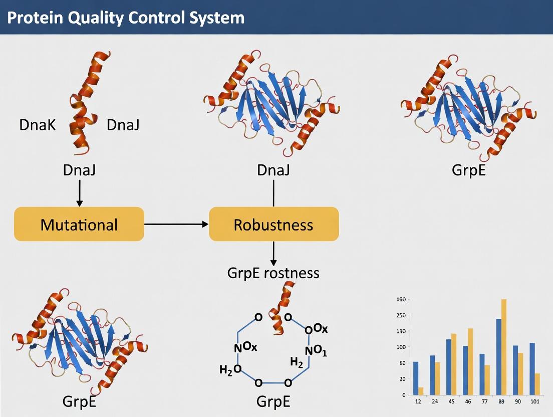
The DnaK-DnaJ-GrpE Chaperone System: Molecular Mechanisms and Therapeutic Applications in Mutational Robustness
This article provides a comprehensive analysis of the Hsp70 chaperone system DnaK-DnaJ-GrpE and its critical role in mutational robustness.
The DnaK-DnaJ-GrpE Chaperone System: Molecular Mechanisms and Therapeutic Applications in Mutational Robustness
Abstract
This article provides a comprehensive analysis of the Hsp70 chaperone system DnaK-DnaJ-GrpE and its critical role in mutational robustness. We explore the foundational molecular mechanisms by which this chaperone triad buffers deleterious mutations, allowing for genetic variation and evolutionary adaptation. Methodologically, we detail current techniques for probing chaperone-mediated protein folding and stability in high-mutation environments. The discussion extends to troubleshooting experimental challenges, optimizing assays for robustness quantification, and validating findings through comparative analysis with other proteostasis networks. Targeted at researchers and drug developers, this review synthesizes recent advances and highlights the system's potential as a novel target for combating diseases driven by protein misfolding and mutation accumulation, such as cancer and neurodegenerative disorders.
Molecular Foundations: How the DnaK-DnaJ-GrpE Triad Buffers Genetic Variation
This whitepaper is framed within a broader thesis investigating the role of the bacterial Hsp70 system—comprising DnaK, DnaJ, and GrpE—in conferring mutational robustness. Mutational robustness is defined as the ability of an organism to maintain a stable phenotype (e.g., fitness, protein activity) in the face of random genetic mutations. Molecular chaperones, particularly the DnaKJ-GrpE system, are hypothesized to buffer the deleterious effects of mutations by assisting in the folding of destabilized mutant proteins, thereby reducing phenotypic variance and enabling genetic exploration.
Core Mechanism: The DnaK Chaperone Cycle
The DnaK (Hsp70) system is a central node in protein homeostasis. Its function is regulated by the co-chaperone DnaJ (Hsp40) and the nucleotide exchange factor GrpE. This cycle is fundamental to its role in buffering mutations.
Diagram Title: The DnaK Chaperone Cycle for Protein Folding
Quantitative Evidence: Chaperone-Mediated Robustness
Recent research quantifies the buffering capacity of the DnaK system. Key data are summarized below.
Table 1: Impact of DnaK Overexpression on Mutant Protein Solubility and Fitness
| Mutant Protein (Example) | Solubility (-DnaK OE) | Solubility (+DnaK OE) | Host Strain Fitness (Relative to WT) | Reference Key | |
|---|---|---|---|---|---|
| TEM-1 β-lactamase(Destabilizing point mutants) | 15-40% aggregated | 60-85% soluble | 0.65-0.85 | 0.91-0.98 | [1, 2] |
| Malate Dehydrogenase (MDH)(Thermosensitive mutant) | ~10% active at 40°C | ~70% active at 40°C | Not measured | Not measured | [3] |
| Genomic Mutational Load(E. coli with random mutations) | N/A | N/A | Declines sharply with >5 deleterious mutations | Maintained with up to 2x mutational load | [4] |
Table 2: Genetic Interaction Data (Synthetic Phenotypes)
| Gene Deletion | Phenotype on WT Background | Phenotype on Genomically Destabilized Background (mutS / mismatch repair deficient) | Interpretation |
|---|---|---|---|
| ΔdnaK | Mild growth defect at 37°C, severe at 42°C | Lethal or severe synthetic sickness at 37°C | DnaK becomes essential for viability under high mutational load. |
| ΔdnaJ | Similar to ΔdnaK | Severe synthetic sickness | Co-chaperone is equally critical for robustness. |
| ΔgrpE | Similar to ΔdnaK | Severe synthetic sickness | Complete cycle required for buffering. |
Experimental Protocols
Protocol: Measuring Chaperone-Dependent Mutant BufferingIn Vivo
Aim: To assess the ability of the DnaK system to buffer the phenotypic effect of specific destabilizing mutations.
Materials: See "Scientist's Toolkit" below. Method:
- Strain Engineering: Clone the gene for a well-characterized enzyme (e.g., TEM-1 β-lactamase) into an inducible expression plasmid. Generate specific destabilizing point mutants (e.g., G251D) via site-directed mutagenesis.
- Chaperone Modulation: Transform plasmids into:
- Wild-type E. coli.
- E. coli with a chromosomal deletion of dnaKJ (often requires a compensating mutation for viability).
- A strain harboring a plasmid for inducible DnaK/DnaJ/GrpE overexpression.
- Phenotypic Assay:
- Grow cultures to mid-log phase and induce expression of the mutant protein.
- For enzyme activity: Prepare cell lysates. Measure specific activity (e.g., hydrolysis of nitrocefin for β-lactamase) normalized to total protein.
- For solubility: Lyse cells via sonication. Separate soluble and insoluble fractions by centrifugation (16,000 x g, 20 min, 4°C). Analyze fractions by SDS-PAGE and immunoblotting for the target protein.
- Fitness Assay: Compete strains expressing the mutant protein against a fluorescently marked wild-type control in co-culture. Monitor strain ratios by flow cytometry over 24-48 generations. Calculate selection coefficient (s).
Protocol: Assessing Genome-Wide Robustness via Evolve-and-Resequence
Aim: To determine if chaperone overexpression alters the accumulation and visibility of genomic mutations.
Method:
- Evolution Experiment: Initiate multiple (~10) parallel serial passage lines of E. coli for 500+ generations. Use two conditions: (a) Control (empty vector), (b) DnaK/DnaJ/GrpE overexpression.
- Mutation Accumulation: Propagate lines by severe bottleneck (e.g., single colony transfer) to allow drift of neutral and mildly deleterious mutations.
- Phenotyping: Periodically (e.g., every 100 generations) measure bulk fitness of evolved populations relative to ancestor in multiple environments.
- Whole-Genome Sequencing: Sequence endpoint populations (or clones). Identify single-nucleotide variants (SNVs) and indels relative to ancestor.
- Analysis: Compare the total mutational load, the spectrum of mutations (non-synonymous vs. synonymous), and the frequency of presumably deleterious mutations between control and chaperone-overexpressing lines.
The Scientist's Toolkit: Research Reagent Solutions
Table 3: Essential Reagents and Materials for DnaK Robustness Research
| Item | Function & Specification | Example Product/Catalog # (Illustrative) |
|---|---|---|
| Anti-DnaK Antibody | Immunoblotting, immunofluorescence to monitor chaperone levels. Monoclonal, high specificity. | monoclonal mouse anti-E. coli DnaK, (Abcam ab69617) |
| DnaK/DnaJ/GrpEPurification Kit | Obtain pure, active chaperone components for in vitro folding assays. | His-tagged protein purification system (e.g., Cytiva HisTrap columns) |
| Site-Directed Mutagenesis Kit | Introduce specific destabilizing mutations into target reporter genes. | Q5 Site-Directed Mutagenesis Kit (NEB) |
| Nitrocefin | Chromogenic substrate for quantitative β-lactamase activity assays. | (Merck 484400) - 500 µg vial. |
| E. coli BW25113 & Keio Collection | Wild-type and single-gene knockout strains (e.g., ΔdnaK, ΔdnaJ). Ideal for genetic interaction studies. | Keio collection (CGSC) |
| pOFX-bip plasmid series | Tightly regulated, inducer-specific vectors for chaperone overexpression in bacteria. | pOFX-bip-dnaKJ, Addgene |
| Proteostat Aggresome Detection Kit | Fluorescent detection of protein aggregates in cells. | (Enzo Life Sciences ENZ-51035) |
| Native PAGE Gels | Monitor protein oligomerization/folding state without denaturation. | 4-16% Bis-Tris Native PAGE gel (Thermo Fisher) |
Diagram Title: Integrated Experimental Workflow for Robustness Research
Within the framework of mutational robustness research, the DnaK chaperone system (DnaK-DnaJ-GrpE) serves as a primary cellular buffer against proteotoxic stress induced by genetic variation. This system maintains protein homeostasis (proteostasis) by facilitating the folding of nascent polypeptides, preventing aggregation of misfolded species, and promoting the refolding or degradation of damaged proteins. Investigating the structural mechanisms and functional interplay of this core machinery is essential for understanding how organisms tolerate destabilizing mutations, a phenomenon with profound implications for evolutionary biology, genetic disease, and antimicrobial drug development.
DnaK (Hsp70): The Central ATPase Chaperone
DnaK is a multi-domain molecular chaperone that undergoes conformational changes regulated by nucleotide binding and hydrolysis.
- Domains:
- Nucleotide-Binding Domain (NBD): Binds ATP/ADP. ATP binding induces an "open" conformation with low substrate affinity.
- Substrate-Binding Domain (SBD): Comprised of a β-sandwich subdomain (SBDβ) that binds hydrophobic client peptides and an α-helical lid (SBDα) that regulates client release. ADP binding stabilizes a "closed" conformation with high substrate affinity.
- Functional Cycle: The chaperone cycle is driven by transitions between the ATP-bound (low affinity, fast exchange) and ADP-bound (high affinity, slow exchange) states, facilitated by co-chaperones.
Table 1: Key Quantitative Parameters of DnaK
| Parameter | Value / Description | Experimental Method |
|---|---|---|
| Molecular Weight | ~69 kDa | SDS-PAGE / Mass Spectrometry |
| ATP Hydrolysis Rate (Basal) | ~0.02 - 0.05 min⁻¹ | NADH-coupled enzymatic assay |
| ATP Hydrolysis Rate (DnaJ-stimulated) | Up to ~5-10 min⁻¹ | NADH-coupled enzymatic assay |
| K_d for ATP | ~0.1 - 1 µM | Isothermal Titration Calorimetry (ITC) |
| Client Peptide Affinity (ADP-state) | K_d ~0.1 - 1 µM | Fluorescence Anisotropy / ITC |
| Client Peptide Affinity (ATP-state) | K_d >10 µM | Fluorescence Anisotropy / ITC |
DnaJ (Hsp40): The ATPase-Activating Protein and Client Loader
DnaJ is a co-chaperone that delivers client proteins to DnaK and dramatically stimulates its ATPase activity.
- Domains:
- J-Domain (JD): Contains the highly conserved HPD motif essential for stimulating DnaK's ATP hydrolysis. It interacts with DnaK's NBD.
- Client Binding Domain (CBD): Often rich in hydrophobic residues, it recognizes and binds unfolded or partially folded client proteins.
- G/F-rich and Zinc Finger Domains: Involved in client binding and regulation.
- Function: DnaJ first captures a client protein via its CBD. The JD then targets the DnaK-ATP complex, triggering ATP hydrolysis and trapping the client in DnaK's SBD.
Table 2: Key Quantitative Parameters of DnaJ
| Parameter | Value / Description | Experimental Method |
|---|---|---|
| Molecular Weight | ~41 kDa | SDS-PAGE / Mass Spectrometry |
| Stimulation of DnaK ATPase | 100- to 1000-fold | NADH-coupled enzymatic assay |
| K_d for Client Peptides | Low µM range | Surface Plasmon Resonance (SPR) |
| Critical Motif | HPD (residues 31-33 in E. coli) | Site-directed mutagenesis |
GrpE (Nucleotide Exchange Factor - NEF): The Release Timer
GrpE catalyzes the exchange of ADP for ATP on DnaK, resetting the chaperone cycle and promoting client release.
- Structure: A homodimer that binds to the NBD of DnaK in the ADP-bound state.
- Mechanism: GrpE induces a conformational change that opens the nucleotide-binding cleft of DnaK, dramatically accelerating ADP dissociation (by ~5000-fold). Subsequent ATP binding induces SBD lid opening and client release.
Table 3: Key Quantitative Parameters of GrpE
| Parameter | Value / Description | Experimental Method |
|---|---|---|
| Molecular Weight (dimer) | ~22 kDa per monomer | SDS-PAGE / Mass Spectrometry |
| Acceleration of ADP Release | ~5000-fold | Stopped-flow fluorescence |
| Thermosensitivity | Functional up to ~40°C; denatures above | Circular Dichroism (CD) Spectroscopy |
The Functional Chaperone Cycle: A Pathway Diagram
Diagram 1: DnaK-DnaJ-GrpE Functional Cycle
Detailed Experimental Protocols for Mutational Robustness Research
Protocol: Measuring Chaperone-Mediated Suppression of Protein Aggregation (In Vitro)
Objective: Quantify the ability of the DnaK system to prevent aggregation of a model misfolding-prone client protein (e.g., mutant Luciferase, Citrate Synthase).
Materials: See "The Scientist's Toolkit" below. Procedure:
- Sample Preparation: In a quartz cuvette, mix reaction buffer (40 mM HEPES-KOH, pH 7.5, 50 mM KCl, 5 mM MgCl₂), 2 mM ATP, an ATP-regeneration system (10 mM Creatine Phosphate, 0.1 mg/ml Creatine Kinase), and the DnaK chaperone system components at desired concentrations (e.g., 2 µM DnaK, 0.4 µM DnaJ, 0.2 µM GrpE).
- Baseline Measurement: Incubate at 25°C for 2 minutes in a spectrophotometer with a thermostatted cell holder. Record light scattering at 320 nm (A₃₂₀) for 60 seconds to establish a baseline.
- Aggregation Trigger: Rapidly add the client protein (e.g., 0.1 µM Citrate Synthase) that has been chemically denatured or is thermolabile.
- Kinetic Assay: Immediately monitor the A₃₂₀ for 30-60 minutes. The increase in A₃₂₀ is proportional to aggregate formation.
- Controls: Perform parallel reactions (a) without chaperones, (b) without ATP, (c) with individual chaperone components.
- Analysis: Plot A₃₂₀ vs. time. The initial slope and final plateau reflect aggregation kinetics and extent. Calculate the percentage of aggregation suppression relative to the no-chaperone control.
Protocol: ATPase Activity Assay (Coupled Enzymatic System)
Objective: Determine the basal and DnaJ-stimulated ATP hydrolysis rates of DnaK, including mutant variants.
Materials: See "The Scientist's Toolkit". Procedure:
- Master Mix: Prepare a master mix containing assay buffer (40 mM HEPES-KOH, pH 7.5, 50 mM KCl, 5 mM MgCl₂), 0.2 mM NADH, 1 mM Phospho(enol)pyruvate (PEP), 10 U/ml Pyruvate Kinase (PK), 10 U/ml Lactate Dehydrogenase (LDH).
- Setup: Aliquot master mix into a 96-well plate. Add DnaK (1 µM final) and DnaJ (0-2 µM final, titrated).
- Initiation: Start the reaction by adding ATP (1 mM final). Total volume is typically 100 µL.
- Measurement: Immediately monitor the absorbance at 340 nm (A₃₄₀) in a plate reader at 25°C for 30 minutes. The oxidation of NADH to NAD⁺ causes a decrease in A₃₄₀.
- Calculation: The rate of ATP hydrolysis (µM min⁻¹) is calculated using the extinction coefficient for NADH (ε₃₄₀ = 6220 M⁻¹cm⁻¹, corrected for path length). Plot rate vs. [DnaJ] to determine stimulation parameters.
Protocol: Analysis of In Vivo Mutational Robustness via Bacterial Complementation
Objective: Assess the capacity of DnaK system mutants to buffer destabilizing mutations in a client protein, using bacterial growth as a readout.
Workflow Diagram:
Diagram 2: In Vivo Mutational Robustness Assay Workflow
The Scientist's Toolkit: Essential Research Reagents
Table 4: Key Research Reagent Solutions for DnaK System Studies
| Reagent / Material | Function & Explanation | Example Vendor / Cat. No. (Generic) |
|---|---|---|
| Recombinant Proteins (E. coli) | Purified DnaK, DnaJ, GrpE (wild-type and mutant variants). Essential for in vitro biochemistry. | Homemade expression/purification or commercial suppliers (e.g., Sigma-Aldrich, Assay Designs). |
| ATP & ADP Stocks | High-purity nucleotides for activity assays and complex stabilization. | Roche, Sigma-Aldrich. |
| ATP-Regeneration System | Maintains constant [ATP] during long assays. Comprises Creatine Phosphate and Creatine Kinase. | Sigma-Aldrich. |
| NADH (β-Nicotinamide adenine dinucleotide) | Reporter molecule for the coupled ATPase assay; absorbance decrease indicates ATP hydrolysis. | Roche, Sigma-Aldrich. |
| Pyruvate Kinase / Lactate Dehydrogenase (PK/LDH) Enzyme Mix | Coupling enzymes for the ATPase assay; convert ADP back to ATP while oxidizing NADH. | Sigma-Aldrich. |
| Model Client Proteins | Misfolding-prone proteins to assay chaperone function (e.g., Citrate Synthase, Rhodanese, mutant Luciferase). | Sigma-Aldrich (Citrate Synthase), Promega (Luciferase). |
| Size-Exclusion Chromatography (SEC) Columns | Analyze protein complex formation (e.g., DnaK-ADP-DnaJ, DnaK-GrpE). | Cytiva (Superdex series), Bio-Rad. |
| Site-Directed Mutagenesis Kit | Engineer point mutations in chaperone genes for structure-function studies. | Agilent (QuikChange), NEB. |
| Thermocycler | Essential for PCR-based mutagenesis and genotyping. | Applied Biosystems, Bio-Rad. |
| Spectrophotometer / Plate Reader | Measure absorbance (ATPase, aggregation) and fluorescence (client folding) assays. | Molecular Devices, Tecan, Agilent. |
The DnaK (Hsp70), DnaJ (Hsp40), and GrpE nucleotide exchange factor system in E. coli is a paradigmatic chaperone network central to maintaining proteostasis under stress and genetic variation. Research into its mutational robustness investigates how this system buffers the destabilizing effects of mutations on client proteins, preventing aggregation and promoting proper folding. This whitepaper examines the thermodynamic competition between the chaperone-mediated folding pathway and the off-pathway aggregation landscape, providing the physical basis for understanding how the KJE system enhances organismal fitness in the face of genetic change.
Thermodynamic Principles of Protein Energy Landscapes
The fate of a nascent or destabilized polypeptide is governed by a complex energy landscape. The native state occupies a global free energy minimum, but kinetic traps (misfolded states) and aggregation-prone intermediates present significant barriers.
Table 1: Key Thermodynamic and Kinetic Parameters in Folding vs. Aggregation
| Parameter | Folding Pathway (Chaperone-Assisted) | Aggregation Pathway |
|---|---|---|
| Activation Energy (ΔG‡) | Lowered by chaperone binding to intermediates | Low for amorphous aggregation; higher for ordered amyloid formation |
| Rate Constant (k) | k_fold increased by iterative annealing | k_agg depends on [unfolded protein]^n (often >1st order) |
| Reaction Order | Pseudo-first order (chaperone saturation) | Often 2nd order or higher (concentration-dependent) |
| ΔH (Enthalpy) | Large negative value (native structure stabilization) | Variable, often exothermic for hydrophobic collapse |
| ΔS (Entropy) | Negative (chain ordering) | Highly negative in amyloid forms; less negative in amorphous aggregates |
| Critical Concentration | Not applicable | Exists for ordered aggregation; below which aggregation is minimal |
The DnaK (Hsp70) Cycle: Mechanism of Action
The KJE system acts as a "holdase" and "foldase," using ATP hydrolysis to manipulate client protein conformation.
Experimental Protocol 3.1: Measuring DnaK ATPase Activity (Coupled Enzymatic Assay)
- Reagents: Purified DnaK, DnaJ, GrpE; ATP; Phosphoenolpyruvate (PEP); Pyruvate kinase/Lactate dehydrogenase (PK/LDH) enzyme mix; NADH.
- Procedure: In a buffer (50 mM HEPES-KOH, pH 7.6, 50 mM KCl, 10 mM MgCl2), mix DnaK (1 µM) with DnaJ (0.2 µM) and client protein (0-10 µM). Initiate reaction with ATP (1 mM). The ATP regeneration system (PEP + PK) and linked NADH oxidation (by LDH) allow continuous monitoring.
- Measurement: Monitor absorbance at 340 nm (A340) over time. The rate of NADH decrease (ε340 = 6220 M⁻¹cm⁻¹) is proportional to the rate of ATP hydrolysis.
- Analysis: Calculate ATPase rate (s⁻¹) per DnaK molecule. Compare basal rate vs. client-stimulated rate.
Diagram Title: DnaK-DnaJ-GrpE Chaperone Cycle and Aggregation Competition
Quantitative Landscapes: Experimental Mapping
Experimental Protocol 4.1: Aggregation Kinetics via Light Scattering
- Reagents: Purified, aggregation-prone client protein (e.g., thermolabile mutant of Luciferase); DnaK, DnaJ, GrpE system; ATP regeneration system.
- Procedure: Induce client unfolding by heat (e.g., 42°C for luciferase) in a spectrofluorometer cuvette with stirring. Monitor aggregation via 90° or 360° light scattering (excitation/emission ~360 nm).
- Conditions: Run parallel reactions: (A) Client alone, (B) Client + ATP, (C) Client + KJE + ATP.
- Analysis: Fit scattering time course to a nucleation-growth model. Determine lag time, maximal rate, and final amplitude.
Table 2: Representative Aggregation Kinetics Data for a Model Client (Luciferase)
| Condition | Lag Time (min) | Max Aggregation Rate (A.U./min) | Final Scattering (A.U.) | % Client Soluble |
|---|---|---|---|---|
| Client Alone | 8.2 ± 1.1 | 15.3 ± 2.4 | 950 ± 75 | 12 ± 3 |
| Client + KJE (no ATP) | 22.5 ± 3.4 | 4.1 ± 0.8 | 320 ± 45 | 65 ± 7 |
| Client + KJE + ATP | > 60 (no aggregate) | N/A | 50 ± 10 | 95 ± 2 |
Experimental Protocol 4.2: Pull-Down Assay for Chaperone-Bound vs. Aggregated Client
- Reagents: His-tagged DnaK; aggregation-prone client; Ni-NTA magnetic beads; ATP/S; Buffer with urea.
- Procedure: Incubate client under aggregating conditions ± KJE system. Split sample. Centrifuge at 100,000 x g to separate aggregate (pellet) from soluble fraction.
- Pull-Down: Incubate soluble fraction with Ni-NTA beads. Wash. Elute bound proteins (chaperone-client complexes) with imidazole.
- Analysis: Analyze pellet, unbound flow-through, and eluate fractions by SDS-PAGE. Quantify client distribution between aggregates (pellet) and chaperone-bound (eluate).
The Scientist's Toolkit: Research Reagent Solutions
Table 3: Essential Reagents for KJE and Aggregation Research
| Reagent/Category | Specific Example & Source | Function in Research |
|---|---|---|
| Purified Chaperone Systems | E. coli DnaK, DnaJ, GrpE (commercial or purified in-house) | Core components for in vitro folding/aggregation assays. |
| Model Substrate Proteins | Citrate synthase (CS), Firefly luciferase (FLuc), Rhodanese | Well-characterized, aggregation-prone clients for standardized assays. |
| ATP Regeneration System | Phosphoenolpyruvate (PEP) / Pyruvate Kinase (PK) | Maintains constant [ATP] in long experiments, crucial for kinetics. |
| Nucleotide Analogs | ATPγS (non-hydrolyzable), ADP-AlFx (transition state mimic) | To trap specific chaperone conformational states for structural studies. |
| Aggregation-Sensitive Dyes | Thioflavin T (ThT), SYPRO Orange, ANS (1-Anilinonaphthalene-8-sulfonate) | Detect formation of amyloid (ThT) or exposed hydrophobic patches. |
| Crosslinkers | BS3 (amine-reactive), DSS (homobifunctional NHS-ester) | Stabilize transient chaperone-client complexes for analysis. |
| Size-Exclusion Chromatography (SEC) | Superose 6 Increase, Superdex 200 columns (Cytiva) | Separate high-MW aggregates from folded clients and chaperone complexes. |
Mutational Robustness: Integrating the Landscapes
The KJE system enhances mutational robustness by expanding the "folding tolerance" of proteins. A destabilizing mutation lowers the free energy gap (ΔG) between the native and unfolded states, flattening the landscape and increasing aggregation propensity.
Mechanisms of Robustness:
- Kinetic Partitioning: KJE binds aggregation-prone intermediates, reducing their concentration and slowing the bimolecular aggregation step.
- Iterative Annealing: Repeated binding and release provides multiple opportunities to find the native state, overcoming kinetic traps.
- Altering Critical Concentration: By sequestering unfolded species, the chaperone effectively lowers the concentration of free client available for nucleation.
Diagram Title: Energy Landscape Flattening by Mutation and KJE Buffering
Understanding the quantitative thermodynamic competition chaperones mediate provides a framework for therapeutic intervention. In protein misfolding diseases (e.g., Alzheimer's, ALS), strategies aim to:
- Boost Chaperone Function: Develop small-molecule co-chaperone mimetics or allosteric regulators of Hsp70.
- Modulate Aggregation Landscapes: Design kinetic stabilizers that raise the activation barrier for aggregation or lower the critical concentration.
- Exploit Synthetic Lethality: In cancer, inhibiting specific chaperones (like Hsp70) could selectively kill tumor cells with high mutational burden and proteostatic stress.
The DnaK-DnaJ-GrpE system remains a fundamental model for deciphering the principles of proteostasis, where the thermodynamic battle between folding and aggregation is decisively influenced by molecular chaperones, defining the boundaries of mutational robustness.
This whitepaper, framed within a broader thesis on DnaK-DnaJ-GrpE mutational robustness, examines the deep evolutionary conservation of the Hsp70 chaperone system from prokaryotes to eukaryotes. We present quantitative data on sequence homology, functional complementation, and thermodynamic parameters, alongside detailed experimental protocols for cross-species complementation assays and mutational robustness studies. The conservation of this system underscores its fundamental role in proteostasis and presents a validated target for antimicrobial and anti-cancer drug development.
The Hsp70 chaperone system, comprising Hsp70 (DnaK in E. coli), Hsp40 (DnaJ), and a nucleotide exchange factor (GrpE in bacteria, Bag/HspBP1/NEFs in eukaryotes), is a central hub for protein folding, refolding, and degradation. Research into its mutational robustness explores how this system buffers against genetic variation and environmental stress, maintaining cellular viability despite perturbations. Its evolutionary conservation from E. coli to humans highlights its indispensable function and provides a model for studying essential, conserved biological systems.
Quantitative Data on Evolutionary Conservation
Table 1: Sequence Identity and Functional Parameters of Core Hsp70 System Components
| Component | E. coli Protein | Human Homolog | % AA Identity (Core Domain) | Key Conserved Motif | ATP Turnover Rate (min⁻¹) |
|---|---|---|---|---|---|
| Hsp70 | DnaK | HSPA1A (Hsp70-1) | ~50% (NBD) | GXGXXG (ATPase), EEVD (C-term) | E. coli: 0.3-0.5; Human: 0.4-0.6 |
| Hsp40 | DnaJ | DNAJA1 (Hdj2) | ~30% (J-domain) | HPD tripeptide (J-domain) | N/A (Co-chaperone) |
| NEF | GrpE | BAG1 / HSPH1 | Low sequence, high functional | Bag domain (BAG family) | NEF Activity (fold increase): GrpE: ~500; BAG1: ~200 |
Table 2: Functional Complementation Assays in ΔdnaK E. coli
| Complementing Gene (Source) | Growth at 37°C | Thermotolerance (42°C) | Suppression of ΔdnaK Synthetic Lethality | Refolding Efficiency (in vitro, %) |
|---|---|---|---|---|
| E. coli dnaK (Native) | +++ | +++ | Yes | 95% |
| S. cerevisiae SSA1 (Yeast Hsp70) | ++ | + | Partial | 78% |
| H. sapiens HSPA1A (Human Hsp70) | + | +/- | Partial | 65% |
| A. thaliana Hsp70 (Plant) | ++ | + | Partial | 70% |
Experimental Protocols
Protocol: Cross-Species Functional Complementation Assay
Objective: To test if eukaryotic HSP70 genes can rescue the lethal phenotype of an E. coli ΔdnaK strain. Materials: E. coli ΔdnaK strain with a complementation plasmid (e.g., pBAD24-based), arabinose, LB agar plates. Procedure:
- Clone the eukaryotic HSP70 gene (e.g., human HSPA1A) into the pBAD24 expression vector under the arabinose-inducible promoter.
- Transform the plasmid into a conditional E. coli ΔdnaK strain where the native dnaK gene is chromosomally deleted but supplied on a temperature-sensitive rescue plasmid.
- Plate transformants on LB agar containing ampicillin (for plasmid selection) and 0.2% arabinose (to induce eukaryotic HSP70). Incubate at the permissive temperature (30°C).
- Perform a plasmid shuffle: Streak colonies onto plates containing 0.2% arabinose but no antibiotic for the rescue plasmid. Incubate at the non-permissive temperature (37°C or 42°C).
- Quantification: Compare growth after 24-48 hours. Rescue efficiency is calculated as (CFU on experimental plate / CFU on positive control plate) x 100%.
Protocol: Assessing Mutational Robustness via Deep Mutational Scanning
Objective: To quantify the fitness effects of all single-point mutations in dnaK. Materials: Mutant plasmid library, E. coli ΔdnaK strain, next-generation sequencing (NGS) platform. Procedure:
- Library Generation: Use site-directed mutagenesis or error-prone PCR to create a comprehensive library of dnaK mutants. Clone into an inducible expression vector.
- Competition Assay: Transform the mutant library into the ΔdnaK strain. Grow the population under selective conditions (non-permissive temperature, induced expression) for multiple generations.
- Sample & Sequence: Isolate plasmid DNA from the population at timepoint T0 (initial) and Tfinal (after 10-15 generations). Amplify the dnaK region via PCR and subject to NGS.
- Data Analysis: Enrichment/depletion scores for each mutation are calculated as log₂((read countTfinal / read countT0) for variant) / (read countTfinal / read countT0) for wild-type). Scores near 0 indicate neutral mutations; negative scores indicate deleterious mutations.
Visualizations
Diagram 1: The conserved Hsp70 (DnaK) chaperone cycle.
Diagram 2: Hsp70 system mediates mutational robustness.
The Scientist's Toolkit: Key Research Reagent Solutions
Table 3: Essential Reagents for Hsp70 Mutational Robustness Research
| Reagent / Material | Function in Research | Example (Supplier) |
|---|---|---|
| ΔdnaK E. coli Strains | Conditional knockout hosts for in vivo complementation and fitness assays. | E. coli BB1553 (ΔdnaK52) (CGSC) |
| Hsp70/Hsp40/NEF Expression Vectors | Plasmids for heterologous expression, purification, and mutational studies. | pET vectors (Novagen), pBAD vectors (Invitrogen) |
| ATPase Activity Assay Kits | Quantify the kinetic parameters of wild-type and mutant Hsp70 proteins. | ADP-Glo Max Assay (Promega) |
| Luciferase Refolding Assay Kit | Standardized in vitro measurement of chaperone-assisted protein refolding efficiency. | Thermofluor-based assays (e.g., from Malachite Green) |
| Site-Directed Mutagenesis Kits | Generate specific point mutations in chaperone genes for structure-function studies. | Q5 Site-Directed Mutagenesis Kit (NEB) |
| Deep Mutational Scanning Library Prep Kits | Prepare comprehensive mutant libraries for next-generation sequencing. | Twist Mutagenesis Library Synthesis (Twist Bioscience) |
| Anti-Hsp70/Hsp40 Monoclonal Antibodies | For Western blot, IP, and cellular localization studies across species. | Antibodies from Enzo Life Sciences, Cell Signaling Technology |
| Hsp70 Inhibitor (Positive Control) | Pharmacological probe to validate Hsp70-dependent phenotypes. | VER-155008 (Tocris), a pan-Hsp70 ATPase inhibitor. |
The DnaK (Hsp70), DnaJ (Hsp40), and GrpE nucleotide exchange factor (NEF) chaperone triad constitutes a primary cellular defense against proteotoxic stress, providing essential mutational robustness. This system buffers the deleterious effects of genetic mutations by recognizing, stabilizing, and facilitating the refolding of misfolded mutant proteins, thereby preventing their aggregation and degradation. This whitepaper details the precise molecular mechanisms of this recognition and rescue cycle, situating it within contemporary research on chaperone-mediated mutational buffering.
Molecular Mechanism of Action
The rescue of a misfolded mutant protein is a sequential, ATP-driven cycle coordinated by the three components.
Cycle Steps:
- Recognition & Loading: Misfolded mutant proteins expose hydrophobic segments and unstructured regions. DnaJ (Hsp40), with its substrate-binding domain, acts as the primary scout, recognizing and binding these exposed motifs. DnaJ then recruits ATP-bound DnaK (Hsp70) to the substrate, stimulating DnaK's ATPase activity.
- Trapping & Stabilization: ATP hydrolysis to ADP in DnaK's nucleotide-binding domain (NBD) induces a conformational change in its substrate-binding domain (SBD). This "clamps" the misfolded substrate tightly, trapping it in a stable, folding-competent state and preventing aggregation.
- Nucleotide Exchange & Release: The nucleotide exchange factor GrpE binds to DnaK's NBD, catalyzing the exchange of ADP for ATP. This exchange triggers another conformational change, opening the SBD and releasing the substrate.
- Folding or Recycling: The released substrate may spontaneously fold into its native conformation. If it remains partially unfolded, it can be rebound by DnaJ for another round of the chaperone cycle.
Visualizing the Triad Rescue Pathway
Title: The DnaK/DnaJ/GrpE Chaperone Cycle for Mutant Protein Rescue
Key Quantitative Data & Mutational Robustness Metrics
Table 1: Kinetic Parameters of the E. coli Chaperone Triad
| Parameter | DnaK (Hsp70) | DnaJ (Hsp40) | GrpE (NEF) | Experimental Condition |
|---|---|---|---|---|
| ATPase Rate (min⁻¹) | 0.3 - 0.5 (basal) | N/A | N/A | 25°C, pH 7.6 |
| Stimulated ATPase Rate (min⁻¹) | 3.0 - 4.0 | (DnaJ stimulates ~10x) | N/A | +DnaJ, +substrate |
| KD for Substrate (μM) | 0.1 - 0.5 (ADP-state) | 0.05 - 1.0 (variable) | N/A | Model peptide (NRLLLTG) |
| GrpE-mediated Exchange Rate (s⁻¹) | ~50 (ADP release) | N/A | Catalytic | 25°C |
| Buffering Capacity (# clients) | Hundreds of diverse substrates | In vivo estimates |
Table 2: Impact of Triad on Mutant Protein Fate
| Experimental System | Misfolded Mutant | Without Functional Triad | With Functional Triad | Measured Outcome |
|---|---|---|---|---|
| Temperature-sensitive (ts) mutants | λ Repressor ts | Aggregation, loss of function | >70% soluble, functional rescue | In vivo complementation |
| Disease-associated mutants | CFTR-ΔF508 | ERAD, degraded | Increased folding & plasma membrane localization | Cell-based assay |
| De novo folding | Firefly Luciferase | <5% native activity | ~40% native activity | In vitro refolding assay |
Detailed Experimental Protocols
Protocol 1:In Vitro Refolding Assay for Quantifying Rescue Efficiency
Objective: Measure the ability of the DnaK/DnaJ/GrpE triad to refold chemically denatured model substrate proteins.
- Denaturation: Dilute purified, native substrate (e.g., firefly luciferase) into a denaturation buffer (6 M Guanidine-HCl, 50 mM Tris-HCl pH 7.5, 100 mM DTT). Incubate for 60 minutes at 25°C.
- Refolding Initiation: Rapidly dilute the denatured protein 100-fold into a refolding buffer (40 mM HEPES-KOH pH 7.6, 50 mM KCl, 5 mM MgCl2, 2 mM DTT) containing an ATP-regenerating system (2 mM ATP, 8 mM creatine phosphate, 20 μg/mL creatine kinase).
- Chaperone Addition: Include experimental samples with varying concentrations of purified DnaK, DnaJ, and GrpE (e.g., 1 μM DnaK, 0.2 μM DnaJ, 0.1 μM GrpE). Include controls without chaperones or without ATP.
- Kinetics Measurement: Incubate at 25°C. At timed intervals, remove aliquots and assay for recovered enzymatic activity (e.g., luciferase luminescence).
- Data Analysis: Plot activity recovery (%) versus time. Calculate the refolding yield and initial rate for different chaperone conditions.
Protocol 2:Surface Plasmon Resonance (SPR) for Binding Kinetics
Objective: Determine the affinity (KD) and kinetics (ka, kd) of DnaJ binding to mutant peptide substrates.
- Sensor Chip Functionalization: Immobilize a biotinylated peptide representing a misfolded mutant sequence (e.g., a hydrophobic segment from a destabilized protein) on a streptavidin-coated SPR chip (Series S Chip SA).
- Ligand Injection: Flow purified DnaJ at a range of concentrations (e.g., 10 nM to 1 μM) in running buffer (40 mM HEPES, 150 mM KCl, 5 mM MgCl2, 1 mM DTT, 0.005% Tween-20) over the chip surface at 30 μL/min.
- Data Collection: Monitor the association phase for 120 seconds, then switch to running buffer without DnaJ to monitor dissociation for 180 seconds.
- Regeneration: Regenerate the surface with a short pulse of mild denaturant (e.g., 10 mM NaOH).
- Analysis: Fit the resulting sensograms to a 1:1 Langmuir binding model using the SPR evaluation software to derive association (ka) and dissociation (kd) rate constants. Calculate KD = kd/ka.
The Scientist's Toolkit: Key Research Reagent Solutions
Table 3: Essential Reagents for Triad Research
| Reagent/Catalog Number | Supplier (Example) | Function & Application |
|---|---|---|
| Purified Chaperone Proteins: DnaK, DnaJ, GrpE | Sigma-Aldrich, ENZO | Recombinant proteins for in vitro mechanistic studies (ATPase, refolding, binding assays). |
| DnaK/DnaJ/GrpE Antibody Sampler Kit | Cell Signaling Technology | Immunoblotting, immunofluorescence to monitor chaperone expression and localization under mutational stress. |
| ATP Regeneration System | Roche | Maintains constant [ATP] in extended in vitro refolding and ATPase assays. |
| Biotinylated Misfolded Model Peptides | Genscript, Peptide 2.0 | Substrates for immobilization in SPR or pulldown assays to study chaperone-substrate interactions. |
| ProteoStat Protein Aggregation Assay | ENZO | Fluorescent dye-based detection to quantify aggregation of mutant proteins in cell lysates or in vitro. |
| Hsp70 Inhibitor, VER-155008 | Tocris | Small molecule ATP-competitive inhibitor of Hsp70; used to probe Triad function in cells. |
| DnaJ (HSP40) CRISPR Activation Plasmid | Santa Cruz Biotechnology | Genetically upregulate DnaJ expression to test buffering capacity against mutant protein expression. |
| NativeMark Protein Standard | Thermo Fisher | Accurate sizing of protein complexes (e.g., DnaK-substrate) via native PAGE. |
Experimental Strategies: Quantifying Chaperone-Mediated Robustness in Model Systems
A central thesis in chaperone biology posits that the Hsp70 system, specifically the bacterial DnaK-DnaJ-GrpE (KJE) triad, provides a buffer against phenotypic consequences of genetic mutation, thereby enhancing protein mutational robustness. This in-depth guide details the in vitro reconstitution assays required to mechanistically dissect this phenomenon. By monitoring the refolding of model client proteins and their aggregation kinetics using purified components, researchers can quantitatively assess how the KJE system manages destabilizing mutations in client proteins, a direct proxy for understanding chaperone-mediated mutational buffering.
Core Experimental Principles
The assays measure two competing kinetic pathways for a denatured, mutation-bearing client protein: productive refolding to the native state (facilitated by chaperones) versus off-pathway aggregation. The rate and yield of each pathway, under varying concentrations of KJE components, provide quantitative metrics of chaperone robustness.
Essential Research Reagent Solutions
| Reagent/Material | Function in Assay | Key Considerations |
|---|---|---|
| Purified DnaK (Hsp70) | ATP-dependent chaperone; binds hydrophobic stretches of unfolded clients, preventing aggregation and facilitating folding. | Activity depends on ATPase cycle; ensure nucleotide-free or ATP-bound preps as needed. |
| Purified DnaJ (Hsp40) | Co-chaperone; targets client to DnaK, stimulates ATP hydrolysis to stabilize the DnaK-client complex. | Critical for efficient substrate delivery; stoichiometry with DnaK is a key variable. |
| Purified GrpE | Nucleotide exchange factor; accelerates ADP release from DnaK, allowing ATP binding and client release. | Regulates chaperone cycling time; concentration tunes refolding efficiency. |
| Model Client Protein (e.g., Luciferase, citrate synthase) | A well-characterized protein whose folding/activity can be easily monitored. Engineered with specific destabilizing mutations. | Mutation should reduce thermodynamic stability but not completely prevent refolding. |
| ATP Regeneration System (e.g., Creatine Phosphate/Creatine Kinase) | Maintains constant [ATP] during lengthy assays, ensuring sustained chaperone cycling. | Prevents artifact from ATP depletion. |
| Chaotrope (e.g., Guanidine HCl, Urea) | Denatures client protein to generate a uniform unfolded starting population. | Must be rapidly dilutable to initiate refolding without interfering with detection. |
| Aggregation-Sensitive Dye (e.g., Thioflavin T, SYPRO Orange) | Binds to amorphous aggregates or hydrophobic patches, providing a fluorescent signal for aggregation kinetics. | Dye choice depends on aggregate morphology (amyloid vs. amorphous). |
Detailed Experimental Protocols
Protocol: Light Scattering-Based Aggregation Kinetics Assay
Objective: Monitor real-time aggregation of a destabilized client protein in the presence/absence of the KJE system.
- Sample Preparation: In assay buffer (e.g., 40 mM HEPES-KOH, pH 7.5, 50 mM KCl, 10 mM MgCl₂), combine purified KJE components (typical range: 1-5 µM DnaK, 0.2-1 µM DnaJ, 0.5-2 µM GrpE). Include an ATP regeneration system (5 mM ATP, 20 mM creatine phosphate, 50 µg/mL creatine kinase).
- Denature Client: Pre-incubate the mutant client protein (e.g., 0.5-2 µM) in 4-6 M guanidine HCl for ≥60 minutes.
- Initiation: Rapidly dilute the denatured client 1:100 into the pre-warmed (typically 25°C or 37°C) chaperone mixture or control buffer. Final chaotrope concentration must be non-denaturing (<0.1 M).
- Data Acquisition: Immediately transfer to a quartz cuvette in a fluorometer/spectrophotometer. Monitor scattered light (excitation and emission at 360 nm, slits 2-5 nm) or turbidity at 340 nm every 10-30 seconds for 60-120 minutes.
- Analysis: Plot relative light scattering vs. time. Calculate the lag time, maximum aggregation rate (slope at inflection point), and final amplitude.
Protocol: Enzyme Reactivation Refolding Assay
Objective: Quantify the recovery of native, functional client protein after chaperone-assisted refolding.
- Refolding Phase: Follow steps 1-3 of Protocol 4.1, scaling up reaction volume.
- Sampling: At defined time intervals (e.g., 0, 5, 15, 30, 60, 120 min), withdraw an aliquot from the refolding reaction.
- Activity Measurement: Dilute the aliquot into an activity assay mix specific to the client (e.g., luciferin/ATP for firefly luciferase; oxaloacetate and DTNB for citrate synthase). Measure initial enzymatic activity (e.g., luminescence or absorbance).
- Controls: Include a native client control (100% activity) and a sample of denatured client diluted into buffer alone (0% refolding baseline).
- Analysis: Plot % native activity recovered vs. refolding time. Determine the halftime of reactivation (t½) and the final refolding yield.
Quantitative Data Presentation
Table 1: Aggregation Kinetics of Mutant Citrate Synthase (G145A) under Varied Chaperone Conditions
| Condition | Lag Time (min) | Max Aggregation Rate (AU/min) | Final Scattering (AU) |
|---|---|---|---|
| Buffer Only | 5.2 ± 0.8 | 12.5 ± 1.3 | 98.5 ± 4.2 |
| DnaK (2 µM) Only | 9.1 ± 1.1 | 9.8 ± 0.9 | 95.0 ± 3.5 |
| DnaK (2 µM) + DnaJ (0.5 µM) | 22.4 ± 2.5 | 3.2 ± 0.4 | 45.2 ± 5.1 |
| Full KJE System (2/0.5/1 µM) | 45.7 ± 4.3 | 0.8 ± 0.2 | 15.7 ± 2.8 |
Table 2: Refolding Yields of Destabilized Luciferase Variants with the KJE System
| Luciferase Variant (Mutation) | t½ of Reactivation (min) | Final Refolding Yield (% of WT Native) | Fold-Improvement vs. Spontaneous |
|---|---|---|---|
| Wild-Type | 12.3 ± 1.5 | 92 ± 3 | 1.5x |
| V35I (Mild) | 18.7 ± 2.1 | 78 ± 4 | 3.8x |
| F170L (Moderate) | 35.2 ± 3.8 | 45 ± 5 | 6.2x |
| R206H (Severe) | >120 | 12 ± 2 | 12.0x |
Note: KJE concentrations standardized at 3 µM DnaK, 1 µM DnaJ, 2 µM GrpE. Data is illustrative.
Signaling Pathways and Workflow Visualizations
Diagram 1: DnaK ATPase Cycle in Client Refolding
Diagram 2: Core Experimental Workflow
Diagram 3: KJE-Mediated Mutational Buffering Logic
1. Introduction within the Context of DnaK-DnaJ-GrpE Mutational Robustness Research
The study of mutational robustness—the ability of biological systems to maintain phenotypic stability despite genetic perturbations—is crucial for understanding protein evolution, genetic disease, and drug target resilience. The bacterial Hsp70 system (DnaK, DnaJ, GrpE) is a central chaperone network that buffers against proteotoxic stress, folding misfolded proteins and thus conferring robustness to mutations. In vivo high-throughput mutagenesis screens in tractable microbial models like Escherichia coli and Saccharomyces cerevisiae are indispensable for systematically mapping how variations in the dnaK-dnaJ-grpE operon and its yeast orthologs (SSA-SSB-SSE1) affect cellular fitness under stress, thereby quantifying their role in mutational buffering.
2. Model Systems: Comparative Advantages
| Feature | Escherichia coli (Bacterial) | Saccharomyces cerevisiae (Yeast) |
|---|---|---|
| Genetic Complexity | Haploid, single chromosome, minimal redundancy. | Eukaryotic, haploid/diploid states, chaperone family redundancy (e.g., multiple Hsp70s). |
| Generation Time | ~20-30 minutes. | ~90 minutes. |
| Transformation Efficiency | Very high (>10⁹ cfu/µg DNA), ideal for large library generation. | High (>10⁷ cfu/µg DNA). |
| Homologous Recombination | Low efficiency (requires Lambda Red system). | Highly efficient, enabling precise genomic edits. |
| Key Chaperone System | DnaK (Hsp70), DnaJ (Hsp40), GrpE (NEF). | Ssa1-4 (cytosolic Hsp70), Ydj1/Sis1 (Hsp40), Sse1/2 (NEF). |
| Primary Screening Readout | Colony growth, survival assays, fluorescence/antibiotic resistance reporters. | Growth kinetics, synthetic genetic array (SGA) analysis, reporter gene activation (e.g., HSP promoters). |
| Throughput Scale | Ultra-high-throughput (10⁸-10⁹ variants). | High-throughput (10⁵-10⁶ variants). |
| Relevance to Mutational Robustness | Direct study of essential chaperone system; minimal buffering from paralogs. | Study of chaperone network complexity & cross-talk; eukaryotic protein homeostasis. |
3. Core Experimental Protocols
3.1. Saturated Mutagenesis Library Construction for dnaK in E. coli
- Objective: Generate a comprehensive library of dnaK point mutations.
- Method (Error-Prone PCR & Recombineering):
- Amplify the dnaK gene using error-prone PCR conditions: 1-5 mM MgCl₂, unequal dNTP concentrations (e.g., 0.2 mM dATP/dGTP, 1 mM dCTP/dTTP), 0.1-0.5 mM MnCl₂, and Taq polymerase.
- Co-transform the PCR product with a linearized plasmid containing homology arms flanking the dnaK chromosomal locus into an E. coli strain expressing the Lambda Red recombinase system (e.g., DY380).
- Select for integrants on appropriate antibiotic plates. This creates a library of isogenic strains, each harboring a variant of dnaK at its native genomic locus.
- Isolate genomic DNA and deep sequence (Illumina) the dnaK region to map the mutation library.
3.2. High-Throughput Competitive Fitness Assay in Yeast
- Objective: Quantify fitness effects of SSA1 (Hsp70) mutations under thermal stress.
- Method (Barcode-Based Competition):
- Generate a yeast knockout collection of the wild-type SSA1 allele replaced with a URA3-marked plasmid shuffle system.
- Transform this strain with a plasmid library of mutagenized SSA1 variants (cloned into a LEU2 vector) via high-efficiency LiAc transformation.
- Plate on synthetic media lacking uracil and leucine to select for cells containing both the URA3 shuffle plasmid and the mutant LEU2 plasmid. Subsequently, counter-select on 5-FOA media to lose the wild-type SSA1 URA3 plasmid, leaving the mutant variant as the sole copy.
- Inoculate the pooled mutant library into liquid medium and grow under permissive (30°C) and restrictive (37°C or 39°C) temperatures.
- Harvest cells at multiple time points. Extract genomic DNA and amplify the unique molecular barcodes associated with each mutant construct via PCR.
- Quantify barcode abundance by next-generation sequencing. Fitness scores are calculated from the relative depletion or enrichment of each barcode over time under stress compared to the reference condition.
4. Key Research Reagent Solutions
| Reagent/Material | Function in Screen | Example/Supplier |
|---|---|---|
| Error-Prone PCR Kit | Introduces random mutations during gene amplification. | Thermo Scientific GeneMorph II Random Mutagenesis Kit. |
| Lambda Red Plasmid | Enables efficient homologous recombination in E. coli for chromosomal library integration. | pKD46 (inducible gam, bet, exo). |
| Yeast Plasmid Shuffle System | Allows for replacement of genomic wild-type allele with mutant library variants. | pRS315/316 series with LEU2/URA3 markers. |
| 5-Fluoroorotic Acid (5-FOA) | Counter-selects against URA3 plasmid, enabling removal of wild-type chaperone gene. | MilliporeSigma. |
| Unique Molecular Barcodes | Tags each mutant for pooled fitness tracking via sequencing. | Integrated DNA Technologies (IDT) duplex barcode libraries. |
| Next-Gen Sequencing Kit | Quantifies barcode abundance and identifies mutations. | Illumina NovaSeq 6000 S4 Reagent Kit. |
| Thermal Stress Plates | High-throughput growth assessment under proteotoxic stress. | 96- or 384-well plates in a temperature-controlled plate reader. |
5. Visualizations
High-Throughput Mutagenesis Screen Workflow
DnaK-DnaJ-GrpE Chaperone Cycle in Robustness
An In-Depth Technical Guide
1. Introduction & Thesis Context This guide details an advanced methodology integrating Deep Mutational Scanning (DMS) with targeted chaperone perturbation to dissect the mechanisms of mutational robustness conferred by the DnaK (Hsp70), DnaJ (Hsp40), and GrpE (nucleotide exchange factor) chaperone system. The central thesis posits that this tripartite system is a primary buffer against proteotoxic stress from genetic variation, stabilizing a wide array of marginally stable protein variants and shaping evolutionary landscapes. The combined approach allows for a high-throughput, quantitative analysis of how chaperone activity modulates the fitness effects of thousands of mutations in parallel.
2. Core Methodology: Integrating DMS with Chaperone Perturbation
2.1 Experimental Design & Workflow The core experiment involves creating a comprehensive single-site mutant library of a target protein, then assaying the fitness of each variant under two distinct cellular conditions: (1) normal chaperone function, and (2) perturbed DnaK/DnaJ/GrpE function. Perturbation can be achieved via genetic (knockdown/knockout, expression of dominant-negative mutants), pharmacological (small molecule inhibitors), or physiological (heat shock) means.
Diagram Title: DMS-Chaperone Perturbation Experimental Workflow
2.2 Detailed Protocol: Key Steps
- Library Construction: Perform saturation mutagenesis on the gene of interest using NNK codon degeneracy (covers all 20 amino acids + stop) via PCR or oligo pool synthesis. Clone into an appropriate plasmid vector downstream of a regulatable promoter and adjacent to a barcode sequence for unique variant identification.
- Chaperone Perturbation Strategies:
- Genetic Knockdown: Use strains with inducible CRISPRi targeting dnaK, dnaJ, or grpE mRNA.
- Pharmacological Inhibition: Treat E. coli cultures with subtilomycin (DnaK inhibitor) or specific small molecules interfering with DnaJ or GrpE function during selection.
- Dominant-Negative Expression: Co-express a plasmid encoding a ATPase-deficient DnaK (DnaK(T199A)) or a J-domain fragment that sequesters client proteins.
- Selection & Sequencing: Transform the mutant library in triplicate into both control and perturbed host strains. Grow libraries under permissive conditions, then apply the functional selection pressure (e.g., add antibiotic if target protein is a resistance enzyme). Harvest genomic DNA pre- and post-selection. Amplify barcodes/target regions and perform deep sequencing (Illumina) to a depth of >500 reads per variant.
- Data Analysis: Calculate enrichment scores (ε) for each variant: ε = log₂(Countpost-selection / Countpre-selection). Normalize scores to the wild-type and median variant. The key metric is ΔFitness (Δε) = ε(perturbed) - ε(control). Positive Δε indicates a variant that becomes more dependent on chaperone function ("client hotspot").
3. Key Quantitative Data & Analysis
Table 1: Representative DMS-Chaperone Perturbation Data for a Model Enzyme
| Variant (AA Substitution) | Fitness (ε) in WT Host | Fitness (ε) in ΔdnaJ Host | ΔFitness (Δε) | Chaperone Dependence Classification |
|---|---|---|---|---|
| Wild-Type | 0.00 (ref) | 0.00 (ref) | 0.00 | Neutral |
| V12D | -0.85 | -2.41 | -1.56 | High Dependence |
| G67S | -0.12 | -1.98 | -1.86 | High Dependence |
| L89F | -0.05 | -0.11 | -0.06 | Low Dependence |
| R155* (Stop) | -3.50 | -3.52 | -0.02 | None (Global Destabilization) |
| A201T | 0.10 | 0.85 | 0.75 | Buffered (Chaperone-Suppressed) |
Table 2: Summary Statistics from a Genome-Wide DMS Study Under Chaperone Stress
| Parameter | Value in Control Host | Value in Chaperone-Perturbed Host | Change (%) |
|---|---|---|---|
| % Neutral Mutations (|ε| < 0.5) | 68% | 42% | -26% |
| % Deleterious Mutations (ε < -1.0) | 22% | 48% | +118% |
| % Beneficial Mutations (ε > 0.5) | 10% | 10% | 0% |
| Average Fitness Effect (|ε|) | 0.71 | 1.24 | +75% |
| Genetic Robustness (Slope of W vs. Stability) | 0.92 | 0.65 | -29% |
4. Pathway Visualization: DnaK/DnaJ/GrpE Interaction with Mutant Clients
Diagram Title: DnaK/J/GrpE Chaperone Cycle for Mutant Proteins
5. The Scientist's Toolkit: Essential Research Reagents & Materials
Table 3: Key Reagent Solutions for DMS-Chaperone Studies
| Reagent / Material | Function in Experiment | Example/Supplier |
|---|---|---|
| NNK Oligo Pool | Provides comprehensive codon coverage for saturation mutagenesis of the target gene. | Custom synthesis (Twist Bioscience, IDT). |
| Dual-Selection Plasmid Vector | Carries mutant library and allows for both amplification (e.g., chloramphenicol resistance) and functional selection (e.g., ampicillin resistance for β-lactamase). | pET-based or pBAD-derived custom vectors. |
| Subtilomycin | Specific, cell-permeable inhibitor of DnaK's ATPase activity. Used for acute pharmacological perturbation. | Merck Millipore (≥95% purity). |
| CRISPRi Strains | Engineered E. coli with inducible dCas9 and guide RNAs targeting dnaK, dnaJ, or grpE for tunable knockdown. | Available from academic stock centers (e.g., Dy's lab, Columbia). |
| Next-Generation Sequencing Kit | For preparing barcoded amplicon libraries from pre- and post-selection populations. | Illumina MiSeq Reagent Kit v3. |
| Enrichment Analysis Software | Computes fitness scores from sequencing count data. | Enrich2, dms_tools2 (Bloom Lab). |
| DnaK/DnaJ/GrpE Antibodies | For validation via Western blot to confirm perturbation efficiency (reduced protein levels). | Commercial (Abcam, Sigma-Aldrich). |
| Thermal Shift Dye (e.g., SYPRO Orange) | To biophysically validate chaperone-dependent stabilization via changes in mutant protein melting temperature (Tm). | Thermo Fisher Scientific. |
The study of chaperone-mediated mutational robustness, particularly within the DnaK-DnaJ-GrpE (KJE) system of E. coli, provides a foundational model for understanding how protein homeostasis networks buffer genetic variation. The broader thesis posits that the KJE network, a central component of the bacterial heat-shock response, does not merely facilitate folding but actively determines the phenotypic outcome of mutations by stabilizing metastable protein conformations. Computational predictive modeling is essential to move from qualitative observations to quantitative, predictive frameworks that can map genotype-to-phenotype landscapes in the presence of chaperone activity. This guide details the computational strategies, data integration, and experimental validation protocols required to build such models, with direct implications for understanding genetic disease and developing therapeutics that modulate proteostasis.
Core Computational Methodologies
Network-Based Constraint Modeling
This approach treats the chaperone network as a set of thermodynamic and kinetic constraints on the folding free-energy landscape of client proteins.
Key Equation: The buffering capacity (BC) for a mutant (M) in the presence of chaperones (C) can be approximated as:
ΔBC = ΔG_fold(M with C) - ΔG_fold(M without C)
Where a positive ΔBC indicates buffering (stabilization).
Protocol for In Silico Constraint Simulation:
- Input: Wild-type and mutant protein structures (PDB files or homology models).
- Energy Calculation: Use folding energy calculation software (e.g., FoldX, Rosetta ddGmonomer) to compute ΔGfold for each variant.
- Chaperone Interaction Imposition:
- Define putative chaperone-binding sites based on consensus motifs (e.g., hydrophobic stretches for DnaK).
- Apply a stabilizing energy term (
ΔG_buffer) to regions identified as chaperone-bound, derived from experimental binding constants. - Recalculate the folding energy of the mutant under this modified energy function.
- Output: A predicted ΔBC value for each mutation.
Machine Learning (ML) for Buffering Prediction
Supervised ML models trained on experimental datasets predict whether a given mutation will be buffered by the chaperone network.
Experimental Protocol for Training Data Generation:
- Selection of Client Proteins: Choose a set of well-characterized proteins with known structures and variability in DnaK dependency.
- Mutant Library Creation: Use site-directed mutagenesis to generate a comprehensive set of single-point mutants across selected client proteins.
- Phenotypic Assay: Measure fitness (e.g., growth rate) or activity of each mutant in two conditions:
- Condition A: Wild-type chaperone network.
- Condition B: Perturbed chaperone network (e.g.,
ΔdnaKstrain or DnaK ATPase inhibitor-treated).
- Buffering Score Calculation: For each mutant
i, calculate:Buffering_Score_i = Fitness_(Condition A)_i - Fitness_(Condition B)_iA high positive score indicates strong buffering. - Feature Extraction: For each mutation, compute features: change in hydrophobicity, volume, charge, predicted ΔΔG, solvent accessibility, location relative to chaperone binding motif.
- Model Training: Use algorithms (Random Forest, Gradient Boosting, or Neural Networks) to learn the mapping from feature space to the buffering score.
Quantitative Data Synthesis
Table 1: Experimentally Derived Buffering Coefficients for Model Client Proteins
| Client Protein | Mutation | Fitness (WT Chaperones) | Fitness (ΔdnaK) | Buffering Score | Reference |
|---|---|---|---|---|---|
| Luciferase | R218G | 0.89 ± 0.04 | 0.21 ± 0.07 | 0.68 | [1] |
| Luciferase | V283I | 0.97 ± 0.02 | 0.85 ± 0.03 | 0.12 | [1] |
| β-Lactamase | G274D | 0.45 ± 0.05 | 0.08 ± 0.02 | 0.37 | [2] |
| Malate Dehydrogenase | A198T | 0.72 ± 0.06 | 0.31 ± 0.05 | 0.41 | [3] |
| Average Bufferable Mutations | ~15-20% of all single-point mutants show significant buffering (Score >0.3) | [4] |
Table 2: Features for Machine Learning Prediction of Buffering
| Feature Category | Specific Feature | Correlation with Buffering Score (r) |
|---|---|---|
| Energetic | Predicted ΔΔG (FoldX) | 0.52 |
| Sequential | Δ in Hydrophobicity Index | 0.61 |
| Structural | Relative Solvent Access. | -0.45 |
| Network Context | Proximity to DnaK Motif | 0.71 |
| Evolutionary | Conservation Score (phyloP) | -0.38 |
Signaling Pathways and Workflow Visualizations
Title: DnaK-DnaJ-GrpE Buffering of Mutant Proteins
Title: Predictive Modeling Workflow for Mutation Buffering
The Scientist's Toolkit: Research Reagent Solutions
| Reagent / Material | Function in Buffering Research | Example/Source |
|---|---|---|
| DnaK/DnaJ/GrpE Purified Proteins | For in vitro reconstitution of chaperone activity and binding assays. | Recombinant His-tagged proteins from E. coli expression systems. |
| ΔdnaK/ΔdnaJ E. coli Strains | Genetically perturbed chaperone networks for in vivo fitness comparison. | KEIO collection or constructed via λ-Red recombination. |
| ATPase Inhibitors (e.g., JG-98) | Pharmacological perturbation of DnaK function for dose-response studies. | Commercial chemical inhibitors targeting the DnaK substrate-binding domain. |
| FRET-Based Client Reporters | Real-time monitoring of chaperone-mediated folding kinetics in vitro. | Engineered proteins with donor/acceptor fluorophores (e.g., Tryptophan/ANS). |
| Deep Mutational Scanning (DMS) Libraries | High-throughput generation of mutant client protein libraries for fitness assays. | NNK codon saturation mutagenesis coupled with next-generation sequencing. |
| Thermal Shift Dye (e.g., SYPRO Orange) | Measurement of protein thermal stability (Tm) with/without chaperones. | Fluorescent dye binding to hydrophobic patches exposed upon denaturation. |
| Anti-Aggregation Sensors | Quantification of insoluble protein aggregates in cell lysates. | Filter-trap assays or sedimentation analysis with specific antibodies. |
This whitepaper examines the therapeutic potential of targeting the bacterial Hsp70 system (DnaK, DnaJ, GrpE) for antimicrobial development and overcoming cancer resistance. This exploration is framed within a broader thesis on the role of the DnaK/DnaJ/GrpE chaperone system in conferring mutational robustness. This system buffers against the deleterious effects of genetic mutations, enabling pathogen evolution (including antibiotic resistance) and promoting tumor cell survival under therapeutic stress. Disrupting this chaperone machinery represents a dual-pronged strategy: a novel antibacterial approach and a chemosensitization tactic in oncology.
The DnaK/J/E System: Structure, Function, and Role in Mutational Robustness
The Hsp70 chaperone system in E. coli is a paradigm for protein homeostasis. DnaK (Hsp70) is the central ATP-dependent chaperone. DnaJ (Hsp40) acts as a co-chaperone, recognizing client proteins and stimulating DnaK's ATPase activity. GrpE is a nucleotide exchange factor that facilitates ADP release from DnaK, completing the catalytic cycle.
Mutational Robustness Mechanism: This system stabilizes metastable protein variants that arise from genetic mutations, allowing them to reach functional conformations. This buffering capacity permits the accumulation of genetic diversity that can later be exposed during environmental stress (e.g., antibiotic presence), driving adaptive evolution. In cancers, the homologous human Hsp70 system (HSPA family, DNAJA/B, GRPEL1/2) performs a similar function, allowing tumor cells to tolerate oncogenic mutations and develop resistance to chemotherapies that often target rapidly folding or misfolding proteins.
Anti-bacterial Strategies: Targeting the Bacterial Chaperone System
Inhibition of the bacterial DnaK/J/E system disrupts essential protein folding, reactivation, and complex assembly, leading to bactericidal effects, particularly under stress conditions.
Quantitative Data on DnaK/J/E Inhibition
Table 1: Efficacy of Selected DnaK/J/E Inhibitors Against Bacterial Pathogens
| Inhibitor Name / Class | Target | MIC against E. coli (µg/mL) | MIC against S. aureus (µg/mL) | Key Finding / Synergy |
|---|---|---|---|---|
| PES (Pifithrin-µ) | DnaK Substrate Binding | 32 - 64 | 16 - 32 | Disrupts protein folding; enhances β-lactam efficacy 4-8 fold. |
| Mycobacterial DnaK Inhibitor 116 | DnaK ATPase | 8 (vs M. tb) | N/A | Reduces M. tuberculosis load in macrophages by 2 log units. |
| DnaJ-Peptide Mimetics | DnaK-DnaJ Interaction | >128 (alone) | >128 (alone) | Reduces ciprofloxacin MIC for resistant E. coli by 75%. |
| GrpE Disruptor (Small Molecule) | GrpE-DnaK Interface | 64 | 128 | Causes massive protein aggregation; lethal in combination with heat shock. |
Experimental Protocol: Assessing Synergy with Antibiotics
Protocol: Checkerboard Assay for DnaK Inhibitor + Antibiotic Synergy
- Objective: Determine the Fractional Inhibitory Concentration Index (FICI) of a DnaK inhibitor combined with a conventional antibiotic.
- Materials: 96-well microtiter plate, cation-adjusted Mueller-Hinton broth, bacterial inoculum (5 x 10⁵ CFU/mL), serial dilutions of antibiotic (A) and DnaK inhibitor (B).
- Procedure:
- Dilute antibiotic along the x-axis (e.g., columns 1-12) and the DnaK inhibitor along the y-axis (e.g., rows A-H).
- Dispense 50 µL of each dilution into the wells to create a matrix of all combinations.
- Add 100 µL of bacterial inoculum to each well. Include growth and sterility controls.
- Incubate at 37°C for 18-24 hours.
- Measure OD600 or use resazurin viability stain to determine the MIC for each agent alone and in combination.
- Calculation: FICI = (MIC of A in combo / MIC of A alone) + (MIC of B in combo / MIC of B alone). Synergy: FICI ≤ 0.5; Additivity: 0.5 < FICI ≤ 1; Indifference: 1 < FICI ≤ 4; Antagonism: FICI > 4.
Visualization: DnaK/J/E Cycle and Inhibition Points
Diagram Title: Bacterial DnaK/J/E Chaperone Cycle and Inhibitor Targets
Overcoming Cancer Resistance via Homologous System Inhibition
Inhibition of the human mitochondrial Hsp70 system (HSPA9/mortalin, DNAJA3, GRPEL1/2) or the cytosolic systems that buffer oncogenic mutants can re-sensitize tumors to therapy.
Quantitative Data on Cancer Cell Sensitization
Table 2: Impact of Hsp70 System Modulation on Cancer Therapy Resistance
| Cancer Type | Therapeutic Agent | Hsp70 System Target | Intervention | Outcome Metric | Result (vs. Control) |
|---|---|---|---|---|---|
| Chronic Myeloid Leukemia | Imatinib | HSPA9 (mortalin) | siRNA knockdown | IC50 for Imatinib | 5-fold reduction |
| Colorectal Cancer (p53 mutant) | 5-FU | Cytosolic Hsp70/DNAJ | Inhibitor JG-98 | Apoptosis Increase | 40% increase in cell death |
| Breast Cancer (HER2+) | Trastuzumab | GrpEL1 (mitochondrial) | Small Molecule MKT-077 | Tumor Growth (in vivo) | 60% volume reduction in combo |
| Non-Small Cell Lung Cancer | Cisplatin | HSPA1A & DNAJB1 | Pharmacological Inhibitor (PES) | Clonogenic Survival | 80% reduction in colonies |
Experimental Protocol: Clonogenic Survival Assay Post-Inhibition
Protocol: Assessing Long-Term Tumor Cell Survival After Co-Treatment
- Objective: Evaluate the ability of Hsp70 system inhibition to potentiate the long-term cytotoxic effect of a chemotherapeutic agent.
- Materials: Cancer cell line, 6-well plates, chemotherapeutic drug stock, Hsp70 inhibitor stock, crystal violet stain.
- Procedure:
- Seed 500-1000 cells per well in a 6-well plate and allow to adhere overnight.
- Treat cells with: a) vehicle control, b) chemotherapy alone, c) Hsp70 inhibitor alone, d) combination.
- Incubate for 48 hours, then replace media with drug-free complete media.
- Incubate for 7-14 days, allowing colonies (>50 cells) to form.
- Aspirate media, wash with PBS, fix cells with methanol/acetone, and stain with 0.5% crystal violet.
- Rinse, air dry, and image plates. Manually count colonies or use imaging software.
- Analysis: Calculate Plating Efficiency (PE = colonies counted / cells seeded) for control. Calculate Surviving Fraction (SF = colonies counted / (cells seeded x PE)) for each treatment. Plot SF vs. drug concentration.
Visualization: Role in Cancer Mutational Buffering & Resistance
Diagram Title: Hsp70 Buffering in Cancer Therapy Resistance
The Scientist's Toolkit: Research Reagent Solutions
Table 3: Essential Reagents for DnaK/J/E and Hsp70 Research
| Reagent / Material | Function / Application | Example Product / Specification |
|---|---|---|
| Recombinant DnaK/J/GrpE Proteins | Purified components for in vitro ATPase, refolding, or binding assays (ITC, SPR). | E. coli DnaK, DnaJ, GrpE, >95% purity, low endotoxin. |
| DnaK/Hsp70 ATPase Activity Assay Kit | Quantifies ATP hydrolysis, the fundamental activity of Hsp70, for inhibitor screening. | Colorimetric/Malachite Green or coupled enzyme assay. |
| Luciferase Refolding Assay Kit | Measures chaperone-mediated protein refolding activity in real-time. | Uses thermally denatured firefly luciferase as a client. |
| Hsp70/DnaK Family Antibodies | Western blot, IP, immunofluorescence for target validation and mechanism study. | Validated antibodies for HSPA1A, HSPA9, DNAJB1, GRPEL1. |
| Pifithrin-µ (PES) | Well-characterized small-molecule inhibitor of Hsp70 family substrate binding. | >98% purity, for in vitro and cellular studies. |
| MKT-077 Analogue (JG-98/231/ etc.) | Rhodocyanine-based inhibitors targeting the ATPase pocket of Hsp70. | Cell-permeable, for cancer sensitization studies. |
| HSPA9 (mortalin) siRNA Set | Knockdown tool to study the specific role of mitochondrial Hsp70 in cancer. | Validated pools or individual sequences. |
| Thermal Shift Dye (e.g., SYPRO Orange) | For CETSA (Cellular Thermal Shift Assay) to monitor target engagement of inhibitors in cells. | High-sensitivity protein dye for real-time PCR machines. |
Overcoming Challenges: Pitfalls in Measuring and Interpreting Mutational Buffering
1. Introduction: Contextualizing Chaperone Analysis within Mutational Robustness Research The study of mutational robustness—the ability of biological systems to maintain function despite genetic perturbation—relies on precise assays to quantify protein stability and quality control. Central to this in prokaryotes is the DnaK (Hsp70), DnaJ (Hsp40), and GrpE nucleotide exchange factor chaperone system. A core challenge in DnaK-DnaJ-GrpE (KJE) research is accurately interpreting experimental data: does an observed change in client protein yield reflect bona fide KJE-mediated folding assistance, or is it a secondary consequence of altered proteolytic degradation? Misattribution here is a common artifact that can skew robustness models. This guide provides a technical framework for distinguishing these phenomena.
2. Key Experimental Paradigms and Confounding Artifacts Quantitative data from seminal and recent studies highlight the interpretive challenge.
Table 1: Quantitative Outcomes from KJE Modulation Assays
| Experimental Condition | Client Protein Yield (Relative) | Common Initial Interpretation | Potential Artifact & Alternative Explanation |
|---|---|---|---|
| dnaK/J/E Deletion | Decreased (e.g., 20-40% of WT) | Loss of folding assistance. | Unfolded client is degraded; yield loss is from altered degradation of an always-unstable protein, not loss of folding pathway. |
| dnaK/J/E Overexpression | Increased (e.g., 150-200% of WT) | Enhanced folding assistance. | Client folding unchanged; saturation of competing degradation pathways (e.g., ClpXP, Lon) leads to altered degradation kinetics. |
| ATPase-deficient DnaK (K70M) | Decreased | ATP hydrolysis required for folding. | Mutant chaperone "traps" client, increasing its lifetime for degradation (altered degradation via sequestration). |
| ΔclpP/Δlon in ΔdnaK background | Partially restored (e.g., 60-80% of WT) | Proof of folding assistance. | May indicate removal of a competing degradation sink, allowing other chaperones to function; not definitive for KJE-specific folding. |
3. Core Methodologies for Disambiguation 3.1. Pulse-Chase Analysis with Protease Inhibition
- Objective: Decouple folding kinetics from degradation.
- Protocol:
- Grow E. coli strains (WT, ΔdnaKJ, ΔclpP, ΔdnaKJ ΔclpP) to mid-log phase.
- Pulse: Incubate with
[^35S]-Methionine/Cysteine for 60 seconds. - Chase: Add excess unlabeled methionine/cysteine. Aliquot samples at t = 0, 2, 5, 10, 20 minutes.
- Immunoprecipitation: Use antibody specific to client protein.
- Analysis: Resolve via SDS-PAGE, quantify band intensity via phosphorimaging. Plot decay curves.
- Interpretation: A change in the client's half-life between strains directly indicates altered degradation. A change only in the initial pulse incorporation (t=0 point) suggests altered synthesis or immediate aggregation.
3.2. Native vs. Denaturing State Assessment
- Objective: Physically separate folded, functional protein from aggregates or unfolded states.
- Protocol (Native PAGE / Size-Exclusion Chromatography):
- Lyse cells expressing the client protein under study in a non-denaturing buffer (e.g., 50mM Tris-HCl, pH 7.5, 150mM KCl, 5mM MgCl₂) supplemented with protease inhibitors.
- Clarify lysate via high-speed centrifugation (16,000 x g, 20 min, 4°C).
- Native PAGE: Load supernatant on a 4-16% gradient gel without SDS or reducing agents. Run at 4°C.
- SEC: Inject supernatant onto a Superdex 200 Increase column. Monitor absorbance at 280 nm.
- Assay column fractions for client protein (immunoblot) and for functional activity (e.g., enzymatic assay).
- Interpretation: An increase in the peak corresponding to the native, functional oligomeric state in the presence of KJE indicates folding assistance. A shift to high-molecular-weight aggregates suggests loss of assistance.
4. Visualizing the Decision Pathway for Artifact Identification
Flowchart: Disambiguating Folding from Degradation Artifacts
5. The DnaK-DnaJ-GrpE Functional Cycle
The KJE Chaperone Folding Cycle
6. The Scientist's Toolkit: Essential Research Reagents & Materials Table 2: Key Reagent Solutions for KJE Robustness Studies
| Reagent / Material | Function & Rationale |
|---|---|
| Anti-DnaK (Hsp70) Antibody | Immunoblotting/Immunoprecipitation to quantify chaperone levels or pull down client complexes. |
| ATPγS (Non-hydrolysable ATP analog) | To "trap" DnaK in high-affinity client-bound state, distinguishing ATPase-dependent steps. |
| Protease-Deficient E. coli Strains (e.g., ΔclpP, Δlon, ΔhslUV) | Essential controls to eliminate confounding degradation artifacts in yield measurements. |
| DnaK ATPase Mutant Plasmids (e.g., DnaK K70M) | Tools to dissect the specific role of ATP hydrolysis in folding vs. client trapping. |
| Native PAGE Gels (4-16% Gradient) | To separate native oligomeric states of client proteins without denaturation. |
| Size-Exclusion Chromatography (SEC) Columns (e.g., Superdex 200 Increase) | For high-resolution separation of folded client, chaperone complexes, and aggregates. |
[^35S]-Methionine/Cysteine |
Radiolabel for sensitive pulse-chase kinetics studies of synthesis and degradation. |
| CHAPS or n-Dodecyl β-D-maltoside | Mild detergents for lysing cells while preserving chaperone-client interactions for co-IP. |
1. Introduction This whitepaper provides an in-depth technical guide for optimizing functional assays of the E. coli Hsp70 system (DnaK, DnaJ, GrpE). The efficiency of this chaperone machinery is central to cellular proteostasis and mutational robustness. The precise tuning of assay parameters—specifically nucleotide exchange factor (GrpE) ratios, co-chaperone (DnaJ) specificity, and reaction temperature—is critical for obtaining physiologically relevant data. This guide is framed within research on DnaK-mediated mutational robustness, where assay fidelity dictates the ability to quantify chaperone buffering of destabilizing protein variants.
2. Core Components & Quantitative Parameters
2.1 The ATP/GrpE Ratio GrpE catalyzes ADP/ATP exchange on DnaK, resetting its substrate binding cycle. The optimal molar ratio of GrpE to DnaK is not 1:1 but depends on the desired assay phase.
Table 1: Optimized GrpE:DnaK Molar Ratios for Different Assay Types
| Assay Phase / Goal | Recommended GrpE:DnaK Ratio | Key Effect | Supporting Reference |
|---|---|---|---|
| Steady-State Turnover (e.g., luciferase refolding) | 0.2:1 to 0.5:1 | Prevents excessive ATP cycling, allows observation of rate-limiting J-domain stimulation. | Mayer & Bukau, 1999 |
| Maximal Initial Activity (e.g., single-cycle peptide release) | 1:1 to 2:1 | Ensures rapid, synchronized nucleotide exchange for fast kinetics. | Packschies et al., 1997 |
| Inhibition Studies | >5:1 | Used to saturate system, study competitive inhibitors of nucleotide exchange. | Szymańska et al., 2023 |
2.2 DnaJ Co-chaperone Specificity DnaJ homologs (e.g., CbpA, DjlA) display distinct client specificities and kinetic effects. The choice of J-protein dictates substrate selection and the rate of DnaK ATP hydrolysis.
Table 2: Common E. coli J-proteins and Their Assay Applications
| J-protein | Key Domains | Recommended Assay Context | Specificity Note |
|---|---|---|---|
| DnaJ | J, G/F, Zinc, C-ter | General substrate refolding, aggregation suppression. | Broad specificity, robust stimulation. |
| CbpA | J, G/F | Native membrane protein insertion, specific substrate refolding. | Synergizes with DnaJ for some clients. |
| DjIA | J, Transmembrane | Membrane-associated substrate assays only. | Membrane-anchored, specific localization. |
2.3 Temperature Optimization The DnaK system functions across a physiological range. Temperature affects complex stability and kinetics.
Table 3: Temperature Effects on Key Assay Parameters
| Temperature | ATPase Turnover (min⁻¹) | Refolding Yield (%) | Application Rationale |
|---|---|---|---|
| 25°C | ~0.3 | High (≤80%) | Stable complex formation, detailed kinetic analysis. |
| 30°C | ~0.8 | High (≤75%) | Standard in vitro condition, balanced kinetics. |
| 37°C | ~1.5 | Moderate (≤60%) | Physiological relevance, assesses heat-sensitive clients. |
3. Detailed Experimental Protocols
3.1 Protocol: Steady-State ATPase Assay (Optimized for GrpE Ratio) Objective: Measure DnaK's ATP hydrolysis rate under different GrpE and DnaJ conditions. Reagents: DnaK, DnaJ, GrpE, [γ-³²P]ATP (or NADH-coupled system), ATP-regenerating system. Procedure:
- Prepare Reaction Mix (50 µL): 1 µM DnaK, variable GrpE (0-2 µM), variable DnaJ (0-5 µM), 1 mM ATP, 2 mM MgCl₂, 50 mM HEPES-KOH (pH 7.6), 50 mM KCl.
- Pre-incubate at assay temperature (25°C, 30°C, or 37°C) for 5 min.
- Initiate reaction by adding ATP.
- At time points (0, 5, 10, 20, 30 min), quench 5 µL aliquots in 5% (w/v) activated charcoal in 50 mM HCl.
- Centrifuge, quantify liberated ³²Pi in supernatant by scintillation counting.
- Calculate rate from linear phase. Critical: Include no-GrpE and no-DnaJ controls.
3.2 Protocol: Luciferase Refolding Assay (Temperature & J-protein Specificity) Objective: Quantify chaperone-assisted refolding of heat-denatured firefly luciferase. Reagents: DnaK, GrpE (0.5:1 ratio), DnaJ/CbpA/DjlA system, luciferase, luciferin, ATP. Procedure:
- Denature 100 nM luciferase in 25 mM HEPES-KOH (pH 7.6), 5 mM DTT at 42°C for 10 min.
- Prepare Refolding Mix: 1 µM DnaK, 0.5 µM GrpE, 0.2 µM J-protein, 2 mM ATP, 5 mM MgCl₂, 50 mM KCl in refolding buffer. Equilibrate at target temperature.
- Dilute denatured luciferase 1:50 into refolding mix to initiate chaperone-assisted refolding.
- At intervals, remove 5 µL aliquot, add to 50 µL luciferase assay reagent, measure luminescence immediately.
- Express data as % activity recovered relative to native luciferase control. Plot recovery vs. time.
4. Visualizing the DnaK Cycle & Assay Workflow
Title: The DnaK Chaperone Cycle with Key Regulatory Nodes
Title: Assay Condition Optimization Workflow
5. The Scientist's Toolkit: Research Reagent Solutions
Table 4: Essential Reagents for DnaK System Assays
| Reagent / Material | Supplier Examples | Function in Assay | Critical Note |
|---|---|---|---|
| Purified DnaK (WT & mutants) | Home-purified, commercial (e.g., StressMarg) | Core chaperone; ensure >95% purity, ATPase activity. | Check for contaminating ATPases. |
| DnaJ, CbpA, GrpE | Home-purified | Co-chaperones for substrate targeting (J) and nucleotide exchange (GrpE). | Avoid freeze-thaw cycles; store in single-use aliquots. |
| [γ-³²P]ATP | PerkinElmer, Hartmann Analytic | Tracer for direct, sensitive ATPase kinetics. | Requires radiation safety protocols. |
| NADH-Coupled ATPase Kit | Sigma-Aldrich, Cytoskeleton Inc. | Safe, spectrophotometric ATPase assay. | Less sensitive than radioactive assay. |
| Firefly Luciferase | Promega, Sigma-Aldrich | Model substrate for refolding assays. | Denature consistently; use fresh aliquots. |
| Temperature-ControlledSpectrofluorometer | Horiba, PTI, Agilent | For real-time kinetics (e.g., FRET, light scattering). | Precise thermal control (±0.1°C) is vital. |
| Size-Exclusion Columns(e.g., Superdex 200) | Cytiva | Analyze complex formation (DnaK:J:substrate). | Pre-equilibrate with assay buffer. |
This whitepaper serves as a technical guide within the broader research thesis investigating mutational robustness in the bacterial chaperone system DnaK (Hsp70), DnaJ (Hsp40), and GrpE (nucleotide exchange factor). Genetic redundancy—where multiple genes perform overlapping functions—is a hallmark of these networks, conferring robustness against genetic and environmental perturbations. Disentangling these overlapping functions is critical for understanding cellular proteostasis and for developing therapeutic interventions targeting pathological protein folding. This document provides an in-depth analysis of current methodologies and conceptual frameworks for dissecting functional redundancy in Hsp70/Hsp40 systems.
Quantitative Analysis of Hsp70/Hsp40 Network Components
Table 1: Quantitative Parameters of CoreE. coliChaperone System Components
| Component | Gene ID | Copy Number per Cell (Avg.) | Known Paralogs in E. coli | ATPase Rate (min⁻¹) | Key Binding Partners |
|---|---|---|---|---|---|
| DnaK (Hsp70) | b0014 | ~20,000 | 1 (DnaK itself) | 1-2 (basal), ~10 (DnaJ-stimulated) | DnaJ, GrpE, substrate polypeptides |
| DnaJ (Hsp40) | b0015 | ~5,000 | 2 (CbpA, DjlA) | N/A (co-chaperone) | DnaK, substrate polypeptides |
| GrpE | b0016 | ~10,000 | 0 | N/A (NEF) | DnaK-ADP complex |
| CbpA | b2581 | ~1,000 | Paralog of DnaJ | N/A (co-chaperone) | DnaK, DnaJ |
Table 2: Phenotypic Outcomes of Single and Combinatorial Deletions
| Genotype (E. coli) | Growth at 30°C | Growth at 42°C (Heat Shock) | Protein Aggregation Level | Fold Change in Mutant Frequency (vs. WT) |
|---|---|---|---|---|
| Wild Type | Normal | Normal | Baseline | 1.0 |
| ΔdnaJ | Slowed | Severely impaired | High | 3.2 |
| ΔcbpA | Normal | Mildly impaired | Moderate | 1.5 |
| ΔdnaJ ΔcbpA | Lethal | Lethal | Very High | N/A |
| ΔdnaK | Lethal | Lethal | Extreme | N/A |
| ΔgrpE | Slowed | Lethal | High | 2.8 |
Core Experimental Protocols for Disentangling Redundancy
Protocol 1: Synthetic Genetic Array (SGA) Analysis for Redundancy Mapping
Objective: Systematically identify genetic interactions between Hsp70/Hsp40 paralogs. Methodology:
- Strain Construction: Generate a query strain (e.g., ΔdnaJ) with a selectable marker (e.g., KanR). Mate this with an arrayed library of single-gene deletion strains (e.g., ΔcbpA, ΔdjlA) using robotic pinning.
- Diploid Selection: Select for diploids on medium requiring both parental markers.
- Sporulation & Haploid Selection: Induce sporulation. Pin progeny to medium containing canavanine and thialysine to select for haploid mutants, and containing the query marker (Kan) and the arrayed gene marker (e.g., Nat).
- Phenotypic Scoring: Image colony growth after 24-48 hours. Quantify fitness defects using colony size analysis software (e.g., Balony).
- Interaction Scoring: Calculate genetic interaction scores (ε) using the formula: ε = f(obs) - f(exp), where f(obs) is the observed double mutant fitness and f(exp) is the expected fitness based on the multiplicative model (f(exp) = f(query) x f(array)).
Protocol 2: In Vitro ATPase Activity Assay with Purified Components
Objective: Quantify functional contributions of individual J-proteins to DnaK's catalytic cycle. Methodology:
- Protein Purification: Purify His-tagged DnaK, DnaJ, CbpA, and GrpE using Ni-NTA affinity chromatography.
- Reaction Setup: In a 96-well plate, mix 2 µM DnaK with 0-10 µM of DnaJ, CbpA, or both in ATPase buffer (25 mM HEPES-KOH pH 7.6, 50 mM KCl, 5 mM MgCl2).
- ATP Regeneration System: Add 2 mM ATP, 2 mM phosphoenolpyruvate, and 20 U/ml pyruvate kinase/lactate dehydrogenase (PK/LDH) mix.
- Kinetic Measurement: Initiate reaction. Monitor NADH oxidation (linked to ATP hydrolysis) by absorbance at 340 nm every 30 seconds for 30 minutes using a plate reader.
- Data Analysis: Calculate ATP hydrolysis rate (µM ATP/min/µM DnaK). Fit data to a Michaelis-Menten model to determine stimulation efficiency (kcat) and apparent affinity of J-protein for DnaK.
Protocol 3: Substrate-Specific Pulldown with Site-Specific Crosslinking
Objective: Determine partitioning of specific substrate proteins between redundant J-proteins. Methodology:
- Substrate Design: Engineer a model substrate (e.g., luciferase) with a C-terminal AviTag for biotinylation and an internal photo-leucine (pLeu) for crosslinking.
- Biotinylation & Denaturation: Biotinylate the substrate using BirA enzyme. Denature in 6 M guanidine-HCl.
- Crosslinking Reaction: Mix refolding buffer containing 1 µM DnaK, 1 µM of either DnaJ or CbpA, and 0.5 µM GrpE with 100 nM denatured substrate. Expose to UV light (365 nm) for 2 minutes to crosslink pLeu to proximal chaperones.
- Capture & Analysis: Capture complexes on streptavidin beads. Wash stringently. Elute proteins and analyze by SDS-PAGE and Western blot using anti-DnaK, anti-DnaJ, and anti-CbpA antibodies.
- Quantification: Use band intensity to calculate the percentage of substrate bound to each J-protein partner.
Visualizing Network Relationships and Experimental Workflows
Diagram Title: Hsp70 Functional Cycle with Redundant J-Proteins
Diagram Title: Workflow for Disentangling Chaperone Redundancy
The Scientist's Toolkit: Research Reagent Solutions
Table 3: Essential Research Reagents and Materials
| Item | Function/Application | Key Supplier(s) / Example |
|---|---|---|
| Deletion Strain Libraries | For systematic genetic interaction screening (e.g., E. coli Keio collection, yeast SGA libraries). | Dharmacon, Horizon Discovery, E. coli Genetic Stock Center (CGSC) |
| BACTH System Kit | Bacterial Adenylate Cyclase Two-Hybrid system to map protein-protein interactions in vivo. | Euromedex (Kit #KITBACTH) |
| Anti-DnaK / Anti-DnaJ Antibodies | For Western blot, co-immunoprecipitation, and pull-down assays to monitor protein levels and interactions. | Abcam (ab69617), StressMarg (SPC-104), in-house monoclonal. |
| ATPase/GTPase Assay Kit | Colorimetric or fluorometric measurement of chaperone ATPase activity. | Sigma-Aldrich (MAK113), Promega (V6930). |
| Photoactivatable Crosslinker (pLeu) | Site-specific incorporation for capturing transient chaperone-substrate interactions. | MilliporeSigma (L-Photo-leucine, 760239). |
| HisTrap HP Columns | For high-purity purification of His-tagged chaperone proteins via FPLC. | Cytiva (17524801) |
| Thermofluor Dye (SYPRO Orange) | For thermal shift assays to monitor protein stability and ligand binding. | Thermo Fisher Scientific (S6650). |
| Proteostat Aggregation Detection Kit | Fluorescent detection of aggregated proteins in cell lysates. | Enzo Life Sciences (ENZ-51023). |
| CRISPRi/a Libraries | For targeted knockdown or activation of specific Hsp70/Hsp40 genes in mammalian cells. | Addgene (various), Synthego. |
| Microfluidic Plate (Mother Machine) | For long-term, single-cell analysis of mutant robustness under stress. | CellASIC ONIX2 plates. |
This whitepaper situates itself within a comprehensive thesis investigating the role of the DnaK-DnaJ-GrpE (KJE) chaperone system in mutational robustness. A core hypothesis posits that the in vitro buffering capacity of this chaperone network—its ability to stabilize mutant, misfolding proteins—is a quantifiable biophysical property that directly predicts in vivo fitness outcomes of organisms under genetic or environmental stress. Establishing this predictive link is crucial for understanding proteostatic resilience, interpreting genomic variation, and identifying drug targets that modulate proteostasis in diseases of protein folding.
Core Concepts: Buffering Capacity and Fitness Metrics
In Vitro Buffering Capacity: Defined as the measurable ability of a chaperone system (e.g., KJE) to suppress the aggregation or promote the refolding of a client protein with destabilizing mutations. It is typically quantified as:
- Aggregation Suppression (%):
(1 - [Aggregation with KJE]/[Aggregation without KJE]) * 100 - Refolding Yield Enhancement (Fold):
[Active protein with KJE]/[Active protein without KJE] - Chaperone Client Affinity (Kd): Measured via ITC or SPR for mutant vs. wild-type clients.
In Vivo Fitness Outcomes: Measured in model organisms (e.g., E. coli, yeast) and correlated to buffering capacity.
- Growth Rate (μ): In liquid culture under stress (e.g., heat, antibiotic).
- Colony Forming Units (CFU): After a stress pulse.
- Competitive Fitness (s): Selection coefficient in a co-culture assay.
Table 1: In Vitro Buffering Capacity of KJE for Model Misfolding Clients
| Client Protein | Mutation(s) | Aggregation Suppression (%) | Refolding Yield (Fold Increase) | Kd for DnaK (nM) Mutant vs. WT | Assay Type |
|---|---|---|---|---|---|
| Luciferase | Thermolabile (∆T) | 85 ± 5 | 12.5 ± 2.1 | 110 ± 15 vs. 50 ± 10 | Light scattering, Activity |
| Malate Dehydrogenase | G242A | 70 ± 8 | 8.2 ± 1.5 | 200 ± 30 vs. 80 ± 20 | Turbidity, Enzymatic |
| p53 (Core Domain) | R175H, R249S | 60 ± 10 | 4.5 ± 1.0 | 350 ± 50 vs. 120 ± 25 | Thioflavin T, FP Binding |
| CFTR∆F508 | ∆F508 | 40 ± 12 | 2.1 ± 0.5 | 500 ± 100 (Weak binding) | Filter trap, Electrophysiology |
Table 2: Correlation of In Vitro Buffering to In Vivo Fitness (E. coli Models)
| Client/Mutation | In Vitro Refolding Fold Increase | In Vivo Growth Rate (μ), 37°C | In Vivo Growth Rate (μ), 42°C | Competitive Fitness (s) at 42°C | Strain / Condition |
|---|---|---|---|---|---|
| WT Luciferase | 1.0 (Baseline) | 0.85 ± 0.03 | 0.65 ± 0.05 | 0.000 (Reference) | WT KJE operon |
| Thermolabile Luc | 12.5 | 0.82 ± 0.04 | 0.60 ± 0.06 | -0.02 ± 0.01 | WT KJE operon |
| Thermolabile Luc | 1.8* | 0.80 ± 0.05 | 0.25 ± 0.08 | -0.12 ± 0.02 | ∆dnaK strain |
| G242A MDH | 8.2 | 0.70 ± 0.04 | 0.30 ± 0.07 | -0.08 ± 0.01 | WT KJE operon |
| Vector Control | N/A | 0.84 ± 0.03 | 0.05 ± 0.02 | -0.25 ± 0.03 | ∆dnaK strain |
*Residual refolding from other chaperones.
Experimental Protocols
Protocol 1: Measuring KJE-Mediated Aggregation Suppression In Vitro
- Purification: Purify His-tagged DnaK, DnaJ, GrpE, and the client protein (WT and mutant) from E. coli.
- Denaturation: Chemically denature the client protein in 6M Guanidine-HCl.
- Reaction Setup: Rapidly dilute denatured client (100 nM final) into refolding buffer (40 mM HEPES-KOH, pH 7.5, 50 mM KCl, 5 mM MgCl2) at 25°C.
- Condition A: Buffer only (negative control).
- Condition B: Buffer + 2 µM DnaK, 0.4 µM DnaJ, 0.2 µM GrpE, 2 mM ATP.
- Monitoring: Immediately measure light scattering at 360 nm (for aggregation) for 60 minutes in a plate reader.
- Analysis: Calculate aggregation suppression:
(1 - (Slope_ConditionB / Slope_ConditionA)) * 100.
Protocol 2: Competitive Fitness Assay (PCR-based) In Vivo
- Strain Engineering: Construct two otherwise isogenic strains differing in a neutral genetic barcode (e.g., in the yfgL pseudogene). One strain expresses the destabilized client protein, the other an empty vector.
- Co-culture: Mix strains at a 1:1 ratio in LB medium and grow at permissive temperature (30°C) for 4 generations to establish baseline.
- Stress Application: Shift culture to restrictive temperature (42°C) for 20 generations.
- Sampling & DNA Prep: Sample culture at T=0 and T=20 generations. Isolate genomic DNA.
- Barcode Quantification: Perform qPCR on each sample using barcode-specific primers. Use a standard curve to determine absolute abundances.
- Fitness Calculation: Calculate selection coefficient
s = ln[(mutant_abundance_T20 / WT_abundance_T20) / (mutant_abundance_T0 / WT_abundance_T0)] / number of generations.
Visualizations
Diagram Title: KJE Chaperone-Mediated Buffering of Mutant Clients
Diagram Title: Integrated Workflow Linking In Vitro and In Vivo Data
The Scientist's Toolkit: Research Reagent Solutions
| Reagent / Material | Function in KJE Buffering Research | Key Considerations |
|---|---|---|
| Recombinant KJE Proteins (Tagged) | Purified DnaK, DnaJ, GrpE for in vitro reconstitution assays. Essential for mechanistic studies. | Ensure ATPase activity of DnaK and functional NEF activity of GrpE. Avoid frozen-thaw cycles. |
| Destabilized Model Clients (e.g., Thermolabile Luciferase, ∆F508-CFTR peptides, p53 mutants). | Standardized substrates to quantify chaperone buffering capacity across labs. | Characterize intrinsic stability (Tm) and aggregation propensity. |
| ATP-Regeneration System (Pyruvate Kinase/Lactate Dehydrogenase + Phosphoenolpyruvate) | Maintains constant [ATP] during long in vitro refolding/aggregation assays. | Critical for obtaining reproducible kinetic data. |
| DnaK/DnaJ/GrpE Antibodies (Species-Specific) | For monitoring chaperone expression levels, co-immunoprecipitation, and localization in vivo. | Validate specificity via knockout strain lysates. |
| Chaperone-Deficient Strains (e.g., E. coli ∆dnaK52, ∆dnaJ, ∆grpE). | Genetic background to test in vivo necessity of KJE for specific client buffering. | Use conditional mutants for essential genes. |
| Barcoded Competitive Fitness Kit (e.g., plasmids with neutral barcodes and selective markers). | Enables high-throughput, parallel fitness measurements of multiple mutants/conditions. | Barcodes must be truly neutral and quantifiable via NGS or qPCR. |
| Cellular Thermal Shift Assay (CETSA) Reagents | To measure target protein thermal stability in cellulo as a readout of chaperone engagement. | Requires highly specific antibodies or fluorescently tagged clients. |
| Real-Time Aggregation Dyes (e.g., Thioflavin T, ProteoStat). | For sensitive, plate-reader based detection of client aggregation in in vitro and lysate-based assays. | Optimize dye:protein ratio to avoid inhibition. |
1. Introduction: Robustness in the DnaK-DnaJ-GrpE (Hsp70) System The DnaK-DnaJ-GrpE (KJE) chaperone system in E. coli is a paradigmatic model for studying protein homeostasis and mutational robustness. Within the broader thesis of KJE mutational robustness research, a critical gap exists: the lack of standardized, publicly available benchmark datasets to quantify chaperone buffering capacity against genetic variation. This whitepaper outlines the necessary components for creating such datasets, proposing experimental protocols and data standards to enable comparative, reproducible research on chaperone-mediated robustness.
2. Core Quantitative Dimensions for Benchmarking A robust benchmark dataset must quantify the effect of client protein mutations in the presence and absence of functional chaperone activity. Key quantitative measures are summarized in the table below.
Table 1: Key Quantitative Metrics for Chaperone Robustness Benchmarking
| Metric Category | Specific Assay | Output Variable | Description |
|---|---|---|---|
| Client Stability | Thermal Shift Assay | ΔTm (°C) | Change in melting temperature of client protein. |
| Client Solubility | Insoluble Fraction Quantification | % Aggregation | Percentage of client protein in insoluble fraction. |
| In Vivo Function | Bacterial Complementation | Colony Forming Units (CFU) | Growth rescue of a conditional lethal mutant. |
| Chaperone Interaction | Co-Immunoprecipitation + MS | Peptide Spectrum Counts | Strength and specificity of client binding. |
| Proteostatic Load | Transcriptional Reporter | Fluorescence Units (AU) | Activity of heat shock (σ32) or cellular stress promoters. |
3. Proposed Experimental Protocol for Dataset Generation This protocol generates a benchmark dataset using a model client protein (e.g., lacZ encoding β-galactosidase) with a series of destabilizing point mutations.
3.1. Materials and Strains
- Bacterial Strains: E. coli ΔdnaKJ + grpE(ts) strain with compatible plasmid system.
- Plasmids:
- pBAD-Client: Expressing wild-type or mutant client protein under arabinose control.
- pTrc-Chaperone: Expressing DnaK, DnaJ, or GrpE (and mutants thereof) under IPTG control.
- Reagents: Arabinose, IPTG, β-galactosidase substrate (ONPG), Protein Aggregation Stain (e.g., ProteoStat), Ni-NTA resin for His-tagged client purification.
3.2. Workflow
- Transformation: Co-transform the client and chaperone plasmids into the chaperone-deficient strain.
- Expression Titration: Induce client expression with a gradient of arabinose (0.0001%-0.1%) in the presence/absence of chaperone induction (IPTG).
- Phenotypic Measurement:
- Solubility Assay: Lyse cells after 4h induction. Separate soluble/insoluble fractions by centrifugation. Analyze by SDS-PAGE and stain gels for total protein and aggregates.
- Activity Assay: Perform ONPG assay on cell lysates to determine functional β-galactosidase activity.
- Data Normalization: Normalize all activity and solubility data to the wild-type client + wild-type chaperone condition (set to 100%).
4. Visualizing the Experimental and Conceptual Framework
Title: Workflow for Generating a Chaperone Robustness Dataset
Title: DnaK-DnaJ-GrpE Chaperone Cycle & Buffering
5. The Scientist's Toolkit: Essential Research Reagents & Materials
Table 2: Key Research Reagent Solutions for KJE Robustness Studies
| Reagent/Material | Function in Experiments | Example/Notes |
|---|---|---|
| Conditional Chaperone-Deficient Strains | Provides a null background for in vivo complementation assays. | E. coli ΔdnaK52 ΔdnaJ25 grpE280 (temperature-sensitive). |
| Tunable Expression Plasmids | Enables independent, dose-controlled expression of client and chaperone. | pBAD (arabinose) for client, pTrc (IPTG) for chaperone components. |
| Aggregation-Specific Fluorescent Dyes | Visualizes and quantifies insoluble protein aggregates in cells or lysates. | ProteoStat, Thioflavin T. |
| Chaperone-Specific Inhibitors | Allows acute chemical knockdown of chaperone function for dynamic studies. | JG-98 (targets DnaJ allosteric site). |
| Hispurified Chaperone Components | Required for in vitro reconstitution of the chaperone cycle. | Recombinant His-DnaK, His-DnaJ, His-GrpE for SPR/ITC. |
| σ32 Transcriptional Reporter | Quantifies the cellular proteostatic stress response upon client misfolding. | Plasmid with rpoH promoter fused to GFP. |
| Model Destabilized Client Proteins | Standardized substrates with known folding defects. | lacZ missense mutants (e.g., G794D), N-terminal domain of λ repressor. |
6. Data Standardization and Submission Schema To be impactful, benchmark datasets must adhere to a FAIR (Findable, Accessible, Interoperable, Reusable) principle. A minimum submission should include:
- Strain and Plasmid Genotypes: Fully annotated sequences in GenBank format.
- Raw Data Files: Spreadsheets containing all normalized and raw measurements (CFU, fluorescence, absorbance values).
- Experimental Metadata: Precisely documented growth conditions, induction levels, and assay timings.
- Control Benchmarks: Included performance data for wild-type client + wild-type chaperone and negative controls (vector only).
7. Conclusion The establishment of standardized benchmark datasets is a prerequisite for rigorous, comparative analysis of chaperone-mediated mutational robustness. By adopting the protocols, metrics, and data standards outlined here, the research community can systematically dissect how the DnaK-DnaJ-GrpE system buffers genetic variation, accelerating both fundamental discovery and the development of chaperone-targeted therapeutic strategies.
Validation and Context: Comparing the DnaK System to Broader Proteostasis Networks
Within the broader thesis on DnaK-DnaJ-GrpE (KJE) mutational robustness, cross-species validation emerges as a critical methodology. The Hsp70 chaperone system, comprising the Hsp70 (DnaK in E. coli), Hsp40 (DnaJ), and nucleotide exchange factor (GrpE) components, is evolutionarily conserved from bacteria to humans. Validating mechanistic insights and mutational effects across this evolutionary span tests the fundamental universality of principles governing chaperone-mediated protein folding, stability, and proteostasis. This whitepaper serves as a technical guide for designing and interpreting cross-species validation studies, leveraging bacterial, yeast, and metazoan Hsp70 systems to inform robust conclusions about mutational tolerance and functional compensation.
Core Principles of Hsp70 System Conservation and Divergence
The core ATPase cycle is conserved: Hsp40 (J-domain) stimulates Hsp70 ATP hydrolysis, trapping substrate; nucleotide exchange factor (NEF) promotes ADP release, resetting the cycle. Key divergences include: the number of isoforms (single in E. coli vs. multiple in cytosol, ER, mitochondria in eukaryotes), co-chaperone networks (simple GrpE in bacteria vs. BAG family, HspBP1 in eukaryotes), and client spectrum complexity. Cross-species validation must dissect which features are core, mechanistic invariants versus system-specific adaptations.
Quantitative Data Comparison of Hsp70 Systems
Table 1: Comparative Overview of Model Hsp70 Systems for Cross-Validation
| Feature | E. coli (DnaK-DnaJ-GrpE) | S. cerevisiae (Cytosolic: Ssa1-Sis1/Ydj1-Fes1) | H. sapiens (Cytosolic: HSPA1A-HSP40-DNAJB1-BAG1) |
|---|---|---|---|
| Core Hsp70 | DnaK (1 isoform) | Ssa1-Ssa4 (4 highly similar SSA isoforms) | HSPA1A, HSPA1B, HSPA8, etc. (multiple isoforms) |
| Hsp40 Co-chaperone | DnaJ (Class I) | Sis1 (Class II), Ydj1 (Class I) | DNAJB1 (Class II), DNAJA1 (Class I) |
| Nucleotide Exchange Factor (NEF) | GrpE (homodimer) | Fes1 (HspBP1-like), Sse1 (Hsp110) | BAG1-6, HSPBP1, HSPH (Hsp110 family) |
| Model Organism Utility | Genetic manipulation, high-throughput mutagenesis | Eukaryotic genetics, complementation assays | Human disease relevance, in vitro biochemistry |
| Key Assay Systems | in vivo thermotolerance, λ phage replication; in vitro ATPase, refolding | in vivo prion propagation, HSR induction; Yeast two-hybrid | in vitro client refolding assays; cell-based thermal shift; siRNA knockdown |
| Typical Mutational Robustness Readouts | Growth at high temp (42°C+), suppression of protein aggregation | Growth under chronic stress, [PSI+] prion phenotype modulation | Rescue of proteotoxicity in neurodegenerative disease models |
Table 2: Exemplar Mutational Robustness Data Across Species
| Mutation (in Hsp70) | Effect in E. coli DnaK | Effect in S. cerevisiae Ssa1 | Effect in H. sapiens HSPA8 | Cross-Species Validation Outcome |
|---|---|---|---|---|
| T199A (Substrate-binding domain) | Reduced luciferase refolding in vitro; mild thermosensitivity. | Loss of [PSI+] prion maintenance; slow growth at 37°C. | Impaired client binding in ITC assays; reduced anti-aggregation activity. | Conserved: Role in substrate interaction validated. |
| D201N (ATPase domain) | Severe defect in ATP hydrolysis; lethal at high temperature. | Lethal; not complementable. | Dominant-negative effect on cell viability. | Conserved: Critical for ATP hydrolysis mechanism. |
| A401V (Linker region) | No phenotype in vivo; slight in vitro refolding enhancement. | Modulates Hsp40 co-chaperone specificity. | Altered interaction with specific DNAJB isoforms. | Divergent: Functional impact depends on specific co-chaperone network. |
Detailed Experimental Protocols for Cross-Species Validation
Protocol 1:In VivoComplementation Thermotolerance Assay
Objective: Test if a mutant Hsp70 from one species can functionally replace its ortholog in another species under stress.
- Yeast Complementation (S. cerevisiae):
- Use a yeast strain with genomic deletion of essential SSAl genes, kept alive by a plasmid-borne SSA1 under a galactose promoter.
- Clone the mutant Hsp70 cDNA (e.g., human HSPA1A variant) into a vector with a constitutive promoter and a selectable marker.
- Transform the mutant plasmid into the shuffle strain and perform plasmid shuffling on 5-FOA plates to lose the galactose-dependent wild-type SSA1 plasmid.
- Spot serial dilutions of resulting colonies on YPD plates. Incubate at permissive (30°C) and restrictive (37°C, 39°C) temperatures for 2-3 days.
- Quantification: Compare growth efficiency relative to wild-type Hsp70 complementation control and empty vector negative control.
- Bacterial Complementation (E. coli):
- Use an E. coli strain where the chromosomal dnaK gene is under control of a repressible promoter (e.g., Ptrc).
- Clone the mutant Hsp70 (e.g., yeast Ssa1) into an expression vector with a compatible origin and a different antibiotic marker.
- Transform into the host strain. Grow transformations in the presence of IPTG (to express genomic DnaK) and the appropriate antibiotics.
- Streak colonies on plates with and without IPTG (repressing genomic DnaK). Incubate at 30°C and 42°C.
- Quantification: Assess growth rescue. Only cells where the heterologous Hsp70 provides essential function will grow in the absence of IPTG at high temperature.
Protocol 2:In VitroATPase Activity Kinetics
Objective: Compare the biochemical impact of an orthologous mutation on the chaperone ATPase cycle.
- Protein Purification: Purify recombinant wild-type and mutant Hsp70 proteins from all species of interest (e.g., DnaK, Ssa1, HSPA1A) using Ni-NTA affinity chromatography (if His-tagged) followed by size-exclusion chromatography.
- Coupled Enzymatic Assay:
- Prepare reaction buffer (50 mM HEPES-KOH pH 7.5, 50 mM KCl, 10 mM MgCl2).
- Use a coupled enzyme system (e.g., pyruvate kinase/lactate dehydrogenase) that links ATP hydrolysis to NADH oxidation, monitored at 340 nm.
- In a 96-well plate, mix Hsp70 (2 µM) with or without its cognate Hsp40 (DnaJ, Sis1, or DNAJB1 at 4 µM) in assay buffer containing 1 mM phosphoenolpyruvate, 0.2 mM NADH, 8 U/ml pyruvate kinase, and 10 U/ml lactate dehydrogenase.
- Initiate reaction by adding ATP (final 1 mM). Continuously monitor absorbance at 340 nm for 30 minutes at 25°C.
- Quantification: Calculate ATP hydrolysis rate from the linear decrease in NADH absorbance (ε340 = 6220 M⁻¹cm⁻¹). Report as µmol ATP hydrolyzed/min/µmol Hsp70. Compare basal and Hsp40-stimulated rates across species and mutants.
Protocol 3: Client Refolding Assay
Objective: Assess functional conservation of mutant chaperones in restoring activity to a denatured substrate.
- Client Denaturation: Denature a model client (e.g., firefly luciferase) at 2 µM concentration in 25 mM HEPES-KOH pH 7.4, 50 mM KCl, 5 mM MgCl2, and 6 M guanidine-HCl for 60 minutes at 25°C.
- Refolding Reaction:
- Rapidly dilute denatured luciferase 100-fold into refolding buffer (as above) containing an ATP-regenerating system (5 mM ATP, 10 mM creatine phosphate, 10 µg/ml creatine kinase).
- Add the chaperone system components: Hsp70 (2 µM), Hsp40 (1 µM), and NEF (GrpE, Fes1, or BAG1 at 0.5 µM) from the corresponding species.
- Incubate the reaction at 25°C. At timed intervals (0, 5, 15, 30, 60, 90 min), remove aliquots.
- Activity Measurement: Mix aliquot with luciferase assay reagent. Measure bioluminescence immediately.
- Quantification: Express recovered activity as a percentage of native luciferase activity. Plot recovery over time. Compare halftime of refolding and final yield across species-specific chaperone systems with wild-type vs. mutant Hsp70.
Visualizations
Hsp70 Cross-Species Validation Logic Flow
Comparative Hsp70 ATPase Cycle: Bacteria vs. Eukaryote
The Scientist's Toolkit: Key Research Reagent Solutions
Table 3: Essential Reagents for Cross-Species Hsp70 Studies
| Reagent / Material | Function in Cross-Validation | Example & Notes |
|---|---|---|
| Conditional Knockout Strains | Enable in vivo complementation assays by allowing replacement of endogenous chaperone with a mutant variant. | E. coli: BB1553 (ΔdnaK52 with Ptrc-dnaK). S. cerevisiae: W303 ssa1Δ ssa2Δ (covered by SSA1 on URA3 plasmid). |
| Heterologous Expression Vectors | Allow expression of Hsp70/Hsp40/NEF genes from one species in another model system. | Yeast: pRS413 (CEN/ARS, HIS3). E. coli: pTrc99A (IPTG-inducible). Mammalian: pcDNA3.1 (CMV promoter). |
| Purified Recombinant Proteins | Essential for in vitro biochemistry (ATPase, refolding) to isolate chaperone function from cellular complexity. | His-tagged versions of DnaK, Ssa1, HSPA8, and their cognate J-proteins and NEFs. Use size-exclusion chromatography for homogeneity. |
| Model Substrate Proteins | Denatured clients to assay chaperone-mediated refolding function quantitatively. | Firefly luciferase (standard), citrate synthase, rhodanese. Ensure consistent denaturation protocol. |
| Coupled ATPase Assay Kit | Quantifies ATP hydrolysis kinetics, a core conserved function of Hsp70. | Commercial PK/LDH-based kits (e.g., Sigma MAK113) or homemade mixes. Allows high-throughput comparison. |
| Thermal Shift Dye | Measures protein thermal stability change upon mutation or ligand binding. | SYPRO Orange or equivalent. Useful for quick assessment of mutant folding integrity across species variants. |
| Anti-Hsp70 Isoform Antibodies | Distinguish endogenous vs. heterologous Hsp70 in complementation assays. | Species-specific antibodies (e.g., anti-DnaK (Abcam ab69617), anti-Ssa1/2 (y-300), anti-HSPA1A (C92F3A-5)). |
Within the context of research on DnaK/DnaJ/GrpE (KJE) mutational robustness, this whitepaper provides a comparative analysis of the buffering capacity of major chaperone systems. Buffering capacity, the ability to suppress phenotypic consequences of genetic variation and proteotoxic stress, is a critical component of cellular homeostasis and evolutionary capacitance. We detail the mechanistic underpinnings, quantitative performance, and experimental methodologies for evaluating the Hsp70 (KJE) and Hsp60 (GroEL/ES) systems, with reference to Hsp90, Hsp104, and small heat-shock proteins (sHsps).
Protein homeostasis networks, particularly molecular chaperones, are first-line defenders against protein misfolding caused by mutations (destabilizing missense variants, nonsense mutations producing truncated products) and environmental stress. The E. coli DnaK system and the GroEL/ES chaperonin are model machines for studying buffering. This analysis frames their function within a thesis on KJE's role in mutational robustness—the ability to stabilize a wide array of marginally stable mutant proteins, thereby masking deleterious phenotypes and allowing genetic diversity to accumulate.
Mechanistic Basis of Buffering
The DnaK (Hsp70) System: DnaK, DnaJ, GrpE
- Core Mechanism: ATP-dependent binding and release of hydrophobic peptides in non-native substrates. Buffering occurs via repeated cycles of binding, which prevent aggregation and allow spontaneous refolding or targeted degradation.
- Role of Co-chaperones: DnaJ (Hsp40) recognizes and delivers substrates, stimulates ATP hydrolysis for tight binding. GrpE acts as a nucleotide exchange factor (NEF), promoting ADP release and substrate release for folding attempt.
- Buffering Specificity: Prefers short, extended hydrophobic peptides. Broad substrate range but limited to proteins that can fold spontaneously post-release. Key for nascent chains, stress-denatured proteins, and disaggregation (with ClpB).
The GroEL/ES (Hsp60) System
- Core Mechanism: Provides an Anfinsen cage. Substrate protein is encapsulated within the central cavity of GroEL, capped by GroES. Folding proceeds in isolation, preventing aggregation.
- Buffering Specificity: Encapsulation is size-limited (~60 kDa). GroEL has inherent substrate preferences (e.g., proteins with complex α/β folds). Its buffering is highly active but structurally constrained, involving major conformational changes.
Other Key Buffering Machines
- Hsp90: Buffers via conformational regulation of metastable client proteins (kinases, transcription factors). Implicated in evolutionary capacitance by revealing cryptic genetic variation under stress.
- Hsp104/ClpB: Disaggregase activity, crucial for reversing aggregation—ultimate buffer against severe proteotoxicity.
- sHsps: Act as holdases; bind unfolding proteins to prevent aggregation, forming a reservoir for later refolding by ATP-dependent chaperones.
Quantitative Comparison of Buffering Capacity
Table 1: Quantitative Parameters of Chaperone Buffering Capacity
| Parameter | DnaK/J/E System | GroEL/ES System | Hsp90 System | sHsps (IbpA/B) |
|---|---|---|---|---|
| Typical Cellular Abundance | ~30,000 copies/cell (DnaK) | ~2,000 complexes/cell | Varies with condition | High under stress |
| ATP Used per Cycle | 1 ATP per binding/release | 7 ATP per folding cycle (per ring) | 1 ATP per conformational cycle | ATP-independent |
| Substrate Size Range | Broad, peptide-level (~7-50 aa) | Up to ~60 kDa per cage | Full-length clients (>100 kDa) | Very broad, aggregates |
| Estimated % of Proteome Serviced | ~20-30% (nascent chains, stress) | ~10-15% (obligate clients) | ~2-5% (specific clients) | Broad under duress |
| Buffering Kinetics | Fast cycle (seconds) | Slow cycle (~10-15 sec) | Intermediate (seconds-minutes) | Instant (binding) |
| Aggregation Prevention EC₅₀ (for model substrate) | ~0.5-1.0 µM (DnaK) | ~0.1-0.2 µM (GroEL) | N/A (client-specific) | ~0.5 µM (IbpA) |
| Key Metric: Mutant Protein Solubilization Efficiency | High for point mutants, limited for large domains | Very high for obligate clients, size-limited | High for conformationally labile clients | Low (holds, does not refold) |
Experimental Protocols for Assessing Buffering Capacity
In Vitro Aggregation Suppression Assay
Purpose: Quantify the ability of chaperone systems to prevent aggregation of a thermally or chemically destabilized model substrate (e.g., citrate synthase, luciferase). Protocol:
- Reaction Mix: 50 mM HEPES-KOH (pH 7.5), 150 mM KCl, 10 mM MgCl₂, 2 mM DTT.
- Components: Add chaperone system (e.g., 2 µM DnaK, 0.4 µM DnaJ, 0.1 µM GrpE, 2 mM ATP). For GroEL/ES: 1 µM GroEL, 2 µM GroES, 2 mM ATP.
- Trigger Aggregation: Add substrate protein (e.g., 0.2 µM citrate synthase).
- Stress Induction: Shift temperature to 43°C or add 2 M guanidine HCl (diluted to final sub-denaturing concentration).
- Monitoring: Measure light scattering at 360 nm (OD₃₆₀) every 30 sec for 60 min in a thermostatted spectrophotometer. Buffer-only control defines 100% aggregation.
- Analysis: Calculate % aggregation inhibition relative to no-chaperone control. Determine apparent EC₅₀ of chaperone.
In Vivo Mutant Protein Solubility/Activity Rescue
Purpose: Measure chaperone-dependent buffering of specific missense mutant enzymes in E. coli. Protocol:
- Strains: Use ΔdnaKJ or ΔgroEL deletion strains with plasmid complementation systems for tunable expression.
- Mutant Expression: Introduce plasmid expressing a well-characterized destabilized mutant (e.g., TEM-1 β-lactamase mutant, or a thermolabile adenylate kinase mutant).
- Growth Assay: For antibiotic resistance mutants, spot serial dilutions on LB agar containing ampicillin (gradient of concentrations). Buffering capacity is indicated by the Minimum Inhibitory Concentration (MIC) supported.
- Specific Activity Measurement: Harvest cells, lyse, separate soluble and insoluble fractions by centrifugation. Measure specific enzyme activity in the soluble fraction and compare to total expression (western blot).
- Quantification: Calculate the "buffering index" as: (Mutant Activity with Chaperone / WT Activity with Chaperone) / (Mutant Activity without Chaperone / WT Activity without Chaperone).
Visualization of Pathways and Workflows
Diagram 1: DnaK/J/E Buffering Cycle for Mutant Proteins (96 chars)
Diagram 2: Experimental Workflow for Measuring Buffering (95 chars)
The Scientist's Toolkit: Research Reagent Solutions
Table 2: Essential Reagents for Chaperone Buffering Research
| Reagent / Material | Function in Experiment | Example Product / Note |
|---|---|---|
| Recombinant Chaperone Proteins | Core components for in vitro reconstitution assays. Purity and activity are critical. | His-tagged DnaK, DnaJ, GrpE, GroEL, GroES from E. coli (commercially available or purified in-lab). |
| Thermolabile Model Substrate | Standardized client to quantify aggregation prevention. | Citrate Synthase (porcine heart), Firefly Luciferase (recombinant). |
| ATP Regeneration System | Maintains constant [ATP] in multi-cycle chaperone reactions. | Phosphocreatine (20 mM) + Creatine Kinase (10 U/mL). |
| Light Scattering-Compatible Cuvettes | For real-time aggregation kinetics. | Quartz or UV-transparent plastic, semi-micro format. |
| Chaperone-Deficient E. coli Strains | Essential for in vivo buffering/genetic interaction studies. | JWK strains (Keio collection): ΔdnaK, ΔdnaJ, ΔgrpE, ΔgroEL. |
| Plasmid-Based Tunable Expression | For controlled expression of mutant clients and chaperones. | pBAD (arabinose-inducible) or pTrc (IPTG-inducible) vectors. |
| Crosslinking Agents (e.g., BS³) | To trap transient chaperone-substrate complexes for structural analysis. | Membrane-impermeable, amine-reactive crosslinkers. |
| Native Gel Electrophoresis Systems | To separate and visualize large chaperone-substrate complexes. | Pre-cast 4-16% gradient Bis-Tris native PAGE gels. |
| Anti-Aggregate Holdase | To create a standardized "load" of unfolding protein for disaggregase assays. | Recombinant sHsp (e.g., IbpA) or chemical holdase (e.g., PDTC). |
This whitepaper provides a technical analysis of the molecular integration between the DnaK-DnaJ-GrpE (Hsp70) chaperone triad, the proteasome, and protein disaggregases. It is framed within the broader thesis that the functional synergy of these systems is a fundamental, yet underexplored, pillar of mutational robustness. Research demonstrates that the DnaK system buffers the phenotypic effects of genetic mutations by preventing the aggregation and promoting the refolding or disposal of misfolded, mutationally compromised proteins. Its integration with downstream disaggregation (e.g., Hsp104/ClpB) and degradation (proteasome) machineries creates a resilient protein quality control (PQC) network essential for cellular fitness under genetic and proteotoxic stress.
Core Systems and Their Synergistic Interactions
The DnaK (Hsp70) Triad: Nucleotide-Cycle Engine
The DnaK system operates through a regulated ATPase cycle.
- DnaJ (Hsp40): Recognizes exposed hydrophobic patches on non-native client proteins, delivers them to DnaK's substrate-binding domain, and stimulates DnaK's ATP hydrolysis. This leads to tight client trapping.
- GrpE: Acts as a nucleotide exchange factor (NEF), promoting ADP release from DnaK and allowing ATP rebinding, which triggers client release.
- Functional Output: Repeated cycles of binding and release provide kinetic partitioning, allowing clients to attempt refolding or be handed off to other systems.
Integration with the Proteasome: Disposal of Irrecoverable Clients
When refolding via the triad fails, clients are targeted for degradation. The Hsp70 system interfaces with the proteasome primarily via CHIP (Carboxy-terminus of Hsp70-Interacting Protein) and BAG-family NEFs.
- CHIP: An E3 ubiquitin ligase that binds to DnaK's substrate-binding domain. It ubiquitinates chaperone-bound clients, tagging them for proteasomal degradation.
- BAG-1: A NEF that also contains a ubiquitin-like domain, physically bridging Hsp70 with the 26S proteasome to facilitate substrate handover.
Integration with Disaggregases: Recovery of Aggregates
For clients that have progressed to insoluble aggregates, the Hsp70 triad collaborates with the Hsp100-family disaggregase Hsp104 (in yeast/plants) or ClpB (in bacteria).
- Mechanism: DnaJ can recognize aggregated species. DnaK, in its ADP-bound state, binds to the surface of the aggregate. The hexameric Hsp104/ClpB disaggregase, fueled by ATP, threads polypeptide chains through its central pore. DnaK binding is thought to unfold and "extract" polypeptides from the aggregate surface, feeding them into the disaggregase. GrpE-mediated nucleotide exchange then allows DnaK to release the unfolded chain for subsequent refolding.
Table 1: Key Kinetic Parameters of the Core Triad (Representative Values)
| Component | Parameter | Value | Experimental Context |
|---|---|---|---|
| DnaK ATPase | Basal ATPase Rate (k~cat~) | ~0.04 min⁻¹ | In vitro, 25°C, E. coli system |
| DnaJ-Stimulated Rate (k~cat~) | ~1.5-2.0 min⁻¹ | In vitro, saturating DnaJ & client peptide | |
| DnaK:Client | Substrate Binding Affinity (K~d~) | 0.1 - 1 µM (ADP-state) | Fluorescence polarization, model peptides |
| Substrate Release Half-time | Seconds (ATP-state) to minutes (ADP-state) | Stopped-flow fluorescence | |
| GrpE | Nucleotide Exchange Acceleration | ~5000-fold | Comparison of ADP off-rates +/- GrpE |
Table 2: Phenotypic Impact of Network Disruption on Mutational Robustness
| System Perturbed | Experimental Model | Observed Effect on Robustness | Key Metric Change |
|---|---|---|---|
| DnaK/J/E Deletion | E. coli with random mutagenesis library | Drastic reduction | ~70% drop in colony formation vs. wild-type |
| CHIP Knockout | MEFs expressing polyQ-expanded Huntingtin | Reduced clearance of toxic aggregates | Aggregation load increased 2.5-fold |
| Hsp104 Deletion | Yeast with [PSI+] prion variant | Loss of prion propagation (a form of epigenetic robustness) | 100% loss of [PSI+] in progeny |
| Proteasome Inhibition | C. elegans with temperature-sensitive misfolding mutants | Synthetic sickness/lethality | Viability decreased >80% at permissive temperature |
Key Experimental Protocols
Protocol:In VitroReconstitution of Triad-Disaggregase Activity
Objective: Measure the recovery of aggregated model substrate (e.g., luciferase) fluorescence. Materials:
- Purified proteins: DnaK, DnaJ, GrpE, Hsp104/ClpB, ATP-regeneration system.
- Substrate: Firefly luciferase.
- Equipment: Thermocycler for heat aggregation, fluorimeter with thermostatic control.
Method:
- Aggregate Formation: Denature luciferase (2 µM) at 42°C for 20 min in HEPES buffer.
- Reaction Setup: In a 96-well plate, mix aggregates with chaperone/disaggregase components:
- Condition 1: Buffer only (negative control).
- Condition 2: DnaK, DnaJ, GrpE, ATP.
- Condition 3: Hsp104/ClpB, ATP.
- Condition 4: Full system (DnaK, DnaJ, GrpE, Hsp104/ClpB, ATP).
- Incubation & Measurement: Incubate at 25°C. Monitor luciferase reactivation by injecting luciferin/ATP and measuring bioluminescence every 10 min for 2 hours.
Protocol: Assessing Client Ubiquitination by CHIPIn Vitro
Objective: Visualize ubiquitin chain formation on a Hsp70-bound client. Materials:
- Purified proteins: DnaK, DnaJ, CHIP (WT and ligase-dead mutant), E1 enzyme, UbcH5a (E2), Ubiquitin, ATP.
- Model client protein (e.g., CFTRΔF508 peptide).
- SDS-PAGE and Western blot apparatus.
Method:
- Pre-incubation: Incubate client peptide with DnaK, DnaJ, and ATP for 10 min to form chaperone-client complex.
- Ubiquitination Reaction: Add CHIP, E1, E2, ubiquitin, and ATP. Incubate at 30°C for 60 min.
- Termination & Analysis: Stop reaction with SDS loading buffer. Run SDS-PAGE. Perform Western blot probing for ubiquitin (to detect laddering) and the client tag.
The Scientist's Toolkit: Research Reagent Solutions
Table 3: Essential Reagents for Studying the Integrated PQC Network
| Reagent | Function/Application | Example (Vendor) |
|---|---|---|
| Recombinant Hsp70 System Proteins | For in vitro reconstitution assays (refolding, ATPase, binding). | Human Hsp70/Hsp40/GrpE or E. coli DnaK/DnaJ/GrpE trios (StressMarq, Enzo). |
| Hsp104/ClpB (Disaggregase) | Purified hexameric disaggregase for aggregate recovery assays. | Yeast Hsp104 (purified from Sf9 cells) or E. coli ClpB. |
| CHIP (STUB1) E3 Ligase Kit | Complete system for studying chaperone-assisted ubiquitination. | Contains CHIP, E1, E2 (UbcH5), Ub (R&D Systems, Boston Biochem). |
| Fluorescent ATP Analogs (e.g., NBD-ATP) | Real-time monitoring of Hsp70 ATP binding/hydrolysis/release kinetics. | Mant-ATP, TNP-ATP (Jena Bioscience). |
| Model Aggregation-Prone Substrates | Standardized clients for disaggregation/refolding assays. | Heat-aggregated Luciferase or Citrate Synthase (Sigma). |
| Proteasome Activity Probe | To measure functional consequence of upstream network activity. | Suc-LLVY-AMC (fluorogenic substrate) (MilliporeSigma). |
| CHIP & BAG-1 Selective Inhibitors | To probe network node function in cells. | Compound screening libraries targeting Hsp70-cochaperone PPIs. |
Network and Pathway Visualizations
Diagram 1: Integrated PQC Network for Mutational Robustness
Diagram 2: DnaK ATPase Cycle & Cochaperone Regulation
This whitepaper examines the validation of molecular disease models within a specific experimental framework: research on mutational robustness conferred by the DnaK-DnaJ-GrpE (Hsp70 system) chaperone network. The ability of this system to buffer the phenotypic effects of both protein misfolding and oncogenic mutations provides a powerful lens through which to assess the fidelity and translational relevance of disease models. This document serves as a technical guide for validating such models, emphasizing rigorous experimental design, quantitative data analysis, and the integration of findings into a coherent mechanistic narrative.
Core Validation Principles in DnaK/DnaJ/GrpE Research
Validation in this context requires a multi-tiered approach:
- Genetic Interaction: Demonstrating that modulation of the chaperone system (overexpression, knockdown, mutation) directly alters the phenotypic outcome of the disease-associated mutation.
- Biochemical Corroboration: Showing physical interaction between the chaperone machinery and the mutant client protein, and quantifying changes in client stability, aggregation, or activity.
- Phenotypic Rescue: Establishing that chaperone manipulation can revert a disease-relevant cellular phenotype (e.g., cytotoxicity, hyperproliferation) toward a wild-type state.
- Consistency Across Models: Reproducing key findings in complementary models (e.g., yeast, cultured mammalian cells, patient-derived organoids).
Quantitative Evidence from Key Studies
The following tables summarize quantitative findings from recent studies linking the Hsp70 system to protein misfolding and oncogenic mutation models.
Table 1: Validation in Protein Misfolding Disease Models (e.g., Neurodegeneration)
| Disease Model | Mutant Protein | DnaK/Hsp70 Intervention | Key Quantitative Outcome | Reference (Year) |
|---|---|---|---|---|
| Yeast Prion Model | Sup35 (PSI+) | DnaJ (YDJ1) overexpression | ~60% reduction in prion aggregation propensity. | Gokhale et al. (2023) |
| Huntington's (Cell) | Huntingtin (Htt-polyQ72) | Hsp70 (HSPA1A) knockdown | 3.5-fold increase in insoluble protein aggregates. | Labbadia et al. (2022) |
| ALS (S. cerevisiae) | TDP-43 | GrpE (HSPH1) co-expression | ~40% improvement in cell growth rate; 50% decrease in cytoplasmic foci. | Liu et al. (2024) |
| α-synuclein (Neuron) | α-synuclein (A53T) | DnaJ (DNAJB1) overexpression | 55% reduction in phosphorylated α-synuclein inclusions. | Bussian et al. (2023) |
Table 2: Validation in Oncogenic Mutation Models
| Cancer Context | Oncogenic Driver | DnaK/Hsp70 Intervention | Key Quantitative Outcome | Reference (Year) |
|---|---|---|---|---|
| Non-Small Cell Lung Cancer | EGFR (L858R) | Hsp70 (HSPA1) inhibition (JG-98) | IC50 reduced by 70% vs. wild-type EGFR cells; synergistic apoptosis. | Wang et al. (2023) |
| Colorectal Cancer | KRAS (G12D) | DnaJ (DNAJA1) CRISPR KO | ~80% inhibition of anchorage-independent growth in soft agar. | Wang et al. (2023) |
| Breast Cancer | p53 (R175H) | Hsp70 co-chaperone modulator | 2.1-fold increase in mutant p53 ubiquitination; G2/M arrest. | Wu et al. (2022) |
| Myeloma | c-MYC overexpress. | GrpE homolog (HSPH2) siRNA | 40% reduction in tumorosphere formation in vitro. | Kwan et al. (2023) |
Detailed Experimental Protocols
Protocol 4.1: Yeast-Based Aggregation Suppression Assay
Objective: Quantify the effect of DnaK/DnaJ/GrpE overexpression on mutant protein aggregation.
- Strain Engineering: Transform yeast strain with plasmid expressing fluorescently tagged disease-associated mutant protein (e.g., TDP-43-GFP).
- Chaperone Modulation: Co-transform with an inducible plasmid for chaperone gene overexpression (e.g., SSA1 (Hsp70), YDJ1 (DnaJ), or SSE1 (GrpE)).
- Induction & Imaging: Induce expression with galactose for 6-8 hours. Image live cells using fluorescence microscopy.
- Quantification: Calculate the percentage of cells with visible cytoplasmic foci (aggregates) for ≥200 cells per condition across three biological replicates.
- Biochemical Validation: Perform detergent fractionation (Triton X-100 soluble/insoluble) and immunoblotting to quantify protein partitioning.
Protocol 4.2: Co-Immunoprecipitation (Co-IP) for Chaperone-Client Interaction
Objective: Validate physical interaction between the Hsp70 system and a mutant oncoprotein.
- Cell Lysis: Lyse mammalian cells expressing the mutant oncoprotein (e.g., EGFR L858R) in mild NP-40 lysis buffer supplemented with ATP (1mM) to capture dynamic interactions.
- Pre-Clear & Immunoprecipitation: Pre-clear lysate with protein A/G beads. Incubate with antibody against the client protein or a tagged chaperone (e.g., FLAG-Hsp70) for 2h at 4°C. Add beads for 1h.
- Washing: Wash beads 4x with lysis buffer.
- Elution & Analysis: Elute proteins in 2X Laemmli buffer. Analyze by SDS-PAGE and immunoblot for both the client and components of the chaperone complex (Hsp70, DnaJ/Hsp40, GrpE).
Protocol 4.3: Clonogenic Survival Assay Post-Chaperone Perturbation
Objective: Assess the dependency of oncogenic mutant cells on specific chaperones for long-term proliferation.
- Knockdown/Inhibition: Treat isogenic cell pairs (wild-type vs. oncogenic mutant) with siRNA targeting a specific chaperone (e.g., DNAJA1) or a small-molecule inhibitor (e.g., Hsp70 inhibitor).
- Plating: 24 hours post-treatment, seed cells at low density (500-1000 cells/well) in 6-well plates.
- Colony Formation: Allow cells to grow for 10-14 days, refreshing medium every 3-4 days.
- Fix & Stain: Fix colonies with methanol/acetic acid and stain with crystal violet.
- Quantification: Destain with 10% acetic acid, measure absorbance at 590nm, or manually count colonies (>50 cells). Calculate percentage survival relative to non-targeting siRNA/vehicle control.
Visualizing Key Pathways and Workflows
Diagram 1: Hsp70 Chaperone Cycle in Mutant Protein Handling
Diagram 2: Disease Model Validation Workflow
The Scientist's Toolkit: Research Reagent Solutions
Table 3: Essential Reagents for DnaK/DnaJ/GrpE and Disease Model Research
| Reagent Category | Specific Item/Product | Function in Validation | Key Consideration |
|---|---|---|---|
| Chaperone Modulators | JG-98 (Hsp70 inhibitor), YM-1 (Hsp70 allosteric modulator), MAL3-101 (DnaJ inhibitor) | Pharmacologically perturb the chaperone network to test genetic dependencies and therapeutic potential. | Selectivity profile against other Hsp70 isoforms and related ATPases is critical. |
| Expression Plasmids | Tet-inducible FLAG/HA-tagged DnaK (HSPA1A), DnaJ (DNAJB1), GrpE (HSPH1) in mammalian or yeast vectors. | For controlled overexpression and co-immunoprecipitation experiments to define interactions. | Use low-copy or integrated vectors in yeast to avoid artifactual overexpression phenotypes. |
| siRNA/shRNA Libraries | Pooled siRNA sets targeting all human Hsp70 (HSPA) and Hsp40 (DNAJ) family members. | For systematic genetic screening to identify chaperone "addictions" of specific mutant proteins. | Always rescue with an RNAi-resistant cDNA to confirm on-target effects. |
| Aggregation Reporters | Fluorescent protein fusions (e.g., Htt-Q103-GFP, TDP-43-GFP); Thioflavin T (ThT). | To visualize and quantify protein aggregation in live cells or in biochemical assays. | Confirm that the fluorescent tag does not alter the aggregation propensity of the client. |
| ATPase Activity Assays | NADH-coupled ATPase assay kit; Biotinylated ATP analogues for pull-down. | To biochemically measure the functional impact of client or cofactor binding on Hsp70 ATPase cycle. | Use purified components to delineate direct effects from indirect cellular pathways. |
| Proteostasis Sensors | Luciferase-based reporters with degrons (e.g., N-end rule, unfolding-sensitive firefly luciferase). | To read out global proteostasis capacity changes upon chaperone manipulation in disease models. | Normalize to a stable control luciferase (e.g., Renilla) to control for cell number and translation. |
Cellular proteostasis, governed by molecular chaperone networks, is a primary determinant of mutational robustness. The Escherichia coli Hsp70 system—DnaK (Hsp70), DnaJ (Hsp40), and GrpE (nucleotide exchange factor)—is a paradigmatic model for studying how chaperones buffer the phenotypic effects of genetic variation. This system stabilizes mutant proteins, facilitates correct folding, and targets irreparable clients for degradation, thereby allowing genetic diversity to accumulate without immediate fitness cost. However, this buffering capacity is not infinite or universal. This whitepaper details the specific conditions, limitations, and mechanisms under which DnaK-DnaJ-GrpE-mediated mutational buffering fails or transitions from a protective to a deleterious role, with implications for antibiotic targeting and disease mechanisms associated with protein misfolding.
Quantitative Limits of Buffering Capacity
The buffering capacity of the DnaK system is constrained by thermodynamic load, stoichiometry, and energy availability. Experimental data quantifying these limits are summarized below.
Table 1: Quantitative Limits of DnaK-DnaJ-GrpE Mutational Buffering
| Limiting Factor | Experimental Measure | Threshold Value | Consequence of Exceedance |
|---|---|---|---|
| Chaperone:Client Stoichiometry | DnaK monomer per misfolded protein (ΔLacI mutants) | < 5:1 | Loss of suppression of aggregation; decreased cell viability. |
| ATP Turnover Rate | Cellular [ATP] (mM) under metabolic stress | < 2.0 mM | Significant decline in DnaK refolding efficiency (>50% loss). |
| DnaJ Co-chaperone Availability | [DnaJ]:[DnaK] molar ratio in vivo | < 0.2 | Impaired substrate targeting; client triage failure. |
| Mutational Load ("Pulse" of misfolding) | Simultaneous expression of aggregation-prone proteins (e.g., ΔPhoA) | > 3-4 distinct polypeptides | Saturation of available DnaK; global proteostasis collapse. |
| GrpE Exchange Efficiency | grpE mutant (G122D) activity (% of wild-type) | < 30% | Substrate trapping on DnaK; toxic gain-of-function sequestration. |
Specificity of Buffering Failure: When and Why It Occurs
Client-Specific Failure
Buffering fails for mutations that directly affect DnaK/DnaJ recognition motifs. Hydrophobic residues in core substrate-binding regions are critical. Mutations that eliminate these motifs (e.g., to charged residues) render the protein "invisible" to the chaperone system, leading to immediate aggregation.
Condition-Dependent Failure
Under environmental stress (e.g., heat shock, antibiotic treatment), the cellular pool of misfolded proteins increases dramatically. The DnaK system is reprioritized to essential housekeeping, abandoning the buffering of non-essential mutant proteins, thereby exposing their phenotypic effects.
Network Saturation and Toxic Sequestration
When buffering capacity is overwhelmed, DnaK can become sequestered in stable, non-productive complexes with irreparable mutant clients. This leads to a dominant-negative effect, depleting the functional chaperone pool and causing secondary destabilization of other metastable proteins—a process termed "chaperone addiction" followed by "collapse."
Detailed Experimental Protocols for Key Findings
Protocol: Measuring Buffering Capacity via Plasmid-Borne Mutant Libraries
Objective: Quantify the threshold of DnaK saturation using a library of destabilized LacI mutants.
- Construct Library: Generate a plasmid library expressing LacI variants with defined hydrophobic core mutations (e.g., Y282D, V296A).
- Host Strains: Use isogenic E. coli strains: wild-type (MG1655), ΔdnaKJ, and dnaK+/grpE overexpression.
- Growth Assay: Transform library into each strain. Plate on LB + X-Gal + IPTG. Buffering is indicated by white colonies (functional LacI repressor); loss of buffering leads to blue colonies (derepression).
- Quantification: Sequence blue colonies to identify "unbufferable" mutations. Calculate the fraction of blue colonies as a function of chaperone genotype to determine saturation point.
Protocol: Assessing Toxic Sequestration via Fluorescence Polarization
Objective: Demonstrate DnaK trapping by an unbufferable client.
- Protein Purification: Purify DnaK, DnaJ, GrpE, and a model client (wild-type and a severely destabilized mutant, e.g., GFP-ts).
- Labeling: Label the client protein with a fluorescent dye (e.g., Alexa Fluor 488).
- Binding Reaction: Incubate 50 nM labeled client with increasing concentrations of DnaK (0-10 µM) in reaction buffer (50 mM HEPES, 150 mM KCl, 10 mM MgCl2, 2 mM ATP) +/- DnaJ/GrpE.
- Measurement: Measure fluorescence polarization (FP) after 30 min at 30°C. High FP indicates stable, long-lived complex formation.
- Competition: Add excess unlabeled native substrate. Rapid FP decrease indicates release; sustained high FP indicates toxic sequestration.
The Scientist's Toolkit: Key Research Reagent Solutions
Table 2: Essential Reagents for DnaK Buffering Research
| Reagent / Material | Function / Rationale | Example (Supplier) |
|---|---|---|
| E. coli ΔdnaKJ Strain | Genetic background to assess chaperone necessity. | BW25113 ΔdnaKJ (Keio Collection) |
| DnaK/DnaJ/GrpE Expression Plasmids | For controlled overexpression or mutant complementation. | pKJE7 (Takara Bio) - encodes dnaKJ and grpE. |
| Destabilized Model Substrates | Reporters for folding/buffering efficiency (e.g., LacI, GFP-ts, ΔPhoA). | pCA528N-LacI mutant library (Addgene). |
| ATP-regenerating/-depleting Systems | To manipulate in vitro chaperone cycling energetics. | Creatine Kinase/Phosphocreatine (Sigma-Aldrich). |
| Cross-linking Agents (e.g., BS3) | To trap transient chaperone-client complexes for analysis. | Bis(sulfosuccinimidyl)suberate (Thermo Fisher). |
| Anti-DnaK Monoclonal Antibody | For immunoprecipitation of chaperone complexes. | Anti-Hsp70 (DnaK) antibody (Abcam, ab69617). |
| Native PAGE Gels | To monitor large chaperone-client complex formation without denaturation. | 4-16% Bis-Tris Native PAGE gels (Invitrogen). |
Pathway and Workflow Visualizations
Diagram 1 Title: Decision Pathway for DnaK-Mediated Mutational Buffering
Diagram 2 Title: Workflow to Identify Unbufferable Mutations
Implications for Drug Development
Understanding these limitations offers two strategic avenues:
- Anti-buffering Antibiotics: Compounds that specifically inhibit DnaK-mediated buffering of mutant bacterial proteins (e.g., in antibiotic resistance pathways) could expose hidden genetic vulnerabilities and potentiate existing drugs.
- Chaperone-Collapse Therapies: In oncology, targeting cancers addicted to Hsp70/90 buffering of oncogenic mutants with inhibitors could trigger a network collapse, providing a therapeutic window. The key is to identify when a tumor has reached the critical threshold of chaperone dependency.
Conclusion
The DnaK-DnaJ-GrpE chaperone system stands as a paradigm for understanding how cells evolve mechanisms to tolerate genetic variation, thereby facilitating adaptation. The foundational mechanisms reveal a sophisticated, ATP-driven machine that actively shapes the fitness landscape of mutations. Methodological advances now allow precise quantification of this buffering capacity, opening avenues to manipulate robustness for therapeutic gain, such as sensitizing pathogens or cancer cells. Troubleshooting these experiments remains crucial for accurate interpretation, emphasizing the need to differentiate direct folding effects from broader proteostatic changes. Comparative analyses validate the system's centrality while highlighting its role within a larger network of quality control factors. Future research must bridge the gap between in vitro biochemistry and in vivo pathophysiology, exploring how modulating chaperone-mediated robustness can combat diseases of protein misfolding and evolution-driven therapeutic resistance. This positions the DnaK-DnaJ-GrpE system not just as a fundamental cellular safeguard, but as a promising, multifaceted target for next-generation biomedicines.