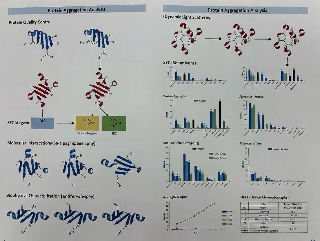
DLS vs SEC for Protein Aggregation: A 2024 Guide for Biopharmaceutical Scientists
This comprehensive guide compares Dynamic Light Scattering (DLS) and Size Exclusion Chromatography (SEC) for analyzing protein aggregation, a critical parameter in biopharmaceutical development.
DLS vs SEC for Protein Aggregation: A 2024 Guide for Biopharmaceutical Scientists
Abstract
This comprehensive guide compares Dynamic Light Scattering (DLS) and Size Exclusion Chromatography (SEC) for analyzing protein aggregation, a critical parameter in biopharmaceutical development. We cover foundational principles, detailed methodologies, and troubleshooting for both techniques. The article provides direct comparative analysis on key metrics like size resolution, sensitivity, and sample requirements, empowering researchers to select and optimize the right method for their specific application, from early-stage formulation to final product quality control.
Understanding Protein Aggregation: Why DLS and SEC Are Essential Analytical Tools
Protein aggregation is a critical physicochemical degradation pathway in biopharmaceuticals, posing significant challenges to drug efficacy and safety. Within the broader research context comparing Dynamic Light Scattering (DLS) and Size Exclusion Chromatography (SEC) for protein aggregation analysis, this whitepaper provides a technical guide to aggregation mechanisms, characterization, and its direct impact on product quality.
Mechanisms of Protein Aggregation
Protein aggregation is a multi-step process initiated by the destabilization of a protein's native conformation. The primary mechanisms include:
- Chemical Instability: Involves covalent modifications such as deamidation, oxidation, hydrolysis, and disulfide bond scrambling. These alterations can reduce conformational stability, promoting partial unfolding and aggregation-prone intermediate states.
- Physical Instability: Involves non-covalent processes driven by thermodynamic factors. This includes:
- Surface-Induced Aggregation: Adsorption to interfaces (air-liquid, solid-liquid) causing denaturation.
- Shaken/Shear-Induced Aggregation: Mechanical stress leading to unfolding.
- Concentration-Dependent Aggregation: Self-association driven by high protein concentration.
- Nucleation-Dependent Polymerization: A process where a slow nucleation phase is followed by rapid growth, characteristic of amyloid fibril formation.
The general pathway proceeds from native monomer → destabilized/unfolded monomer → aggregation-prone intermediate → small soluble oligomers → larger sub-visible aggregates → visible particles/precipitates.
Title: Nucleation-Dependent Aggregation Pathway
Types of Protein Aggregates
Aggregates are classified by size, reversibility, and structure. The table below summarizes key types and their characteristics relevant to analysis.
Table 1: Classification and Properties of Protein Aggregates
| Aggregate Type | Size Range | Reversibility | Structure | Primary Analytical Method |
|---|---|---|---|---|
| Soluble Oligomers | 1 - 100 nm | Often Reversible | Amorphous or Ordered | SEC, AUC, native-PAGE, DLS |
| Sub-visible Particles | 0.1 - 10 μm | Irreversible | Amorphous, Fibrillar | MFI, RMM, Flow Imaging |
| Visible Particles | > 10 μm | Irreversible | Amorphous, Precipitate | Visual Inspection, LM |
| Amorphous Aggregates | Variable | Irreversible | Disordered, Random | SEC, DLS, Spectroscopy |
| Amyloid Fibrils | nm width, μm length | Irreversible | Cross-β-sheet rich | TEM, CD, ThT Fluorescence |
Impact on Drug Efficacy and Safety
Impact on Efficacy
Aggregation directly reduces the concentration of active monomeric protein, diminishing therapeutic activity. Large aggregates can alter pharmacokinetics (e.g., rapid clearance) and hinder delivery. Additionally, aggregates can act as a depot, leading to unpredictable release profiles.
Impact on Safety
Protein aggregates are a major immunogenicity risk factor. They can break immune tolerance by providing repetitive epitopes for B-cell activation or acting as adjuvants, potentially leading to Anti-Drug Antibody (ADA) formation. ADAs can neutralize drug activity, alter pharmacokinetics, or cause cross-reactivity with endogenous proteins.
Title: Aggregate-Induced Immunogenicity Pathway
Analytical Techniques: DLS vs. SEC in Aggregation Analysis
A core thesis in characterization is the complementary use of DLS and SEC.
Table 2: Comparative Analysis of DLS and SEC for Aggregation Assessment
| Parameter | Dynamic Light Scattering (DLS) | Size Exclusion Chromatography (SEC) |
|---|---|---|
| Principle | Measures fluctuations in scattered light to determine hydrodynamic radius (Rh) via diffusion coefficient. | Separates species based on hydrodynamic volume as they elute through a porous column. |
| Size Range | ~0.3 nm to 10 μm (theoretically). Best for 1 nm - 1 μm. | Limited by column pore size. Typically resolves ~1 nm - 50 nm radius. |
| Key Output | Intensity-based size distribution (Z-average, PDI). | Concentration-based profile (UV/VIS/RI signal). |
| Advantages | Fast, minimal sample prep, measures in native formulation, detects large aggregates/oligomers. | Gold standard for quantifying soluble %monomer/aggregate, high resolution for small oligomers. |
| Limitations | Low resolution, biased towards large particles (intensity ∝ d⁶), cannot separate species. | Potential column interactions, shear stress, dilution, may miss large aggregates stuck in column. |
| Role in Thesis | Primary tool for early, formulation-stage screening and detecting large/ subvisible aggregates. | Primary tool for precise quantification of soluble aggregates for lot release and stability studies. |
Experimental Protocols
Protocol 1: High-Throughput DLS Screening for Aggregation Propensity
- Sample Prep: Dilute protein to target concentration (e.g., 1 mg/mL) in formulation buffer. Filter using a 0.1 μm syringe filter (for small volume) or 0.22 μm filter.
- Instrument Setup: Load sample into low-volume quartz cuvette or 96-well plate. Equilibrate to measurement temperature (e.g., 25°C) for 2 minutes.
- Measurement: Run measurement with automatic attenuation selection. Perform minimum 10-15 runs per sample.
- Data Analysis: Review correlation function fit. Report Z-average diameter (d.nm) and Polydispersity Index (PDI). Analyze intensity size distribution for peak populations.
Protocol 2: Quantitative SEC for Monomer Purity
- Column Equilibration: Equilibrate SEC column (e.g., TSKgel UP-SW3000) with mobile phase (e.g., PBS + 200 mM arginine, 0.02% azide) at 0.5 mL/min until stable baseline.
- System Calibration: Inject protein standard mix to confirm resolution and retention time.
- Sample Analysis: Inject 10-100 μL of sample at 0.5-1 mg/mL. Run isocratic elution at 0.5 mL/min with UV detection at 280 nm.
- Data Integration: Integrate peak areas. Calculate % monomer as (Monomer Peak Area / Total Peak Area) * 100.
The Scientist's Toolkit: Key Research Reagent Solutions
Table 3: Essential Materials for Protein Aggregation Studies
| Item | Function / Role | Example/Note |
|---|---|---|
| SEC Columns | High-resolution separation of monomer from soluble aggregates. | TOSOH TSKgel (e.g., G3000SWxl) or Waters columns. Use with appropriate mobile phase additives. |
| DLS Plates/Cuvettes | Low-volume, disposable containers for light scattering measurements. | Malvern ZEN0040 45μL micro cuvette or Black 96-well plates with clear bottom. |
| Formulation Buffers | To screen excipient effects on aggregation stability. | Polysorbate 20/80, Sucrose, Trehalose, Arginine, Histidine buffers at various pH. |
| Chemical Stressors | To induce controlled aggregation for stability studies. | Guanidine HCl, Urea (denaturants); H2O2 (oxidizer); Elevated temperature. |
| Aggregation-Specific Dyes | Detect and characterize amyloid or amorphous aggregates. | Thioflavin T (ThT) for amyloid fibrils; Bis-ANS for exposed hydrophobic patches. |
| Protein Standards | Calibrate SEC columns and validate DLS size measurements. | Gel Filtration Markers Kit (e.g., from Sigma); NISTmAb for monoclonal antibody studies. |
Title: Decision Flow: Choosing DLS vs SEC
Within the comparative analysis of DLS versus Size Exclusion Chromatography (SEC) for protein aggregation studies, DLS provides a critical, non-invasive, and absolute measurement of hydrodynamic diameter and size distribution in native solution conditions. Unlike SEC, which separates species based on hydrodynamic volume under shear forces and requires column calibration, DLS measures the time-dependent fluctuation of scattered light from particles undergoing Brownian motion, yielding the diffusion coefficient directly. This makes DLS indispensable for detecting large, fragile aggregates and submicron particles that may be lost in SEC columns, though it lacks the resolving power of SEC for mono-disperse mixtures. This whitepaper details the core principles, protocols, and applications of DLS specifically for characterizing protein size and polydispersity in biopharmaceutical development.
Core Theoretical Principles
Brownian Motion and Diffusion
Particles in suspension undergo random Brownian motion. The diffusion coefficient (D) is inversely related to particle size via the Stokes-Einstein equation: D = kT / (3πηd_H) where:
- k = Boltzmann constant
- T = Absolute temperature
- η = Solvent viscosity
- d_H = Hydrodynamic diameter
Light Scattering and Fluctuation
A monochromatic laser illuminates the sample. Scattered light intensity fluctuates over time due to constructive and destructive interference from moving particles. Smaller particles move faster, causing rapid intensity fluctuations.
Autocorrelation Function Analysis
The core of DLS is the calculation of the intensity autocorrelation function (ACF), g²(τ): g²(τ) = 〈I(t) * I(t+τ)〉 / 〈I〉² where I is intensity and τ is delay time. The ACF decays from a value of ~2 to 1; the decay rate is proportional to the diffusion coefficient.
From Correlation to Size Distribution
The normalized field ACF, g¹(τ), is derived from g²(τ) via the Siegert relation. It is fitted using algorithms (e.g., Cumulants, CONTIN, NNLS) to extract a distribution of decay rates (Γ), which are converted to a distribution of diffusion coefficients and, via Stokes-Einstein, to hydrodynamic size.
Key Quantitative Parameters and Data Presentation
Table 1: Core DLS Output Parameters and Interpretation for Protein Samples
| Parameter | Typical Symbol | Definition | Interpretation in Protein Aggregation Context |
|---|---|---|---|
| Z-Average Size | d_H (Z-avg) | Intensity-weighted mean hydrodynamic diameter. | Robust mean size indicator. Sensitive to large aggregates. |
| Polydispersity Index | PDI or PI | Width parameter from Cumulants analysis (μ₂/Γ²). | 0.0-0.05: Monodisperse (e.g., pure mAb). 0.05-0.7: Mid-polydisperse. >0.7: Very broad distribution. |
| Intensity Size Distribution | – | Particle size distribution based on scattered light intensity. | Highly sensitive to large particles (scales with d⁶). A small number of aggregates can dominate. |
| Volume/Number Distribution | – | Derived from intensity using Mie theory. Assumes spherical, known RI. | Caution: Model-dependent. Can underestimate aggregates. Used for qualitative comparison. |
| Peak Analysis | Mode(s) | Identified maxima in the size distribution. | Identifies dominant populations (e.g., monomer at 10 nm, aggregate at 100 nm). |
Table 2: Comparative Metrics: DLS vs. SEC for Protein Aggregation
| Analytical Aspect | Dynamic Light Scattering (DLS) | Size Exclusion Chromatography (SEC) |
|---|---|---|
| Principle | Fluctuations in scattered light (Brownian motion). | Hydrodynamic separation on a porous column. |
| Sample State | Measurement in native buffer, no dilution/concentration. | Often requires buffer exchange, dilution, shear stress. |
| Size Range | ~0.3 nm – 10 μm (ideal: 1 nm – 1 μm). | ~1 – 100 nm (depending on column). |
| Aggregate Recovery | High for large, fragile aggregates. | Potential for column adsorption/filter loss. |
| Resolution | Low. Cannot resolve species with size differences < 2-3x. | High. Can resolve monomer, dimer, trimer. |
| Primary Output | Hydrodynamic diameter, PDI, distribution profiles. | Chromatogram (elution volume), relative quantification. |
| Concentration Sensitivity | Works at low concentrations (≥0.1 mg/mL for proteins). | Requires higher loading (often ≥0.5 mg/mL). |
Experimental Protocols
Protocol: Standard DLS Measurement for Protein Formulations
Objective: Determine the hydrodynamic size distribution and polydispersity of a protein therapeutic candidate.
Materials: (See "Scientist's Toolkit" below) Procedure:
- Sample Preparation:
- Clarify protein solution using a 0.02 μm or 0.1 μm syringe filter (Anotop or similar) or centrifuge at 10,000-15,000 x g for 10-20 minutes.
- Critical: Filter or centrifuge the buffer blank separately using the same method.
- Measurement Setup:
- Equilibrate DLS instrument (e.g., Malvern Zetasizer) at 25°C for at least 15 minutes.
- Use a low-volume disposable cuvette (e.g., 45 μL, Brand ZEN2112) or a quartz cuvette.
- Load clarified buffer as the blank. Perform a measurement to confirm it is free of dust/particulates (count rate < 10 kcps typical).
- Load protein sample (typically 30-50 μL). Ensure no bubbles are present.
- Data Acquisition:
- Set measurement angle to 173° (backscatter, NIBS default) for highest sensitivity and reduced multiple scattering.
- Set automated attenuation selection.
- Set number of runs to 10-15, with duration automatically determined.
- Perform a minimum of 3-5 technical replicates per sample.
- Data Analysis (Cumulants Method Primary):
- Inspect the correlation function: Should be a smooth, single decay for monodisperse samples. Multiple decays indicate polydispersity.
- Record the Z-Average diameter and Polydispersity Index (PDI) from the Cumulants analysis.
- Examine the Intensity size distribution plot. Use the CONTIN or NNLS size distribution report to identify peak modes.
- Always report Z-Avg ± standard deviation and PDI ± SD from replicates.
Protocol: Assessing Thermal Stability via DLS (Melting Point, T_m)
Objective: Identify protein unfolding/aggregation onset temperature. Procedure:
- Prepare sample as in 4.1.
- In software, configure a temperature ramp (e.g., from 20°C to 90°C, with 2-5°C increments).
- Set equilibration time (e.g., 60-120 s) at each temperature.
- Perform DLS measurement at each step, recording Z-Avg and PDI.
- Plot Z-Avg vs. Temperature. The T_m (aggregation) is defined as the temperature at which a sharp, irreversible increase in size is observed.
Visualizations
Diagram 1: DLS Measurement and Analysis Workflow (93 chars)
Diagram 2: Decision Logic: DLS vs SEC for Aggregation (95 chars)
The Scientist's Toolkit: Essential Research Reagent Solutions
Table 3: Key Materials for DLS Protein Analysis
| Item/Reagent | Function & Importance in DLS |
|---|---|
| Disposable Cuvettes (e.g., ZEN2112) | Low-volume, disposable cells to minimize cross-contamination and eliminate cleaning artifacts. Essential for high-throughput screening. |
| Syringe Filters (Anotop, 0.02/0.1 μm) | For sample clarification. Removes dust and large particulates that can invalidate measurements. 0.02 μm is preferred for small proteins. |
| Standard Reference Material (e.g., NIST-traceable latex spheres) | For instrument validation and performance qualification. Confirms accuracy and alignment of the system. |
| Viscosity Standard (e.g., Sucrose/Toluene) | Used to calibrate or verify solvent viscosity settings, critical for accurate size calculation via Stokes-Einstein. |
| Formulation Buffers (PBS, Histidine, etc.) | Must be filtered (0.02 μm) prior to use. Buffer scattering properties (RI, viscosity) are baseline for measurement. |
| Quartz Cuvettes (e.g., Hellma) | Required for measurements at high temperatures (>90°C) or with organic solvents not compatible with disposable plastics. |
| Protein Stability Kits (e.g., ExProt) | Pre-formulated buffers and excipients for systematic screening of formulation conditions using DLS thermal stability assays. |
DLS operates on fundamental principles of Brownian motion and light scattering, providing rapid, non-destructive measurements of hydrodynamic size and polydispersity. Within the thesis of DLS vs. SEC for protein aggregation, DLS's strength lies in its sensitivity to large aggregates and its ability to analyze proteins in their native formulation state without separation forces. While SEC offers superior resolution for quantifying specific oligomers, DLS is the frontline tool for stability assessment, aggregation screening, and characterizing polydisperse systems. The protocols and best practices outlined herein, when combined with the orthogonal data from SEC, form the cornerstone of a robust analytical strategy for ensuring the safety and efficacy of biopharmaceutical products.
Within the analytical framework of protein aggregation analysis, Size Exclusion Chromatography (SEC) stands as a cornerstone technique for separating biomolecules based on their effective size in solution—their hydrodynamic volume. This whitepaper delineates the core principles of SEC, positioning it as a complementary and often orthogonal technique to Dynamic Light Scattering (DLS) in biopharmaceutical research. While DLS provides a rapid, ensemble measurement of size distribution in a native sample, SEC offers high-resolution separation and quantification of individual species (monomer, aggregates, fragments), making it indispensable for purity assessment and stability studies in drug development.
Fundamental Principles: Separation by Hydrodynamic Volume
SEC separates molecules as they pass through a column packed with porous, inert beads (stationary phase). The separation mechanism is based on differential access to the pore network:
- Large molecules (with a hydrodynamic volume larger than the pore size) cannot enter the pores and are excluded. They elute first in the void volume (V₀).
- Intermediate molecules partially access the pore network, leading to delayed elution.
- Small molecules fully access the pores, eluting last at the total volume (Vₜ).
The key parameter is the distribution coefficient, Kd, which relates elution volume (Ve) to void and total column volume: Kd = (Ve - V₀) / (Vₜ - V₀) A molecule's elution volume (Ve) is determined by its Stokes radius (Rh), a measure of hydrodynamic volume, not directly by molecular weight. Calibration with known standards is required to estimate molecular size or weight.
Diagram Title: SEC Separation Mechanism by Hydrodynamic Volume
Comparative Context: SEC vs. DLS for Aggregation Analysis
SEC and DLS provide complementary data in aggregation analysis. The following table summarizes their core attributes.
Table 1: Key Comparison of SEC and DLS for Protein Aggregation Analysis
| Parameter | Size Exclusion Chromatography (SEC) | Dynamic Light Scattering (DLS) |
|---|---|---|
| Primary Measurement | Elution volume (related to Rh) | Fluctuation in scattered light (related to Rh) |
| Separation Capability | Yes, physical separation of species. | No, measures the entire ensemble in the sample. |
| Resolution | High. Can resolve monomer, dimer, oligomers, fragments. | Low. Difficult to distinguish similar sizes or complex mixtures. |
| Sample State | Dilute, filtered. May disrupt weak aggregates. | Near-native, concentrated possible. Non-invasive. |
| Quantification | Direct (peak area) for separated species. | Indirect (intensity/volume distribution). Less accurate for minor species. |
| Key Output | Chromatogram with peaks for each species. | Hydrodynamic radius distribution (intensity/volume/mass). |
| Typical Application | Purity/aggregate quantification for release assays. | Early formulation screening, stability assessment. |
Detailed Experimental Protocol for Analytical SEC
The following protocol is standard for protein aggregation analysis in biopharmaceutical development.
Materials & Instrumentation:
- SEC-HPLC System with isocratic pump, autosampler, column oven, and UV/VIS detector.
- SEC Column: e.g., Tosoh TSKgel UP-SW300, Waters Acquity UPLC BEH200, or comparable.
- Mobile Phase: Typically 0.1-0.2 M sodium phosphate, 0.1-0.3 M sodium chloride, pH 6.8-7.4. Filter (0.22 µm) and degas.
- Protein Standards: Gel Filtration Calibration Kit (e.g., from Cytiva or Sigma-Aldrich).
- Samples: Protein sample at 0.5-2 mg/mL, centrifuged and filtered (0.22 µm).
Procedure:
- System Equilibration: Flush the system with at least 2 column volumes (CV) of mobile phase at the recommended flow rate (e.g., 0.2-0.5 mL/min for analytical columns). Stabilize baseline.
- Void Volume Determination: Inject a high-MW molecule that is totally excluded (e.g., Blue Dextran 2000 kDa). Record the elution volume of the peak center as V₀.
- Column Calibration: Inject individual standards from the calibration kit. Record the elution volume (Ve) for each. Plot log(MW) vs. Ve (or Kd) to create a calibration curve.
- Sample Analysis: a. Set detector wavelength (typically 280 nm for protein). b. Inject sample (typically 5-50 µL). c. Run isocratic elution for 1-2 CV. d. Integrate peaks.
- Data Analysis: Identify peaks based on elution volume relative to standards. Calculate aggregate percentage as (Area of aggregate peaks / Total peak area) x 100%.
Diagram Title: Standard Analytical SEC Workflow
The Scientist's Toolkit: Essential SEC Reagents and Materials
Table 2: Key Research Reagent Solutions for SEC Analysis
| Item | Function & Critical Notes |
|---|---|
| SEC Column | Porous silica or polymeric beads with defined pore size. Selection (e.g., 125Å, 200Å, 300Å) determines separation range. Must be compatible with mobile phase pH. |
| Aqueous Mobile Phase Buffers | Maintain protein stability and prevent non-size interactions. Common: Phosphate Buffered Saline (PBS), Sodium Phosphate + NaCl. Additives (e.g., 5% ethanol) inhibit microbial growth. |
| Protein Standard Kits | Set of globular proteins with known molecular weight. Essential for creating a calibration curve to relate elution volume to hydrodynamic size/MW. |
| Sample Filtration Units (0.22 µm) | Removes particulates that could clog the column. Spin filters are commonly used for small sample volumes. |
| HPLC-grade Water | Used for buffer preparation to minimize UV-absorbing impurities that increase background noise. |
| System Suitability Standards | A well-characterized protein mixture (e.g., monoclonal antibody monomer/aggregate mix) run daily to monitor column performance and system precision. |
Advanced Considerations and Method Development
- Non-Ideal Interactions: Electrostatic or hydrophobic interactions with the stationary phase distort elution. Mitigation includes adjusting ionic strength, pH, or adding modifiers (e.g., arginine).
- Flow Rate Optimization: Lower flow rates improve resolution but increase run time and diffusion. Typical flow rates are 0.2-1.0 mL/min.
- Sample Load: Overloading (by mass or volume) leads to peak broadening and loss of resolution. Must be empirically determined.
- Coupling with Multi-Angle Light Scattering (MALS): SEC-MALS directly determines absolute molecular weight and root-mean-square radius (Rg) independent of elution volume, providing definitive aggregation characterization.
In the critical assessment of protein therapeutics, SEC's principle of separation by hydrodynamic volume provides an indispensable, quantitative, and high-resolution profile of aggregation state. When integrated with the ensemble sizing data from DLS, researchers gain a comprehensive analytical picture—from early-stage formulation screening (DLS) to precise, regulated quality control (SEC). Continuous advancements in column chemistry and coupling with detectors like MALS further solidify SEC's role as a foundational pillar in biopharmaceutical research and development.
The Critical Role of Aggregation Analysis in Biopharmaceutical Development Pipelines
The formation of protein aggregates—from dimers to subvisible and visible particles—poses a significant risk to the safety, efficacy, and stability of biopharmaceuticals. Aggregation analysis is therefore a non-negotiable, critical component throughout the development pipeline, from early candidate selection and formulation development to process optimization, quality control, and regulatory filing. This technical guide frames the discussion within the ongoing research thesis comparing two principal orthogonal techniques: Dynamic Light Scattering (DLS) and Size Exclusion Chromatography (SEC). The selection between these methods, or their strategic combination, is fundamental to developing a robust Control Strategy for a biologic drug product.
The Analytical Challenge: DLS vs. SEC
The core challenge in aggregation analysis is the polydisperse, heterogeneous, and often unstable nature of protein aggregates. No single analytical method provides a complete picture, necessitating an orthogonal approach. DLS and SEC serve as foundational, yet philosophically different, techniques.
Dynamic Light Scattering (DLS) measures fluctuations in scattered light intensity from particles undergoing Brownian motion to derive a hydrodynamic diameter (Z-average) and a polydispersity index (PdI). It is a primary, non-invasive, and absolute size technique requiring no columns or standards. Its strength lies in analyzing native, unfractionated samples in formulation buffers, providing a rapid assessment of overall sample polydispersity. However, it has limited resolution for mixtures and is biased towards larger, more strongly scattering particles.
Size Exclusion Chromatography (SEC) is a high-resolution, separation-based technique that fractionates species based on their hydrodynamic volume as they pass through a porous column matrix. It is typically coupled with UV, fluorescence, or light scattering detectors. SEC provides quantitative, population-based data (e.g., % monomer, % aggregate). Its primary limitation is the potential for column interactions, shear-induced artifacts, or dilution of labile aggregates during separation.
The thesis context posits that DLS is superior for early-stage, high-throughput screening and stability assessment under native conditions, while SEC is indispensable for quantitative release and stability testing once method conditions are rigorously controlled to avoid artifacts.
Quantitative Data Comparison: DLS vs. SEC
Table 1: Core Technical Comparison of DLS and SEC
| Parameter | Dynamic Light Scattering (DLS) | Size Exclusion Chromatography (SEC) |
|---|---|---|
| Measured Principle | Hydrodynamic radius (Rh) via Brownian motion | Hydrodynamic volume via column retention time |
| Sample State | Native, in solution (minimal preparation) | Often requires buffer exchange to mobile phase |
| Key Outputs | Z-average diameter (d.nm), Polydispersity Index (PdI), Intensity/Volume/Number Distributions | Chromatogram with quantified peak areas (% Monomer, % LMW, % HMW) |
| Detection Limit for Aggregates | ~0.1% (by mass) for large aggregates (>100 nm); poor for small oligomers | ~0.1-1% (by mass) for soluble aggregates near monomer size |
| Analysis Time | ~1-3 minutes per sample | ~10-30 minutes per sample (plus column equilibration) |
| Sample Consumption | Low (typically 2-50 µL) | Moderate (typically 10-100 µL) |
| Key Advantage | Rapid, native state, measures wide size range, detects large aggregates/particulates | High resolution, quantitative, separates co-existing species |
| Key Limitation | Low resolution, intensity-weighted bias, sensitive to dust/particulates | Risk of column interactions, shear disruption, non-ideal separation |
Table 2: Application in Biopharmaceutical Development Pipeline
| Development Stage | Primary Aggregation Questions | Preferred Technique(s) & Rationale |
|---|---|---|
| Early Discovery / Candidate Selection | Does the protein exhibit innate aggregation propensity? | DLS: Rapid screening of thermal/chemical stability (e.g., Tm, Tagg). |
| Formulation Development | Which buffer/excipient best suppresses aggregation? | DLS & SEC (orthogonal): DLS for high-throughput stability (e.g., temp-ramp), SEC for quantitative ranking. |
| Process Development | Do purification steps or hold times induce aggregation? | SEC-HPLC: Quantify soluble aggregate levels. DLS/MALS: For absolute size without standards. |
| Drug Product & Fill-Finish | Does freezing, thawing, or shear cause aggregation? | Micro-Flow Imaging (MFI) & DLS: For subvisible/visible particles. SEC for soluble aggregates. |
| QC & Release Testing | Does the product meet pre-defined aggregate specifications? | Validated SEC-HPLC: Required for GMP compliance, precise quantification. |
| Stability Studies | How do aggregate profiles change over time under storage? | SEC-HPLC & DLS: SEC for trend analysis, DLS as a complementary native-state check. |
Experimental Protocols for Key Analyses
Protocol 1: High-Throughput Aggregation Propensity Screening via DLS
Objective: To determine the apparent aggregation temperature (Tagg) and compare stability of different protein candidates or formulations. Materials: Monoclonal antibody (mAb) candidates (1 mg/mL in various buffers), 384-well plate, plate-based DLS instrument (e.g., Wyatt DynaPro Plate Reader). Procedure:
- Sample Prep: Dispense 25 µL of each protein formulation into individual wells of a 384-well plate. Include buffer blanks for background subtraction. Seal plate with optical film.
- Instrument Setup: Set temperature ramp from 20°C to 80°C at a rate of 0.5°C/min. Configure laser power and acquisition time for optimal signal-to-noise.
- Data Acquisition: The instrument automatically measures scattering intensity and correlation function at each temperature step.
- Analysis: Plot scattered light intensity (or derived Rh) vs. temperature. Tagg is identified as the inflection point where a sharp increase in intensity signals rapid aggregate formation.
- Output: Rank-order candidates/formulations by their Tagg; higher Tagg indicates greater conformational stability against aggregation.
Protocol 2: Quantitative Aggregate Profiling by SEC-HPLC with MALS Detection
Objective: To accurately quantify the percentage of high-molecular-weight (HMW) aggregates and low-molecular-weight (LMW) fragments in a final drug product batch. Materials: mAb drug product, SEC mobile phase (e.g., 100 mM sodium phosphate, 150 mM sodium chloride, pH 6.8, 0.02% sodium azide), SEC column (e.g., TSKgel G3000SWxl), HPLC system with UV, MALS, and dRI detectors. Procedure:
- System Preparation: Equilibrate SEC column with mobile phase at 0.5 mL/min for at least 30 minutes until baseline is stable. Normalize MALS/dRI detectors according to manufacturer protocol.
- Sample Preparation: Centrifuge drug product vial at 14,000 x g for 5 minutes to remove any pre-existing particulates. Dilute sample to 2 mg/mL with mobile phase.
- Chromatography: Inject 20 µL of sample. Run isocratic elution at 0.5 mL/min for 30 minutes. Monitor UV absorbance at 280 nm.
- Data Collection & Analysis: UV chromatogram identifies elution peaks. MALS/dRI data provide absolute molecular weight for each peak, confirming aggregate identity (e.g., dimer, trimer).
- Quantification: Integrate peak areas for monomer, HMW, and LMW species. Report %HMW = (AreaHMW / Total Area) x 100%. Validate method for precision, accuracy, and limit of detection/quantitation.
Visualizing the Analytical Decision Pathway
Decision Workflow for Aggregation Analysis
The Scientist's Toolkit: Key Research Reagent Solutions
Table 3: Essential Materials for Aggregation Analysis Experiments
| Item / Reagent | Function / Purpose | Example Product/Criteria |
|---|---|---|
| High-Purity SEC Columns | Separates monomer from aggregates based on size. Critical for resolution and reproducibility. | TSKgel SuperSW series (Tosoh), AdvanceBio SEC columns (Agilent). |
| SEC Mobile Phase Kits | Pre-formulated, consistent buffers minimize column interactions and method variability. | Thermo Scientific SEC Buffer Kits, Waters SEC Mobile Phase Kits. |
| Protein Stability Dyes | Fluorescent dyes (e.g., Sypro Orange) used with DLS/DSF for high-throughput thermal stability screening. | Protein Thermal Shift Dye (Thermo Fisher). |
| Nanoparticle Size Standards | Calibrates and validates DLS instrument performance across a defined size range. | NIST-traceable polystyrene or silica nanospheres. |
| Aggregate Positive Controls | Proteins with known aggregation profiles (e.g., heat-stressed mAbs) used for method development. | In-house stressed samples or commercial reference materials. |
| Low-Protein-Bind Consumables | Minimizes sample loss and false aggregate formation from surface adsorption. | Polypropylene tubes/plates, MAXYMum Recovery vials (Waters). |
| Inline Detectors (MALS, dRI) | Provides absolute molecular weight and concentration without relying on column calibration. | DAWN MALS detector (Wyatt), Optilab dRI (Wyatt). |
| Formulation Excipient Library | A panel of stabilizers (sugars, surfactants, amino acids) for screening aggregation suppressors. | High-purity sucrose, polysorbate 80, L-histidine. |
Aggregation analysis is not a single check-box activity but a strategic, multi-technique endeavor embedded across the biopharmaceutical development pipeline. The DLS-vs-SEC thesis underscores that the techniques are complementary, not competitive. DLS acts as a vigilant, native-state sentinel for rapid risk assessment, while SEC serves as the quantitative workhorse for definitive characterization and compliance. Integrating data from both, alongside other orthogonal methods, builds the profound understanding required to ensure the delivery of safe, stable, and effective biologic medicines to patients. The future lies in advanced, automated platforms that seamlessly combine these principles for real-time, at-line monitoring during bioprocessing.
In protein aggregation analysis research, the strategic choice between Dynamic Light Scattering (DLS) and Size Exclusion Chromatography (SEC) as complementary or standalone techniques forms a foundational thesis. DLS provides rapid, volume-weighted hydrodynamic size distribution in solution, while SEC separates species by hydrodynamic volume, offering mass-based quantification. The core thesis posits that DLS excels as a primary, high-throughput stability screening tool, whereas SEC delivers orthogonal, quantitative validation for critical quality attributes in biopharmaceutical development. Their synergistic use is mandated for regulatory filings, yet strategic deployment depends on the development stage, sample throughput requirements, and the specific aggregation questions being addressed.
Technical Comparison: DLS vs. SEC
The quantitative and operational characteristics of DLS and SEC are compared in the tables below.
Table 1: Core Performance Metrics
| Parameter | Dynamic Light Scattering (DLS) | Size Exclusion Chromatography (SEC) |
|---|---|---|
| Size Range | ~0.3 nm to ~10 µm | ~1 kDa to ~10 MDa (column dependent) |
| Sample Volume | Low (10-50 µL) | Moderate (10-100 µL injection) |
| Analysis Time | Fast (1-3 minutes per sample) | Slow (10-30 minutes per run) |
| Primary Output | Hydrodynamic diameter (Z-average), PDI, intensity-size distribution | Elution profile, molecular weight (via calibration), % monomer/aggregate |
| Key Advantage | No separation, minimal sample prep, measures in formulation buffer | High resolution of coexisting species, quantitative mass-based data |
| Key Limitation | Low resolution for polydisperse samples; intensity bias for large aggregates | Potential column interactions, sample dilution, buffer exchange required |
Table 2: Application-Specific Suitability
| Research Context | Recommended Primary Technique | Rationale |
|---|---|---|
| High-Throughput Formulation Screening | DLS (Standalone) | Rapid assessment of colloidal stability under various conditions. |
| Quantifying <1% High-Molecular-Weight Aggregates | SEC (Primary) | Superior sensitivity and quantification for low-abundance species. |
| Characterizing Subvisible Particles (>1 µm) | DLS (Standalone) | SEC columns typically exclude large particles; DLS range is suitable. |
| Stability Indicating Method for Release | SEC (Primary), DLS (Complementary) | SEC provides quantitative, validated data; DLS offers orthogonal quick check. |
| Analysis of Irreversible Aggregates | DLS (Primary) | SEC may cause column fouling; DLS measures in native state. |
Experimental Protocols
Protocol 1: DLS for High-Throughput Stability Screening
Objective: Rapidly assess the impact of buffer pH and excipients on monoclonal antibody (mAb) colloidal stability. Materials: Purified mAb, 96-well plate, DLS plate reader, formulation buffers. Method:
- Prepare mAb at 1 mg/mL in 20 different formulation buffers (varying pH 5.0-8.0 and excipient types).
- Centrifuge all samples at 15,000 x g for 10 minutes to remove dust.
- Pipette 40 µL of each supernatant into a clean, low-volume 96-well plate.
- Equilibrate plate in instrument to 25°C.
- Perform DLS measurement with 3 acquisitions of 10 seconds each.
- Record Z-average diameter and Polydispersity Index (PDI).
- Data Interpretation: Formulations yielding the smallest Z-average and lowest PDI (<0.1) indicate optimal colloidal stability.
Protocol 2: SEC for Quantitative Aggregate Profiling
Objective: Precisely quantify the monomer and aggregate content of a therapeutic protein product. Materials: HPLC system with UV detector, SEC column (e.g., Tosoh TSKgel G3000SWxl), mobile phase (e.g., 100 mM sodium phosphate, 150 mM NaCl, pH 6.8), protein sample. Method:
- Equilibrate SEC column with mobile phase at 0.5 mL/min until stable baseline.
- Prepare protein sample at 1-2 mg/mL in mobile phase. Centrifuge/filter (0.22 µm).
- Inject 10-20 µL onto the column. Run isocratically at 0.5 mL/min for 30 min.
- Monitor UV absorbance at 280 nm.
- Integrate peak areas for monomer, dimer, and high-molecular-weight (HMW) species.
- Calculate %HMW = (Area of HMW peaks / Total peak area) x 100.
- Calibration: Use a protein standard mix to generate a calibration curve for molecular weight estimation.
Visualizing the Strategic Workflow
The logical decision pathway for choosing between DLS and SEC is outlined below.
Decision Pathway: DLS vs. SEC Selection
The complementary data integration from both techniques is shown in the workflow below.
Complementary DLS-SEC Analysis Workflow
The Scientist's Toolkit: Key Research Reagent Solutions
| Item | Function in DLS/SEC Analysis |
|---|---|
| SEC Columns (e.g., Tosoh TSKgel series) | Silica-based hydrophilic resin for separating biomolecules by size with minimal nonspecific interaction. |
| DLS Quartz Cuvettes (Low Volume) | High-quality, disposable cuvettes for minimizing sample volume and reducing dust/scattering interference. |
| Protein Stability & Aggregation Standards | Monodisperse proteins (e.g., BSA, lysozyme) for instrument performance verification and SEC calibration. |
| SEC Mobile Phase Additives | Buffers with controlled ionic strength (e.g., phosphate, NaCl) and modifiers (e.g., 5% ethanol) to prevent column interactions. |
| Ultracentrifugation Filters (0.1/0.22 µm) | For critical sample clarification prior to DLS or SEC to remove particulates and dust. |
| 96-Well DLS Microplates | Specialized plates with clear, flat-bottom wells for high-throughput DLS screening in plate readers. |
| HPLC-Grade Water & Buffers | Essential for preparing mobile phases to minimize background scattering and UV absorbance noise. |
| Column Storage Solution (0.05% NaN3) | Bacteriostatic agent for long-term SEC column storage to prevent microbial growth and column degradation. |
Step-by-Step Protocols: Best Practices for DLS and SEC in Aggregation Studies
1. Introduction: The Critical Role of Preparation in DLS vs. SEC Analysis
Within the comparative research thesis of Dynamic Light Scattering (DLS) versus Size Exclusion Chromatography (SEC) for protein aggregation analysis, sample preparation is the foundational variable that dictates data integrity. While both techniques analyze hydrodynamic size, their operational principles impose distinct preparation demands. SEC, a separation technique, can tolerate minor particulates but requires precise sample volume and buffer compatibility with the column matrix. DLS, a non-invasive, ensemble measurement in a cuvette, is exquisitely sensitive to any contaminant, dust, or air bubble, as it cannot distinguish between a protein aggregate and a dust particle. Therefore, rigorous preparation—specifically buffer matching, filtration, and concentration optimization—is not merely a recommendation for DLS; it is an absolute prerequisite for generating reliable, publishable data that can be meaningfully correlated with SEC results. This guide details the protocols to achieve this.
2. Core Principles & Quantitative Guidelines
2.1 Buffer Matching and Exchange
The solvent for DLS measurement must be optically clean and have a known, low viscosity. Incompatibility between the sample buffer and the DLS instrument's cleaning solvent or previous sample is a common source of contamination.
- Primary Goal: Use the exact same buffer for the sample and the blank. The blank must be the filtrate passed through the same filter used for the sample.
- Key Consideration: For samples in high-viscosity buffers (e.g., with >10% glycerol or sucrose) or buffers with high salt concentration (>500 mM), viscosity and refractive index corrections are essential for accurate size determination.
Table 1: Buffer Compatibility and Preparation Guidelines for DLS
| Buffer Component | Recommended Maximum Concentration for DLS | Preparation Note | Primary Risk |
|---|---|---|---|
| Glycerol/Sucrose | ≤ 5% (v/v or w/v) | Required for viscosity correction in software. | Increased viscosity inflates apparent size. |
| Salts (e.g., NaCl) | ≤ 200 mM (ideal) | Filter buffer (0.02 µm) before adding to sample. | Can cause salt crystals, scattering artifacts. |
| Detergents (e.g., CHAPS, DDM) | ≥ CMC (Critical Micelle Concentration) | Must be above CMC to prevent protein destabilization. | Micelles contribute to scattering signal. |
| Reducing Agents (DTT, TCEP) | As needed for stability | Prepare fresh; DTT can oxidize and form particles. | Oxidation products create particulates. |
| Imidazole | ≤ 50 mM (from His-tag purification) | Higher concentrations can increase scattering noise. | Contributes to background signal. |
Protocol: Buffer Exchange via Desalting Column for DLS
- Equilibrate a PD-10 or equivalent desalting column with at least 25 mL of your target DLS buffer (pre-filtered through 0.02 µm).
- Apply up to 2.5 mL of your protein sample to the column.
- Elute the protein with 3.5 mL of the target buffer, collecting the colored/opalescent fraction (~1.5 mL).
- Filter the eluted sample immediately as described in Section 2.2.
2.2 Filtration and Clarification
This is the single most critical step to remove dust and pre-existing aggregates that would dominate the DLS signal.
- Filter Selection: Use syringe-driven, low-protein-binding filters.
- Pore Size: 0.02 µm or 0.1 µm for monodisperse proteins. A 0.02 µm filter removes small particulates and micron-scale aggregates but may retain large protein oligomers.
- Process: Always filter the buffer first, then use that filtrate to rinse the cuvette. Filter the sample last. For precious samples, pre-wet the filter with a small volume of buffer to minimize adsorption losses.
Protocol: Standard Filtration for DLS
- Assemble a sterile, low-protein-binding 0.02 µm or 0.1 µm syringe filter.
- Draw up 1-2 mL of your prepared buffer into a clean syringe. Attach the filter and expel the buffer to waste. This pre-wets the filter.
- Draw up another 1-2 mL of buffer, filter it, and use this ultra-clean filtrate to rinse the DLS cuvette three times.
- Draw up your protein sample into a new, clean syringe. Filter the sample directly into the rinsed DLS cuvette, or into a low-binding microcentrifuge tube if concentration measurement is needed first.
2.3 Concentration Optimization
Protein concentration must be tailored to avoid interparticle interference (concentration-dependent aggregation) and ensure a strong signal-to-noise ratio.
Table 2: Protein Concentration Guidelines for DLS Analysis
| Analysis Goal | Recommended Starting Concentration | Rationale |
|---|---|---|
| Size Measurement (Monodisperse) | 0.1 - 0.5 mg/mL | Minimizes intermolecular interactions, provides ideal signal for most instruments. |
| Aggregation Detection | 0.5 - 2.0 mg/mL | Increases signal from low-abundance aggregates, but requires verification of non-concentration-dependent effects. |
| High-Throughput Screening | 0.2 - 0.3 mg/mL | Balances signal quality with material conservation. |
| Critical Rule: | Measure across a dilution series (e.g., 2.0, 1.0, 0.5 mg/mL) to confirm size stability. | If the hydrodynamic radius (Rh) decreases with dilution, the sample is experiencing concentration-dependent aggregation or repulsive interactions. |
3. The Scientist's Toolkit: Essential Research Reagent Solutions
Table 3: Key Materials for DLS Sample Preparation
| Item | Function & Rationale |
|---|---|
| 0.02 µm Anotop or Whatman Syringe Filters | Gold standard for final sample clarification. Removes sub-micron particulates. |
| Low-Protein-Binding Microcentrifuge Tubes (e.g., PP, PMP) | Prevents sample loss and spurious aggregation on container walls. |
| Disposable, Pre-Cleaned DLS Cuvettes (e.g., quartz, glass) | Ensures no carryover contamination; quartz is required for UV analysis in some instruments. |
| Size Exclusion Desalting Columns (e.g., GE PD-10, Zeba Spin) | For rapid buffer exchange into an ideal, low-viscosity DLS buffer. |
| Optically Clean Buffer Components | Use HPLC-grade water and highest purity salts to minimize background scattering. |
| Concentration Measurement Device (Nanodrop, Qubit) | Accurately determine protein concentration post-filtration for dilution series. |
4. Experimental Workflow and Decision Pathway
The following diagram outlines the logical workflow for preparing a sample for DLS analysis within a comparative study context.
Diagram Title: DLS Sample Prep and Quality Control Workflow
5. Conclusion: Integrating Preparation with Analytical Strategy
In the context of a thesis comparing DLS and SEC, standardized, meticulous sample preparation is the linchpin for cross-method validation. A sample prepared using the above guidelines for DLS—effectively matched, filtered, and at an optimized concentration—will not only yield robust DLS data but will also be inherently suitable for subsequent SEC analysis, ensuring that observed differences in aggregation profiles are analytical and not preparative artifacts. Mastery of these foundational steps transforms DLS from a simple size check into a powerful, orthogonal tool for comprehensive protein aggregation analysis.
Within a comparative research thesis on Dynamic Light Scattering (DLS) versus Size Exclusion Chromatography (SEC) for protein aggregation analysis, DLS serves as a critical, rapid technique for assessing hydrodynamic size distribution and aggregation state in solution. This guide details the precise execution of a DLS measurement, focusing on the critical parameters that define data quality and reproducibility.
Core DLS Measurement Parameters & Settings
Optimal parameter configuration is essential for acquiring accurate, meaningful data. Incorrect settings can lead to artifacts or misinterpretation of aggregation states.
Diagram 1: Hierarchical overview of critical DLS measurement parameters.
Key Parameter Tables
Table 1: Standard DLS Parameter Settings for Protein Analysis
| Parameter | Typical Setting | Purpose & Rationale |
|---|---|---|
| Temperature | 25°C (or physiological 37°C) | Controls sample stability and Brownian motion. Must be stable (±0.1°C). |
| Equilibration Time | 60-120 seconds | Ensures thermal homogeneity before measurement. |
| Measurement Angle | 173° (backscatter) or 90° | Backscatter reduces sensitivity to dust/large aggregates and is optimal for concentrated or absorbing samples. |
| Laser Wavelength | 633 nm (He-Ne) or 830 nm (NIR) | NIR minimizes fluorescence and absorption from proteins/buffers. |
| Measurement Duration | 10-30 seconds per run | Balances signal averaging and sample stability. |
| Number of Runs | 5-15 consecutive runs | Provides statistics (mean size, PDI) and checks for time-dependent aggregation. |
| Attenuator | Automated or set for 200-800 kcps | Optimizes count rate to avoid detector saturation or low signal. |
Table 2: Impact of Key Parameter Variations on DLS Results
| Parameter Change | Potential Effect on Apparent Size | Risk of Artifact |
|---|---|---|
| Insufficient Equilibration | Increasing trend over runs | False positive for aggregation. |
| Count Rate Too High | Artificially small size, low PDI | Detector saturation, corrupted correlation. |
| Count Rate Too Low | Noisy correlation function | Unreliable size distribution. |
| Too Few Runs | Poor statistical reliability | Misinterpretation of sample polydispersity. |
Determining the Optimal Run Number
"Run Number" refers to the quantity of discrete, consecutive measurements performed on a single sample aliquot. It is not the number of replicates (separate sample preparations).
Protocol: Establishing Run Number and Data Sufficiency
- Initial Setup: Load sample, set temperature, and allow full equilibration.
- Pilot Measurement: Configure instrument for 10-15 runs of 10-second duration.
- Data Acquisition: Execute the series without pause.
- Analysis:
- Plot the Z-Average Diameter and Polydispersity Index (PDI) versus run number.
- A stable, non-trending Z-Average indicates no time-dependent change (e.g., aggregation, settling).
- Calculate the standard deviation and coefficient of variation (CV%) of the Z-Average across runs.
- For a stable, monodisperse protein standard (e.g., BSA), the CV% of the Z-Average should be < 2%.
- Determination: The sufficient run number is the point after which the mean and CV% stabilize. This is often 5-10 runs. Additional runs enhance statistics for polydisperse samples.
Diagram 2: Logical workflow for determining the optimal DLS measurement run number.
Data Acquisition & Primary Correlation Data
The fundamental output of a DLS instrument is the intensity-intensity time autocorrelation function (ACF), g²(τ).
Experimental Protocol: Raw Correlation Function Acquisition
- Correlator Configuration: The correlator should be set for a sufficient number of delay time channels (τ), typically spanning from microseconds to seconds, to capture the full decay.
- Measurement Execution: For each run, the instrument measures fluctuating scattered light intensity, I(t), and computes: g²(τ) = ⟨I(t)·I(t+τ)⟩ / ⟨I(t)⟩² where ⟨⟩ denotes time average.
- Output: The raw g²(τ) curve is stored for each run. A smooth, mono-exponential decay indicates a monodisperse sample. A stretched or multi-phasic decay indicates polydispersity or aggregation.
- Cumulative Analysis: The software often averages the correlation functions from all specified runs before fitting to improve signal-to-noise.
The Scientist's Toolkit: DLS Research Reagent Solutions
Table 3: Essential Materials for Robust DLS Protein Analysis
| Item | Function & Importance in DLS |
|---|---|
| Size Standard (e.g., 100nm latex beads) | Validates instrument performance, alignment, and data processing parameters. Provides a known reference for size and PDI. |
| Protein Standard (e.g., BSA, IgG) | Monodisperse protein sample to verify buffer compatibility, filter integrity, and measurement protocol. |
| Syringe Filters (0.02µm or 0.1µm, low protein binding) | Critical for removing dust and pre-existing aggregates from solvents and protein samples. Anisopore filters are preferred. |
| Ultra-Pure, Filtered Buffers | All buffers must be filtered through a 0.02µm filter to eliminate particulate background signal. |
| Low-Volume, Disposable Cuvettes (e.g., 12µL, 45µL) | Minimizes sample volume, reduces protein consumption, and prevents cross-contamination. Ensure they are dust-free. |
| Quality Control Sample (Stressed Antibody) | A sample with a known, stable level of aggregation for inter-experiment and inter-instrument comparison. |
Diagram 3: Complementary roles of DLS and SEC workflows in protein aggregation analysis.
Within the broader research thesis comparing Dynamic Light Scattering (DLS) and Size Exclusion Chromatography (SEC) for protein aggregation analysis, SEC-HPLC stands out for its quantitative resolution of monomers, fragments, and oligomers. While DLS excels at rapid, native-state sizing, SEC-HPLC provides a high-resolution, quantitative profile critical for biopharmaceutical characterization. The success of this separation hinges on three pillars of sample preparation: mobile phase selection, column equilibration, and load optimization. This guide details the protocols and rationale behind these critical steps.
Mobile Phase Selection: Composition and Optimization
The mobile phase in SEC-HPLC must maintain protein stability, prevent non-specific interactions with the column matrix, and enable accurate size-based separation.
Key Components and Functions:
| Component | Typical Concentration/Type | Primary Function | Critical Consideration |
|---|---|---|---|
| Buffer Salt | 20-100 mM phosphate, citrate, or Tris | Maintains pH and ionic strength to minimize protein-stationary phase interactions. | Ionic strength >150 mM is often needed to shield electrostatic interactions. |
| Salt Additive | 100-300 mM NaCl or K₂SO₄ | Further reduces ionic interactions between analyte and column. | K₂SO₄ can be more effective than NaCl for some proteins. |
| pH Adjuster | pH 6.0-7.5 (protein dependent) | Maintains protein solubility and stability. | Must be at least 1.0 pH unit away from protein's pI to ensure charge repulsion. |
| Organic Modifier | ≤5% v/v Acetonitrile or Isopropanol | Reduces hydrophobic interactions. | Use sparingly; can denature proteins or alter column bed. |
| Stabilizer/Chelant | 0.1-1 mM EDTA, 5-10% Sucrose | Prevents metal-catalyzed oxidation and stabilizes conformation. | Sucrose can increase viscosity, affecting backpressure. |
Experimental Protocol for Mobile Phase Screening:
- Prepare a candidate buffer at 50 mM strength, pH 7.0 (±0.5 from protein pI).
- Add NaCl to 150 mM as a starting point.
- Filter through a 0.22 µm PVDF or cellulose membrane and degas.
- Inject a standard protein mix (e.g., thyroglobulin, BSA, ribonuclease A) and the target protein.
- Evaluate: Peak symmetry (tailing factor <1.5), recovery (>90% by peak area vs. injection), and separation resolution between monomer and dimer.
- If recovery is low or tailing occurs, systematically adjust: a) Increase salt to 300 mM, b) Adjust pH by ±0.3 units, c) Add 2% isopropanol.
- Finalize the composition that yields optimal recovery, resolution, and run-to-run reproducibility.
Column Equilibration: Protocols for Stability and Reproducibility
Proper equilibration ensures the column is at a consistent, stable state, critical for accurate retention time and aggregation quantitation.
Quantitative Equilibration Criteria:
| Parameter | Target Value | Measurement Method |
|---|---|---|
| Retention Time Stability | ≤±0.1 min variation for main peak | Consecutive injections of a standard. |
| Backpressure Stability | ≤±5% fluctuation from baseline | System pressure monitor. |
| Baseline UV Absorbance | Stable, flat baseline (λ=280 nm) | Observe detector output over time. |
| Theoretical Plates (N) | Consistent with column certificate (±15%) | Calculate from a small molecule standard (e.g., acetone). |
Detailed Equilibration Protocol:
- Connect the new or stored column to the HPLC system with the inlet disconnected.
- Flush with at least 3 column volumes (CV) of deionized water at 50% of the method flow rate to remove storage solvent.
- Connect inlet to the prepared SEC mobile phase.
- Equilibrate with at least 10 CV of mobile phase at the analytical flow rate (e.g., 0.5-1.0 mL/min for a 7.8 x 300 mm column).
- Verify equilibration by making 3-5 consecutive injections of a stable, non-valuable protein standard (e.g., 10 µL of 2 mg/mL BSA).
- The column is considered equilibrated when the retention time of the standard monomer peak varies by less than 0.1 minutes and the peak area varies by less than 2% across three consecutive injections.
- For long-term use, a guard column of identical chemistry is strongly recommended to protect the analytical column.
Load Optimization: Balancing Resolution and Sensitivity
Injection load is a critical yet often overlooked variable. Overloading distorts peaks and reduces resolution, while underloading compromises aggregate detection.
Quantitative Load Optimization Data:
| Column Dimension (ID x L mm) | Typical Pore Size (Å) | Optimal Protein Mass Load* | Optimal Injection Volume* | Impact of Overloading (>2x Optimal Mass) |
|---|---|---|---|---|
| 7.8 x 300 | 150-300 | 50-100 µg | 10-25 µL | Peak fronting, loss of dimer/monomer resolution. |
| 4.6 x 300 | 150-300 | 10-20 µg | 5-10 µL | Increased backpressure, skewed peak shapes. |
| 2.1 x 300 | 150-300 | 1-5 µg | 1-3 µL | Severe loss of efficiency and resolution. |
*For a typical monoclonal antibody (~150 kDa). Values scale with protein molecular weight.
Experimental Protocol for Determining Optimal Load:
- Prepare a concentrated, filtered (0.1 µm) sample of the target protein in the final SEC mobile phase.
- Perform a series of injections at the same volume (e.g., 10 µL) but with increasing protein concentration (e.g., 1, 2, 5, 10 mg/mL).
- In a separate series, inject a constant mass (e.g., 50 µg) in increasing volumes (e.g., 5, 10, 25, 50 µL) by diluting the stock.
- Analyze chromatograms for:
- Retention Time Shift: >0.1 min earlier indicates overloading.
- Peak Asymmetry (As): As >1.5 indicates volume or mass overload.
- Resolution (Rs) between monomer and nearest aggregate: Rs <1.5 indicates compromised separation.
- The optimal load is the maximum mass and volume that does not cause a significant shift in retention time or a reduction in resolution.
The Scientist's Toolkit: Research Reagent Solutions
| Item | Function in SEC-HPLC Sample Prep |
|---|---|
| SEC Column (e.g., TSKgel UP-SW300, AdvanceBio SEC) | Silica- or polymer-based stationary phase with defined pore size for size-based separation of biomolecules. |
| 0.1 µm Ultrafiltration Device (PES or Centrifugal) | Critical for final sample filtration to remove particulates and pre-formed large aggregates that could block the column. |
| HPLC-Grade Salts & Buffers | High-purity chemicals to prepare mobile phase, minimizing UV-absorbing impurities and column contamination. |
| 0.22 µm PVDF Membrane Filters | For mobile phase filtration to remove particles and microorganisms. |
| Protein Stability Additives (e.g., Sucrose, Arginine) | Used in sample buffer to prevent artificial aggregation induced by dilution or handling prior to injection. |
| System Suitability Standard | A stable protein mixture containing monomer and a defined oligomer to validate column performance daily. |
| Guard Column (matched chemistry) | Protects the expensive analytical column from contaminants, extending its lifetime. |
Visualization: SEC-HPLC Optimization Workflow
Diagram Title: SEC-HPLC Method Development & Optimization Workflow
The rigorous optimization of sample preparation for SEC-HPLC—through tailored mobile phases, meticulous equilibration, and precise load determination—generates the high-fidelity aggregation data required for a meaningful comparison with DLS. While DLS offers a rapid, low-consumable assessment of hydrodynamic size in native conditions, the optimized SEC-HPLC method provides a quantitative, resolved profile of co-existing species. This allows the thesis to critically evaluate the strengths (sensitivity to small aggregates, quantitation) and limitations (artifacts, solution conditions) of each technique, guiding scientists toward a complementary analytical strategy for protein aggregation in drug development.
Within the context of a thesis comparing Dynamic Light Scattering (DLS) and Size Exclusion Chromatography (SEC) for protein aggregation analysis, SEC stands out for its unparalleled ability to resolve and quantify distinct oligomeric states. While DLS excels at rapid, native-state size distribution analysis, SEC provides a high-resolution, quantitative profile of monomer, fragments, and aggregates under denaturing or native conditions. This guide details the critical parameters for establishing a robust SEC method, focusing on flow rate, multi-detector setups, and calibration.
Method Foundation: Flow Rate & Column Selection
The flow rate is intrinsically linked to column dimensions and stationary phase. Optimal flow rates balance resolution, analysis time, and shear stress that could disrupt weak aggregates.
| Column Dimension (ID x Length) | Typical Particle Size | Recommended Flow Rate (for proteins) | Impact on Analysis |
|---|---|---|---|
| 7.8 x 300 mm (Analytical) | 5 µm, 10 µm | 0.5 - 1.0 mL/min | Standard for high resolution; longer run times. |
| 4.6 x 300 mm (Narrow-bore) | 3 µm, 5 µm | 0.2 - 0.35 mL/min | Higher sensitivity, lower solvent consumption. |
| 2.1 x 150 mm (UHPLC) | 1.7 µm, 2 µm | 0.1 - 0.25 mL/min | Maximum resolution & speed; higher backpressure. |
Protocol: Determining Optimal Flow Rate for Resolution
- Column Equilibration: Equilibrate the selected SEC column (e.g., 7.8 x 300 mm, 5 µm) with at least 5 column volumes of mobile phase (e.g., PBS, pH 7.4).
- Standard Injection: Inject a protein standard mix containing a monomer and a known aggregate (e.g., BSA monomer and dimer).
- Flow Rate Series: Perform sequential injections at 0.5, 0.75, and 1.0 mL/min.
- Data Analysis: Calculate the resolution (Rs) between the two peaks at each flow rate: Rs = 2*(tR2 - tR1) / (w1 + w2), where tR is retention time and w is peak width at baseline. Select the flow rate providing Rs > 1.5.
Detection Strategies: UV, MALS, and RI
A multi-detector array is essential for comprehensive characterization beyond mere retention time.
| Detector Type | Key Measured Parameter | Role in Aggregation Analysis | Key Advantage | Key Limitation |
|---|---|---|---|---|
| UV (PDA) | Absorbance (280 nm) | Quantification of eluted protein mass per species. | Universal, sensitive, quantitative. | Molar mass independence; requires chromophore. |
| Multi-Angle Light Scattering (MALS) | Absolute Molar Mass | Direct, in-line measurement of molar mass for each eluting species. | Absolute mass without calibration; detects aggregates. | Sensitive to dust; requires precise concentration input. |
| Refractive Index (RI) | Refractive Index Change | Measures concentration of any eluting species; essential for MALS. | Universal detection. | Low sensitivity; sensitive to temperature/flow changes. |
Protocol: Establishing a UV-MALS-RI Method
- System Setup: Connect detectors in series: HPLC → UV detector → MALS detector → RI detector. Ensure all flow cells are compatible and have minimal dead volume.
- Normalization & Alignment: Run a narrow, monodisperse protein standard (e.g., BSA monomer) at the chosen method flow rate. Use MALS software to normalize detector angles and align the volumetric delay between UV, MALS, and RI signals.
- Calibration: Calibrate the MALS detector using pure toluene or a specified standard according to the manufacturer's protocol. Calibrate the RI detector with a known concentration of a standard (e.g., 1 mg/mL BSA).
- Analysis: For each sample, the UV provides the chromatogram, MALS provides absolute molar mass across each peak, and RI provides concentration for mass calculation. The combined data confirms if an early-eluting peak is a true aggregate (high MALS mass) or a non-aggregated conformational variant (similar MALS mass).
Calibration Standards
Calibration relates retention volume to hydrodynamic size. Two primary approaches exist.
| Calibration Standard Type | Purpose | Common Standards | Critical Consideration |
|---|---|---|---|
| Protein-Based (Traditional) | Create a calibration curve of log(Molar Mass) vs. Retention Volume. | Thyroglobulin, IgG, BSA, Ovalbumin, Ribonuclease A, Vitamin B12. | Assumes globular protein shape; inaccurate for unfolded or extended aggregates. |
| Broad Standard + QELS (SEC-MALS) | MALS provides absolute mass; Quasi-Elastic Light Scattering (QELS) module provides hydrodynamic radius (Rh). | Polystyrene sulfonate or Pullulan standards (for non-proteinaceous polymers). | Provides direct Rh measurement for each eluting slice, independent of shape. |
Protocol: Traditional Calibration Curve Generation
- Prepare Standards: Dissolve individual protein standards at 1-2 mg/mL in the SEC mobile phase. Filter (0.1 µm).
- Inject Separately: Inject each standard onto the equilibrated SEC column using the established method.
- Record Retention Time: Note the retention volume at the peak apex for each standard.
- Generate Curve: Plot log10(Molar Mass) vs. Retention Volume. Fit with a 3rd- or 4th-order polynomial. Use this curve to estimate the molar mass of unknown peaks based on their retention.
The Scientist's Toolkit: SEC Aggregation Analysis Essentials
| Research Reagent / Material | Function in SEC Analysis |
|---|---|
| SEC Columns (e.g., TSKgel, Superdex, AdvanceBio) | Porous silica or polymeric beads that separate molecules based on hydrodynamic size. |
| HPLC-Grade Buffers & Salts | Form the mobile phase; must be filtered (0.22 µm) and degassed to prevent column damage & baseline noise. |
| Protein Calibration Standard Kits | Pre-qualified sets of proteins for generating traditional SEC calibration curves. |
| MALS Detector (e.g., Wyatt DAWN, Malvern OMNISEC) | Provides absolute molar mass and size (Rg) for each eluting species without calibration. |
| In-line DLS/QELS Module | Attaches to MALS to measure hydrodynamic radius (Rh) for direct size comparison with DLS data. |
| UV/Vis Photodiode Array Detector | Monitors protein elution via absorbance (280 nm) and checks for spectral purity of peaks. |
| Refractive Index Detector | Provides concentration data for each eluting species, required for accurate MALS analysis. |
| 0.1 µm centrifugal filters | For clarifying protein samples and mobile phases to remove particulates that clog columns or scatter light. |
| Autosampler Vials with Low-Protein-Binding Inserts | Prevents sample loss via adsorption to vial surfaces. |
Diagram: SEC-MALS Workflow for Aggregation Analysis
Diagram: DLS vs SEC Decision Pathway
Dynamic Light Scattering (DLS) and Size Exclusion Chromatography (SEC) are cornerstone techniques for analyzing protein size and aggregation. Within the thesis context of DLS vs. SEC, a critical distinction emerges: DLS excels in high-throughput, label-free, and minimal-sample screening of colloidal stability and hydrodynamic size, while SEC remains the gold standard for quantifying low levels of soluble aggregates with superior size resolution. This guide positions DLS not as a replacement for SEC, but as a powerful complementary tool for rapid, early-stage formulation screening, where speed, low sample consumption, and the ability to analyze opaque formulations are paramount.
Core Principles: DLS for Formulation Screening
DLS measures the time-dependent fluctuations in scattered light from particles undergoing Brownian motion. The diffusion coefficient is extracted via an autocorrelation function, which is then used to calculate the hydrodynamic radius (Rh) via the Stokes-Einstein equation. Key parameters for formulation screening include:
- Z-Average Diameter (d.nm): The intensity-weighted mean hydrodynamic size.
- Polydispersity Index (PdI): A dimensionless measure of sample heterogeneity (0-0.05: monodisperse; 0.05-0.7: mid-range; >0.7: very polydisperse).
- Size Distribution by Intensity: Reveals sub-populations of monomers, oligomers, and aggregates.
High-Throughput DLS Experimental Workflow
The following diagram illustrates a standardized high-throughput (HT) DLS screening protocol for formulation development.
HT-DLS Formulation Screening Workflow
Detailed Experimental Protocol
Aim: To screen 96 different buffer/excipient conditions for their ability to suppress aggregation of a monoclonal antibody (mAb) at 1 mg/mL.
Materials: See "The Scientist's Toolkit" below. Method:
- Plate Design: Prepare a 96-deep well stock plate with varying excipients (e.g., sugars, surfactants, salts) across pH 4.0-8.0. Include replicates and controls.
- Sample Preparation: Using a liquid handling robot, transfer 195 µL of each buffer from the stock plate to a 96-well clear-bottom DLS measurement plate. Add 5 µL of concentrated mAb stock (40 mg/mL) to each well to achieve a final concentration of 1 mg/mL in 200 µL. Seal and mix via orbital shaking (500 rpm, 60 sec).
- Stress Incubation: Incubate the sealed plate at 40°C for 24-72 hours in a thermostatted incubator.
- Clarification: Centrifuge the plate at 3000 × g for 10 minutes to sediment large, sub-visible particles.
- DLS Measurement: Load plate into a plate-based DLS instrument equilibrated at 25°C. Measurement parameters per well: 3-5 acquisitions of 10 seconds each. Automatic attenuation selection.
- Data Processing: Software calculates Z-average, PdI, and intensity size distribution for each well. Results are exported to a spreadsheet for analysis.
Key Data and Performance Metrics
DLS provides rapid, quantitative readouts for comparative screening. The following table summarizes typical data from a hypothetical screening study.
Table 1: Example DLS Data from HT mAb Formulation Screen (Post 72h at 40°C)
| Formulation Condition | pH | Key Excipient | Z-Avg (d.nm) | PdI | Dominant Peak (nm) | Interpretation |
|---|---|---|---|---|---|---|
| Control (Citrate) | 6.0 | None | 12.8 | 0.32 | 10, 120, >1000 | High aggregation |
| Hit 1 | 6.0 | 0.1% PS80 | 10.2 | 0.05 | 10 | Excellent stability |
| Hit 2 | 5.5 | 250mM Sucrose | 10.5 | 0.08 | 10 | Good stability |
| Candidate 3 | 7.0 | 150mM Arg-HCl | 11.1 | 0.15 | 10, 40 | Minor oligomerization |
| Candidate 4 | 8.0 | 100mM NaCl | 14.5 | 0.45 | 10, >1000 | Poor stability |
Table 2: Comparative Analysis: DLS vs. SEC for Formulation Screening
| Parameter | High-Throughput DLS | Analytical SEC |
|---|---|---|
| Sample Throughput | High (96-384 samples/day) | Low (4-12 samples/day) |
| Sample Volume | Low (2-20 µL) | Moderate (50-100 µL) |
| Analysis Time | Fast (1-5 min/sample) | Slow (15-30 min/sample) |
| Size Range | ~1 nm - 10 µm | ~1 nm - ~50 nm (column-dependent) |
| Aggregate Resolution | Low (broad distributions) | High (resolves monomer, dimer, etc.) |
| Quantification | Semi-quantitative (intensity-weighted) | Fully quantitative (mass/UV concentration) |
| Formulation Compatibility | High (tolerates viscosities, some particulates) | Low (column clogging risk) |
| Primary Screening Role | Rapid identification of stable conditions | Confirmatory analysis of % monomer/aggregate |
Integrating DLS and SEC Data in a Development Thesis
The strategic workflow leverages the strengths of both techniques, as shown in the following decision pathway.
Integrated DLS & SEC Analysis Pathway
The Scientist's Toolkit: Essential Research Reagents & Materials
Table 3: Key Reagents and Materials for HT-DLS Formulation Screening
| Item | Function & Importance |
|---|---|
| Multi-Angle DLS Plate Reader | Enables simultaneous DLS measurement from 96- or 384-well plates, enabling true high-throughput. |
| Clear-Bottom Microplate | Low-volume, optical quality plates compatible with DLS measurements. |
| Liquid Handling Robot | Automates precise, reproducible dispensing of buffers, proteins, and excipients. |
| Monoclonal Antibody Standard | A well-characterized protein (e.g., NISTmAb) for system calibration and protocol validation. |
| Polymer Nanosphere Standards | Latex beads of known size (e.g., 50nm, 100nm) for routine instrument performance verification. |
| Phosphate & Citrate Buffer Stocks | For creating a broad-range pH matrix. Must be sterile-filtered (0.22 µm) to remove dust. |
| Excipient Library | Stocks of surfactants (PS80, PS20), sugars (sucrose, trehalose), amino acids (Arg, Gly), and salts. |
| 0.22 µm Spin Filters | For critical clarification of protein and buffer stocks to remove particulate interferents before DLS. |
Thesis Context: The analysis of protein aggregates, sub-visible particles, and monomeric populations is critical for biopharmaceutical development, where stability, efficacy, and immunogenicity are paramount. A central thesis in analytical biophysics contrasts Size Exclusion Chromatography (SEC) with Dynamic Light Scattering (DLS). While DLS excels at rapid, non-invasive size distribution analysis in native solution, SEC provides superior quantitative resolution for separating and quantifying monomer from oligomeric and sub-visible aggregated species, making it the gold standard for release and stability testing. This guide details the application of SEC for this precise quantification.
Principles of SEC for Aggregate Analysis
SEC separates molecules based on their hydrodynamic radius as they pass through a porous stationary phase. Larger aggregates are excluded from pores and elute first, followed by smaller oligomers, and finally the monomeric protein. Coupled with ultraviolet (UV), fluorescence, or multi-angle light scattering (MALS) detection, it provides a quantitative profile of the protein population.
Experimental Protocols for High-Resolution SEC
Protocol 1: Standard Quantitative SEC-UV for Monomer Purity
- Column Selection: Use a column with a pore size appropriate for your protein's molecular weight (e.g., TSKgel SuperSW3000 for mAbs).
- Mobile Phase: Filter (0.1 µm) and degas a solution typically composed of 25-100 mM phosphate or citrate buffer, 100-300 mM NaCl, pH 6.8-7.4. The ionic strength must be sufficient to minimize non-specific interactions with the column matrix.
- System Equilibration: Equilibrate the HPLC system and column at a constant flow rate (e.g., 0.2-0.5 mL/min for analytical columns) until a stable baseline is achieved (~20 column volumes).
- Sample Preparation: Dialyze or buffer-exchange the protein sample into the mobile phase. Centrifuge at 14,000-16,000 x g for 10 minutes to remove any pre-existing large particles. Load 10-100 µg of protein.
- Chromatography: Isocratic elution at constant flow rate and temperature (20-25°C). Monitor elution at 214 nm, 254 nm, and/or 280 nm.
- Data Analysis: Integrate peak areas. The monomer purity is calculated as: (Monomer Peak Area / Total Integrated Peak Area) x 100%. Aggregate percentage is the sum of all peak areas eluting before the monomer peak.
Protocol 2: SEC-MALS for Absolute Size and Mass Determination
- Follow Protocol 1 for sample preparation and chromatography.
- Detector Configuration: Connect in series: SEC column → UV detector → MALS detector (with 16-18 angles) → Refractive Index (RI) detector.
- System Calibration: Normalize MALS detectors using a toluene standard or a monodisperse protein (e.g., BSA). Determine the inter-detector delay volume and band broadening.
- Analysis: Use dedicated software (e.g., ASTRA, OMNISEC) to calculate the absolute molecular weight and root-mean-square radius (Rg) for each elution slice. This confirms the identity of aggregates (dimers, trimers, etc.) independent of elution time.
Data Presentation: SEC vs. DLS for Aggregation Analysis
Table 1: Quantitative Comparison of SEC and DLS for Protein Aggregation Analysis
| Parameter | Size Exclusion Chromatography (SEC) | Dynamic Light Scattering (DLS) |
|---|---|---|
| Primary Measurement | Hydrodynamic radius via chromatographic separation. | Hydrodynamic radius via intensity fluctuations of scattered light. |
| Quantification | Direct and quantitative. Peak area provides exact % monomer, dimer, HMW species. | Indirect and semi-quantitative. Intensity-based sizing; highly biased towards larger aggregates. |
| Size Range | ~1-100 nm (limited by column pore size and non-specific interactions). | ~0.3 nm - 10 µm (broad, but resolution poor for polydisperse samples). |
| Resolution | High. Can resolve monomer, dimer, trimer, and small oligomers. | Low. Provides an intensity-weighted size distribution (z-average); cannot resolve similar sizes. |
| Sample Consumption | Low to moderate (µg to mg). | Very low (µL volume, µg mass). |
| Key Advantage | Quantitative purity analysis; orthogonal method for identity (with MALS). | Rapid, native-state analysis; detects large, sub-visible particles (>1 µm). |
| Key Limitation | Potential for on-column interactions or shear-induced aggregation. | Poor resolution for heterogeneous mixtures; cannot quantify monomer loss directly. |
| Ideal Application | Stability-indicating method for lot release, formulation screening. | Early-stage formulation screening, thermal stability (via melting temperature Tm), detecting large aggregates. |
Table 2: Example SEC-UV Data from a Stressed Monoclonal Antibody Sample
| Peak Identity | Retention Time (min) | Peak Area (% of Total) | Assigned Species (via SEC-MALS) |
|---|---|---|---|
| High Molecular Weight (HMW) Aggregates | 8.2 - 9.5 | 2.1% | Large soluble aggregates (>500 kDa) |
| Dimer | 9.6 - 10.2 | 1.8% | Dimer (~300 kDa) |
| Monomer | 10.3 - 11.5 | 95.5% | Intact mAb (~150 kDa) |
| Low Molecular Weight (LMW) Fragments | 12.0 - 13.0 | 0.6% | Fab fragments, half-antibodies |
Visualizing Method Selection and Workflow
Decision Flow: Choosing Between SEC and DLS (63 chars)
SEC Quantitative Analysis Workflow (49 chars)
The Scientist's Toolkit: Research Reagent Solutions
| Item | Function & Rationale |
|---|---|
| SEC Columns (e.g., TSKgel SuperSW, AdvanceBio, Zenix) | Silica- or polymer-based columns with specific pore sizes designed for biomolecule separation. Minimize non-specific adsorption. |
| HPLC-Grade Buffers & Salts | High-purity components for mobile phase preparation. Minimize UV absorbance and particulates that can clog columns or generate noise. |
| 0.1 µm or 0.22 µm Membrane Filters | For sterilizing and clarifying mobile phases and samples. Removes particulates that can damage the column or create false aggregate signals. |
| Protein Standards for Calibration | Monodisperse proteins (e.g., thyroglobulin, BSA, ribonuclease A) or protein mixture standards to validate column performance and resolution. |
| In-line Degasser or Sonicator | Removes dissolved air from mobile phase to prevent bubble formation in pumps, detectors, and columns, ensuring stable baseline. |
| Multi-Angle Light Scattering (MALS) Detector | Provides absolute molecular weight and size (Rg) for each eluting species, orthogonal confirmation of aggregate identity independent of retention time. |
| Refractive Index (RI) Detector | Measures concentration of eluting species; essential for accurate mass calculation in SEC-MALS and for detecting non-UV absorbing components. |
| Autosampler Vials with Low-Protein-Binding Inserts | Minimizes sample loss due to adsorption to vial walls, critical for accurate quantification of low-concentration species. |
Solving Common Problems: Optimizing DLS and SEC Data Quality and Reliability
Dynamic Light Scattering (DLS) is a cornerstone technique for the hydrodynamic size analysis of proteins and nanoparticles in solution. Within the context of biopharmaceutical development, where protein aggregation is a critical quality attribute, DLS is often employed alongside Size Exclusion Chromatography (SEC). While SEC provides a separation-based, quantitative assessment of monomer and aggregate populations, DLS offers rapid, non-invasive, and absolute size measurement in the native formulation. Its primary advantages are speed, minimal sample consumption, and sensitivity to large, sub-visible aggregates that may be excluded from SEC columns. However, DLS data interpretation is highly susceptible to specific artifacts. This guide provides an in-depth technical framework for troubleshooting three pervasive challenges: dust/particulate artifacts, multiple scattering, and viscosity effects, thereby ensuring data robustness in comparative research against SEC.
Core Challenge 1: Dust and Particulate Artifacts
Dust and large, non-sample particulates are the most common source of error in DLS. A single, large contaminant can dominate the scattered light intensity (which scales with the sixth power of the diameter, ~d⁶), leading to a gross overestimation of the sample's hydrodynamic radius (Rh).
Troubleshooting Protocols & Solutions:
Sample Filtration/Centrifugation: The primary preventive method.
- Protocol: Filter all buffers and protein samples through 0.1 µm or 0.02 µm syringe filters (e.g., Anotop or PVDF membranes) directly into the ultraclean DLS cuvette. For sensitive or adsorbing proteins, low-protein-binding centrifugal filters (100 kDa MWCO) can be used.
- Validation: Measure the filtered buffer blank first. The correlation function should decay completely due to solvent noise, and the derived size distribution should be flat.
Use of Ultraclean Cuvettes:
- Protocol: Use only certified disposable or meticulously cleaned quartz cuvettes. Cleaning involves successive rinses with filtered ethanol, filtered 1M HCl, and copious amounts of filtered, deionized water. Dry in a particle-free environment.
Data Analysis Robustness:
- Software Tools: Utilize the "Dust Reject" or " Spike Removal" algorithms present in modern instruments (e.g., Zetasizer software). These functions identify and ignore large, transient spikes in the scattering intensity.
- Statistical Validation: Perform a minimum of 10-15 measurement repeats. Examine the correlation function overlay; runs contaminated by dust will show clear, irregular deviations. Discard these outliers and rely on the consistent majority.
Table 1: Impact of a Single 1 µm Dust Particle on Apparent DLS Size
| Sample True Composition | Intensity-Weighted Mean (Z-Avg) Without Dust | Intensity-Weighted Mean (Z-Avg) With One 1µm Particle | Distortion |
|---|---|---|---|
| 100% 5 nm Monomer | ~5 nm | > 100 nm | Extreme |
| 95% 5 nm Monomer, 5% 100 nm Aggregate | ~30-50 nm | > 300 nm | Severe |
| 50% 5 nm Monomer, 50% 100 nm Aggregate | ~70-90 nm | ~200-250 nm | Major |
Core Challenge 2: Multiple Scattering
In concentrated or turbid samples, the scattered photon may be scattered multiple times before reaching the detector. This "multiple scattering" shortens the measured decay time of the correlation function, leading to an artificially small apparent Rh.
Troubleshooting Protocols & Solutions:
Sample Dilution: The first-line approach.
- Protocol: Perform a dilution series (e.g., from 10 mg/mL to 0.1 mg/mL for a monoclonal antibody). Plot the measured Z-Average Rh vs. concentration. The plateau region at low concentrations, where Rh becomes independent of concentration, represents the correct value. Extrapolate to zero concentration if necessary.
Specialized Instrumentation:
- Backscatter Detection (NIBS): Use instruments equipped with Non-Invasive Back-Scatter (NIBS) optics. Detecting at an angle of 173° (near 180°) minimizes the path length through the sample, thereby reducing the probability of multiple scattering events.
- Dual-Angle or 3D-DLS: Cross-validate measurements at two angles (e.g., 90° and 173°). Consistent results indicate minimal multiple scattering. 3D-DLS systems use a rapidly rotating detection scheme to average out effects from large, static particles and some multiple scattering artifacts.
Referenced Techniques:
- Protocol for Transmission Measurement: For very turbid samples, measure the optical attenuation (attenuance) at the laser wavelength. A transmission < ~80% indicates a high likelihood of multiple scattering, necessitating dilution or the use of a specialized cell.
Diagram 1: Multiple Scattering Cause and Solutions (87 chars)
Core Challenge 3: Viscosity Effects
The Stokes-Einstein equation (Rh = kT / 6πηD) directly links the calculated Rh to the sample viscosity (η). Using an incorrect viscosity value is a systematic error, shifting all size results.
Troubleshooting Protocols & Solutions:
Accurate Viscosity Measurement:
- Protocol for Using a Viscometer: Measure the absolute viscosity of the exact sample buffer/formulation at the experimental temperature using a capillary or micro-volume rotational viscometer. Do not rely on water viscosity or estimates.
- Protocol for Viscosity Estimation from Composition: For complex formulations, use literature values or predictive models for components like sucrose, glycerol, or arginine. Validate with measurement if possible.
Software Input Verification:
- Protocol: Always manually check and input the correct viscosity and temperature values in the DLS software before measurement. The default is often pure water at 25°C.
Internal Consistency Check:
- Protocol: Measure a known standard (e.g., 100 nm polystyrene latex) in the buffer of interest. If the measured size deviates from the certified value, inaccurate viscosity or refractive index parameters are the likely cause.
Table 2: Impact of Viscosity Error on Calculated Hydrodynamic Radius
| Actual Sample Viscosity (cP) | Viscosity Used in Software (cP) | Apparent Rh vs. True Rh | Error for a 10 nm True Particle |
|---|---|---|---|
| 1.0 (Water, 20°C) | 1.0 | Correct | 10.0 nm |
| 1.5 (Sucrose Buffer) | 1.0 (Water) | Underestimated | Apparent Rh = 6.7 nm |
| 1.0 (Water) | 1.5 (Sucrose Buffer) | Overestimated | Apparent Rh = 15.0 nm |
| 2.0 (High Conc. Excipient) | 1.0 (Water) | Severely Underestimated | Apparent Rh = 5.0 nm |
Integrated Experimental Workflow for Robust DLS Analysis
Diagram 2: Integrated DLS Best Practice Workflow (79 chars)
The Scientist's Toolkit: Essential Research Reagent Solutions
| Item/Category | Example Product/Brand | Function in DLS Troubleshooting |
|---|---|---|
| Syringe Filters | Whatman Anotop 25 (0.02 µm), Millex-GV (0.22 µm PVDF) | Removal of dust and aggregates from buffers and protein samples prior to measurement. |
| Ultraclean Cuvettes | Disposable plastic micro cuvettes (ZEN0040), Quartz cuvettes (Hellma) | Minimizes introduction of particulates from the measurement cell itself. |
| Viscosity Standards | NIST-traceable viscosity oils, Cannon certified standards | Calibration of viscometers used to determine exact buffer viscosity. |
| Nanoparticle Size Standards | NIST-traceable polystyrene latex beads (e.g., 60 nm, 100 nm) | Validation of instrument performance and accuracy of viscosity/RI parameters. |
| Protein Stabilizers | High-purity sucrose, trehalose, histidine, polysorbate 20 | Formulation components that increase viscosity; require precise measurement. |
| Centrifugal Filters | Amicon Ultra (100 kDa MWCO, low-binding) | Gentle concentration and buffer exchange while removing large aggregates. |
| Data Analysis Software | Malvern Zetasizer Pro, Wyatt Dynamics, ALV Correlator | Provides advanced algorithms for dust rejection, distribution analysis, and quality metrics. |
Effective troubleshooting of dust, multiple scattering, and viscosity transforms DLS from a simple "size check" into a robust, orthogonal method for protein aggregation analysis. While SEC excels at quantifying resolved, stable oligomers, a properly executed DLS experiment provides critical information on larger, potentially fragile aggregates and the overall size distribution in a native state. By adhering to the rigorous protocols outlined above, researchers can generate reliable DLS data that meaningfully complements and contextualizes SEC chromatograms, leading to a more comprehensive understanding of protein solution behavior in biopharmaceutical research.
Dynamic Light Scattering (DLS) is a cornerstone technique for analyzing the size distribution and aggregation state of proteins and nanoparticles in solution. A critical output from DLS analysis is the Polydispersity Index (PDI), a dimensionless measure of the breadth of the size distribution derived from the autocorrelation function analysis. This guide provides an in-depth technical framework for interpreting PDI values, delineating when the derived size distribution is trustworthy, particularly within the context of comparative analysis with Size Exclusion Chromatography (SEC) for protein aggregation studies in biopharmaceutical development.
Theoretical Foundation of PDI
The PDI is calculated from the cumulant analysis of the intensity autocorrelation function. The second-order cumulant (µ₂) relates to the variance of the distribution of diffusion coefficients, and PDI is defined as µ₂/Γ², where Γ is the average decay rate. The PDI scale ranges from 0 (perfectly monodisperse) to 1 (highly polydisperse), though values >0.7 indicate a very broad distribution where the intensity-weighted mean size (Z-average) may be unreliable.
Table 1: Standard Interpretation of DLS PDI Values
| PDI Range | Interpretation | Reliability of Z-Average & Distribution |
|---|---|---|
| 0.00 – 0.05 | Near-monodisperse | Excellent. Reported size distribution is highly reliable. |
| 0.05 – 0.10 | Moderately narrow | Good. Z-average is robust; distribution modal peaks are accurate. |
| 0.10 – 0.20 | Moderately polydisperse | Moderate. Z-average is usable but distribution may contain multiple populations. |
| 0.20 – 0.30 | Quite polydisperse | Low. Z-average is approximate; distribution requires validation (e.g., via SEC). |
| >0.30 | Very polydisperse/broad | Unreliable. DLS alone is insufficient; orthogonal methods (SEC, MALS) are essential. |
DLS vs. SEC for Protein Aggregation: Core Context
The reliability of DLS PDI must be evaluated relative to SEC, the gold standard for quantifying soluble aggregates. DLS provides an ensemble, intensity-weighted measurement in a native state but suffers from low resolution for mixtures. SEC separates species by hydrodynamic volume but can suffer from column interactions. A low PDI (<0.1) typically correlates with a single, dominant SEC peak. A high PDI (>0.2) often, but not always, signals multiple species resolvable by SEC. Critically, DLS is sensitive to large aggregates (due to the r⁶ intensity weighting) that may be lost on an SEC column, making them complementary.
Key Experimental Protocols for Validating DLS PDI
Protocol: Standard DLS Measurement for Protein Samples
Objective: To obtain a reliable Z-average diameter and PDI.
- Sample Preparation: Clarify protein solution via centrifugation (e.g., 10,000-14,000 x g, 10 min, 4°C) or filtration (0.1 µm or 0.22 µm syringe filter, protein-compatible).
- Instrument Setup: Equilibrate instrument (e.g., Malvern Zetasizer, Wyatt DynaPro) at 25°C (or relevant temperature) for 10 min.
- Loading: Load 12-50 µL of sample into a low-volume quartz cuvette or a disposable microcuvette. Avoid bubbles.
- Measurement Parameters: Set measurement angle to 173° (backscatter, NIBS) to minimize dust interference. Set automatic attenuation and measurement duration (typically 10-15 runs of 10 seconds each).
- Data Acquisition: Perform a minimum of 3-5 technical replicates. Check correlation function integrity (should be smooth, single exponential decay for monodisperse samples).
- Analysis: Use "General Purpose" or "Multiple Narrow Modes" analysis algorithm in software. Record Z-average (d.nm) and PDI from the cumulant fit.
Protocol: Orthogonal Validation Using SEC-MALS
Objective: To resolve and quantify oligomeric species implied by an elevated PDI.
- Column Selection: Use a suitable SEC column (e.g., Tosoh TSKgel G3000SWxl, Superdex 200 Increase).
- System Setup: Equilibrate SEC system coupled to Multi-Angle Light Scattering (MALS) and Refractive Index (RI) detectors with mobile phase (e.g., PBS + 200 mM NaCl, pH 7.4).
- Sample Injection: Inject 10-100 µL of the same sample prepared for DLS.
- Data Collection: Monitor UV (280 nm), light scattering, and RI signals.
- Analysis: Use ASTRA or equivalent software to determine absolute molar mass and hydrodynamic radius (from MALS) for each eluting peak. Compare the relative peak areas (UV-based) to the intensity-weighted distribution from DLS.
Visualizing the Decision Workflow
Diagram 1: Workflow for Interpreting DLS PDI (76 chars)
Diagram 2: Complementary Nature of DLS and SEC Analysis (78 chars)
Quantitative Comparison Data
Table 2: Case Study – Monoclonal Antibody (mAb) Under Stress Conditions
| Sample Condition | DLS Z-Average (d.nm) | DLS PDI | SEC Main Peak (%) | SEC Dimer (%) | SEC HMW (%) | Trust DLS Distribution? |
|---|---|---|---|---|---|---|
| Native (4°C) | 10.8 ± 0.2 | 0.06 ± 0.02 | 99.5 | 0.4 | 0.1 | Yes |
| Thermal (45°C, 1h) | 11.5 ± 0.3 | 0.15 ± 0.03 | 95.2 | 3.8 | 1.0 | With Caution |
| Agitation Stress | 42.1 ± 15.3 (main peak) | 0.48 ± 0.10 | 92.1 | 2.5 | 5.4* | No – Requires SEC-MALS |
Note: DLS detected a large aggregate population (>100 nm) not fully recovered in SEC-HMW, likely due to column filtration or adhesion.
The Scientist's Toolkit: Research Reagent Solutions
Table 3: Essential Materials for DLS & SEC Protein Aggregation Studies
| Item | Supplier Examples | Function & Critical Note |
|---|---|---|
| DLS Cuvettes | Malvern, Wyatt, Hellma | Disposable or quartz microcuvettes for sample containment. Must be scrupulously clean and free of dust. |
| 0.1 µm Spin Filters | Pall, Millipore | For critical sample clarification prior to DLS, removing particulates that skew results. |
| SEC Columns | Cytiva (Superdex), Tosoh Bioscience, Agilent | High-resolution columns for separating monomer, dimer, and higher-order aggregates. |
| SEC-MALS Mobile Phase | N/A (Lab-prepared) | Typically PBS with added salt (e.g., 150-300 mM NaCl) to minimize non-specific protein-column interactions. Must be filtered (0.1 µm). |
| Protein Standards | Wyatt, Agilent | Monodisperse proteins (e.g., BSA, thyroglobulin) for calibrating SEC retention time and validating DLS instrument performance. |
| Stabilization Buffers | Thermo Fisher, Formulation Libraries | Excipients (e.g., Sucrose, Polysorbate 20) used in control samples to benchmark against stressed samples and ensure sample integrity during measurement. |
Within the ongoing research discourse comparing Dynamic Light Scattering (DLS) and Size Exclusion Chromatography (SEC) for protein aggregation analysis, SEC remains the benchmark for quantifying soluble aggregates and fragments due to its superior resolving power and ability to separate species by hydrodynamic size. However, its accuracy is critically dependent on mitigating three pervasive challenges: non-size interactions, column fouling, and shear-induced aggregation. This guide details advanced troubleshooting strategies to ensure data integrity.
Minimizing Non-Size Interactions
Non-size interactions, such as electrostatic or hydrophobic interactions between the analyte and the column stationary phase, cause skewed elution profiles, leading to inaccurate size and quantitation estimates.
Experimental Protocol for Diagnosing & Mitigating Interactions:
- Diagnosis (Elution Volume Shift Test): Inject the same sample under two different mobile phase conditions.
- Condition A: Standard formulation buffer (e.g., PBS, pH 7.4).
- Condition B: Modified buffer with increased ionic strength (e.g., +150-300 mM NaCl) and/or 2-5% organic modifier (e.g., acetonitrile, isopropanol) for hydrophobic issues.
- Interpretation: A significant shift (>0.2 mL) in the monomer peak elution volume between conditions indicates non-size interactions.
- Mitigation Strategies:
- Adjust Mobile Phase Ionic Strength: Increase salt concentration (e.g., NaCl, KCl) to 150-500 mM to shield electrostatic interactions.
- Modify pH: Adjust pH away from the protein's pI to increase net charge and reduce hydrophobic/ionic interactions. A deviation of ±1 pH unit is a common starting point.
- Add Modifiers: Incorporate arginine (10-50 mM) to suppress protein-surface interactions, or low percentages of organic solvent for hydrophobic effects.
- Select Alternative Stationary Phase: Switch to a column with a different surface chemistry (e.g., more hydrophilic, charged, or inert coatings like diol groups).
Quantitative Data on Interaction Mitigation:
Table 1: Impact of Mobile Phase Modifiers on Apparent Recovery of an IgG1 Monomer
| Mobile Phase Modification | Monomer Peak Recovery (%) | Observed Elution Volume (mL) | Theoretical Elution Volume (mL) |
|---|---|---|---|
| 20 mM Phosphate, pH 6.8 | 78% | 7.8 | 8.2 |
| + 250 mM NaCl | 95% | 8.1 | 8.2 |
| + 20 mM L-Arginine | 98% | 8.2 | 8.2 |
Preventing Column Fouling
Fouling degrades resolution, increases backpressure, and reduces column lifetime. It is caused by adsorption of aggregates, irreversible binding of sticky proteins, or contaminants.
Experimental Protocol for Column Cleaning-in-Place (CIP):
- Prevention: Always use a guard column. Filter all samples (0.1 or 0.22 µm) and buffers (0.22 or 0.45 µm).
- Regular Maintenance Wash: After each batch (or daily), flush with 2-3 column volumes (CV) of a solution stronger than the mobile phase (e.g., 0.1% TFA, 4-6 M Guanidine HCl, or 20-30% Isopropanol).
- Diagnosis of Fouling: Monitor a rise in system backpressure (>10% increase from baseline) or a loss of plate count (>15% decrease).
- CIP Procedure: For severe fouling, reverse-flush the column at a low flow rate (0.2 mL/min) with 5-10 CV of a stringent cleanser (e.g., 0.1-0.5 M NaOH for silica-based columns rated for high pH, or 6 M guanidine HCl + 20% IPA). Re-equilibrate thoroughly with starting buffer (≥10 CV).
Quantitative Data on Fouling Prevention:
Table 2: Effect of Guard Column and CIP on Column Performance Over Time
| Condition | Initial Plate Count (N/m) | Plate Count After 100 Injections | Backpressure Increase (%) |
|---|---|---|---|
| No Guard Column, No CIP | 15,000 | 9,500 | +85 |
| With Guard Column, Weekly CIP | 14,800 | 14,200 | +12 |
Mitigating Shear Effects
The shear forces generated in the HPLC system (e.g., at pump heads, injection valves, and frits) can artificially induce protein aggregation, producing misleading results.
Experimental Protocol for Shear Stress Assessment:
- Control Experiment: Prepare a sample aliquot and keep it stationary in the autosampler vial.
- Shear-Exposed Experiment: Subject an identical aliquot to a simulated "pre-injection" workflow: pass it through an empty piece of PEEK tubing (length and ID mimicking the system path) using the instrument pump at the analytical flow rate (e.g., 0.5 mL/min), collecting it into a vial.
- Analysis: Immediately analyze both aliquots by SEC and by a low-shear method (e.g., DLS or analytical ultracentrifugation).
- Interpretation: A significant increase in high molecular weight (HMW) species in the shear-exposed sample versus the control indicates shear sensitivity.
Mitigation Strategies:
- Use wider internal diameter (ID) tubing (e.g., 0.005" vs. 0.003") for system connections.
- Reduce system flow rate during analysis, if possible.
- Bypass the autosampler and inject manually for critical samples.
- Use low-volume, well-polished injection valves.
- Consider capillary-based or microfluidic SEC systems for extremely shear-sensitive proteins.
The Scientist's Toolkit: Key Research Reagent Solutions
Table 3: Essential Materials for Robust SEC Analysis
| Item | Function / Purpose |
|---|---|
| SEC Columns (e.g., BEH200, AdvanceBio) | Silica-based or polymeric particles with tailored pore sizes for biomolecule separation. |
| Guard Column (matching chemistry) | Protects the expensive analytical column from irreversible fouling and contaminants. |
| HPLC-Grade Salts (e.g., NaCl, KCl) | Modifies mobile phase ionic strength to minimize electrostatic protein-column interactions. |
| L-Arginine Hydrochloride | A versatile mobile phase additive that suppresses protein-surface and protein-protein interactions. |
| Sodium Hydroxide Solution (0.1-0.5 M) | Effective cleaning-in-place (CIP) agent for removing adsorbed biological material (use only with pH-stable columns). |
| Organic Modifiers (e.g., IPA, ACN) | Used in low percentages (2-5%) to mitigate hydrophobic interactions or for column cleaning. |
| Low-Protein Binding Filters (0.1 µm) | For critical sample preparation to remove pre-existing aggregates without sample loss. |
| Wide-ID PEEK Tubing (0.005" or 0.03" ID) | Reduces shear stress in the system flow path compared to standard narrow tubing. |
Visualizing SEC Troubleshooting Workflows
Diagram 1: SEC Troubleshooting Decision Pathway
Diagram 2: DLS vs SEC Comparative Analysis
Within the framework of protein aggregation analysis, Size Exclusion Chromatography (SEC) and Dynamic Light Scattering (DLS) serve as orthogonal, complementary techniques. While DLS excels at rapid, non-invasive size distribution analysis in solution, SEC provides critical quantitative separation and fractionation of monomeric species from aggregates and fragments. This guide details the optimization of SEC resolution—a cornerstone for achieving reliable, reproducible data in biopharmaceutical development, where accurate quantitation of aggregates is a critical quality attribute.
Core Principles: The Relationship Between Resolution, Selectivity, and Efficiency
SEC resolution (Rs) is fundamentally governed by the interplay of column efficiency (N, theoretical plates), selectivity (α, separation factor), and retention (k, capacity factor), approximated for closely eluting peaks by: [ R_s \approx \frac{1}{4} \sqrt{N} \left( \frac{\alpha - 1}{\alpha} \right) \left( \frac{k}{1 + k} \right) ] In ideal SEC, where separation occurs exclusively in the pore volume, k is constrained, making optimization of N and α paramount.
The Optimization Triad: Column, Particle, and Pore
Column Selection: Chemistry and Dimensions
Column chemistry dictates non-specific interactions. Modern columns for proteins employ diol-bonded silica or hybrid particles to minimize ionic and hydrophobic interactions. Column dimensions (length L, internal diameter ID) directly impact efficiency, backpressure, and sample loading capacity.
Table 1: Impact of Column Dimensions on SEC Performance
| Parameter | Increase in Column Length | Increase in Column Diameter |
|---|---|---|
| Resolution (Rs) | Increases ∝ √L | Decreases (due to increased eddy diffusion) |
| Theoretical Plates (N) | Increases linearly with L | Decreases |
| Backpressure (ΔP) | Increases linearly with L | Decreases with (ID)4 |
| Sample Load Capacity | Slight increase | Increases ∝ (ID)2 |
| Mobile Phase Use | Increases | Increases |
Particle Size (dp): The Driver of Efficiency
Smaller particles dramatically reduce band broadening by minimizing the van Deemter C term (mass transfer) and A term (eddy diffusion). Modern UHPLC-SEC utilizes sub-2 µm particles.
Table 2: Effect of Particle Size on Column Performance
| Particle Size (µm) | Typical Plate Height (H, µm) | Max Operating Pressure | Application Context |
|---|---|---|---|
| 10 | ~20-30 | < 600 psi | Traditional, low-pressure LC |
| 5 | ~10-15 | < 1000 psi | High-performance (HPLC) SEC |
| 3 | ~6-9 | < 3000 psi | Ultra-high-performance starts |
| 1.7 | ~4-6 | > 10,000 psi | UHPLC-SEC, maximum resolution |
Pore Geometry and Size Distribution
Pore geometry (shape, connectivity) and size distribution are primary determinants of selectivity (α). A pore size offering a linear selectivity curve across the target molecular weight range is ideal. For monoclonal antibodies (∼150 kDa), 150-300 Å pores are standard. Broader pore distributions can resolve a wider MW range but may exhibit non-linear calibration.
Table 3: Pore Size Selection Guide for Proteins
| Target Protein Size (kDa) | Recommended Pore Size (Å) | Key Separation Goal |
|---|---|---|
| 5 - 50 | 100 - 150 | Peptides, small proteins |
| 50 - 1000 | 150 - 300 | mAbs, large proteins |
| 500 - 10,000 | 300 - 1000 | Oligomers, large aggregates |
| Mixed populations | Mixed-bed or broad-pore | Screening unknown aggregates |
Integrated Experimental Protocol for SEC Method Development
Protocol: Systematic SEC Method Optimization for mAb Aggregation Analysis
1. Column Screening:
- Equilibrate three columns with identical particle size (~3 µm) but varying pore geometry (e.g., 150 Å, 250 Å, 450 Å) in mobile phase (e.g., 50 mM NaPhosphate, 150 mM NaCl, pH 6.8).
- Inject 10 µL of a standard mixture containing mAb monomer, dimer, and high molecular weight (HMW) aggregate.
- Measure retention times and calculate resolution (Rs) between monomer-dimer and dimer-HMW peaks. Select column with highest Rs for critical pair.
2. Flow Rate Optimization (for selected column):
- Perform isocratic runs at flow rates of 0.3, 0.5, 0.75, and 1.0 mL/min.
- Plot plate count (N) vs. linear velocity (u). Identify the flow rate at the apex of the van Deemter curve (maximum N). In practice, a slightly higher flow may be chosen for throughput.
3. Sample Load Optimization:
- Inject the monomer standard at increasing concentrations (0.5, 1, 2, 5 mg/mL) with constant injection volume.
- Plot peak width at half height vs. load. Identify the point where peak broadening deviates from linearity (<10% increase). This is the maximum load for optimal resolution.
The Scientist's Toolkit: Essential Research Reagent Solutions
Table 4: Key Reagents and Materials for High-Resolution SEC
| Item | Function & Critical Specification |
|---|---|
| UHPLC-SEC Column (1.7µm, 250Å) | Provides high-efficiency separation. Key specs: Particle size, pore size, bonding chemistry (e.g., diol), column dimensions (e.g., 4.6 x 150 mm). |
| SEC-Compatible Mobile Phase Buffers | Maintain protein stability and minimize non-specific interactions. Often phosphate or citrate buffers with 100-250 mM NaCl, pH 6.0-6.8. Must be filtered (0.22 µm). |
| Protein Stability Additives | Reduce adsorption and aggregation on-column. Examples: 5% Isopropanol, 100-200 mM Arginine. |
| NIST-traceable MW Standards | For column calibration and validation. A mix of well-characterized globular proteins covering the separation range. |
| Low-Protein-Binding Vials & Filters | Prevent sample loss. Use polypropylene vials and 0.1 µm PVDF filters for mobile phase/sample preparation. |
| In-line DLS Detector | Orthogonal Tool: Coupled post-column to provide direct hydrodynamic radius measurement of each eluting peak, confirming SEC separation fidelity. |
Visualizing the Optimization Workflow and DLS-SEC Synergy
Optimizing SEC resolution through deliberate column selection, reduction of particle size, and matching pore geometry to the target analyte is a systematic process. When executed within the context of protein aggregation analysis, it elevates SEC from a simple purity check to a robust, quantitative technique. Its synergy with DLS—where SEC provides quantitative separation of stable species and DLS probes the native state and detects large, labile aggregates—creates a powerful orthogonal framework essential for confident characterization in biopharmaceutical research and development.
Dynamic Light Scattering (DLS) and Size Exclusion Chromatography (SEC) are cornerstone techniques for characterizing protein size, oligomeric state, and aggregation propensity in biopharmaceutical development. Each technique provides complementary but distinct data, and misinterpretation arises when their inherent limitations and analytical pitfalls are not rigorously considered. This guide details these pitfalls within the experimental workflow, from sample preparation to data interpretation.
Core Principles and Inherent Limitations
Dynamic Light Scattering (DLS)
DLS measures time-dependent fluctuations in scattered light intensity from particles in Brownian motion to derive a hydrodynamic radius (Rh) via the Stokes-Einstein equation. It is a rapid, non-destructive solution-state technique.
Key Pitfalls:
- Intensity-Weighted Bias: The scattering intensity is proportional to the particle diameter to the sixth power (~d⁶). A minute volume fraction of large aggregates can dominate the signal, masking the presence of the main monomeric species.
- Polydispersity & Resolution: DLS has poor resolution for polydisperse samples. It cannot reliably distinguish species with size differences less than a factor of 2-3.
- Viscosity & Refractive Index: Accurate Rh calculation requires precise values for solvent viscosity and sample refractive index, which are often approximated.
Size Exclusion Chromatography (SEC)
SEC separates particles based on their hydrodynamic volume as they elute through a porous column matrix. Detection (typically by UV absorbance) yields a chromatogram where elution volume correlates with size.
Key Pitfalls:
- Non-Size-Based Interactions: Adsorptive or electrostatic interactions between the protein and the column matrix can retard elution, leading to an underestimation of size.
- Shear & Dilution Effects: The flow-induced shear and significant sample dilution during separation can disrupt labile aggregates or, conversely, induce aggregation.
- Column Loading & Resolution: Overloading the column compromises resolution, while underloading challenges detection limits. Calibration is required for absolute size determination.
Quantitative Comparison of Technique Parameters
Table 1: Core Technical Comparison of DLS and SEC
| Parameter | Dynamic Light Scattering (DLS) | Size Exclusion Chromatography (SEC) |
|---|---|---|
| Size Range | ~0.3 nm to 10 μm | ~1 kDa to 10 MDa (column-dependent) |
| Sample Consumption | Low (μL volumes) | Moderate (tens of μL) |
| Measurement Time | Seconds to minutes per sample | Minutes to tens of minutes per run |
| Key Output | Hydrodynamic radius (Rh), polydispersity index (PDI) | Elution profile (relative abundance vs. time/volume) |
| Size Resolution | Low (factor of 2-3) | Moderate to High (dependent on column) |
| Sample State | Native solution (minimal dilution) | After column separation (significant dilution) |
| Primary Weighting | Intensity-weighted (biased toward large particles) | Mass/Concentration-weighted (UV signal ~mass) |
| Artifact Sources | Dust, air bubbles, viscosity errors, multiple scattering | Column interactions, shear forces, dilution |
Table 2: Common Data Interpretation Pitfalls and Cross-Validation Strategies
| Pitfall Scenario | DLS Data Presentation | SEC Data Presentation | Recommended Cross-Check |
|---|---|---|---|
| Presence of large aggregates | High PDI (>0.2); obscure monomer peak. | Potential pre-peak or column entry exclusion. | Use SEC with light scattering (MALS) detection. |
| Protein-column interaction | Normal Rh in buffer. | Asymmetric peak tailing; delayed elution. | Modify mobile phase (ionic strength, pH). |
| Shear-induced aggregation | Normal distribution pre-SEC. | Appearance of aggregates only in SEC chromatogram. | Compare DLS pre- and post-SEC fraction collection. |
| Presence of small oligomers | May be unresolved from monomer. | May co-elute as a shoulder on main peak. | Use high-resolution SEC columns; Analytical Ultracentrifugation (AUC). |
Experimental Protocols for Cross-Validated Analysis
Protocol 4.1: Integrated DLS-SEC Workflow for Aggregation Assessment
Objective: To characterize the size distribution and aggregation state of a therapeutic monoclonal antibody (mAb) candidate.
Materials: See "The Scientist's Toolkit" below. Sample: Purified mAb at 1-10 mg/mL in formulation buffer.
Part A: DLS Analysis (Pre-SEC)
- Sample Preparation: Clarify the mAb solution by centrifugation at 14,000 × g for 10 minutes at 4°C. Filter using a 0.1 μm syringe filter (low protein binding).
- Instrument Setup: Equilibrate DLS instrument at 25°C. Use solvent parameters (viscosity, refractive index) for the specific formulation buffer.
- Measurement: Load 30-50 μL of sample into a low-volume quartz cuvette. Perform a minimum of 10 measurements, each lasting 10-30 seconds.
- Data Acquisition: Record the intensity autocorrelation function. Perform a Cumulants analysis to obtain the Z-average diameter (Z-avg) and Polydispersity Index (PDI). Use a distribution algorithm (e.g., NNLS) to view the intensity size distribution.
Part B: SEC Analysis
- Column Equilibration: Equilibrate the SEC column (e.g., Agilent AdvanceBio SEC 300Å) with at least 5 column volumes of mobile phase (e.g., 100 mM Sodium Phosphate, 150 mM NaCl, pH 6.8) at a flow rate of 0.5 mL/min.
- Sample Injection: Inject 20-50 μg of the same centrifuged/filtered sample used in Part A.
- Chromatography: Isocratic elution at 0.5 mL/min with UV detection at 280 nm.
- Data Acquisition: Record the chromatogram. Integrate peak areas for monomer and aggregates. Calibrate the column using a protein standard mixture to assign apparent molecular weights.
Part C: Post-SEC DLS Analysis (Validation)
- Fraction Collection: Collect the main monomer peak eluting from SEC in a low-binding tube.
- Concentration: Gently concentrate the fraction using a centrifugal filter unit (10 kDa MWCO) to approximate the original sample concentration.
- Repeat DLS: Perform DLS analysis as in Part A on the concentrated SEC fraction.
Interpretation: Compare the DLS size distribution pre- and post-SEC. A clean monomeric peak by SEC coupled with a reduced Rh and PDI in the post-SEC DLS confirms the removal of aggregates and validates that the primary species is monomeric. Discrepancies indicate technique-specific artifacts.
Visualizing Workflows and Relationships
Title: Integrated DLS-SEC Workflow for Aggregation Analysis
Title: Diagnostic Logic for Interpreting DLS-SEC Discrepancies
The Scientist's Toolkit
Table 3: Essential Research Reagent Solutions & Materials
| Item | Function/Benefit | Example (Vendor/Type) |
|---|---|---|
| Low-Protein-Binding Filters | Removes particulates and large aggregates prior to analysis without significant adsorptive loss of protein. | 0.1 μm Millex-VV (PVDF) or Anotop (Alumina) syringe filters. |
| SEC Columns (High-Resolution) | Provides optimal separation of monomer from small oligomers (dimers, trimers). | Agilent AdvanceBio SEC 300Å, 2.7 μm or Tosoh TSKgel G3000SWXL. |
| Aqueous SEC Mobile Phase | Buffer must minimize protein-column interactions (non-specific or ionic) and maintain protein stability. | 100-200 mM phosphate buffer with 100-300 mM NaCl, pH ~6.8. |
| Protein Size Standards | Calibrates SEC columns for apparent molecular weight determination. | Gel Filtration Markers Kit (e.g., from Sigma-Aldrich or Bio-Rad). |
| Low-Binding Microtubes | Prevents loss of protein, especially aggregates, to container walls during sample handling. | Eppendorf Protein LoBind or Axygen Maxymum Recovery tubes. |
| Quartz DLS Cuvettes | Provides optimal optical clarity for light scattering with minimal sample volume. | Hellma 105.251-QS or similar (50 μL volume). |
| Concentrator Devices | For gentle post-SEC fraction concentration to enable follow-up analysis (e.g., DLS). | Amicon Ultra centrifugal filters (appropriate MWCO). |
| Viscosity Standard | Calibrates/verifies DLS instrument settings for accurate hydrodynamic radius calculation. | Certified polystyrene nanospheres or glycerol solutions. |
Head-to-Head Comparison: DLS vs. SEC on Sensitivity, Resolution, and Regulatory Fit
This technical guide provides a comparative analysis of Dynamic Light Scattering (DLS) and Size Exclusion Chromatography (SEC) for characterizing protein aggregates. Within the broader thesis of DLS vs. SEC for protein aggregation analysis, this document focuses on the core parameters of size range capabilities and detection limits, which are critical for selecting the appropriate analytical technique in biopharmaceutical development.
Core Principles: DLS and SEC
Dynamic Light Scattering (DLS) measures temporal fluctuations in scattered light intensity caused by Brownian motion of particles in solution to derive a hydrodynamic diameter via the Stokes-Einstein equation. It is a non-separative, ensemble technique.
Size Exclusion Chromatography (SEC) separates molecules based on their hydrodynamic volume as they pass through a porous stationary phase. Larger molecules elute first, as they are excluded from pores, while smaller molecules penetrate pores and elute later. It is typically coupled to concentration-sensitive detectors (e.g., UV, fluorescence).
Quantitative Comparison: Size Range and Detection Limits
Table 1: Comparative Technical Specifications of DLS and SEC for Aggregate Analysis
| Parameter | Dynamic Light Scattering (DLS) | Size Exclusion Chromatography (SEC) |
|---|---|---|
| Effective Size Range | ~0.3 nm to ~10 μm (1,000 nm) | ~1 nm to ~70 nm (varies with column pore size) |
| Optimal Size Range | 1 nm – 200 nm | 5 nm – 50 nm (for typical protein analysis columns) |
| Mass Concentration Detection Limit (for aggregates) | ~0.1 mg/mL (ensemble average) | ~0.001 mg/mL (species-specific, dependent on detector) |
| Number Concentration Sensitivity | High for large particles (>100 nm); poor for small aggregates in presence of monomers. | Low; mass-sensitive, not particle-counting. |
| Low Abundance Species Detection | Poor (typically requires >5-10% by mass for reliable detection). | Excellent (can detect <0.1% aggregate species with optimized methods). |
| Resolution | Low (provides distribution, not discrete peaks). | High (can resolve monomers, dimers, trimers, HMW species). |
| Sample Throughput | High (minutes per sample, minimal preparation). | Low to Medium (10-30 minutes per run, requires method development). |
Table 2: Advantages and Limitations in Context of Detection
| Aspect | DLS | SEC |
|---|---|---|
| Primary Strength | Rapid, native-state analysis of polydispersity & mean size. | Quantitative separation and quantification of distinct oligomeric states. |
| Key Limitation | Cannot resolve similar sizes; biased towards larger, scattering-intensive particles. | Risk of column interactions, shear-induced artifacts, or on-column dissociation. |
| Sample Requirement | Minimal volume (≈ 2-50 μL), wide buffer compatibility. | Larger volume (≈ 10-100 μL), requires mobile phase compatibility. |
| Aggregate Sub-Type Focus | Best for large, sub-visible particles (>1 μm) and nanoparticles. | Best for soluble, stable low-molecular-weight (LMW) and high-molecular-weight (HMW) aggregates. |
Detailed Experimental Protocols
Protocol 1: Standard DLS Measurement for Protein Aggregates
Objective: Determine the hydrodynamic size distribution and polydispersity index (PdI) of a protein sample.
Materials: Purified protein sample, appropriate buffer, DLS instrument (e.g., Malvern Zetasizer, Wyatt DynaPro), disposable microcuvettes or quartz cuvettes, 0.02 μm or 0.1 μm syringe filters.
Procedure:
- Sample Preparation: Centrifuge the protein solution at 10,000-15,000 x g for 10 minutes or filter through a 0.1 μm (or 0.02 μm for small aggregates) filter to remove dust.
- Instrument Setup: Turn on the instrument and laser, allowing for warm-up (~30 min). Set measurement temperature (typically 20-25°C). Select appropriate material (protein) refractive index (≈1.45) and dispersant (buffer) properties.
- Loading: Pipette 20-50 μL of the clarified sample into a low-volume quartz cuvette or disposable cuvette. Avoid introducing bubbles.
- Measurement: Place the cuvette in the instrument. Set number of runs (10-15) and run duration (typically 10 seconds each). Initiate measurement.
- Data Analysis: The instrument software uses an autocorrelation function to derive the diffusion coefficient and calculates the intensity-based size distribution via the Stokes-Einstein equation. Record the Z-average diameter (intensity-weighted mean hydrodynamic size) and the Polydispersity Index (PdI). A PdI <0.1 is considered monodisperse; >0.3 indicates a broad distribution.
- Volume/Number Distribution: Use software algorithms to convert intensity distributions to volume or number distributions for qualitative assessment of populations.
Protocol 2: Quantitative SEC Analysis for Protein Aggregates
Objective: Separate and quantify monomeric protein from aggregate species (dimers, HMW) and fragments.
Materials: HPLC/UHPLC system with isocratic pump, autosampler, SEC column (e.g., Tosoh TSKgel, Waters Acquity), UV/Vis or fluorescence detector, mobile phase (e.g., 25 mM sodium phosphate, 150 mM sodium chloride, pH 6.8, 0.02% sodium azide), protein standards for calibration.
Procedure:
- Column Equilibration: Install appropriate SEC column (e.g., 300 mm x 7.8 mm for analytical scale). Flush with at least 5 column volumes (≈ 30 mL) of filtered (0.22 μm) and degassed mobile phase at the recommended flow rate (e.g., 0.5-1.0 mL/min). Ensure stable baseline on the detector.
- System Suitability: Inject a standard protein mixture (e.g., thyroglobulin, BSA, ovalbumin) to check resolution, peak symmetry, and plate count.
- Sample Preparation: Centrifuge or filter protein sample (≈ 1-5 mg/mL) to remove particulates. Use same buffer as mobile phase or ensure compatibility to avoid on-column precipitation.
- Injection and Separation: Load 10-100 μL of sample onto the autosampler. Set run time to ensure full elution (typically 15-30 min). Start the run.
- Detection: Monitor elution at an appropriate wavelength (e.g., 280 nm for UV absorption).
- Data Analysis: Integrate peak areas for monomer, aggregates (eluting earlier), and fragments (eluting later). Calculate % aggregate as: (Area of aggregate peaks / Total area of all protein peaks) x 100. Use a calibration curve with protein standards for approximate size assignment.
Visualization of Workflows and Relationships
DLS Data Analysis Workflow
SEC Analytical Workflow
Technique Selection Logic Tree
The Scientist's Toolkit: Essential Research Reagent Solutions
Table 3: Key Materials and Reagents for Aggregate Analysis
| Item | Function in Analysis | Example Vendor/Product |
|---|---|---|
| SEC Columns | Porous stationary phase for size-based separation of biomolecules. Critical for resolution. | Tosoh TSKgel SW/SWxl series; Waters Acquity UPLC BEH; Agilent AdvanceBio. |
| DLS Quality Control Standards | Latex/nanoparticle standards of known size for instrument verification and performance qualification. | Malvern Polystyrene Nanosphere Standards; NIST-traceable size standards. |
| SEC Protein Standards | Mixture of proteins with known molecular weights for column calibration and system suitability tests. | Gel Filtration Calibration Kits (e.g., from Cytiva or Bio-Rad). |
| Mobile Phase Additives | Salts (NaCl, Na2SO4) to control ionic strength; modifiers (arginine) to mitigate protein-column interactions. | High-purity salts (Sigma-Aldrich); L-Arginine HCl. |
| Sample Filters | Remove dust and pre-existing large particulates to prevent artifacts in DLS and SEC column clogging. | PVDF or PES membrane syringe filters (0.1 μm or 0.02 μm pore size). |
| Low Protein-Binding Consumables | Minimize sample loss, especially for low-concentration aggregates, during preparation and transfer. | Polypropylene tubes & tips; specific low-binding microcuvettes for DLS. |
DLS and SEC offer complementary capabilities for protein aggregate analysis. DLS excels in providing a rapid, overall assessment of size distribution and detecting large aggregates/sub-visible particles (size range ~1 nm - 10 μm), but suffers from poor resolution and low sensitivity to minor species. SEC excels in resolving and quantifying specific low-abundance oligomeric states (detection limit <0.1%) within a more limited size range (~1-70 nm), albeit with risks of method-induced artifacts. An integrated analytical strategy employing both techniques is often essential for comprehensive characterization in biopharmaceutical research and development.
The choice between Dynamic Light Scattering (DLS) and Size Exclusion Chromatography (SEC) is pivotal in protein aggregation analysis, a critical quality attribute for biotherapeutics. This whitepaper positions these techniques within a core thesis: SEC is the definitive tool for quantitative, high-resolution separation and absolute quantitation of distinct oligomeric states, while DLS excels as a rapid, sensitive tool for sizing and monitoring aggregation trends in native, undisturbed samples. The complementary yet distinct roles of each technique underpin robust aggregation profiling in research and development.
Core Principles and Quantitative Outputs
Size Exclusion Chromatography (SEC) separates proteins based on hydrodynamic volume as they pass through a porous column matrix. Larger aggregates elute first, followed by monomers and smaller fragments. Its strength lies in quantitative resolution.
Dynamic Light Scattering (DLS) analyzes temporal fluctuations in scattered light from particles undergoing Brownian motion to derive a hydrodynamic size distribution. Its strength lies in native-state sizing and sensitivity to large aggregates.
Table 1: Core Quantitative Outputs Comparison
| Parameter | SEC (with MALS/dRI) | Classic DLS |
|---|---|---|
| Primary Output | Chromatogram (Signal vs. Elution Volume) | Intensity Correlation Function |
| Key Metric | % Area of integrated peaks (e.g., % HMWP, % Monomer, % LMWP) | Z-Average Diameter (d.nm) & Polydispersity Index (PDI) |
| Size Range | ~1-100 nm (column-dependent) | ~0.3 nm - 10 μm |
| Quantitation | Absolute (µg/mL) for separated species via dRI/concentration. | Relative intensity weighting; not mass- or number-based. |
| Aggregate Detection | Resolved peaks for stable oligomers. | Highly sensitive to large, subvisible aggregates. |
| Concentration Requirement | ~0.1-5 mg/mL (injected). | As low as 0.01 mg/mL. |
| Sample Volume | ~10-100 µL (injected). | ~2-50 µL (minimal). |
| Analysis Time | 10-30 minutes per run. | 1-3 minutes per measurement. |
Experimental Protocols for Comparative Analysis
Protocol 3.1: SEC Method for Quantifying Monomer and Aggregates
Objective: To separate and quantify monomeric protein from high- and low-molecular-weight species. Materials: See Scientist's Toolkit. Procedure:
- System Equilibration: Equilibrate SEC column (e.g., AdvanceBio SEC 300Å, 2.7 µm) in mobile phase (e.g., PBS, pH 7.4 + 200 mM NaCl) at 0.35 mL/min for ≥30 minutes until stable baseline.
- Standard Calibration: Inject 10 µL of protein standard mix (e.g., thyroglobulin, BSA, ovalbumin). Record elution volumes to generate a calibration curve.
- Sample Preparation: Centrifuge sample at 14,000 x g for 10 minutes at 4°C to remove particulates. Load into injection vial.
- Sample Analysis: Inject 10 µL of sample (1 mg/mL). Run isocratic elution for 15 minutes. Monitor UV at 280 nm.
- Data Analysis: Integrate peak areas for High-Molecular-Weight Proteins (HMWP), monomer, and Low-Molecular-Weight Proteins (LMWP). Report as percentage of total peak area.
Protocol 3.2: DLS Method for Sizing and Aggregation Trend Analysis
Objective: To determine the hydrodynamic size distribution and detect the presence of aggregates in a native solution. Materials: See Scientist's Toolkit. Procedure:
- Instrument Warm-up: Power on laser and allow to stabilize for 15 minutes.
- Sample Preparation: Filter mobile phase (same as SEC) through 0.02 µm filter. Centrifuge sample at 14,000 x g for 10 minutes. Load 20 µL into ultra-low volume cuvette.
- Measurement Setup: Set temperature to 25°C. Allow 2-minute equilibration.
- Data Acquisition: Perform 10-15 measurements, each 10 seconds in duration.
- Analysis: Use cumulants analysis to obtain Z-Average and PDI. Use intensity-size distribution algorithms (e.g., NNLS) to identify population modes. Report size and % intensity for each peak.
Visualizing Workflows and Data Interpretation
Diagram 1: SEC Quantitative Analysis Workflow (66 chars)
Diagram 2: DLS Sizing and Trend Analysis Workflow (71 chars)
The Scientist's Toolkit: Key Reagents & Materials
Table 2: Essential Research Reagent Solutions
| Item | Function | Typical Example |
|---|---|---|
| SEC Column | Porous matrix for size-based separation. | Agilent AdvanceBio SEC 300Å, 2.7µm, 7.8x300mm. |
| SEC Mobile Phase | Buffered salt solution to maintain protein stability and minimize non-specific interactions. | 50 mM Sodium Phosphate, 150 mM NaCl, pH 7.0. |
| Protein Standards | For SEC column calibration and system suitability. | Thyroglobulin (8.6 nm), BSA (3.8 nm), Ribonuclease A (1.8 nm). |
| DLS Cuvette | High-quality, disposable cell for holding sample with minimal background scattering. | Ultra-low volume (10-50 µL) quartz or disposable plastic cuvette. |
| 0.02 µm Filter | To prepare particle-free buffer for DLS background measurement. | Anotop 10 or similar inorganic membrane syringe filter. |
| Stabilization Buffer | Formulation buffer to prevent artificial aggregation during analysis. | PBS with 0.01% Polysorbate 20. |
| Aggregation Inducer | Positive control for method development (e.g., heat, agitation). | Incubation at 40°C for 24 hours. |
Integrated Data Analysis and Complementary Use
Table 3: Complementary Data from a Stressed Protein Sample
| Analysis | SEC-UV Result | DLS Result | Interpretation Synergy |
|---|---|---|---|
| Native Sample | 99.5% Monomer, 0.5% Dimer | Z-avg: 5.2 nm, PDI: 0.08, Main Peak: 5.0 nm | Excellent agreement on primary monomer size. |
| Heat-Stressed Sample | 92% Monomer, 6% Trimer, 2% >Decamer | Z-avg: 18.7 nm, PDI: 0.35, Peaks: 5.0 nm (95% Int), 45 nm (5% Int) | SEC quantifies stable trimer; DLS detects large, polydisperse aggregates unresolved by SEC. |
| Key Insight | Quantitative: 6% trimer mass concentration. | Sensitive & Native: 5% intensity from large aggregates. | Combined view: Majority is monomer, with small quantifiable oligomer and trace large aggregates. |
Diagram 3: Technique Selection Logic for Aggregation Analysis (73 chars)
Within the thesis of SEC vs. DLS for protein aggregation, neither technique is universally superior. SEC provides the quantitative accuracy and regulatory-accepted data for stable species, while DLS offers unparalleled speed and sensitivity for initial sizing and detecting the onset of aggregation, particularly for large or transient species. A robust analytical strategy leverages DLS for rapid screening and stability trend analysis, followed by SEC for definitive identification and quantitation of resolved species. This synergistic approach ensures a comprehensive understanding of protein aggregation throughout the drug development lifecycle.
This whitepaper serves as a critical technical evaluation within a broader thesis investigating the comparative merits of Dynamic Light Scattering (DLS) and Size Exclusion Chromatography (SEC) for protein aggregation analysis. A pivotal advancement in both techniques is their coupling with advanced detectors for absolute measurements of size and molecular weight (MW) without reliance on column calibration. This guide provides an in-depth analysis of two primary methodologies: SEC coupled with Multi-Angle Light Scattering (SEC-MALS) and DLS, often enhanced by compositional analysis or fractionation modes.
Core Technology Principles
SEC-MALS
- Principle: SEC separates species by hydrodynamic volume. The eluent flows sequentially through a UV/RI detector (for concentration) and a MALS detector. The MALS detector measures the intensity of scattered light at multiple angles, allowing for the absolute determination of molar mass (M) and root mean square radius (Rg) for each elution slice via the Debye plot.
- Strength: Provides resolved, absolute MW and size distributions for individual species in a mixture (monomer, dimer, aggregates). It is the gold standard for quantifying low levels of high molecular weight (HMW) aggregates.
DLS
- Principle: Measures temporal fluctuations in scattered light intensity from a bulk solution to determine the diffusion coefficient (D), which is converted via the Stokes-Einstein equation to the hydrodynamic radius (Rh).
- Strength: Provides a rapid, non-destructive measurement of the average Rh and polydispersity (PdI) in native solution conditions. Advanced implementations like field-flow fractionation coupled with MALS and DLS (FFF-MALS-DLS) add a separation dimension.
- Key Mode: Size Distribution by Intensity: Represents the relative scattering intensity contribution of each size population. Larger aggregates are heavily weighted due to the ~r⁶ dependence of scattering.
Quantitative Comparison Table
Table 1: Comparative Technical Specifications of SEC-MALS and DLS
| Parameter | SEC-MALS | Batch-Mode DLS | FFF-MALS-DLS (Advanced DLS) |
|---|---|---|---|
| Measured Parameter | Molar Mass (Mw), Rg (radius of gyration) | Hydrodynamic Radius (Rh), Polydispersity Index (PdI) | Mw, Rg, Rh (per fraction) |
| Size Range | ~10 kDa – 10⁸ Da, 1 – 200+ nm Rg | ~0.3 nm – 10 μm (Rh) | ~1 kDa – 100 μm |
| Resolution of Mixtures | High (Chromatographic separation) | Low (Bulk measurement) | Medium-High (Flow-based separation) |
| Sensitivity to Aggregates | Extremely high for trace HMW | High, but can be masked by dominant species | High, with separation |
| Sample Concentration | 0.1 – 5 mg/mL (post-column) | 0.01 – 100 mg/mL | 0.01 – 2 mg/mL (injected) |
| Sample Volume | 10 – 100 µL injection | 2 – 50 µL | 10 – 100 µL injection |
| Analysis Time | 20 – 40 minutes | 1 – 5 minutes | 30 – 60 minutes |
| Key Output | Chromatograms with absolute Mw/Rg per slice | Intensity-based size distribution, PdI | Fractograms with Mw, Rg, Rh per slice |
Table 2: Performance in Protein Aggregation Analysis
| Analysis Goal | Recommended Technique | Critical Reasoning |
|---|---|---|
| Quantifying % HMW Aggregates | SEC-MALS | Provides direct, quantitative mass concentration of aggregates separate from monomer. |
| Monitoring Aggregation Kinetics | DLS | Rapid, low-volume measurements ideal for time-course studies (e.g., thermal stress). |
| Characterizing Large/Sub-visible Particles | DLS or FFF-MALS-DLS | DLS for quick assessment; FFF-MALS-DLS for resolving complex mixtures. |
| Determining Absolute MW of Unknowns | SEC-MALS | Direct measurement independent of shape or column calibration. |
| Assessing Native State Size | DLS | Measurement in solution without dilution or column interactions. |
Experimental Protocols
Protocol 1: SEC-MALS for Monomer and Aggregate Quantification
Objective: To separate and absolutely quantify the molecular weight and mass concentration of monomeric and aggregated protein species.
- System Setup: Connect an SEC system (HPLC/UPLC) to sequential detectors: UV (280 nm), MALS, and RI.
- Column Equilibration: Equilibrate a suitable SEC column (e.g., AdvanceBio SEC 300Å) with mobile phase (e.g., PBS, pH 7.4) at 0.5 mL/min until stable baseline.
- Normalization & Calibration: Perform a MALS detector normalization using a monodisperse protein standard (e.g., BSA). Calibrate the RI detector response and determine the inter-detector delay volume using a narrow MW standard.
- Sample Preparation: Centrifuge protein sample (2-5 mg/mL) at 14,000xg for 10 min. Load 50 µL of supernatant.
- Data Acquisition & Analysis: Run isocratic elution. Use ASTRA or equivalent software to calculate absolute Mw and Rg for each elution slice. Integrate peaks to determine mass concentration from the RI or UV signal.
Protocol 2: DLS for Hydrodynamic Size and Stability Screening
Objective: To determine the hydrodynamic size distribution and polydispersity of a protein sample under native conditions.
- Instrument Preparation: Start the DLS instrument (e.g., Malvern Zetasizer) and allow laser warm-up (15 min). Set temperature to 25°C.
- Cell Cleaning: Rinse a disposable microcuvette with filtered buffer, then with filtered deionized water. Dry with clean air.
- Sample Loading: Load 20-50 µL of filtered (0.1 µm or 0.02 µm syringe filter) protein sample (0.1-1 mg/mL) into the cuvette, avoiding bubbles.
- Measurement Settings: Set measurement angle to backscatter (173°), automatic attenuation selection, and 3-12 runs per measurement.
- Data Acquisition: Run measurement. Software (e.g., ZS Xplorer) calculates the correlation function, fits it to obtain the diffusion coefficient, and reports Rh distribution and PdI.
Visualizations
Title: SEC-MALS Instrumental Workflow
Title: DLS Principle: From Fluctuations to Size
Title: Role of This Comparison in Broader Thesis
The Scientist's Toolkit: Essential Research Reagent Solutions
Table 3: Key Materials for SEC-MALS and DLS Experiments
| Item | Function & Importance | Typical Example(s) |
|---|---|---|
| SEC Columns | Separates proteins by hydrodynamic size. Pore size selection is critical for optimal resolution of monomer and aggregates. | AdvanceBio SEC 300Å, TSKgel UP-SW3000, Superdex Increase. |
| MALS-Calibrated Standards | Used to normalize the MALS detector and verify system performance. Must be monodisperse and stable. | Bovine Serum Albumin (BSA), IgM, toluene. |
| Narrow MW Standards | Determines inter-detector delay volume and monitors column performance. | Thyroglobulin, IgG, Ovalbumin, Ribonuclease A. |
| Ultra-Pure Buffers & Salts | Mobile phase preparation. Must be filtered (0.1 µm) to eliminate particulate noise in light scattering. | Phosphate Buffered Saline (PBS), histidine buffer, sodium chloride. |
| Sterile Syringe Filters | Critical for removing dust and large particulates from samples and buffers prior to DLS or SEC-MALS. | 0.1 µm or 0.02 µm PES or Anotop filters. |
| Disposable DLS Cuvettes | Minimizes contamination and sample carryover. Essential for high-sensitivity measurements. | ZEN0040 (Malvern) or equivalent quartz microcuvettes. |
| Protein Stability Additives | Used in stress studies to induce or inhibit aggregation for method validation. | Arginine, surfactants (PS20, PS80), salts. |
This whitepaper provides a technical guide to implementing Dynamic Light Scattering (DLS) and Size Exclusion Chromatography (SEC) for protein aggregation analysis within the rigorous framework of ICH Q2(R1) “Validation of Analytical Procedures” and ICH Q6B “Specifications: Test Procedures and Acceptance Criteria for Biotechnological/Biological Products.” Within the broader research thesis comparing DLS vs. SEC, compliance with these guidelines is not optional but a fundamental requirement for drug development.
Core Regulatory Principles & Mapping to Techniques
ICH Q2(R1) outlines validation characteristics for analytical procedures. ICH Q6B specifically addresses setting specifications for biological products, emphasizing the criticality of assessing size variants and aggregates. The application of these guidelines differs for DLS (often a characterization tool) and SEC (a purity/impurity quantitation method).
Table 1: Mapping of ICH Q2(R1) Validation Characteristics to DLS and SEC
| Validation Characteristic | Dynamic Light Scattering (DLS) | Size Exclusion Chromatography (SEC) |
|---|---|---|
| Accuracy | Not typically applicable for particle size distribution. Verified using certified nanosphere standards (e.g., NIST-traceable latex beads). | Required. Assessed by recovery of spiked protein aggregates or monomers from a mixture or compared to a known reference material. |
| Precision | ||
| - Repeatability | Essential for intensity-based hydrodynamic radius (Rh) and % Polydispersity Index (%PDI). Multiple measurements of the same sample. | Critical for % aggregate reporting. Multiple injections of the same sample preparation. |
| - Intermediate Precision | Required for method robustness. Different days, analysts, instruments. | Required for method transfer. Different days, analysts, columns (same type), instruments. |
| Specificity | Limited. Identifies presence of particles of different sizes but cannot distinguish protein aggregates from silicone oil droplets or air bubbles. Sample filtration is critical. | High. Ability to separate monomer from dimer, HMW aggregates, and fragments. Confirmed by orthogonal methods (e.g., online MALS, native MS). |
| Detection Limit (LOD) / Quantitation Limit (LOQ) | LOD is relevant (~0.1-1% for large aggregates by intensity). Not a quantitative technique for low-level aggregates. | Required for reporting minor species. LOQ for HMW aggregates is typically established (e.g., 0.1% - 0.5%). Determined via signal-to-noise or precision-accuracy profile. |
| Linearity & Range | Not applicable for direct aggregation quantitation. The intensity-weighted distribution is non-linear in concentration. | Required for quantitative assays. Established for monomer and aggregate peaks across a specified concentration range (e.g., 0.5 - 5 mg/mL). |
| Robustness | Evaluated by deliberate variations: sample equilibration time, temperature control, number of measurements, attenuation setting. | Systematically tested via variations: mobile phase pH (±0.1), ionic strength, flow rate (±10%), column temperature, injection volume. |
Table 2: ICH Q6B Considerations for Aggregate Analysis
| ICH Q6B Element | Implication for DLS | Implication for SEC |
|---|---|---|
| Choice of Test Procedure | Supports "multiple analytical techniques." Ideal for early screening, formulation stability, and subvisible particle assessment. | Often the principal method for release and stability testing of drug substance/product for soluble aggregate quantitation. |
| Reference Standards | Use of USP/ISO particle size standards for system suitability. Protein controls for method performance. | Use of well-characterized protein aggregate/monomer mixtures for system suitability. Primary reference standard for identification. |
| Setting Acceptance Criteria | Criteria may be set for mean Rh (Z-average) and %PDI (e.g., PDI < 0.2 for monodisperse) as characterization limits. | Quantitative acceptance criteria for HMW aggregates and LMW species are set (e.g., HMW ≤ 1.0% for release). |
| Validation of Comp. Methods | Required if DLS data is used in a comparative or trending capacity (e.g., for stability). | Always required for a regulatory submission as a control procedure. |
Detailed Experimental Protocols for Validation
Protocol 1: SEC-HPLC Method Validation for Aggregate Quantitation (Per ICH Q2(R1))
- Objective: To validate a SEC method for the separation and quantitation of monomer, high-molecular-weight (HMW) aggregates, and low-molecular-weight (LMW) fragments of a monoclonal antibody.
- Materials: See "The Scientist's Toolkit" below.
- Method:
- System Suitability: Prior to validation runs, inject a system suitability sample (a stressed mAb sample generating consistent low-level aggregates). Criteria: Resolution between monomer and dimer ≥ 1.5, %RSD of retention time for monomer peak ≤ 2.0% over six injections.
- Specificity/Forced Degradation: Analyze samples subjected to stress conditions: heat (e.g., 40°C for 2 weeks), agitation, pH shift. Compare chromatograms to unstressed control. Confirm peak identity with SEC-MALS.
- Linearity & Range: Prepare sample solutions at a minimum of five concentrations spanning the intended range (e.g., 0.5, 1.0, 2.0, 4.0, 5.0 mg/mL). Inject in triplicate. Plot peak area response vs. concentration for monomer and aggregate peaks. Calculate correlation coefficient (R² > 0.998).
- Accuracy/Recovery: Prepare samples with known ratios of monomer and aggregate (via spiking or mixing). Perform assay and calculate % recovery for each species. Target recovery: 95-105%.
- Precision:
- Repeatability: Six independent preparations of the same sample (e.g., 2 mg/mL) by one analyst. Calculate %RSD for %HMW and %Monomer.
- Intermediate Precision: Repeat repeatability study on a different day, with a different analyst and instrument. Compare results via F-test/t-test.
- LOQ/LOD: Serial dilute a sample with known low-level aggregates. LOQ is the lowest concentration where %RSD ≤ 15% and accuracy 80-120%. LOD is typically a concentration yielding a signal-to-noise ratio of 3:1.
- Robustness: Use a Design of Experiments (DoE) approach, varying key parameters (flow rate ±0.05 mL/min, column temp ±3°C, mobile phase pH ±0.1). Monitor impact on critical resolution and %HMW.
Protocol 2: DLS Method Suitability Testing for Characterization (Informed by ICH)
- Objective: To establish a standardized DLS procedure for characterizing protein size and polydispersity in formulation screening.
- Materials: See "The Scientist's Toolkit" below.
- Method:
- Instrument Qualification: Prior to use, measure a certified NIST-traceable latex standard (e.g., 60 nm ± 3 nm). The measured Z-average must be within the certificate's range.
- Sample Preparation: Centrifuge all protein samples at ≥10,000-15,000 x g for 10-15 minutes to remove dust and large aggregates. Filter buffers through a 0.02 µm or 0.1 µm filter.
- Measurement Parameters:
- Equilibrate sample in cuvette at measurement temperature (e.g., 25°C) for 120-300 seconds.
- Set number of measurements to 10-15 runs per sample.
- Set measurement duration to automatic based on achieving desired correlation function statistics.
- Use appropriate laser attenuation to avoid saturation or low signal.
- Data Acquisition & Analysis: Record the Z-average hydrodynamic radius (Rh in nm) and the Polydispersity Index (PDI). Report the mean and standard deviation of the runs. Use intensity-weighted distribution for size assessment. The volume-weighted distribution can be reviewed but is model-dependent.
- Suitability Criteria: For a stable, monodisperse protein: PDI < 0.1 (highly monodisperse), PDI 0.1-0.2 (near monodisperse). PDI > 0.3 indicates a significant polydisperse population. The Z-average should be consistent with expected molecular weight.
Visualizing the Compliance Workflow
Diagram 1: ICH Compliance Pathway for Aggregation Analysis
The Scientist's Toolkit: Essential Research Reagent Solutions
Table 3: Key Materials for SEC and DLS Compliance
| Item | Function & Rationale | Example/Criteria |
|---|---|---|
| SEC Column | Separates proteins by hydrodynamic volume. Critical for resolution. | TSKgel UP-SW300, Waters Acquity UPLC BEH200, Superdex Increase. Must be specified in method. |
| SEC Mobile Phase | Maintains protein integrity and prevents non-size exclusion interactions. | Typically phosphate or citrate buffer with 150-300 mM NaCl, pH 6.8-7.4. Must be filtered (0.22 µm). |
| NIST-Traceable Size Standards | For DLS instrument qualification and SEC column calibration. | Polystyrene nanospheres (e.g., 10 nm, 60 nm, 100 nm) with certified diameter. |
| Protein System Suitability Control | Verifies daily performance of the entire analytical system (SEC or DLS). | A stressed or formulated protein sample generating a consistent, stable level of aggregates. |
| Ultra-pure Water & Filters | Eliminates particulate interference, crucial for DLS and SEC mobile phase. | 0.02 µm or 0.1 µm Anotop or similar syringe filters for samples. 0.22 µm for buffers. |
| Analytical Balance & pH Meter | Fundamental for precise buffer preparation (ICH Q2(R1) robustness). | Calibrated, qualified equipment with appropriate sensitivity (0.1 mg). |
| Reference Standard | Well-characterized protein for identification and quantitative comparison (ICH Q6B). | Primary reference standard of the drug substance with defined purity. |
| Sample Vials & Vial Inserts | Ensure compatibility and prevent leachables. Critical for automated HPLC systems. | Glass vials with low-protein-binding polypropylene inserts. |
Within the critical research domain of biopharmaceutical development and neurodegenerative disease, protein aggregation analysis is paramount. The central thesis of modern analytical strategy posits that Dynamic Light Scattering (DLS) and Size Exclusion Chromatography (SEC) are not interchangeable tools but complementary techniques anchored in distinct physical principles. Selecting the appropriate instrument requires a rigorous decision framework based on the specific biological question, sample properties, and required data output. This guide provides a structured methodology for this selection, ensuring data integrity and experimental efficiency.
Core Principles: DLS vs. SEC
Dynamic Light Scattering (DLS) measures time-dependent fluctuations in scattered light intensity from particles in Brownian motion to derive a hydrodynamic radius (R~h~) via the Stokes-Einstein equation. It is a population-averaged, non-separative technique ideal for native-state analysis in solution.
Size Exclusion Chromatography (SEC) is a fractionating technique that separates species based on their hydrodynamic volume as they elute through a porous column matrix. It provides a resolution-based profile, quantifying relative amounts of monomer, oligomer, and aggregate.
Decision Framework: A Step-by-Step Guide
The following decision logic should be applied sequentially to determine the optimal primary technique.
Diagram Title: Decision Logic for Choosing Between DLS and SEC
Technical Comparison & Data Presentation
Table 1: Core Technical Specifications and Capabilities
| Parameter | Dynamic Light Scattering (DLS) | Size Exclusion Chromatography (SEC) |
|---|---|---|
| Physical Principle | Fluctuation of scattered light | Hydrodynamic volume separation |
| Sample State | Native, in-solution | Often requires eluent exchange |
| Key Output | Hydrodynamic radius (R~h~), PDI | Elution volume, relative quantitation (%) |
| Size Range | ~0.3 nm to 10 μm | ~1 kDa to 10 MDa (column-dependent) |
| Aggregate Resolution | Low (population-averaged) | High (size-resolved) |
| Concentration Sensitivity | Low (μg/mL) | High (mg/mL typical) |
| Speed of Analysis | Fast (minutes) | Slow (10-30 min/run) |
| Primary Use Case | Stability, native size, aggregation screening | Purity, aggregate quantification, oligomer isolation |
Table 2: Quantitative Performance Metrics for Representative Systems
| Analysis Goal | Model Protein | DLS Result (PDI / R~h~) | SEC Result (% Aggregate) | Recommended Tool |
|---|---|---|---|---|
| Forced Degradation | IgG1 mAb, 40°C, 7d | PDI: 0.45 | HMW: 8.2% | SEC (quantitative) |
| Native Oligomerization | Apo-transferrin | R~h~: 5.2 nm (dimer evident) | Monomer: >99% | DLS (native state) |
| Low-Level Aggregate | Formulated Insulin | PDI: 0.12 (insensitive) | HMW: 0.9% | SEC (sensitive) |
| Size Trend Monitoring | BSA in various buffers | R~h~ trend: 3.4nm → 3.8nm | Elution time shift: -0.1 min | DLS (high-throughput) |
Experimental Protocols
Protocol 1: High-Throughput Aggregation Screening via DLS
Application: Formulation screening or stability assessment.
- Sample Prep: Centrifuge all samples at 10,000 x g for 10 min to remove dust. Use low-protein-binding tubes.
- Instrument Setup: Equilibrate plate reader or cuvette holder to 25°C. Use a 830 nm laser, 173° backscatter detection angle.
- Measurement: Load 50 μL sample per well. Perform 12 acquisitions of 10 seconds each.
- Data Analysis: Use cumulants analysis to derive Z-average diameter and PDI. Report intensity-weighted size distribution.
Protocol 2: Quantitative Aggregate Profiling via SEC with MALS
Application: Release testing or critical quality attribute (CQA) measurement.
- Chromatography: Use a TSKgel G3000SWxl column. Isocratic elution with mobile phase (50 mM sodium phosphate, 150 mM NaCl, pH 6.8) at 0.5 mL/min.
- Detection: Employ inline UV (280 nm), Multi-Angle Light Scattering (MALS), and Refractive Index (RI) detectors.
- Calibration: Normalize MALS detectors using a monomeric BSA peak.
- Analysis: Integrate UV chromatogram peaks. Use MALS data to confirm absolute size of eluting species. Report % area of monomer, fragment, and high-molecular-weight (HMW) species.
Diagram Title: Comparative Experimental Workflows: DLS vs. SEC-MALS
The Scientist's Toolkit: Key Research Reagent Solutions
Table 3: Essential Materials for Protein Aggregation Analysis
| Item | Function | Key Consideration for DLS/SEC |
|---|---|---|
| SEC Columns (e.g., TSKgel, Superdex) | Size-based separation of species. | Pore size selection dictates resolution range. UHPLC columns enable faster runs. |
| MALS Detector (e.g., Wyatt DAWN) | Determines absolute molecular weight of eluting species independent of elution time. | Critical for confirming aggregate identity and detecting non-globular structures. |
| Quartz Cuvettes / Low-Volume Plates | Hold sample for light scattering measurement. | Must be scrupulously clean; disposable plates minimize carryover for screening. |
| Mobile Phase Filters (0.1 μm) | Remove particulates from SEC buffers. | Essential for low noise in both SEC (pressure) and DLS (artifacts). |
| Protein Stability Additives | (e.g., Polysorbate 20, Sucrose) Maintain native state during analysis. | Can interfere with SEC columns; DLS allows study in formulation buffer. |
| Reference Materials (NIST mAb) | System suitability and inter-lab comparison. | Provides known aggregation profile for method validation. |
The most robust analytical strategy for protein aggregation employs DLS and SEC orthogonally. DLS serves as a rapid, native-state guardian for stability assessment and early screening, while SEC provides the quantitative, resolution-critical data required for definitive characterization and quality control.
Final Recommendation Workflow:
- Screen with DLS for rapid size and PDI trends under stress.
- Quantify any indicated instability with SEC for precise % aggregation.
- Characterize isolated peaks from SEC using offline DLS or MALS for confirmation.
This decision framework ensures the right tool is used for the right question, driving efficient and conclusive research in biopharmaceutical development.
Conclusion
DLS and SEC are not mutually exclusive but are powerful, complementary tools in the protein aggregation analysis arsenal. DLS excels as a rapid, low-consumption tool for hydrodynamic size, polydispersity assessment, and high-throughput screening, while SEC provides superior quantitative resolution of monomer and aggregate populations, often essential for regulatory filings. The optimal strategy often involves using DLS for early-stage, rapid characterization and formulation development, followed by quantitative SEC (often coupled with MALS) for critical quality attribute monitoring. Future directions point towards increased automation, advanced data analytics, and the integration of orthogonal techniques like microflow imaging (MFI) or mass spectrometry to build a complete aggregate profile, ultimately de-risking biotherapeutic development and ensuring patient safety.