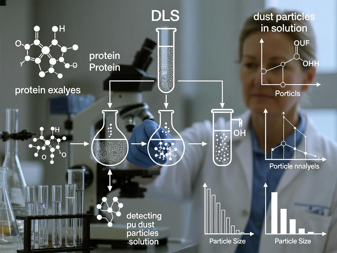
DLS Detection of Dust and Particulate Contamination in Protein Solutions: A Complete Guide for Biopharma Researchers
This article provides a comprehensive guide for researchers, scientists, and drug development professionals on detecting and managing dust and particulate contamination in protein samples using Dynamic Light Scattering (DLS).
DLS Detection of Dust and Particulate Contamination in Protein Solutions: A Complete Guide for Biopharma Researchers
Abstract
This article provides a comprehensive guide for researchers, scientists, and drug development professionals on detecting and managing dust and particulate contamination in protein samples using Dynamic Light Scattering (DLS). We explore the fundamental principles of how DLS differentiates protein signals from contaminant noise. We detail a step-by-step methodological workflow for accurate detection and application in quality control. The guide addresses common troubleshooting scenarios and optimization techniques for sample preparation and instrument settings. Finally, we validate DLS against complementary techniques like NTA and SEC-MALS and discuss its critical role in ensuring protein sample integrity for reliable biophysical characterization, formulation development, and regulatory compliance in therapeutic protein pipelines.
Understanding the Signal: How DLS Detects and Distinguishes Dust from Protein in Solution
Troubleshooting Guides and FAQs
Q1: Why is my DLS intensity autocorrelation function decaying too rapidly, and the derived hydrodynamic radius (Rh) is unrealistically small (~1 nm)?
A: This is a classic indicator of contamination by particulates or dust. Large particles scatter light intensely and dominate the correlation function, causing it to decay rapidly in the initial channels. The algorithm may fit this rapid decay and interpret it as very fast diffusion of small particles.
- Solution: Always clarify and filter your protein samples and buffers. Use 0.02 µm or 0.1 µm syringe filters (compatible with your protein) directly into a meticulously cleaned, dust-free disposable cuvette. Centrifuge the sample at high speed (e.g., 15,000 x g) for 10 minutes prior to measurement if filtration is not possible.
Q2: My measurement shows multiple peaks in the size distribution. Is this sample polydispersity or an artifact?
A: It could be either. True polydispersity indicates a mixture of oligomers. An artifact is often due to a few large aggregates or dust particles coexisting with the monomer.
- Troubleshooting: Check the intensity-weighted distribution versus the volume- or number-weighted distribution. A large, intense peak at >1000 nm in the intensity plot that becomes tiny in the volume plot is likely dust/aggregates. Repeat measurement with filtered sample. Analyze the same sample at multiple angles (if using a multi-angle instrument); dust scattering is more angle-dependent than small proteins.
Q3: What does a poor fit (high residual) of the autocorrelation function mean for my protein sample analysis?
A: A high, structured residual (non-random deviations) suggests the data does not fit the assumed model, often due to: 1. Presence of large, settling aggregates: Creates a non-decaying component. 2. Sample polydispersity exceeding instrument/model limits. 3. Foreign particle contamination. * Action: Visually inspect the sample for settling. Filter/centrifuge. Use a more advanced analysis algorithm (e.g., CONTIN, NNLS) if true polydispersity is expected. Ensure the sample is not convecting due to temperature instability.
Q4: How critical is buffer viscosity and refractive index for accurate Rh determination in protein studies?
A: Critical. The diffusion coefficient (D) from DLS is used in the Stokes-Einstein equation [Rh = kT/(6πηD)]. An incorrect viscosity (η) directly proportionally affects Rh. The refractive index affects the scattering angle calibration.
- Protocol: Always measure the viscosity of your exact buffer composition at the measurement temperature using a viscometer. Use literature or manufacturer values for refractive index increments (dn/dc) for accurate size calculation. Do not assume water viscosity for high-salt or glycerol-containing buffers.
Key Experimental Protocols
Protocol 1: Sample Preparation for Dust-Free DLS of Proteins
- Clean Environment: Perform all preparations in a laminar flow hood, if possible.
- Buffer Preparation: Prepare buffer, then filter through a 0.02 µm membrane filter into a clean flask.
- Protein Handling: Centrifuge protein stock solution at ≥15,000 x g for 15 minutes at 4°C to pellet aggregates.
- Sample Formation: Carefully aspirate the top 80% of the supernatant. Dilute with filtered buffer to desired concentration. Gently mix; avoid vortexing.
- Final Filtration: For stringent studies, filter the diluted sample through a 0.1 µm low-protein-binding syringe filter (e.g., PVDF) directly into the DLS cuvette.
- Cuvette: Use high-quality, disposable plastic cuvettes or thoroughly cleaned quartz cuvettes with dust-free seals.
Protocol 2: Validating Instrument and Sample Quality
- Standard Measurement: Measure a known standard (e.g., 100 nm polystyrene latex beads) to verify instrument performance and alignment.
- Buffer Background: Measure your filtered buffer alone in the same cuvette. The count rate should be very low (<20 kcps for most systems) and the correlation function should show no decay or a very weak, slow decay.
- Sample Measurement: Load your prepared protein sample. The count rate should be significantly higher than the buffer background.
- Repeat Measurements: Perform a minimum of 5-10 sequential runs (duration 10-60 seconds each). Check for consistency in derived size and intensity. An increasing Rh over time suggests aggregation or settling.
Data Presentation
Table 1: Impact of Filtration on Apparent Hydrodynamic Radius of a Monoclonal Antibody
| Sample Preparation Method | Intensity-Weighted Rh (d.nm) | Polydispersity Index (PDI) | Peak 1 (Main, nm) | Peak 2 (Artifact, nm) | Interpretation |
|---|---|---|---|---|---|
| Unfiltered, Vortexed | 12.4 ± 45.1 | 0.45 | 10.2 (92%) | 4200 (8%) | High PDI & large peak indicate dust/aggregates. |
| Centrifuged (15k x g, 15 min) | 10.8 ± 1.2 | 0.05 | 10.8 (100%) | - | Acceptable for stable proteins. |
| Filtered (0.1 µm) | 9.8 ± 0.3 | 0.02 | 9.8 (100%) | - | Optimal, dust-free preparation. |
| Buffer Only (0.02 µm filtered) | N/A | N/A | No meaningful decay | - | Clean background. |
Table 2: Common DLS Artifacts and Their Signatures in Thesis Research on Dust Detection
| Artifact Source | Signature in Intensity Autocorrelation | Effect on Size Distribution (Intensity) | Diagnostic Test |
|---|---|---|---|
| Few Large Dust Particles | Very rapid initial decay, poor fit. | Dominant large peak (>1000 nm), high PDI. | Filter sample; result becomes monomodal. |
| Protein Aggregation/Settling | Decay with a "tail," non-random residuals. | Peak size increases with measurement number. | Measure sequential runs; inspect sample. |
| Insufficient Cleaning | High, variable background count rate. | Unstable baseline, noisy correlation function. | Measure filtered solvent in cuvette. |
| Temperature Fluctuations | Drifting correlation function between runs. | High run-to-run variability in Rh. | Ensure adequate equilibration time (>2 min). |
The Scientist's Toolkit: Research Reagent Solutions
| Item | Function in DLS Protein Analysis |
|---|---|
| 0.02 µm Anotop Syringe Filter | For final filtration of buffers to achieve ultrapure, dust-free background. |
| 0.1 µm PVDF Syringe Filter | For filtering protein samples to remove aggregates >100 nm without significant adsorption. |
| Disposable PMMA Cuvettes | Pre-cleaned, sealed cuvettes minimize introduction of dust from labware. |
| Polystyrene Size Standards (e.g., 30 nm, 100 nm) | Essential for daily validation of instrument performance and alignment. |
| Viscosity Standard (e.g., S800) | Used to calibrate or verify the viscometer for accurate buffer viscosity measurement. |
| BSA Standard (1 mg/mL) | A stable protein standard to check the overall protocol for biologically relevant samples. |
Visualizations
Title: DLS Workflow: From Sample to Rh with Dust Impact
Title: DLS Signature of Dust vs. Clean Protein Samples
Troubleshooting & FAQs
Q1: Why does my DLS measurement of a purified protein sample show a large particle population in the micron range? A: This is a classic indication of dust or other foreign particulates (e.g., aggregated protein fibers, lint) contaminating the sample. Dust particles scatter light intensely (scales with diameter^6) and can dominate the correlation function, obscuring the true protein size distribution. Even a few particles per mL can cause significant artifacts.
Q2: How can I distinguish between real protein aggregates and dust artifacts in my DLS data? A: Analyze the correlation function. Dust often causes a sharp, rapid decay at very short correlation times. Conduct a procedural control: filter your buffer through the same 0.02µm filter used for samples. Measure it alone. A significant signal in the buffer indicates non-sample particulates. Furthermore, dust signals are often inconsistent between replicate measurements, whereas true aggregates are reproducible.
Q3: What is the most effective sample preparation method to eliminate dust for sensitive DLS measurements in protein research? A: A rigorous two-step filtration protocol is essential.
- Ultra-Clean Buffer Preparation: All buffers must be filtered through a 0.02 µm pore-size, low-protein-binding Anotop syringe filter (or equivalent) into a scrupulously cleaned glass vial.
- Sample Filtration: The protein sample, prepared or diluted in the pre-filtered buffer, should be filtered through a separate, new 0.02 µm Anotop filter directly into the ultra-clean DLS cuvette. Note: For proteins > 1 MDa, use a 0.1 µm filter to avoid sample loss.
Q4: My sample volume is very low (< 50 µL). How can I effectively prepare it for DLS? A: Use low-volume, low-protein-binding centrifugal filters (e.g., 100 kDa MWCO). Pre-rinse the filter device with filtered buffer. Spin your sample, then recover it. This concentrates the protein and removes larger particulates. Transfer directly to a micro-cuvette using gel-loading tips, which have a smaller bore to reduce lint pickup.
Q5: How should I clean my DLS cuvettes to avoid introducing artifacts? A: Avoid detergent use. Use a multi-solvent rinse protocol:
- Rinse with filtered >18 MΩ·cm water.
- Rinse with filtered ethanol (HPLC grade).
- Rinse with filtered acetone (HPLC grade).
- Dry in a particle-free environment (laminar flow hood) under a gentle, filtered nitrogen or argon stream. Do not let the cuvette air-dry openly.
Table 1: Impact of Filtration on Apparent Hydrodynamic Radius (Rh) in a Model Monoclonal Antibody Solution (1 mg/mL)
| Sample Preparation Method | Peak 1 Rh (nm) | % Intensity | Peak 2 Rh (nm) | % Intensity | PDI | Interpretation |
|---|---|---|---|---|---|---|
| Unfiltered Sample | 5.2 | 95.2 | 1250 | 4.8 | 0.42 | Dust/aggregates dominate scattering. |
| Buffer Filtered (0.1 µm), Sample Unfiltered | 5.5 | 98.5 | 850 | 1.5 | 0.15 | Reduced but significant dust artifact. |
| Buffer & Sample Filtered (0.02 µm) | 5.8 | 100 | n/a | 0 | 0.05 | True monodisperse protein signal. |
Table 2: Scattering Intensity Contribution by Particle Size (Theoretical Mie Scattering)
| Particle Type | Diameter (nm) | Scattering Intensity (Relative to 10 nm protein) | Notes for DLS |
|---|---|---|---|
| Monomeric Protein | 10 | 1 | The signal of interest. |
| Protein Decamer | 22 | ~ 120 | A real aggregate. |
| Dust / Silicate | 500 | 1.56 x 10^8 | Will completely overwhelm the protein signal. |
| Lint Fiber | 2000 | 1.0 x 10^10 | A single fiber can ruin a measurement. |
Experimental Protocols
Protocol 1: Ultra-Clean Sample Preparation for High-Sensitivity DLS Purpose: To prepare protein samples free of particulate artifacts for accurate hydrodynamic radius determination. Materials: See "Scientist's Toolkit" below. Procedure:
- Place the storage vial of buffer in a laminar flow hood.
- Using a clean syringe, draw buffer and filter through a 0.02 µm Anotop syringe filter into a new, pre-rinsed (with filtered water) glass scintillation vial. Cap immediately.
- Prepare your protein sample at the desired concentration using this filtered buffer.
- Using a new syringe and a new 0.02 µm Anotop filter, filter the protein solution directly into the meticulously cleaned DLS cuvette.
- Cap the cuvette with its sealing lid or parafilm.
- Perform DLS measurement promptly.
Protocol 2: The Buffer Blank Control Experiment Purpose: To diagnose the presence of particulates originating from buffers, cuvettes, or the environment. Procedure:
- Prepare ultra-clean buffer as in Protocol 1, step 2.
- Filter this buffer directly into the cleaned DLS cuvette using a new 0.02 µm filter.
- Measure this "blank" buffer for the same duration and settings as your sample.
- Analyze data. The correlation function should be flat with no decay, and the size distribution should show no peaks. Any signal indicates a failed preparation requiring investigation of the filtration or cleaning process.
Visualizations
Title: How Dust Skews DLS Data Flow
Title: Dust vs. Real Aggregate Diagnostic Tree
The Scientist's Toolkit
Table 3: Essential Research Reagents & Materials for Dust-Free DLS
| Item | Function & Rationale |
|---|---|
| 0.02 µm Anotop Syringe Filters (Inorganic Membrane) | Gold standard for creating particle-free buffers. The aluminum oxide membrane is exceptionally clean and low-binding. |
| Low-Protein-Binding Centrifugal Filters (e.g., 100 kDa MWCO) | For concentrating dilute samples and pre-clearing aggregates from small-volume (< 50 µL) preparations. |
| HPLC-Grade Solvents (Water, Ethanol, Acetone) | Used for cuvette cleaning. HPLC grade ensures minimal particulate contamination. |
| Glass Scintillation Vials | For storing filtered buffer. Glass sheds fewer particles than plastic and is easier to clean. |
| Glass Gas-Tight Syringes | For handling and filtering buffers/samples. Minimizes introduction of rubber/plasticizer particles. |
| Gel-Loading Pipette Tips | Their narrow bore reduces aspiration of airborne lint when loading samples into micro-cuvettes. |
| Laminar Flow Hood (Clean Bench) | A particle-controlled workspace is critical for sample preparation, cuvette drying, and assembly. |
| Particle-Free Cuvette Seals or Parafilm | To seal the cuvette after loading, preventing dust ingress during measurement. |
This technical support center provides guidance for interpreting Dynamic Light Scattering (DLS) data within the context of a thesis focused on detecting dust artifacts in protein sample solutions. Distinguishing between legitimate protein monomers/aggregates and contamination signals is critical for accurate analysis in drug development.
FAQs & Troubleshooting Guides
Q1: My DLS measurement shows a major peak at ~5 nm and a very small, broad peak around 10,000 nm. Is this sample aggregation or contamination? A: A dominant peak at a size consistent with your target protein (e.g., 5 nm) with a very small, sporadic signal in the micron range is highly indicative of dust or foreign particulates. True large-scale protein aggregation would typically show a more defined, repeatable peak at sub-micron scales (e.g., 100-1000 nm). Perform sample filtration (0.02 µm or 0.1 µm) and re-measure. If the large peak disappears or is drastically reduced, it was likely dust.
Q2: How can I differentiate between a true high-molecular-weight aggregate and a dust particle? A: Use both intensity-weighted and volume-weighted distribution views. Dust, being large and scarce, produces a very high scattering intensity but contributes negligible volume. A true aggregate population will be more proportional across intensity and volume distributions. Additionally, perform sequential measurements; dust signals are often inconsistent (non-reproducible) between runs, while aggregates are stable.
Q3: My sample is visibly clear, but DLS shows a significant polydispersity index (PdI) > 0.3. What does this mean? A: A high PdI indicates a broad size distribution. This could be due to:
- True sample heterogeneity (presence of aggregates and monomers).
- A few large contaminants (dust, fibers) skewing the correlation function. Troubleshooting steps: Centrifuge the sample at 10,000-15,000 g for 10 minutes and carefully pipette from the top. Re-measure. If PdI drops significantly, the sample was contaminated. If it remains high, true polydispersity/aggregation is likely.
Q4: What is the best practice for sample preparation to minimize dust artifacts in DLS? A: Follow this protocol:
- Use ultra-pure, filtered buffers (filter through 0.02 µm or 0.1 µm membrane).
- Filter the protein sample using a compatible, low-protein-binding syringe filter (e.g., 0.02 µm for monomeric samples, 0.1 µm for larger complexes).
- Thoroughly clean the cuvette with filtered solvent and use lint-free wipes.
- Centrifuge the sample briefly just before loading to pellet any settled aggregates or particles.
- Perform measurements in a laminar flow hood or clean air environment if possible.
Key Data Tables
Table 1: Characteristic Spectral Signatures in DLS
| Species | Typical Size Range (nm) | Intensity Signal | Volume/Number Signal | Reproducibility Between Runs | Effect of 0.1 µm Filtration |
|---|---|---|---|---|---|
| Protein Monomer | 2 - 10 | Moderate | High | High | Unaffected or slightly lost. |
| Protein Oligomer | 10 - 50 | Moderate to High | Moderate | High | May be lost if size > pore size. |
| Protein Aggregate | 100 - 1000 | Very High | Low to Moderate | High | Often removed. |
| Dust/Particulate | >1000 (1 µm) | Extremely High | Very Low | Low (Erratic) | Removed. |
| Air Bubbles | Variable (large) | Extremely High, Spiky | Negligible | None | Removed by degassing/centrifugation. |
Table 2: Troubleshooting Matrix for Common DLS Issues
| Symptom | Possible Cause | Diagnostic Test | Solution |
|---|---|---|---|
| A single, huge, erratic peak >1 µm | Dust or fiber contamination | Filter sample through 0.1 µm; re-measure clean cuvette with buffer. | Implement rigorous sample filtration and cleaning protocols. |
| Broad PdI (>0.3), multiple peaks | True polydispersity OR few large contaminants | View volume-weighted distribution; centrifuge sample and re-analyze supernatant. | Use centrifugal filtration; consider SEC-MALS for separation. |
| Unstable baseline, noisy correlation function | Air bubbles, insufficient equilibration | Inspect cuvette visually; let sample equilibrate to instrument temperature longer. | Degas buffer; centrifuge sample gently; ensure proper cuvette filling. |
| Peak size shifts between measurements | Sample changing (aggregation/degradation) OR temperature drift | Perform time-course measurements; monitor instrument temperature stability. | Check sample stability; use a temperature-controlled sample chamber. |
Experimental Protocols
Protocol 1: Validating Sample Purity via Sequential Filtration
Objective: To confirm if large-sized signals originate from the protein sample or external contamination.
- Prepare your protein solution in filtered buffer.
- Perform an initial DLS measurement (3-5 repeats). Record intensity distribution.
- Pass the sample through a low-protein-binding, 0.1 µm pore size syringe filter.
- Perform DLS measurement again on the filtered sample (3-5 repeats).
- Interpretation: If the large particle signal disappears and the main protein peak remains, the large particles were contaminants. If both signals reduce proportionally, they may be genuine large aggregates.
Protocol 2: Distinguishing Aggregates from Dust via Signal Proportionality Analysis
Objective: Use the disparity between intensity and volume distributions to identify sparse, large particles.
- Acquire a high-quality DLS measurement with a high number of runs (≥10).
- Analyze the data to generate both intensity-weighted and volume-weighted size distributions.
- Compare the two distributions.
- Interpretation: A peak that is dominant in the intensity plot but minuscule or absent in the volume plot indicates a low concentration of large scatterers (characteristic of dust). A peak present in both distributions suggests a higher population concentration (more characteristic of aggregates).
Diagrams
Title: DLS Peak Interpretation Workflow
Title: Clean DLS Sample Preparation Steps
The Scientist's Toolkit: Essential Research Reagents & Materials
| Item | Function in DLS Sample Preparation |
|---|---|
| Ultra-Pure Water (e.g., Milli-Q) | Minimizes background scattering from ionic impurities and particles in buffer preparation. |
| 0.02 µm & 0.1 µm Syringe Filters (Anotop or similar) | Critical for removing sub-micron and micron-sized particulates from buffers and samples, respectively. |
| Low-Protein-Binding Microcentrifuge Tubes | Prevents loss of sample, especially low-concentration proteins, via adsorption to tube walls. |
| Disposable, Pre-Cleaned Cuvettes (e.g., ZEN0040) | Provides a consistent, low-dust optical path; disposable nature avoids cleaning artifacts. |
| Filtered Buffer Solutions | All buffers must be filtered through a 0.02 µm membrane to eliminate scattering background. |
| Precision Gas-Tight Syringes | Allows for bubble-free, accurate loading of sample into the cuvette, preventing artifact signals. |
| Tabletop Microcentrifuge | For pelleting large aggregates or contaminants prior to filtration and analysis. |
| Lint-Free Laboratory Wipes | For cleaning cuvette exterfaces without introducing fibers. |
Technical Support Center: Troubleshooting DLS Data Quality
Frequently Asked Questions (FAQs)
Q1: My DLS correlation function decays very quickly and shows significant noise or instability, especially at long lag times. What does this indicate? A1: An unstable, noisy correlation decay, particularly at the tail, is a primary indicator of large, scattering contaminants like dust or aggregates. These few large particles cause intense scattering bursts that corrupt the statistical averaging, leading to an unreliable measurement. This directly compromises thesis conclusions on native protein size distribution.
Q2: My calculated Polydispersity Index (PDI) is very high (>0.2) and the size distribution plot shows a significant "tail" towards larger hydrodynamic radii. Is this sample intrinsically polydisperse or is it contaminated? A2: While sample intrinsic polydispersity is possible, a skewed size distribution with a large-particle tail alongside a high PDI is a classic signature of dust contamination. For most purified, monodisperse protein samples, a PDI >0.2 suggests the presence of a second, larger population. Your thesis must differentiate between true sample heterogeneity and artifact.
Q3: What is the most critical step in sample preparation to avoid these indicators? A3: Rigorous clarification of both the solvent and the protein sample is non-negotiable. This involves filtration through ultraclean, protein-low-binding membranes with a pore size of 0.02 µm or 0.1 µm, depending on protein size. Centrifugation immediately prior to loading the cuvette is also essential.
Troubleshooting Guides
Issue: Unstable Correlation Function & High PDI Symptoms: Correlation function does not decay smoothly; poor fit residuals; PDI reported >0.3; size distribution graph is multimodal. Step-by-Step Resolution:
- Prepare Clean Solvent: Filter your buffer (e.g., PBS) through a 0.02 µm Anotop syringe filter directly into an ultraclean vial.
- Clarify Sample: Centrifuge your protein sample at >15,000 x g for 10-15 minutes at the relevant temperature (4°C or room temp).
- Clean Cuvette: Use a dedicated, filtered solvent (e.g., filtered ethanol, then filtered buffer) to rinse the cuvette. Use compressed, filtered air or nitrogen to dry.
- Careful Loading: Pipette only the top 70-80% of your centrifuged supernatant into the clean cuvette, avoiding the pellet.
- Run Controls: Always measure the filtered buffer alone first. Its intensity count rate should be very low (<10% of your sample signal) and its correlation function flat.
Issue: Persistent Large Particle Tails Symptoms: A consistent, low-intensity signal at radii >2x the main peak. Step-by-Step Resolution:
- Verify Filtration: Ensure you are using a membrane with an appropriate pore size. For proteins <100 kDa, 0.02 µm is recommended.
- Environmental Control: Perform all sample handling in a laminar flow hood to minimize airborne dust.
- Instrument Check: Perform a validation measurement using a known standard (e.g., 100 nm latex beads) to confirm instrument performance.
- Data Analysis Review: Apply appropriate analysis algorithms (e.g., Multiple Narrow Modes, General Purpose) and check the "Fit Quality" parameter. A poor fit suggests the model cannot handle the contaminant signal.
Table 1: Impact of Filtration on DLS Metrics for a 150 kDa Protein Sample
| Sample Preparation Method | Mean Rh (nm) | PDI | Peak 1 Intensity (%) | Peak 2 (Tail) Intensity (%) | Correlation Function Quality |
|---|---|---|---|---|---|
| Unfiltered, Uncentrifuged | 8.2 ± 2.1 | 0.45 | 78 | 22 (at 120 nm) | Unstable, noisy tail |
| 0.1 µm Filtered | 6.8 ± 1.5 | 0.28 | 92 | 8 (at 80 nm) | Moderately stable |
| 0.02 µm Filtered & Centrifuged | 5.9 ± 0.3 | 0.12 | 100 | 0 | Smooth, monomodal decay |
Table 2: DLS Signal Thresholds for Contamination Detection
| Indicator | Clean Sample Threshold | Warning Zone | Contamination Likely |
|---|---|---|---|
| Polydispersity Index (PDI) | < 0.1 | 0.1 - 0.2 | > 0.2 |
| Buffer Count Rate (% of sample) | < 5% | 5% - 10% | > 10% |
| Correlation Function Fit Residual | Random, < 2% | Structured, < 5% | Structured, > 5% |
Detailed Experimental Protocols
Protocol 1: Ultraclean Sample Preparation for DLS Objective: To prepare a protein sample free of dust and large aggregates for accurate DLS analysis. Materials: See "The Scientist's Toolkit" below. Method:
- Filter 2 mL of the sample buffer through a 0.02 µm inorganic membrane syringe filter into a clean glass vial.
- Rinse a quartz or glass DLS cuvette thoroughly with the filtered buffer. Dry with filtered, compressed air.
- Centrifuge the protein solution at 18,000 x g for 15 minutes at 4°C.
- Gently pipette the top 80% of the supernatant, avoiding the pellet.
- Load the clarified sample into the pre-rinsed cuvette, cap it, and ensure no bubbles are present.
- Measure the filtered buffer in the same cuvette as a blank prior to the sample.
Protocol 2: Diagnostic DLS Measurement Sequence Objective: To systematically diagnose dust contamination in a sample. Method:
- Buffer Baseline: Measure 3-5 runs of the filtered, blank buffer. Record the average intensity (kcps) and observed diameter.
- Sample Measurement: Measure the prepared protein sample with at least 10-15 repeat runs.
- Data Inspection: Examine the correlation function overlay for stability. Check the derived size distribution for a large-particle tail.
- Comparative Analysis: Subtract the buffer's scattering profile (if instrument software allows). Compare the sample's PDI and mean size to the buffer measurement.
Visualizations
DLS Contamination Diagnosis Workflow
Mechanism of Dust Interference in DLS
The Scientist's Toolkit: Essential Reagents & Materials
| Item | Function & Rationale |
|---|---|
| 0.02 µm Anotop Syringe Filter (Inorganic Membrane) | Gold standard for final buffer filtration. Inert aluminum oxide membrane minimizes protein adsorption and removes sub-100 nm particulates. |
| Ultra-Clear, Disposable Size-Exclusion Columns | For rapid buffer exchange into filtered, dust-free buffer, removing aggregates from the sample. |
| Low-Volume, Quartz DLS Cuvettes | Provide optimal optical quality and minimize the sample volume required, reducing the probability of dust inclusion. |
| Protein-Low-Binding Microcentrifuge Tubes (1.5 mL) | Minimizes sample loss and prevents the introduction of polymeric contaminants during centrifugation steps. |
| Filtered, Compressed Air or Nitrogen Duster | Essential for drying cuvettes without introducing lint or dust from laboratory wipes. |
| Nanopure Water System (0.05 µm filter) | Source of particle-free water for making all buffers and cleaning solutions. |
| Latex Nanosphere Size Standards (e.g., 60 nm, 100 nm) | Used for regular validation of DLS instrument performance and alignment. |
Technical Support Center
Troubleshooting Guide: DLS Data Interpretation Issues
Issue 1: Unusually high polydispersity index (PdI) or multiple peaks in DLS size distribution.
- Check: Sample preparation area and vial cleanliness. Inspect for dust contamination on cuvette windows.
- Action: Filter all buffers through a 0.02 µm or 0.1 µm syringe filter. Centrifuge protein samples at 10,000-15,000 x g for 10 minutes prior to measurement to pellet large, dust-like particulates.
- Verification: Run a blank measurement of filtered buffer. The intensity count rate should be low and stable.
Issue 2: Irreproducible size measurements between replicate samples.
- Check: Consistency of sample handling. Dust ingress can occur during pipetting or cuvette loading.
- Action: Perform measurements in a laminar flow hood or clean air environment. Use high-quality, low-binding, pre-rinsed vials and pipette tips.
- Verification: Implement a standard pre-measurement protocol including buffer filtration and sample centrifugation for all replicates.
Issue 3: Sudden spikes in scattering intensity that distort correlation functions.
- Check: For the presence of a few large, scattering particles (dust) passing through the laser beam.
- Action: Use a cross-correlation DLS instrument (if available) to suppress artifacts from dust. Increase the number of measurement runs to allow the software to identify and statistically exclude spikes.
- Verification: Visually inspect the correlation function for sharp discontinuities, indicative of dust spikes.
Frequently Asked Questions (FAQs)
Q1: How can I distinguish between a true protein aggregate and a dust particle in my DLS measurement? A: True protein aggregates will typically show a concentration-dependent signal and will be present across replicate samples prepared from the same stock. Dust particles are often random, non-reproducible events. Use intensity-based size distributions for detection; dust appears as sporadic, very high-intensity signals in the >1 µm range. Confirm by sample filtration or centrifugation—true large aggregates may be reduced but not eliminated, while dust signals will vanish.
Q2: What is the minimum size of dust that can interfere with DLS analysis of proteins? A: Due to the intensity of scattered light being proportional to the sixth power of the particle diameter (I ∝ d⁶), even sub-micron dust particles (e.g., 0.5 µm) can dominate the signal over nanometer-sized proteins (e.g., 10 nm). The table below quantifies this effect.
Q3: What are the best practices for sample handling to minimize dust contamination for DLS? A: 1. Perform all prep in a laminar flow hood or dedicated clean bench. 2. Filter all buffers through a 0.02 µm or 0.1 µm membrane filter. 3. Centrifuge protein samples before analysis. 4. Use high-quality, disposable cuvettes or meticulously clean quartz cuvettes with filtered solvents. 5. Cap samples when not being measured.
Q4: Can DLS software algorithms completely correct for dust contamination? A: No. While modern algorithms (e.g., multiple narrow modes, spike removal) can identify and ignore sporadic, large-particle events, they cannot salvage data from a heavily contaminated sample. The primary defense is rigorous sample cleaning.
Table 1: Relative Scattering Intensity of Particles in Solution Demonstrates why dust dominates the DLS signal.
| Particle Type | Diameter (nm) | Relative Scattering Intensity (Approx.) |
|---|---|---|
| Monomeric Protein | 10 | 1 (Baseline) |
| Protein Trimer | 15 | 11 |
| Small Aggregate | 100 | 1,000,000 |
| Dust Particle | 500 | 15,625,000,000 |
Table 2: Effect of Sample Preparation on DLS Results for a 1 mg/mL mAb Solution Data from controlled experiments.
| Preparation Method | Z-Average (d.nm) | Polydispersity Index (PdI) | Peak 1 (nm) | Peak 2 (nm) | Result Integrity |
|---|---|---|---|---|---|
| Unfiltered Buffer, No Spin | 12.8 ± 45.1 | 0.48 ± 0.31 | 8.2 | >1000 | Unacceptable |
| Buffer Filtered (0.1 µm), Sample Centrifuged | 10.5 ± 0.3 | 0.05 ± 0.02 | 10.5 | - | High Integrity |
Experimental Protocols
Protocol 1: Standardized DLS Sample Preparation for Dust Minimization
- Buffer Preparation: Prepare formulation buffer. Filter through a 0.02 µm or 0.1 µm pore-size syringe filter directly into a clean, dust-free container.
- Sample Preparation: Dilute protein stock into filtered buffer to desired concentration.
- Clarification: Centrifuge the diluted sample at 10,000-15,000 x g for 10 minutes at the relevant temperature (e.g., 4°C or 25°C).
- Loading: Carefully pipette the top 80-90% of the supernatant into a clean DLS cuvette, avoiding the pellet. Cap the cuvette.
- Measurement: Place cuvette in instrument equilibrated to temperature. Allow 2-5 minutes for temperature equilibration before starting measurement.
Protocol 2: Controlled Dust Contamination Experiment Purpose: To visualize the impact of dust on aggregation analysis.
- Prepare two identical samples of a monodisperse protein (e.g., BSA) using Protocol 1. Measure both via DLS to confirm identical baseline profiles (Record Z-Avg, PdI, distribution).
- Test Sample: Lightly tap a spatula of fine, dried lyophilized buffer powder over the open cuvette of one sample. Gently swirl to partially mix.
- Control Sample: Re-measure the untouched control sample.
- Measurement: Immediately measure the "contaminated" test sample.
- Analysis: Compare correlation functions and size distributions. The contaminated sample will show unstable intensity, poor correlation function fit, and large size artifacts.
Diagrams
Title: Logical Flow of Dust Contamination Impact on DLS Data
Title: DLS Sample Prep & QC Workflow for Dust Mitigation
The Scientist's Toolkit: Research Reagent Solutions
| Item | Function in Dust Mitigation for DLS |
|---|---|
| 0.02 µm or 0.1 µm Syringe Filters | Removes sub-micron particulates and microbial contaminants from buffers and solvents. The primary defense against dust. |
| Ultra-Clear, Low-Binding Microcentrifuge Tubes | Minimizes particle shedding and protein adsorption during sample prep and centrifugation. |
| Disposable, Sealed DLS Cuvettes | Prevents contamination from cuvette cleaning processes and allows for one-time, clean use. |
| Certified Clean Air Enclosure (Laminar Flow Hood) | Provides a particulate-free workspace for sample handling, pipetting, and cuvette loading. |
| High-Speed Microcentrifuge | Pellet's trace aggregates and any introduced dust particles prior to supernatant sampling for DLS. |
| Particle-Free Water & Buffer Solutions | Commercially available, certified fluids for critical dilutions and instrument calibration. |
| Latex/Nitrile Gloves & Lab Coat | Reduces introduction of human-sourced particles and fibers during experimentation. |
Best Practices: A Step-by-Step DLS Protocol for Dust Detection and Clean Protein Analysis
Technical Support Center: Troubleshooting & FAQs
Troubleshooting Guides
Issue: High polydispersity index (PdI) and erratic correlation function in DLS measurement.
- Check 1: Insufficient sample clarification. Repeat centrifugation at recommended force and time. Consider using a smaller pore size syringe filter.
- Check 2: Vial incompatibility. Ensure vials are specifically designed for DLS, are scrupulously clean, and free of dust. Use the correct vial size for your instrument's sample chamber.
- Check 3: Air bubbles introduced during vial loading. Centrifuge loaded vials at a low speed (e.g., 500 x g for 1 min) before measurement.
Issue: Consistent particulate contamination despite filtration.
- Check 1: Filter membrane compatibility. Verify the filter material is not adsorbing your protein or leaching contaminants. Pre-rinse filters with buffer.
- Check 2: Contaminated buffer. Always filter or ultracentrifuge buffer prior to sample preparation.
- Check 3: Dirty vial. Implement a stringent vial cleaning protocol (see below).
Frequently Asked Questions (FAQs)
Q1: What is the optimal centrifugation protocol to remove dust from a 1 mg/mL monoclonal antibody sample prior to DLS? A1: For most protein samples, ultracentrifugation at 100,000 - 150,000 x g for 30-60 minutes at 4°C is considered the gold standard. For routine clarification in a standard microcentrifuge, a protocol of 15,000 - 21,000 x g for 30-60 minutes at 4°C is often sufficient. Always balance rotors carefully.
Q2: Should I use a 0.22 µm or 0.1 µm filter for my protein sample? A2: A 0.22 µm filter is standard for removing microbial contaminants and large aggregates. For aggressive dust removal in sensitive DLS work, a 0.1 µm filter is superior but carries a higher risk of adsorbing larger proteins or protein complexes. Always check for sample loss post-filtration.
Q3: What is the most critical factor in vial selection for DLS? A3: Optical quality and material cleanliness. Vials must have clear, scratch-free optical surfaces. Disposable, certified dust-free cuvettes are preferred. For flow cells, ensure they are compatible with automatic syringe systems and can be cleaned without introducing scratches.
Q4: My sample is very precious and low-volume. What is the minimal clarification workflow? A4: 1) Use pre-filtered buffer. 2) Use a low-protein-binding, 0.1 µm centrifugal filter device (spin at 10,000 x g for 5-10 min). 3) Directly load the filtrate into a low-volume, disposable microcuvette to minimize handling.
Table 1: Centrifugation Protocols for Sample Clarification
| Sample Type | Recommended Force | Time | Temperature | Expected Outcome |
|---|---|---|---|---|
| Standard Buffer (PBS) | 15,000 x g | 30 min | 4°C | Removal of nano-dust & large particulates. |
| Monoclonal Antibody (1-10 mg/mL) | 100,000 x g | 60 min | 4°C | Removal of aggregates > ~200 kDa; clear baseline. |
| Small Protein (< 50 kDa) | 20,000 x g | 45 min | 4°C | Clarification without excessive pelleting of monomer. |
| Viral Vector Prep | 2,000 x g | 10 min | 4°C | Quick removal of cellular debris (pre-filter step). |
Table 2: Filtration Membrane Compatibility
| Membrane Material | Protein Recovery (Typical) | Key Application | Aggregation Risk |
|---|---|---|---|
| Cellulose Acetate (CA) | >95% (for many proteins) | General use, low adsorption. | Low |
| Polyethersulfone (PES) | >90% | Fast flow, high throughput. | Low-Moderate |
| Polyvinylidene Fluoride (PVDF) | Variable | Low protein binding for specific assays. | Moderate |
| Anopore (Aluminum Oxide) | >95% | Precise pore size, DLS standard. | Very Low |
| Regenerated Cellulose (RC) | >90% | Low adsorption for sensitive proteins. | Low |
Experimental Protocols
Protocol 1: Ultracentrifugation for High-Purity DLS Samples
- Prepare Buffer: Filter all buffers through a 0.1 µm Anotop syringe filter into a cleaned, dedicated container.
- Prepare Sample: Dilute protein into pre-filtered buffer to desired concentration.
- Load Tubes: Carefully load sample into ultracentrifuge tubes (e.g., polycarbonate). Precisely balance tube pairs by mass.
- Centrifuge: Run at 120,000 x g for 45 minutes at 4°C using a fixed-angle rotor.
- Recover Sample: Carefully extract tubes. Pipette the top 80% of the supernatant, avoiding the pellet and meniscus.
- Load Vial: Transfer supernatant directly to a cleaned DLS vial or cuvette.
Protocol 2: Rigorous DLS Vial/Cuvette Cleaning
- Rinse: Rinse 3x with ultrapure, 0.1 µm-filtered water.
- Soak: Soak for 15 minutes in 2% Hellmanex III solution.
- Scrub (if applicable): Gently use a dedicated cuvette brush.
- Rinse: Rinse exhaustively (10x) with filtered water.
- Final Rinse: Rinse 3x with 0.1 µm-filtered ethanol or the sample buffer.
- Dry: Air-dry in a laminar flow hood or dust-free environment. Use lint-free wipes for external surfaces only.
Diagrams
DLS Sample Prep Workflow
Dust Interference in DLS Correlation
The Scientist's Toolkit: Research Reagent Solutions
| Item | Function in DLS Sample Prep |
|---|---|
| Anotop 0.1 µm Syringe Filter (Inorganic Membrane) | Provides superior final filtration for buffers and samples with minimal protein adsorption and low particle shedding. |
| Ultra-Clear or Polycarbonate Ultracentrifuge Tubes | Designed for high g-forces; minimal leachables and smooth interiors reduce particle generation during pelleting. |
| Hellmanex III Cleaning Solution | Specifically formulated alkaline solution for cleaning optical components and glassware, effectively removing organic contaminants. |
| Disposable, Dust-Free Microcuvettes (e.g., ZEN0040) | Pre-cleased, sealed cuvettes that eliminate cleaning variability and are ideal for low-volume, precious samples. |
| Low-Protein-Binding Microcentrifuge Tubes (e.g., Protein LoBind) | Minimizes protein loss via surface adsorption during sample handling and centrifugation steps. |
| Certified Particle-Free Water/Buffer | Commercially available buffers guaranteed to have extremely low particulate background for critical baseline measurements. |
| Precision Gas Duster | Used to remove lint and dust from vial exteriors and instrument sample chambers prior to insertion. |
Technical Support & Troubleshooting Guides
FAQ 1: How do I determine the optimal attenuator setting for a protein sample, and what are the signs of incorrect attenuation?
- Answer: The optimal attenuator setting is one where the measured intensity (kcps) is within the instrument's linear range, typically between 100-1000 kcps for most systems. An attenuator that is too low (under-attenuation) will cause detector saturation, indicated by a spike or flat top on the intensity trace and correlation function that decays to zero too quickly. An attenuator that is too high (over-attenuation) yields a weak, noisy signal with a poor signal-to-noise ratio in the correlation function. Protocol: Perform an attenuator scan. Load your sample, set temperature to 25°C, and run sequential 10-second measurements across the available attenuator settings. Plot mean intensity vs. attenuator position to identify the plateau region in the linear range.
FAQ 2: My correlation function is noisy even with clear samples. Could measurement position (z-position) be the issue?
- Answer: Yes. An incorrect measurement position within the cuvette can introduce artifacts from meniscus, bubbles, or dust stuck to the walls. This creates fluctuating scattering and a noisy correlation function. Protocol: Always perform a z-position scan for a new cell type or sample volume. Using a stable, standard (e.g., toluene or a known protein), take short measurements at different depths. Select the position that yields the highest, most stable intensity and the smoothest correlation function decay.
FAQ 3: Why is precise temperature control critical for DLS in protein-dust studies, and how do I verify it?
- Answer: Temperature affects solvent viscosity, protein diffusion coefficient, and can induce aggregation (creating false "dust" signals). Fluctuations cause hydrodynamic radius (Rh) drift. For protein studies, control to ±0.1°C is standard. Protocol: Verify system calibration using a standard with a known, temperature-dependent viscosity (e.g., pure water). Measure its Rh at multiple setpoints (e.g., 15°C, 20°C, 25°C). The calculated Rh should be constant. Drift indicates a calibration issue.
FAQ 4: I suspect my buffer has particulate dust. How can I use instrument setup to diagnose this vs. protein aggregates?
- Answer: Use the combination of attenuator setting and intensity analysis. Dust particles are typically large, few, and scatter intensely. Protocol:
- Measure filtered buffer at an optimal attenuator. Note the mean intensity (Ibuffer) and polydispersity index (PDI).
- Measure your protein sample at the same attenuator. Note intensity (Isample).
- A significant, variable Isample much higher than Ibuffer, with a multi-modal size distribution skewed to high nm/µm range, suggests dust contamination. A consistent, moderate increase in I_sample with a monomodal peak near the expected protein size suggests a clean sample with protein aggregates.
Table 1: Optimal Attenuator Selection Guide
| Sample Type | Expected Intensity Range (kcps) | Recommended Start Attenuator | Key Diagnostic Signal |
|---|---|---|---|
| Filtered Buffer / Solvent | 50 - 200 | Medium-High | Baseline for contamination. |
| Monodisperse Protein (1 mg/mL) | 200 - 600 | Medium | Smooth, single exponential decay. |
| Polydisperse / Aggregating Protein | 300 - 800 | Medium-Low | Multi-modal distribution. |
| Sample Suspected of Dust | 500 - >1000 (variable) | Low (with caution) | Spiking intensity, erratic correlation function. |
Table 2: Troubleshooting Symptoms and Solutions
| Symptom | Possible Cause | Diagnostic Check | Corrective Action |
|---|---|---|---|
| Intensity spikes, then drops. | Large dust particle transient. | Observe raw intensity trace in real-time. | Ultra-centrifuge or filter sample (0.02µm). |
| Correlation function is noisy. | Incorrect z-position or low count rate. | Perform z-scan; check if kcps < 100. | Optimize z-position; decrease attenuator. |
| Rh value drifts over time. | Temperature instability or sample aggregation. | Monitor temperature log; measure buffer standard. | Check thermostat; verify sample stability. |
| High PDI in known standard. | Cuvette defects or dirty optics. | Visually inspect cuvette; clean with solvent. | Replace cuvette; perform optical cleaning cycle. |
Experimental Protocols
Protocol 1: Comprehensive Pre-Measurement Instrument Qualification.
- Objective: Establish a dust-free, optimally configured baseline.
- Materials: Filtered toluene standard, filtered buffer (0.1µm), lint-free gloves, clean cuvettes.
- Methodology:
- Power on instrument and laser, allowing warm-up for 30 minutes.
- Load toluene standard into a pristine cuvette. Set temperature to 25°C.
- Execute automated attenuator and z-position scans. Record optimal settings.
- Perform 5 consecutive measurements (duration: 60 sec each). Record mean Rh, PDI, and intensity.
- Acceptance Criteria: Rh = 1.0 ± 0.1 nm; PDI < 0.05.
- Repeat steps 2-5 with filtered buffer. Acceptance Criteria: Intensity < 200 kcps; no detectable size peaks > 3 nm.
Protocol 2: Differentiating Dust from Protein Aggregates via Attenuator-Dependent Intensity Analysis.
- Objective: Diagnose the source of large scatterers.
- Methodology:
- Prepare three aliquots: (A) filtered buffer, (B) filtered protein sample, (C) unfiltered protein sample.
- Using the optimal z-position, measure each aliquot at three attenuator settings: Low, Optimal, High.
- For each measurement, record the Mean Intensity and Peak Ratio (Intensity-weighted % in the >1000 nm size bin).
- Plot Peak Ratio vs. Mean Intensity. Dust-contaminated samples (C) show high, non-linear increases in Peak Ratio with intensity. Pure aggregates (B) show a more linear relationship.
Diagrams
Title: DLS Experimental Workflow for Dust Detection
Title: Attenuator Selection Troubleshooting Logic
The Scientist's Toolkit: Essential Research Reagent Solutions
| Item | Function in DLS Protein/Dust Research |
|---|---|
| ANAPORE / Ultrafine Filters (0.02µm) | Final sample filtration to remove sub-micron particulate dust without absorbing protein. |
| Sealed, Optical Quality Cuvettes | Minimizes introduction of airborne dust and prevents evaporation during measurement. |
| Toluene or Polystyrene Nanosphere Standard | Provides known size and scattering for daily instrument verification and calibration. |
| High-Purity Water (HPLC Grade) | Prevents contamination from impurities in buffer preparation. |
| Stable, Monodisperse Protein (e.g., BSA) | Positive control for protein sizing, used to distinguish instrument drift from sample issues. |
| Viscosity Standard (e.g., Sucrose Solutions) | Used to validate temperature control accuracy via viscosity-dependent Rh measurements. |
Troubleshooting Guides & FAQs
Q1: How many experimental runs (N) are sufficient for DLS measurements of protein samples to be statistically valid? A: For a standard protein sizing experiment, a minimum of 3-10 consecutive runs per sample is recommended. If you are monitoring aggregation or detecting small particulate populations like dust, increase this to 10-20 runs. This accounts for the stochastic nature of particle diffusion and improves the probability of capturing transient dust events. Statistical confidence is more about the quality and consistency of the correlograms than simply maximizing N. If the calculated intensity or number size distributions vary significantly between runs, it indicates an unstable sample (e.g., ongoing aggregation) or contamination.
Q2: What duration (measurement time per run) should I set for each DLS run when screening for dust? A: The optimal duration balances signal-to-noise with sample stability. For clear protein solutions, 30-60 seconds per run is often adequate. When specifically probing for low levels of large aggregates or dust particles, which scatter light intensely but may be rare, extending the measurement time to 120-180 seconds can improve the probability of their detection. However, excessively long runs (e.g., >5 minutes) risk data distortion from sedimentation, sample degradation, or temperature drift within the cuvette.
Q3: My DLS results show a sporadic large-size peak. Is this dust or protein aggregation? How can I differentiate? A: This is a common issue. Follow this diagnostic protocol:
- Replicate: Immediately perform 5-10 additional runs on the same sample aliquot. Note the reproducibility.
- Filter: Pass a fresh aliquot of your sample through a 0.02 µm or 0.1 µm syringe filter (nanopore filter) compatible with proteins (e.g., Anotop). Re-measure.
- Analyze:
- If the large peak disappears and all subsequent runs are consistent, the signal was likely from dust.
- If the large peak diminishes but is still present inconsistently, it may be a mix of dust and aggregates.
- If the large peak remains and is reproducible across runs, it strongly indicates genuine protein aggregation.
- Control: Always filter your buffer separately and measure it as a background check.
Q4: How many independent sample replicates (biological/technical) are needed for publication-quality data in a DLS study? A: The replication hierarchy is crucial for confidence.
- Technical Replication: Perform ≥3 measurement runs per filled cuvette.
- Sample Replication: Prepare and measure ≥3 independent aliquots of the same protein batch (intra-batch).
- Biological/Batch Replication: For robust conclusions, repeat the experiment with ≥2 independently prepared protein batches or biological samples. A rigorous design reporting the mean hydrodynamic radius (Rh) and polydispersity index (PDI) across these levels is essential for credible data.
Q5: The correlogram decays to baseline too quickly or is noisy. What should I adjust? A: A fast, noisy decay suggests a weak scattering signal.
- Increase Concentration: Optimize protein concentration to be within the instrument's ideal range (often 0.1-1 mg/mL for many proteins). Avoid concentrations so high that intermolecular interactions become significant.
- Check Sample Clarity: Ensure the sample is truly solution-clear. Centrifuge if necessary (see protocols).
- Verify Optics: Clean the exterior of the cuvette with lint-free cloth and ethanol. Ensure no bubbles are in the light path.
- Adjust Duration: Slightly increase measurement time per run to improve averaging.
Data Presentation
Table 1: Recommended DLS Run Parameters for Protein Samples with Dust Detection
| Experimental Goal | Runs per Sample (N) | Duration per Run | Independent Sample Replicates | Key Diagnostic Step |
|---|---|---|---|---|
| Standard Protein Sizing | 3 - 5 | 30 - 60 s | ≥ 3 | Buffer background subtraction |
| Aggregation Kinetics | 5 - 10 | 30 - 120 s | ≥ 2 | Time-point sampling & filtration |
| Low-Level Aggregate/Dust Detection | 10 - 20 | 60 - 180 s | ≥ 3 | Pre-filtration of sample & buffer |
| Formulation Screening | 5 - 10 | 30 - 60 s | ≥ 2 | High-throughput plate calibration |
Table 2: Troubleshooting Summary for Spurious Large-Particle Signals
| Observation | Possible Cause | Immediate Action | Confirmatory Test |
|---|---|---|---|
| Single large peak in one run | Dust/foreign particle | Replicate runs (N≥10) on same aliquot | Peak disappears in subsequent runs |
| Consistent large peak across runs | Protein aggregation | Filter sample (0.02-0.1 µm) | Peak persists post-filtration |
| Variable bimodal distribution | Mix of dust & aggregates | Filter sample, then monitor over time | Filtering removes only the sporadic component |
| Large peak only in sample, not buffer | Sample preparation issue | Centrifuge sample pre-measurement | Peak reduces after centrifugation |
Experimental Protocols
Protocol 1: Sample Preparation for Dust-Free DLS Measurement
- Buffer Preparation: Prepare buffer and filter it through a 0.02 µm or 0.1 µm pore-size syringe filter into a clean flask.
- Protein Solution: Dissolve or dialyze your protein into the filtered buffer.
- Clarification: Centrifuge the protein solution at 10,000 - 20,000 x g for 10-15 minutes at 4°C (or recommended storage temperature) to pellet any large aggregates or insoluble matter.
- Sample Extraction: Carefully pipette the top 80-90% of the supernatant into a new, clean tube. Avoid disturbing the pellet.
- Cuvette Loading: Using a filtered pipette tip, load the required volume into a thoroughly cleaned DLS cuvette. Avoid introducing bubbles.
- Buffer Control: Load filtered buffer into a separate, identically cleaned cuvette for background measurement.
Protocol 2: Systematic Replication & Statistical Confidence Workflow
- Background Measurement: Measure filtered buffer for 5-10 runs. Record the mean intensity and size distribution. Acceptable buffer should show only a low-intensity signal from solvent molecules.
- Primary Sample Measurement:
- Load your prepared protein sample.
- Equilibrate to the set temperature (typically 25°C) for 2-5 minutes.
- Perform Run Set 1 (e.g., 10 consecutive runs). Record the correlogram, Rh, and PDI for each.
- Intra-Sample Replication:
- Empty and clean the cuvette.
- Load a new, independent aliquot from the same prepared sample tube.
- Repeat Step 2 (Run Set 2).
- Inter-Batch Replication:
- Prepare a fresh protein batch from stock or expression system.
- Repeat the entire Sample Preparation protocol and Steps 2-3.
- Data Analysis:
- Calculate the mean and standard deviation of Rh and PDI for each run set and across batches.
- Use statistical tests (e.g., ANOVA) to determine if differences between formulations or conditions are significant relative to the replicate variance.
Mandatory Visualization
DLS Sample Prep & Replication Workflow
DLS Signal Analysis for Dust Detection
The Scientist's Toolkit: Research Reagent Solutions
Table 3: Essential Materials for DLS Protein Analysis
| Item | Function & Importance | Example/Note |
|---|---|---|
| Anotop 25 Syringe Filters (0.02 µm) | Gold-standard for ultrafiltration of buffers to remove nanoscale dust and particulates, creating a clean background. | Inorganic aluminum oxide membrane; low protein binding. |
| Zeta Potential Cells / Disposable Cuvettes | High-quality, optical-grade cuvettes specific to your DLS instrument. Cleanliness is paramount. | Disposable cuvettes prevent cross-contamination. Reusable cells require rigorous cleaning. |
| Size Exclusion Chromatography (SEC) Columns | For orthogonal purification to separate monomeric protein from aggregates prior to DLS analysis. | Superdex or similar media. Used in protocol development. |
| Ultrapure Water System | Produces water with >18 MΩ.cm resistivity, free of particles and organics, for buffer preparation. | Essential for all stock solutions. |
| Non-ionic Surfactant (e.g., Polysorbate 20) | Added at low levels (0.01%) to formulations to minimize protein adsorption to cuvette walls and filters. | Must be pre-filtered. Can affect scattering at high CMC. |
| Nanoparticle Size Standards | Latex or silica beads of known, monodisperse size (e.g., 60 nm, 100 nm). Used for instrument validation and performance checks. | Crucial for SOP verification and troubleshooting. |
| Lint-Free Wipes & HPLC-Grade Solvents | For cleaning cuvettes and instrument optics without introducing fibers or residue. | Methanol, ethanol, or acetone. |
Technical Support Center: Troubleshooting Dynamic Light Scattering (DLS) for Protein Purity Analysis
Troubleshooting Guides & FAQs
Q1: During my DLS experiment on a protein sample, the correlation function decays very rapidly and does not plateau. What does this indicate, and how should I proceed? A1: A rapidly decaying correlation function that fails to plateau often suggests the presence of large, scattering contaminants—such as dust or aggregated protein—dominating the signal. This masks the signal from your protein of interest.
- Action Protocol:
- Filter All Solutions: Pass your protein buffer and sample through a 0.02 µm or 0.1 µm syringe filter (anaerobically if needed) immediately before measurement.
- Centrifuge: Ultracentrifuge your protein sample at high speed (e.g., 100,000 x g for 15 minutes) to pellet large aggregates and dust.
- Clean the Cuvette: Use filtered solvent (e.g., ethanol, then filtered water) and compressed air to clean. Consider using a dedicated, high-quality quartz cuvette.
- Re-measure: Always prepare and measure a filtered buffer blank first to ensure the system and cuvette are clean.
Q2: The measured hydrodynamic radius (Rh) of my known protein is significantly larger than expected. Is this always due to oligomerization? A2: Not necessarily. While oligomerization is one cause, anomalous large Rh values in the context of dust detection research often point to sample preparation artifacts.
- Troubleshooting Checklist:
- Contamination: Repeat the filtration and centrifugation steps from Q1.
- Protein Stability: The sample may have aggregated during purification or storage. Check storage conditions (temperature, buffers, freeze-thaw cycles).
- Buffer Mismatch: Ensure the solvent viscosity and refractive index parameters in the DLS software exactly match your buffer composition. An error here skews the Rh calculation.
- Concentration Too High: Non-ideal scattering effects at high concentrations can distort results. Perform a concentration series to identify the ideal, dilute range for your protein.
Q3: My correlation function is noisy and unstable, even with a clean buffer measurement. What could be wrong with the instrument? A3: This points to instrumental or environmental factors.
- Diagnostic Protocol:
- Validate with a Standard: Measure a certified latex nanosphere standard of known size (e.g., 60 nm or 100 nm). If the result is accurate and the correlation function is smooth, the instrument is functioning.
- Check for Vibrations: Ensure the instrument is on a stable, vibration-isolated table. Even subtle building vibrations can destroy correlation.
- Check Temperature Equilibrium: Allow ample time (10-15 minutes) for the sample chamber to reach the set temperature before measurement.
- Laser Power: Verify the laser is operating at correct power. An aging laser may produce unstable intensity.
Q4: How can I distinguish between a small amount of large protein aggregates and dust particles in my DLS data? A4: This is a critical challenge. They can have similar scattering signatures.
- Experimental Differentiation Method:
- Repeat Measurement Post-Filtration: Aggregates often re-form over time, while dust, once removed by rigorous 0.02 µm filtration, should not reappear in a freshly prepared sample. Monitor the sample over 30-60 minutes.
- Use Complementary Techniques: As per thesis research, combine DLS with Static Light Scattering (SLS) or Turbidimetry. Dust typically has a different refractive index increment (dn/dc) than protein, affecting SLS data interpretation.
- Analyze the Correlation Function Fit: Use a multimodal analysis algorithm (e.g., CONTIN, NNLS). While not definitive, dust particles often appear as a very large, discrete, and variable population compared to more consistent aggregate populations.
Key Experimental Protocol: Pre-Measurement Sample Clarification for Dust-Free DLS
Objective: To prepare a protein sample for DLS analysis that is free of dust and large aggregates, ensuring the correlation function decay reflects only the protein of interest.
Materials: See "Scientist's Toolkit" below. Procedure:
- Prepare Buffers: Dissolve all buffer salts in ultrapure, filtered (0.1 µm) water. Degas if necessary.
- Final Filtration: Filter the complete buffer through a 0.02 µm syringe filter into a clean glass vial.
- Protein Sample Preparation: Dialyze or dilute your protein into the clarified, filtered buffer.
- Clarification: Transfer the protein solution to a compatible ultracentrifuge tube. Centrifuge at 100,000 x g for 15 minutes at 4°C (or your protein's stable temperature).
- Sample Extraction: Carefully pipette the top 70-80% of the supernatant, avoiding the pellet.
- Cuvette Loading: Using a clean pipette tip, load the supernatant into a meticulously cleaned quartz cuvette. Avoid introducing bubbles.
- Blank Measurement: First, measure the filtered buffer blank in the same cuvette to establish a clean baseline. The correlation function should decay very slowly (long decay time), indicating minimal particulate noise.
- Sample Measurement: Proceed with measuring your clarified protein sample.
Table 1: Effect of Clarification Steps on Apparent Hydrodynamic Radius (Rh) of a 50 kDa Protein
| Sample Preparation Method | Apparent Rh (nm) - Main Peak | Polydispersity Index (PDI) % | Correlation Function Quality | Likely Cause of Anomaly |
|---|---|---|---|---|
| Unfiltered, Uncentrifuged | 12.4 ± 0.8 & >1000 | >30% | Noisy, multi-exponential | Dust & aggregates dominate |
| Buffer Filtered (0.1 µm) Only | 8.5 ± 0.5 & ~200 | 22% | Improved, but unstable | Residual dust in sample |
| Sample Centrifuged (15k x g) Only | 10.1 ± 1.2 | 18% | Moderate | Small aggregates remain |
| Full Protocol (0.02 µm filter + 100k x g) | 5.2 ± 0.3 | <10% | Smooth, mono-exponential decay | True monomeric protein signal |
Table 2: Common DLS Artifacts and Their Signatures in Correlation Function Decay
| Anomaly | Correlation Function Signature | Impact on Derived Size | Corrective Action |
|---|---|---|---|
| Dust / Large Particles | Very fast initial decay, no clear baseline | Spurious large size peak | Rigorous filtration & centrifugation |
| Protein Aggregation | Multi-exponential decay, shift over time | High PDI, large Rh peak | Check buffer, stability, concentration |
| Bubbles in Cuvette | Erratic, extremely noisy trace | Unreliable / failed measurement | Careful loading, degas buffer |
| Low Concentration | Weak, noisy signal at long delay times | High error margin | Increase protein concentration if possible |
| Concentration Too High | Non-exponential decay due to interactions | Underestimated Rh | Dilute sample and re-measure |
The Scientist's Toolkit: Research Reagent Solutions for DLS Sample Prep
| Item | Function & Rationale |
|---|---|
| Anotop 25 Syringe Filter (0.02 µm) | Gold-standard for final buffer filtration. Removes >99.9% of dust particles and microbial contaminants. |
| Ultracentrifuge & Compatible Tubes | Pellet sub-micron aggregates and remaining fine particulates post-filtration. Essential for clarifying viscous solutions. |
| High-Purity Quartz Cuvette | Minimizes background scattering from the cuvette walls compared to disposable plastic cuvettes. |
| Certified Nanosphere Size Standards | (e.g., NIST-traceable polystyrene beads). Validates instrument performance and alignment before sample runs. |
| Particle-Free Water & Buffers | Using dedicated, filtered stocks for all preparations prevents introducing new contaminants. |
| Low-Protein-Binding Microcentrifuge Tubes | Prevents loss of precious sample and reduces nucleation sites for aggregation during handling. |
Experimental Workflow and Signal Analysis Diagrams
Technical Support Center & FAQs
Q1: Our DLS measurements for a monoclonal antibody show a significant secondary peak at a high hydrodynamic radius (>1000 nm), suggesting aggregation or dust. How do we differentiate between the two?
A: A sporadic, non-reproducible peak at very large sizes is often indicative of dust. Genuine protein aggregates are typically more reproducible and appear at smaller radii (e.g., 100-500 nm for soluble aggregates). Follow this protocol:
- Centrifuge: Filter the sample buffer (0.02 µm or 0.1 µm syringe filter) and centrifuge the protein sample at 10,000-15,000 x g for 10 minutes to pellet large particles.
- Re-measure: Carefully pipette the supernatant from the top 75% of the tube for DLS analysis.
- Compare: If the high-radius peak disappears or is drastically reduced, it was likely dust/particulates. Persistent peaks suggest true aggregation.
Q2: During in-process control of a viral vector, the polydispersity index (PdI) is consistently high (>0.3), making size interpretation unreliable. What steps should we take?
A: High PdI indicates a broad size distribution. For complex biologics like viral vectors, this can be inherent. To ensure data quality:
- Viscosity Correction: Measure the buffer viscosity at your process temperature (e.g., 25°C) using a viscometer and input the exact value into the DLS software. Cell culture media and lysates have different viscosities than pure water.
- Attenuator & Position: Ensure the attenuator is set optimally (count rate should be in the manufacturer's recommended range, e.g., 100-500 kcps for many systems). Validate the cell position is correct.
- Multiple Measurements: Perform a minimum of 10-12 sequential measurements. Discard any outliers and average the remaining. Use intensity-based size distribution for primary peaks and volume/mass distribution with caution for relative comparison.
Q3: For lot release, our SOP requires reporting the Z-Average (d.nm) and % Intensity of the main peak. The values drift over a 5-minute acquisition. How do we standardize the measurement?
A: Time-dependent drift can indicate sample instability or sedimentation. Use this standardized protocol:
- Equilibration: Allow the sample in the cuvette to thermally equilibrate at the set temperature (e.g., 20°C) for 120 seconds before starting acquisition.
- Acquisition Parameters: Set measurement duration to 60 seconds per run, with 10-15 repeat runs. Enable the "stability" criterion in software (if available) to reject measurements where the baseline or count rate deviates beyond a set threshold (e.g., ±10%).
- Analysis Criteria: Process only the repeats that pass the stability check. Report the mean and standard deviation of the Z-Average and main peak % Intensity from the stable subset.
Experimental Protocols
Protocol 1: Standardized Sample Preparation for DLS to Minimize Dust Interference Objective: To prepare protein samples for DLS analysis in a manner that minimizes particulate contamination. Materials: See "The Scientist's Toolkit" below. Procedure:
- Perform all steps in a laminar flow hood, if possible.
- Filter the buffer or formulation through a 0.02 µm Anotop syringe filter into a clean, glass vial.
- Centrifuge the protein stock solution at 14,000 x g for 10 minutes at the analysis temperature.
- Dilute the protein using the filtered buffer, drawing only from the top portion of the centrifuged stock. Aim for an ideal concentration (e.g., 0.5-1 mg/mL for many antibodies).
- Gently mix by inverting the tube 2-3 times. Do not vortex.
- Load sample into a clean, dust-free quartz or disposable cuvette, avoiding bubbles.
Protocol 2: In-process Control Measurement for a Protein Purification Eluate Objective: To monitor aggregate formation during a chromatography step. Procedure:
- Collect elution fraction directly into a low-protein-binding microcentrifuge tube.
- Centrifuge immediately at 2,000 x g for 2 minutes to remove any potential column bleed or large particles.
- Load supernatant into DLS cuvette.
- Set instrument to method-specific temperature (e.g., 8°C if from cold elution).
- Perform 5 measurements of 70 seconds each.
- Record the Z-Average, PdI, and % Intensity in the monomer and aggregate size ranges. Compare against pre-defined specifications for that process step.
Data Presentation
Table 1: DLS Data Interpretation Guide for Common Issues
| Observation (Intensity Distribution) | Possible Cause | Diagnostic Test | Action for IPC/Lot Release |
|---|---|---|---|
| Single, sharp peak at expected size, PdI < 0.08 | Monodisperse sample, suitable for analysis. | None required. | Report result. |
| Secondary peak at >1000 nm, non-reproducible | Dust or airborne particulates. | Repeat with filtered buffer/centrifuged sample. Peak disappears. | Re-prepare and re-measure sample. |
| Secondary peak at 10-50 nm | Buffer components or protein fragments. | Measure buffer blank. Compare. | Characterize further with SEC if specification is breached. |
| Secondary peak at 100-500 nm, reproducible | Protein aggregates. | Increase temperature; peak may grow. | Quantify % intensity in aggregate peak. Flag if above release limit. |
| Broad primary peak, PdI > 0.3 | Polydisperse sample (e.g., viral vectors, adhesin proteins). | Check viscosity setting. Use number distribution for estimate of predominant population. | May be inherent; use Z-Average with caution. Track trend vs. reference. |
| Drifting size over time | Sample settling, aggregation, or temperature instability. | Check equilibration time. Enable stability criterion. | Use only data from stable period. Investigate sample compatibility. |
Table 2: Example DLS Release Criteria for a Monoclonal Antibody Drug Substance
| Quality Attribute | Method | Specification | Action Limit |
|---|---|---|---|
| Monomer Size | DLS (Z-Average) | 10.5 ± 1.0 nm | Investigate if outside 9.5 - 11.5 nm |
| Polydispersity (PdI) | DLS | ≤ 0.15 | Investigate if > 0.12 |
| Large Particles (>100 nm) | DLS (% Intensity) | ≤ 5.0% | Investigate if > 2.0% |
Visualizations
Diagram 1: DLS Data Analysis Workflow for Dust Identification
Diagram 2: DLS Role in Biopharma Process & Release Thesis Context
The Scientist's Toolkit: Essential Research Reagent Solutions
| Item | Function in DLS for Biopharma |
|---|---|
| 0.02 µm Anotop Syringe Filters | For ultrafiltration of buffers to remove sub-micron particulates that can interfere with measurements. |
| Low-Protein-Binding Microcentrifuge Tubes | To minimize sample loss and surface-induced aggregation during preparation and centrifugation. |
| High-Quality Quartz or Disposable UV Cuvettes | Cuvettes specifically designed for light scattering, ensuring clean optical paths and minimal background. |
| Certified Viscosity Standard | For calibrating and verifying instrument viscosity settings, critical for accurate size calculation in non-aqueous buffers. |
| Size Calibration Standard (e.g., 60 nm, 100 nm latex) | A monodisperse nanoparticle standard to validate instrument performance and alignment weekly or monthly. |
| Stable, Monodisperse Protein Control | A well-characterized protein (e.g., BSA) at a known concentration to act as a system suitability control. |
Solving Common DLS Challenges: Troubleshooting Dust Contamination and Optimizing Signal-to-Noise
Troubleshooting Guides & FAQs
Lab Environment FAQs
Q1: My DLS results show a persistent peak >1µm, suggesting dust. I work in a laminar flow hood. What could be wrong? A: Laminar flow hoods protect samples from external particulates but do not address internally generated contaminants. The likely culprit is compromised lab air quality or contaminated equipment outside the hood. Verify HEPA filter integrity and monitor room particle counts (>0.5 µm particles should be <100,000 per cubic foot for cleanroom ISO 7 standards). Static electricity on plastic consumables can also attract airborne dust during transfer.
Q2: How can I verify if my lab environment is the source of dust contamination? A: Run a systematic negative control experiment:
- Prepare your standard buffer (e.g., PBS, Tris) using standard lab protocols.
- Perform DLS measurement in the intended sample cell.
- Filter the buffer through a 0.02 µm syringe filter directly into a new, pristine cuvette in a particle-minimized environment.
- Perform DLS measurement again. A significant reduction in large diameter counts points to environmental or handling contamination.
Buffers & Reagents FAQs
Q3: I filtered my buffer through a 0.22 µm filter, but DLS still detects large aggregates. Why? A: Standard 0.22 µm filters are insufficient for DLS sample prep. They can shed particles or fail to retain agglomerates. Furthermore, buffer components (salts, excipients) can form nano/micro-crystals or harbor microbial growth. Use ultrapure, low-particulate-grade chemicals and filter through a 0.02 µm inorganic membrane filter (e.g., Anotop) immediately before use.
Q4: My protein buffer contains glycerol and DTT. Could these be culprits? A: Yes. Glycerol is viscous and hygroscopic, which can attract moisture and particulates, and can form complexes. DTT can oxidize and form disulfide-linked dimers or higher-order aggregates, which scatter light. Always prepare fresh DTT stocks and consider using TCEP as a more stable alternative. Filter all additives separately before adding to the buffer.
Sample Handling FAQs
Q5: I am careful, but my sample handling consistently introduces large particles. What are the critical steps? A: The highest risk steps are sample transfer and cuvette loading. Avoid using standard pipette tips; use ultraclean, low-retention, or filtered tips. When loading the cuvette, never let the pipette tip touch the optical windows. Tilt the cuvette and let the sample flow gently down the wall. Always perform a final "pre-measurement" spin in a micro-centrifuge (e.g., 2 min at 10,000 x g) to pellet any introduced particulates.
Q6: Can the cuvette itself be a source of interference? A: Absolutely. Even new, disposable cuvettes can have molding debris or dust. Rinse thoroughly with filtered buffer or solvent (e.g., filtered ethanol) followed by copious filtered water. The gold standard is to use a dedicated, high-quality quartz cuvette that is cleaned with a rigorous protocol (e.g., Hellmanex III, followed by filtered water and acetone rinses).
Table 1: Common Contaminant Sources and Their Typical DLS Signatures
| Contaminant Source | Typical Size Range (DLS) | Polydispersity Index (PDI) Impact | Effect on Cumulants Analysis |
|---|---|---|---|
| Laboratory Dust | 1 - 10 µm | Drastically increases (>0.5) | Obscures protein peak; can cause fit errors |
| Buffer Crystallization | 100 - 500 nm | Moderately increases (0.1-0.4) | Appears as secondary population |
| Filter Shedding | 0.1 - 1 µm | Increases (varies) | Broad distribution, often asymmetric |
| Microbial Growth | 500 nm - 3 µm | Drastically increases | Time-dependent increase in large size mode |
| Protein Aggregates | 100 nm - 1 µm | Increases | Appears as a discrete population post-protein peak |
Table 2: Efficacy of Common Filtration Methods for DLS Sample Prep
| Filtration Method | Pore Size | Recommended For | % Reduction in >100nm Counts* |
|---|---|---|---|
| Cellulose Acetate (Syringe) | 0.22 µm | Rough pre-cleaning of buffers | 40-60% |
| Nylon (Syringe) | 0.22 µm | Aqueous buffers (low protein binding) | 50-70% |
| PVDF (Syringe) | 0.10 µm | Aggressive pre-filtration | 60-80% |
| Anopore (Inorganic, Alumina) | 0.02 µm | Final filtration for DLS | 95-99% |
| Ultrafiltration Spin Concentrator | 10 kDa MWCO | Buffer exchange & aggregate removal | 85-95% (for aggregates) |
*Estimated based on particle counting studies. Actual results depend on initial contaminant load.
Experimental Protocols
Protocol 1: Ultra-Clean Buffer Preparation for DLS
Objective: Prepare 50 mL of particle-minimized phosphate buffer saline (PBS).
- Materials: Ultrapure water (18.2 MΩ·cm), analytical grade salts, 0.02 µm Anotop syringe filters, glassware baked at 500°C for 4h, cleaned magnetic stir bar.
- Procedure: a. Bake all glassware (bottles, beakers) to pyrolyze organic contaminants. b. In the baked beaker, dissolve salts in ultrapure water using the cleaned stir bar. c. Filter the solution through a 0.22 µm PVDF filter into a baked glass bottle to remove large crystals/particles. d. Immediately before use, filter the required buffer volume through a 0.02 µm Anotop filter directly into the final sample vial or cuvette.
- Validation: Run DLS on the final buffer. The intensity autocorrelation function should decay only due to solvent noise; no large particle signature should be detectable.
Protocol 2: Negative Control & Sample Handling Audit
Objective: Diagnose contamination introduced during sample handling.
- Materials: Pre-filtered buffer (from Protocol 1), protein sample, low-retention filtered pipette tips, cleaned cuvettes.
- Procedure: a. Step A (Buffer Blank): Load filtered buffer into a cleaned cuvette using a filtered tip. Measure via DLS. Record size distribution. b. Step B (Handling Test): In a mimic of your standard protocol, use a new aliquot of filtered buffer, but subject it to all typical handling steps (transfers between tubes, vortexing, etc.). Load and measure via DLS. c. Step C (Full Protocol): Prepare your protein sample using the ultra-clean protocol and measure.
- Analysis: Compare results. A large-particle signal in Step B but not Step A pinpoints handling as the contamination source.
Diagrams
Title: DLS Contamination Diagnostic Decision Tree
Title: Ultra-Clean Buffer Prep & Validation Workflow
The Scientist's Toolkit: Research Reagent Solutions
Table 3: Essential Materials for Dust-Minimized DLS Sample Preparation
| Item | Specific Type/Example | Function in DLS Prep |
|---|---|---|
| Water Purification System | Millipore Milli-Q or equivalent (18.2 MΩ·cm) | Provides ultrapure, particle-free water as the universal solvent. |
| Final Filter | Whatman Anotop 25 Plus (0.02 µm inorganic membrane) | Removes sub-100 nm particles and aggregates; gold standard for final buffer clarification. |
| Prefilter | Millex PVDF or Nylon (0.1 or 0.22 µm) | Removes larger particles and crystals to prevent clogging of the final filter. |
| Pipette Tips | Avygen Low-Retention Filtered Tips or equivalent | Prevents aerosol and particle transfer from pipette; low retention ensures accurate volume transfer. |
| Cuvettes | Disposable: Malvern ZEN0040; Reusable: Hellma quartz | Provide clean, scratch-free optical windows. Quartz cuvettes allow rigorous cleaning. |
| Cuvette Cleaner | 2% Hellmanex III solution | Effectively removes protein and organic films from quartz cuvettes without leaving residues. |
| Sample Tubes | Protein LoBind microcentrifuge tubes (Eppendorf) | Minimizes protein adsorption and particle shedding from tube walls. |
| Centrifuge | Microcentrifuge with 10,000 x g capability | Pellet's any residual particulates in the sample immediately before DLS loading. |
Technical Support Center: Troubleshooting & FAQs
Q1: My Dynamic Light Scattering (DLS) measurement of a purified protein sample shows a persistent large-diameter peak (>1000 nm) that I suspect is dust. How can I confirm this is not a real protein aggregate? A: First, validate using the instrument's dust discrimination filter (if available). Then, perform a comparative filtration protocol:
- Prepare two aliquots of your sample.
- Filter Aliquot A through a 0.02 µm Anotop syringe filter. Centrifuge Aliquot B at 10,000 x g for 10 minutes.
- Measure both aliquots using identical DLS settings (laser power, attenuation, number of runs).
- If the large-diameter peak is absent or significantly reduced in Aliquot A but persists in B, it is likely dust introduced during handling. A persistent peak in both after filtration suggests genuine aggregates or sample contamination.
Q2: After enabling advanced baseline correction, my correlation function appears overly smoothed, and the size distribution seems to lose resolution for smaller oligomers. What key parameter should I adjust? A: You are likely over-correcting the baseline. The critical parameter is the "Baseline Fit Region" or "Fit End Point." Do not set it too close to the decay curve. Follow this protocol:
- Disable auto-baseline correction.
- Visually inspect the correlation function plot. The tail (last 10%) should plateau to a constant value.
- Manually set the baseline fit to start only in this plateau region (e.g., the last 20% of the correlation function time axis).
- Re-process the data. The baseline value should be stable and typically between 0.95 and 1.05 for a clean sample. Excessive smoothing occurs if the fit region encroaches on the decaying part of the curve.
Q3: What is the optimal combination of advanced settings for measuring a low-concentration (0.1 mg/mL) protein in a salt-containing buffer to minimize dust artifacts? A: For low-concentration, challenging samples, a multi-pronged settings strategy is required. See the optimized parameters in the table below.
Table 1: Recommended DLS Settings for Low-Concentration Protein Solutions
| Parameter | Recommended Setting | Rationale |
|---|---|---|
| Measurement Angle | Backscatter (173°) | Maximizes signal from small volumes, reduces dust scattering contribution. |
| Attenuator | Automatic (or manual high) | Prevents detector saturation from rare, large dust particles. |
| Number of Runs | 15-20 | Improves statistical averaging to distinguish stochastic dust events. |
| Run Duration | 15-20 seconds | Balances signal averaging with sample stability/time. |
| Dust Filter (Threshold) | Enabled (Set to "High" or 90-95%) | Discards data runs where intensity spikes indicate a dust particle traversing the beam. |
| Baseline Mode | Manual or Restricted Auto | Prevents software from misinterpreting dust spikes as baseline. |
Experimental Protocols
Protocol 1: Systematic Validation of Dust Discrimination Filters Objective: To empirically determine the optimal dust discrimination threshold for your specific instrument and sample cell. Materials: Clean buffer, monodisperse 100 nm polystyrene size standard, protein sample. Method:
- Measure clean, filtered buffer for 50 runs. Record the maximum intensity count rate observed (Imaxbuffer).
- Measure the 100 nm standard for 50 runs. Record the average (Iavgstd) and maximum (Imaxstd) intensity.
- Calculate the Dust Threshold Multiplier (DTM):
DTM = (I_max_buffer / I_avg_std) * 1.5. - For your protein sample, set the dust rejection threshold to:
Threshold = I_avg_protein * DTM. Any run with a maximum intensity exceeding this value is discarded. - This protocol establishes a sample-specific, quantitative threshold rather than relying on manufacturer presets.
Protocol 2: Baseline Correction Calibration Using a Known Monodisperse Sample Objective: To calibrate baseline correction settings for accurate size determination. Method:
- Using a fresh, filtered monodisperse standard (e.g., 30 nm latex), acquire data with default settings.
- Process the data using the CONTIN or NNLS algorithm with baseline correction disabled. Note the polydispersity index (PdI) and peak position.
- Re-process the same data, enabling baseline correction. Adjust the "Baseline Fit End" parameter until the reported PdI is minimized and the peak position matches the result from Step 2.
- This empirically derived "Baseline Fit End" value is now optimized for your instrument's optics and standard cell, and can be used as a starting point for protein samples measured under identical conditions.
Visualization: DLS Data Processing Workflow
Diagram Title: DLS Data Analysis with Dust & Baseline Steps
The Scientist's Toolkit: Key Research Reagent Solutions
Table 2: Essential Materials for DLS Sample Preparation
| Item | Function in Dust Mitigation |
|---|---|
| Anotop Syringe Filters (0.02 µm pore) | Gold-standard for final sample filtration. Inorganic Al2O3 membrane minimizes protein adsorption and leaches minimal particulates. |
| Ultra-Pure Water (e.g., Milli-Q) | Essential for cleaning all glassware and preparing buffers. Low particle count is critical. |
| Particle-Free Disposable Cuvettes | Pre-cleaned, sealed cuettes (e.g., UVette, ZEN0040) eliminate the major source of dust introduction: cell cleaning. |
| Size Standard (Latex Nanospheres) | Monodisperse standards (e.g., 30 nm, 100 nm) are mandatory for validating instrument performance and calibration of settings. |
| Protein Stabilizer/Carrier | For dilute proteins, a carrier like BSA (0.1 mg/mL) can reduce surface adsorption, but must be accounted for in data interpretation. |
| Particle-Free Gloves & Lint-Free Wipes | Prevent introduction of skin cells and fibers during sample handling and cuette drying. |
Technical Support Center: Troubleshooting & FAQs
Ultrafiltration
Q1: My protein recovery yield after ultrafiltration is consistently below 50%. What could be the cause? A: Low recovery is often due to non-specific binding to the membrane. To mitigate:
- Solution: Pre-condition the membrane by filtering a solution of inert protein (e.g., 1% BSA) or the buffer with a low concentration of your protein's primary detergent (e.g., 0.01% Tween-20). Rinse with your sample buffer before processing the actual sample. Use low-protein-binding (LPB) regenerated cellulose membranes instead of cellulose acetate.
- Protocol - Membrane Pre-Conditioning:
- Flush new centrifugal unit with 10 mL of deionized water by centrifugation at 3000 x g for 10 min.
- Prepare 5 mL of 1% BSA in your sample buffer.
- Filter the BSA solution by centrifugation at recommended g-force until ~0.5 mL remains.
- Incubate the unit at room temperature for 15 minutes.
- Discard filtrate and wash with 3 x 10 mL of your sample buffer.
Q2: I see a secondary peak in my DLS data post-ultrafiltration that wasn't there before. A: This indicates contamination or sample aggregation.
- Solution: Ensure all ultrafiltration devices and collection vials are particle-free. Rinse with filtered buffer (0.02 µm filter) immediately before use. Perform the filtration in a laminar flow hood. The new peak may also be due to shear-induced aggregation; avoid high centrifugation speeds. Use the manufacturer's recommended g-force and consider using stirred-cell devices for shear-sensitive proteins.
Degassing
Q3: Is degassing necessary for all DLS measurements, and how do I know if my sample has problematic microbubbles? A: Degassing is critical for measurements at temperatures above the sample storage temperature or when using organic solvents. Microbubbles cause large, spurious scattering events visible as "spikes" in the correlator function or very large, variable hydrodynamic radius (R~h~) readings.
- Solution: Degas your buffer before preparing the final sample. Use a vacuum degassing station (applying 0.5 bar vacuum for 10-15 minutes with gentle stirring) or sonicate the buffer in a vacuum desiccator. For the sample itself, avoid vortexing immediately before measurement; let it settle.
Q4: What is the most effective method to degas a small-volume (50 µL) protein sample without causing concentration or aggregation? A: Direct degassing of small volumes is challenging. The recommended protocol is indirect degassing.
- Protocol - Indirect Degassing for Small Volumes:
- Degas the main buffer stock thoroughly via vacuum filtration (0.1 µm filter) or sonication under vacuum.
- In a cleanroom or laminar flow hood, prepare your sample using the degassed buffer in a particle-free vial.
- Centrifuge the prepared sample vial at 4,000 x g for 5 minutes in a swing-bucket rotor to pellet any residual bubbles or particles.
- Carefully pipette from the middle of the vial for DLS loading.
Cleanroom Practices
Q5: Despite working in a ISO Class 5 hood, my buffer blanks show significant dust counts in DLS. Where is the contamination coming from? A: The contamination likely originates from reagents, sample vials, or pipettes, not the ambient air.
- Solution: Implement a strict filtration protocol. All buffers must be filtered through a 0.02 µm inorganic membrane filter (e.g., Anotop) directly into a pre-rinsed, particle-free vial. Use certified particle-free consumables (vials, pipette tips). Always wear powder-free gloves and clean all bottle exteriors and vial caps with filtered ethanol/water before introducing them into the hood.
Q6: What is the single most impactful cleanroom practice for improving DLS data quality in protein sizing studies? A: Buffer preparation and handling. Using ultrapure, filtered water (18.2 MΩ·cm) and filtering all buffers through a 0.02 µm filter immediately before use reduces the background particle count to negligible levels, allowing the true signal from the protein and any large aggregates to be accurately resolved.
Table 1: Impact of Sample Preparation Steps on DLS Results (Typical Values)
| Preparation Step | Key Variable | Typical Optimal Setting | Effect on Polydispersity Index (PDI) | Effect on Particle Count Rate (kcps) |
|---|---|---|---|---|
| Buffer Filtration | Pore Size | 0.02 µm | Reduces by 60-80% | Reduces background by >90% |
| Ultrafiltration | Membrane Material | Regenerated Cellulose (LPB) | Can lower PDI by removing aggregates | May reduce count by 10-30% (binding loss) |
| Centrifugation | Speed & Time | 10,000 x g, 10 min | Can lower PDI by 20-40% | Minimal effect on monomer count |
| Degassing | Method | Vacuum (0.5 bar, 10 min) | Eliminates spike artifacts | Stabilizes count rate (±5% vs. ±50%) |
| Vial Choice | Type | Certified Particle-Free | Reduces by 10-30% | Reduces background by 50-70% |
The Scientist's Toolkit: Essential Research Reagent Solutions
Table 2: Key Materials for Dust-Free DLS Sample Preparation
| Item | Function & Critical Feature | Example Product/Brand |
|---|---|---|
| 0.02 µm Inorganic Membrane Filter | Final filtration of buffers to remove nanoscale dust particles. Anopore/alumina membranes are preferred for low extractables. | Whatman Anotop 25 (0.02 µm) |
| Low-Protein-Binding Ultrafiltration Devices | Concentrate or buffer-exchange protein samples while minimizing loss and aggregate generation. | Amicon Ultra (30kDa MWCO, Regenerated Cellulose) |
| Particle-Free / Dust-Free Vials | Sample containers that do not shed particulates, crucial for buffer blanks and sample storage. | Malvern Panalytical UVette, HPCL certified glass vials |
| Certified Particle-Free Pipette Tips | Prevent introduction of contaminants during liquid handling. | Aerosol-resistant tips with polymer filter |
| Powder-Free Nitrile Gloves | Prevent contamination from glove powder. Must be worn at all times when handling samples and consumables. | Kimberly-Clark Kimtech Pure G3 |
| Ultrapure Water System | Produce water with minimal ionic/organic content and particle levels suitable for nanoparticle analysis. | Millipore Milli-Q IQ 7000 |
| Degassing Station | Remove dissolved gasses from buffers to prevent microbubble formation during DLS measurement. | Sonication bath in a vacuum desiccator |
Experimental Workflow Diagrams
Title: DLS Sample Prep Workflow
Title: DLS Problem Diagnosis Tree
Troubleshooting Guide & FAQs
Q1: Why does my DLS measurement show a sudden, transient spike in size distribution, often >1 micron? A1: This is a classic signature of a dust particle or foreign fiber passing through the laser beam. True protein aggregates will produce a more consistent, repeatable signal. Perform the following checks:
- Filter all buffers and samples through a 0.02 µm or 0.1 µm syringe filter (non-protein binding, e.g., Anotop or PVDF) immediately before loading into the cuvette.
- Centrifuge the sample at 10,000-15,000 x g for 10 minutes prior to filtration to pellet larger debris.
- Ensure the cuvette is impeccably clean. Use dedicated cuvette cleaning protocols with filtered solvents.
- Run multiple consecutive measurements. Dust spikes are stochastic and will not replicate consistently, while aggregate signals are stable.
Q2: How can I distinguish between a genuine high-molecular-weight (HMW) aggregate population and dust in the autocorrelation function? A2: Analyze the quality of the autocorrelation function (ACF) and the derived size distribution.
- Check the ACF fit. A clean sample yields a smooth, exponentially decaying ACF. Dust causes sharp, irregular decays or "kinks."
- Examine the distribution plot resolution. True aggregates appear as a defined, albeit broad, peak. Dust often manifests as a single, very large size bin with extremely high polydispersity.
- Use intensity vs. number distribution. A few large dust particles dominate the intensity-weighted distribution but are negligible in the number-weighted view. A true HMW aggregate population will be more proportionally represented in both.
Q3: What experimental controls can I implement to rule out dust conclusively? A3: Implement a systematic control experiment.
- Buffer Control: Measure filtered buffer alone under identical conditions. Any signal >10 nm is likely residual contamination from the cuvette or buffer.
- Sample Replication: Prepare and measure at least three independently filtered samples. Statistical consistency indicates aggregates.
- Ultracentrifugation Validation: Subject the sample to high-speed centrifugation (e.g., 100,000 x g, 1 hour). Genuine aggregates will pellet, significantly reducing the large-size signal in the supernatant. Dust may not pellet efficiently.
Q4: My protein is intrinsically large or forms legitimate oligomers. How do I avoid false dust flags? A4: Combine DLS with orthogonal techniques.
- Use Size-Exclusion Chromatography (SEC) coupled with MALS (Multi-Angle Light Scattering). SEC separates aggregates from the main peak, and MALS provides a direct molecular weight measurement without interference from dust in the flow cell.
- Perform Dynamic Light Scattering in plate readers with simultaneous static light scattering (SLS) to determine the radius of gyration (Rg) vs. hydrodynamic radius (Rh). The Rg/Rh ratio can indicate particle morphology and help confirm large, structured assemblies versus compact debris.
| Observation | Indicative of Large Aggregates | Indicative of Dust Particle | Key Differentiator |
|---|---|---|---|
| Signal Replicability | Consistent across replicates (low CV%) | Stochastic, non-replicable spikes | Statistical analysis of ≥3 runs |
| Autocorrelation Function | Smooth, mono- or multi-exponential decay | Irregular decays, sharp drops | Visual & quality-of-fit parameter (e.g., residual) |
| Size Distribution Peak | Defined, possibly broad peak >100 nm | Single bin at extreme size (e.g., >3000 nm) | Peak shape and polydispersity index (PdI) |
| Intensity vs. Number % | Significant intensity % in large size range | High intensity %, negligible number % | Comparative analysis of distribution types |
| Effect of Filtration (0.1 µm) | Signal may persist or reduce slightly | Large-size signal eliminated | Pre- vs. post-filtration measurement |
| Effect of Ultracentrifugation | Large-size signal in supernatant reduced | Little to no change in supernatant signal | Supernatant analysis post-100,000 x g spin |
Experimental Protocols
Protocol 1: Sample Preparation for Dust-Free DLS Measurement
Objective: To prepare a protein sample minimizing particulate interference for reliable DLS analysis. Materials: Protein solution, DLS buffer, 0.1 µm centrifugal filters (non-protein binding), 1.5 mL microcentrifuge tubes, DLS cuvette. Procedure:
- Pre-filter all buffer solutions through a 0.02 µm syringe filter into a clean flask.
- Centrifuge the protein stock solution at 15,000 x g for 10 minutes at 4°C to pellet large aggregates and debris.
- Carefully extract the supernatant without disturbing the pellet.
- Dilute the supernatant with the pre-filtered buffer to the desired concentration.
- Filter the diluted sample again using a 0.1 µm centrifugal filter at 4,000 x g for 2 minutes.
- Load the filtrate directly into a meticulously cleaned DLS cuvette, avoiding bubbles.
- Equilibrate the cuvette in the instrument for 5 minutes at the set temperature before measurement.
Protocol 2: Orthogonal Validation via SEC-MALS
Objective: To confirm the presence of large aggregates separated from potential dust. Materials: HPLC system, SEC column (e.g., Superdex 200 Increase), MALS detector, refractive index (RI) detector, filtered mobile phase (e.g., PBS, 0.1 µm filtered). Procedure:
- Equilibrate the SEC column with filtered mobile phase at a constant flow rate (e.g., 0.5 mL/min) until a stable baseline is achieved.
- Inject 50-100 µL of the centrifuged (15,000 x g) but unfiltered protein sample.
- Monitor the elution using UV, RI, and MALS detectors simultaneously.
- Analyze data using the MALS software (e.g., ASTRA). The MALS detector will provide absolute molecular weight for each eluting peak, confirming if early-eluting species are genuine protein aggregates (high MW) or particulate matter (which typically does not yield a valid MW fit).
Diagrams
Title: DLS Sample Prep & Measurement Workflow
Title: Dust vs Aggregate Diagnostic Decision Tree
The Scientist's Toolkit: Research Reagent Solutions
| Item | Function in DLS Sample Prep |
|---|---|
| 0.02 µm Anotop Syringe Filter (Inorganic Membrane) | Provides final, ultra-fine filtration of buffers to remove nearly all particulate matter without protein adsorption. |
| 0.1 µm PVDF Centrifugal Filter Unit | For gentle final filtration of protein samples to remove sub-micron dust while minimizing shear stress and sample loss. |
| Ultra-Clean, Disposable DLS Cuvettes | Pre-cleaned, sealed cuvettes eliminate the primary source of contamination: improper cleaning of reusable cells. |
| Non-Interacting Storage Buffers | Buffers formulated without stabilizers that can form nano or microparticles (e.g., from polysorbate degradation). |
| Pre-Filtered Bovine Serum Albumin (BSA) Solution | A 0.1 µm filtered BSA solution (1%) for passivating surfaces and cuvettes to minimize non-specific adsorption. |
| Size Standards (Latex Nanospheres) | Monodisperse beads (e.g., 60 nm, 200 nm) for regular instrument performance validation and troubleshooting. |
Technical Support Center: Troubleshooting Dynamic Light Scattering (DLS) for Protein Sample Analysis
Frequently Asked Questions (FAQs)
Q1: The DLS software reports a high polydispersity index (PDI) and a multimodal size distribution. Is this due to dust, or is my protein sample aggregating? A: A high PDI can indicate either dust contamination or true sample heterogeneity. First, check the Quality Factor (QF) metric. A QF > 90% suggests the measurement is internally consistent but may still include dust. Next, examine the Dust Rejection algorithm's report. If a large particle population (>1 µm) is identified and rejected, and the recalculated distribution shows a monodisperse peak, dust is the likely culprit. Verify by ultra-centrifuging or filtering your sample (see Protocol A) and re-measuring.
Q2: After enabling the "Aggressive Dust Filtering" option, my main protein peak's reported size shifts significantly. Is this algorithm distorting my data? A: Overly aggressive filtering can sometimes clip the tail of a legitimate, broad distribution. Do not rely on a single algorithm setting. Perform a stepwise analysis:
- Run the measurement with standard settings and note the intensity-weighted mean size and PDI.
- Apply increasingly stringent dust rejection and plot the reported size vs. the QF.
- Identify the "plateau region" where the reported size stabilizes despite increasing filter stringency. This size is likely accurate. A continued drift suggests the sample itself is polydisperse.
Q3: What does a low Quality Factor (QF < 70%) indicate, and what should I do? A: A low QF indicates poor correlation function fit, casting doubt on all size data. This is rarely due to dust alone. Common causes and actions are detailed in the table below.
| QF Range | Likely Cause | Recommended Troubleshooting Action |
|---|---|---|
| < 50% | Sample concentration is too high, causing multiple scattering. | Dilute sample and re-measure. |
| 50-70% | Sample is undergoing rapid aggregation or sedimentation during measurement. | Check for visual clarity. Reduce measurement duration and temperature equilibration time. |
| Any QF | Presence of very large, sparse aggregates or dust particles. | Enable dust rejection algorithms and/or perform sample filtration/centrifugation. |
| Any QF | Air bubbles or dirt on the cuvette. | Inspect cuvette, clean, and degas sample if necessary. |
Q4: How do I validate that the dust rejection algorithms in my DLS software are working correctly for my protein formulation buffer? A: Use a standardized experimental protocol with a control sample.
Protocol A: Validation of Dust Rejection via Spiked Silica Microspheres
- Prepare:
- Sample: Your protein in its standard formulation buffer.
- Dust Simulant: A dilute suspension of certified silica microspheres (e.g., 2 µm diameter).
- Mix: Spike the protein sample with a known, low volume of the microsphere suspension to simulate dust contamination.
- Measure:
- Run the DLS measurement on the spiked sample with dust rejection algorithms disabled. Record the size distribution.
- Run the same measurement with dust rejection algorithms enabled (e.g., "Auto-Reject" or "Debris Filter").
- Analyze: Compare the two results. A successful algorithm will suppress the >1 µm peak from the report while leaving the native protein size distribution unchanged. The QF may also improve.
Key Experimental Protocols
Protocol B: Standard Operating Procedure for Reliable DLS of Protein Solutions (Pre-Dust Mitigation) Objective: To acquire a DLS measurement minimizing artifacts from dust and aggregates.
- Sample Preparation:
- Filter all buffers using a 0.02 µm (or 0.1 µm) syringe filter directly into a cleaned cuvette.
- Centrifuge the protein sample at >15,000 x g for 10 minutes at the measurement temperature.
- Carefully pipette the top 80% of the supernatant into the filtered buffer in the cuvette. Avoid pipetting from the bottom.
- Instrument Setup:
- Equilibrate the instrument and sample at the measurement temperature for 5 minutes.
- Set measurement duration to 10-15 runs of 10 seconds each (auto-repeat).
- Enable the "Quality Factor" display and threshold alarm (set to >85%).
- Software Configuration:
- Enable the "Dust Reject" or "Statistical Particle Recognition" module.
- Set the size threshold for automatic rejection to 1 µm.
- Select the "Intensity-weighted distribution" and "Z-Average (mean)" as primary outputs.
- Execution & Validation:
- Perform 3 consecutive measurements.
- The Z-Average between measurements should vary by < 2%.
- Confirm the QF is >85% and the correlation function decay is smooth.
Visualizing the DLS Data Analysis Workflow with Dust Rejection
Title: DLS Data Analysis Workflow with QF and Dust Rejection
The Scientist's Toolkit: Research Reagent Solutions for DLS Protein Analysis
| Item | Function & Importance for Dust Mitigation |
|---|---|
| Anopore / Ultrafine Syringe Filters (0.02 µm) | Gold standard for filtering buffers directly into DLS cuvettes. Inorganic membrane minimizes protein adsorption and introduces minimal particulates. |
| Ultra-Clear, Disposable Size Exclusion Chromatography (SEC) Columns | Used for offline sample purification to remove aggregates and debris immediately before DLS measurement, complementing software dust rejection. |
| Certified Nanosphere Size Standards (e.g., 60nm, 100nm) | Essential for validating instrument performance and algorithm accuracy post-maintenance or software update. |
| Low-Protein Binding Microcentrifuge Tubes | Prevents generation of silicone-oil droplets or leaching of polymers that can be misidentified as dust particles by algorithms. |
| High-Purity Water System (Type I, 18.2 MΩ·cm) | Minimizes ionic and particulate background that interferes with correlation functions, improving baseline QF. |
| Stainless Steel or Quartz DLS Cuvettes | Superior to glass for cleanliness and reducing stray reflections that can corrupt correlation data at longer decay times. |
Beyond DLS: Validating Findings with Complementary Techniques and Establishing Robust QC
Troubleshooting Guides & FAQs
Q1: During correlative NTA and DLS analysis for my protein samples, the NTA concentration (particles/mL) is orders of magnitude higher than expected from the protein mass. What could cause this? A: This discrepancy is a key indicator of contamination, often from dust or aggregates. In the context of DLS detecting dust in protein solutions, NTA provides visual confirmation. High particle counts with low scatter intensity in NTA videos often confirm the presence of small, non-proteinaceous contaminants like dust or silicone oil droplets, which DLS may misinterpret due to its intensity-weighting.
Q2: My NTA sample appears "foggy" in the video, and the software fails to track most particles. How can I improve imaging for visual confirmation of dust? A: A foggy background indicates a high concentration of small, scattering particles or soluble contaminants.
- Dilute the sample further using filtered (0.02 µm or 0.1 µm) buffer. NTA optimal concentration is 10^7-10^9 particles/mL.
- Ensure thorough cleaning: Rinse syringe and sample chamber with filtered, deionized water and filtered 70% isopropanol.
- Centrifuge your protein sample at 10,000-15,000 x g for 10 minutes before NTA analysis to pellet large aggregates and some dust.
- Check camera level: Manually adjust the camera gain and detection threshold to optimize for the particles of interest.
Q3: How do I definitively distinguish between protein aggregates and dust particles using correlative NTA and DLS? A: Use a multi-parameter approach:
- NTA Visual Clues: Dust particles often appear as bright, sharp, irregularly shaped specks with high scatter intensity for their size. Protein aggregates are typically more spherical and diffuse.
- Size Correlation: DLS will show a large, dominant peak if big aggregates or dust are present. NTA's single-particle tracking can resolve sub-populations. A population with a high refractive index (bright) but small size (e.g., 100-300 nm) is likely dust.
- Protocol: Analyze the sample (1) as prepared, (2) after centrifugation (2,000 x g, 10 min), and (3) after filtration through a 0.1 µm syringe filter. Dust is often removed by gentle centrifugation/filtration, while protein aggregates may reform or pass through.
Q4: The size distribution from NTA and DLS for the same protein sample are consistently different. Which one is correct? A: Both are correct but measure different principles. This correlation is the core of the analysis.
- DLS is intensity-weighted and highly sensitive to large particles (dust/aggregates). A few large particles can dominate the signal.
- NTA is particle number-weighted and provides direct visual observation. A table comparing results helps identify contamination:
Table 1: Interpreting Discrepancies Between DLS and NTA Data
| Observation | DLS Hydrodynamic Diameter | NTA Mode Diameter | Likely Interpretation |
|---|---|---|---|
| Case 1 | Large peak (> 500 nm) | Majority of particles < 50 nm | Dust/Aggregate Contamination. DLS is dominated by few large contaminants visually confirmed by NTA. |
| Case 2 | Broad distribution (e.g., 10-100 nm) | Broad distribution (e.g., 15-80 nm) | Polydisperse Sample. Good correlation, no single contaminant. |
| Case 3 | ~20 nm peak | ~80 nm peak | Protein Aggregation. NTA may be biased against very small, low-scatter monomers. Check instrument detection settings. |
Q5: What are the critical sample preparation steps to minimize dust for accurate correlative microscopy in protein stability studies? A:
- Buffer Filtration: Always filter buffers through a 0.02 µm or 0.1 µm anisotropic membrane filter into a scrupulously cleaned container.
- Vial Selection: Use low-protein-binding vials (e.g., polypropylene) and avoid glass vials which can shed particles.
- Cleaning Pipettes: Regularly clean pipette shafts and use filtered tips.
- Sample Handling: Perform all sample manipulations in a laminar flow hood to avoid airborne dust.
- Centrifugation: Implement a standard pre-analysis centrifugation step (e.g., 15,000 x g, 10 min) and carefully extract the supernatant without disturbing the pellet.
Experimental Protocol for Correlative DLS-NTA Dust Detection
Title: Protocol for Visual Confirmation of Dust Contaminants in Protein Solutions Using Correlative DLS and NTA.
Objective: To identify and confirm the presence of dust/foreign particles in protein samples using DLS as a primary detector and NTA for visual validation.
Materials: See "Research Reagent Solutions" table below.
Procedure:
- Sample Preparation:
- Prepare protein solution in filtered (0.02 µm) buffer.
- Split into three aliquots: (A) As-is, (B) Post-centrifugation (15,000 x g, 10 min), (C) Post-filtration (0.1 µm syringe filter).
- DLS Analysis:
- Equilibrate DLS instrument at desired temperature (e.g., 25°C).
- Load aliquot A into a clean, low-volume cuvette. Measure 3-5 times for 30-60 seconds each.
- Record the intensity-based size distribution, polydispersity index (PdI), and count rate (kcps).
- Repeat for aliquots B and C.
- NTA Analysis for Visual Confirmation:
- Prime the NTA sample chamber with 1-2 mL of filtered, particle-free water, then with filtered buffer.
- Dilute aliquot A (if necessary) with filtered buffer to achieve ideal particle concentration.
- Inject the sample. Capture 3-5 x 60-second videos, adjusting camera level and detection threshold to capture both bright and dim particles.
- Visually inspect the video for irregular, highly scattering particles against the protein background.
- Record the particle concentration and size distribution (number-weighted).
- Repeat for aliquots B and C.
- Correlative Data Analysis:
- Compare DLS intensity size distribution to NTA number size distribution for each aliquot (See Table 1).
- A significant reduction in large size mode after centrifugation/filtration in both techniques confirms dust.
- Visually identify and note the presence of bright, irregular particles in NTA videos of aliquot A that are absent in B/C.
Visualizations
Correlative DLS-NTA Workflow for Dust Detection
Logical Pathway for Dust Confirmation
Research Reagent Solutions
Table 2: Essential Materials for Correlative DLS-NTA Dust Analysis
| Item | Function & Rationale |
|---|---|
| Anisotropic Syringe Filters (0.02 µm or 0.1 µm) | Removes particles and microorganisms from buffers to eliminate background contamination. Essential for sample preparation. |
| Low-Protein-Binding Microcentrifuge Tubes (e.g., Polypropylene) | Minimizes protein adhesion and particle shedding from container walls during sample handling. |
| Particle-Free Water (HPLC Grade or Filtered) | Used for initial cleaning of the NTA sample chamber and for diluting samples without adding contaminants. |
| Certified Nanoparticle Size Standards (e.g., 100 nm Polystyrene) | Validates the calibration and performance of both DLS and NTA instruments prior to sample analysis. |
| Glass or Disposable Cuvettes (for DLS) | High-quality, clean cuvettes are critical for accurate DLS measurements without introducing scatter from the cell itself. |
| High-Purity, Low-Particulate Buffers | Buffers specifically formulated and packaged for sensitive particle analysis to reduce interference from buffer components. |
Technical Support Center
Troubleshooting Guides & FAQs
Q1: I am using SEC-MALS to verify protein oligomer size and check for aggregates. After switching to a new batch of a protein therapeutic candidate, my MALS signal is consistently noisy with large spikes, even though the UV trace looks smooth. What could be the cause and how can I resolve this?
A: This is a classic symptom of large, scattering particles (like dust or microgels) entering the MALS flow cell. In the context of DLS research detecting dust in protein solutions, SEC-MALS is highly sensitive to these contaminants. The MALS detector responds instantaneously to large scatterers, while the UV detector averages over a longer pathlength and may not resolve these transient spikes.
Protocol for Diagnosis & Resolution:
- Filter All Solutions: Centrifuge your protein sample at 16,000-20,000 x g for 10 minutes at 4°C. Use a 0.1 µm syringe filter (PVDF or cellulose acetate, low protein binding) to filter the supernatant directly into the sample vial. Crucially, also filter your entire SEC mobile phase through a 0.1 µm vacuum filtration system.
- Clean the System: Perform a system clean-in-place. Flush with 0.5 M NaOH for 30-60 minutes, followed by extensive flushing with filtered, deionized water and then filtered mobile phase.
- Run a Blank Injection: Inject a blank of filtered mobile phase. A clean baseline confirms the issue was with the sample or solvent. Persistent spikes suggest a dirty flow cell, requiring professional cleaning.
- Re-analyze Sample: Re-inject the filtered protein sample. The noisy MALS signal should resolve, revealing the true scattering from the protein oligomers/aggregates.
Q2: During FFF-MALS analysis of a viral vector, the recovery is low (<70%), and the measured radius of gyration (Rg) by MALS seems inconsistent across the peak. What are the potential experimental errors?
A: Low recovery in FFF often points to sample loss due to non-ideal membrane interactions or incorrect focus/elution conditions. Inconsistent Rg across the peak can indicate sample degradation, aggregation during the run, or poor fractionation resolution.
Protocol for Optimization:
- Membrane Selection: For viral vectors or adhesive proteins, use a polyethersulfone (PES) or regenerated cellulose membrane instead of standard RC. Pre-condition the membrane by injecting a low-concentration "blank" of carrier fluid with 0.1% BSA to block non-specific sites.
- Optimize Cross-Flow Profile: Implement a stepped or graded cross-flow decay instead of a abrupt drop. This improves recovery of larger, more wall-interacting species. Example for a 100 kDa polysaccharide:
- Step 1: 3.0 mL/min cross-flow for 5 min (focusing).
- Step 2: Maintain 3.0 mL/min for 10 min (elution of smallest materials).
- Step 3: Linearly decay cross-flow from 3.0 to 0.0 mL/min over 15 min.
- Verify MALS Consistency: Ensure the sample concentration (from dRI or UV) is well above the MALS detection limit across the entire peak. Low signal-to-noise leads to poor Rg fits.
Q3: How do I interpret discrepancies between the hydrodynamic radius (Rh) from DLS and the radius of gyration (Rg) from SEC-MALS for the same protein sample, especially when assessing sample purity from dust?
A: This is a fundamental comparison. DLS (Rh) and MALS (Rg) measure different physical dimensions. The ratio ρ = Rg / Rh provides insight into molecular conformation and sample homogeneity, which is critical for distinguishing globular proteins from contaminants.
- For a compact, globular protein: ρ ≈ 0.78.
- For a random coil: ρ ≈ 1.5-2.0.
- A significant sub-population with a high ρ value in SEC-MALS (e.g., >3) can indicate the presence of large, loose aggregates or non-proteinaceous contaminants (like dust fragments), which DLS may over-represent due to its extreme sensitivity to large particles.
Experimental Protocol for Correlative Analysis:
- Perform SEC-MALS-dRI: Obtain absolute molar mass and Rg for each eluting slice.
- Calculate Theoretical Rh: For a known molar mass (M) and assuming a compact globule, estimate Rh using: Rh ≈ (3M / 4πNρ)
1/3, where N is Avogadro's number and ρ is partial specific volume (~0.73 mL/g for proteins). - Compare with DLS: Measure Rh of the filtered sample batch via DLS. Use the intensity distribution to identify the presence of large scatterers (>10% of intensity from species >100 nm suggests significant contamination).
- Analyze the Ratio: Calculate ρ for the main peak. A large deviation from 0.78-1.0 may indicate an unfolded protein or the co-elution of a conformationally different contaminant.
Data Presentation
Table 1: Troubleshooting SEC/FFF-MALS Issues for Sample Verification
| Symptom | Potential Cause | Diagnostic Step | Corrective Action |
|---|---|---|---|
| Noisy MALS spikes, smooth UV | Particulates/Dust in flow cell | Run blank injection; inspect flow cell visually | Filter sample & mobile phase (0.1 µm); clean system with NaOH |
| Low FFF recovery (<70%) | Membrane adsorption | Measure total mass from dRI vs. injected mass | Change membrane type (e.g., to PES); add modifier (0.02% NaN3); optimize cross-flow |
| Rg fit fails across peak | Insufficient scattering signal | Check S/N ratio at peak edges | Increase injected mass/concentration |
| SEC peak fronts/tails | Column overload or solvent mismatch | Reduce injection volume by 50% | Ensure sample solvent matches mobile phase; use lower loading |
| Negative dRI peak | Sample RI < mobile phase RI | Check buffer composition | Adjust buffer salinity or use a different buffer system |
Table 2: Key Size Parameters from Light Scattering Techniques
| Technique | Measured Parameter | Typical Range | Key Sensitivity | Primary Use in Quality Control |
|---|---|---|---|---|
| DLS | Hydrodynamic Radius (Rh) | 0.3 nm - 10 µm | Extreme to largest particles (∝ radius⁶) | Rapid dust/aggregate screening, batch stability |
| SEC-MALS | Radius of Gyration (Rg), Absolute Molar Mass | 10 nm - 500 nm (Rg) | Mass & size of separated species | Confirming oligomeric state, detecting co-eluting aggregates |
| FFF-MALS | Radius of Gyration (Rg), Absolute Molar Mass | 2 nm - >1 µm (Rg) | Very large, fragile complexes | Size analysis of VLPs, gene therapies, large aggregates |
Mandatory Visualizations
Title: SEC-MALS-dRI System Workflow for Size Verification
Title: Integrating DLS Dust Research with SEC/FFF-MALS
The Scientist's Toolkit: Research Reagent Solutions
Table 3: Essential Materials for Robust SEC/FFF-MALS Analysis
| Item | Function | Key Consideration for Dust-Free Analysis |
|---|---|---|
| 0.1 µm PVDF Syringe Filter | Final filtration of protein samples prior to injection. | Low protein binding; removes particulates >100 nm. |
| 0.1 µm Vacuum Filtration System | Filtration of SEC/FFF mobile phase buffers. | Essential for achieving a clean MALS baseline. |
| SEC Columns (e.g., Superdex, TSKgel) | High-resolution size-based separation. | Choose pore size matched to protein size; store in clean, particle-free buffer. |
| FFF Membranes (PES or RC) | Semi-permeable wall in FFF channel. | PES for adhesive samples (viruses, mAbs); pre-condition to improve recovery. |
| MALS & dRI Calibration Standards | Instrument calibration for accurate Mw and Rg. | Use monodisperse, non-aggregating standards (e.g., BSA, pullulan). Filter before use. |
| Particle-Free Vials & Caps | Sample storage and injection. | Use certified "HPLC/LC-MS" grade vials to minimize leachables and particles. |
| NaOH Solution (0.5 M) | System cleaning and sanitization. | Removes adsorbed proteins and biofilms from flow path. Flush thoroughly after use. |
Troubleshooting Guides & FAQs
FAQ 1: During DLS analysis of my protein sample, I consistently get high polydispersity index (PdI) readings (>0.7). What could be the cause and how can I resolve this? A: A high PdI in DLS for protein samples often indicates the presence of large aggregates or contaminating particulates like dust. First, ensure all solvents and buffers are filtered through a 0.02 μm or 0.1 μm syringe filter prior to use. Centrifuge your protein sample at 14,000-16,000 x g for 10-15 minutes at 4°C to pellet any large aggregates or dust particles, and carefully pipette the supernatant for analysis. Always perform measurements in a laminar flow hood or on a clean bench to minimize airborne dust contamination. If the issue persists, consider using an ultra-cleaned cuvette specific for DLS.
FAQ 2: My Nanoparticle Tracking Analysis (NTA) software is failing to track particles correctly, giving erratic concentration values. What steps should I take? A: Erratic tracking in NTA is commonly due to suboptimal camera level or detection threshold settings. Begin by verifying sample concentration is within the ideal instrument range (10^7-10^9 particles/mL). Use 100 nm polystyrene calibration standards to optimize settings: adjust the camera level so particles are clear, bright dots against a dark background, and set the detection threshold to exclude background noise. Ensure the flow cell or sample chamber is clean and free of air bubbles. If using a syringe pump, confirm the flow rate is steady and slow (typically ~30 μL/sec).
FAQ 3: With Resonant Mass Measurement (RMM), my baseline frequency signal is unstable. How do I stabilize it? A: An unstable baseline in RMM (e.g., on a Archimedes system) is frequently related to temperature fluctuations or contaminants in the system. Allow the instrument and all solutions to thermally equilibrate in the measurement room for at least 2 hours. Perform a full system clean according to the manufacturer's protocol, typically involving successive washes with detergent, water, and ethanol. Ensure the measurement buffer is degassed to prevent micro-bubbles from entering the cantilevered sensor (the microchannel), which cause significant noise.
FAQ 4: When comparing particle size distributions from DLS and NTA for the same protein sample, why are the results different? A: DLS and NTA measure different physical principles (hydrodynamic radius vs. direct visualization and Brownian motion) and have different weighting biases. DLS is intensity-weighted and highly sensitive to large particles (e.g., dust, aggregates), which can dominate the signal. NTA provides a number-weighted distribution and can visually discriminate between protein monomers and a few large dust particles. This difference is central to your thesis on dust detection. If DLS shows a larger mean size than NTA, it is strong evidence for the presence of large, scattering contaminant particulates like dust in your sample.
FAQ 5: How can I definitively confirm that a peak in my sub-micron analysis is dust and not protein aggregates? A: A multi-technique approach is key. First, analyze the sample with NTA to visually identify if large, irregularly shaped particles are present. Then, filter the sample through a 0.1 μm filter or ultra-centrifuge it. Re-analyze with DLS. A significant reduction in the measured hydrodynamic radius and PdI suggests the removal of large particulates (dust). RMM can provide buoyant mass, which, when combined with size, allows calculation of density—dust particles often have a density (~2 g/cm³) distinct from protein aggregates (~1.3 g/cm³).
Data Presentation: Comparison of Techniques
Table 1: Core Technical Specifications and Outputs
| Feature | Dynamic Light Scattering (DLS) | Nanoparticle Tracking Analysis (NTA) | Resonant Mass Measurement (RMM) |
|---|---|---|---|
| Size Range | 0.3 nm - 10 μm | 30 nm - 2 μm | 50 nm - 5 μm |
| Measured Parameter | Hydrodynamic radius (Rh) | Radius from Stokes-Einstein | Buoyant Mass |
| Weighting | Intensity-weighted | Number-weighted | Mass-weighted |
| Concentration Range | 0.1 mg/mL - 40 mg/mL (protein) | 10⁷ - 10⁹ particles/mL | 10⁵ - 10⁸ particles/mL |
| Sample Volume | ~12 μL - 3 mL | ~300 μL - 1 mL | ~40 μL |
| Key Strength | High sensitivity to large particles/aggregates; fast. | Visual validation; size & concentration. | Direct mass measurement; size & density. |
| Key Limitation | Cannot resolve polymodal mixtures; biased by dust. | Lower size resolution; user-dependent settings. | Lower throughput; susceptible to bubbles. |
Table 2: Suitability for Detecting Dust in Protein Solutions
| Assessment Criteria | DLS | NTA | RMM |
|---|---|---|---|
| Sensitivity to Trace Dust | Very High (Dominates signal) | Moderate (Can be visually identified) | High (Mass detection) |
| Ability to Discriminate Dust from Aggregate | Poor (Indirect inference) | Good (Visual morphology & relative scatter) | Excellent (Via density from mass/size) |
| Sample Preparation Criticality | Extreme (Filtration/centrifugation vital) | High | High (Degassing critical) |
| Best Used For | Screening for contamination/aggregates. | Identifying & quantifying contaminant sub-populations. | Confirming contaminant nature via density. |
Experimental Protocols
Protocol 1: Sample Preparation for DLS to Minimize Dust Artifacts
- Buffer Preparation: Prepare your desired buffer (e.g., PBS, Tris-HCl). Filter through a 0.02 μm syringe filter into a cleaned, dust-free container.
- Protein Preparation: Centrifuge the protein stock solution at 14,000 x g for 15 minutes at 4°C.
- Sample Dilution: Dilute the supernatant from Step 2 into the filtered buffer from Step 1 to the desired concentration. Avoid vortexing; mix by gentle inversion.
- Cuvette Handling: Use only certified, ultra-clean DLS cuvettes. Rinse the cuvette with filtered buffer 3x before loading 50-80 μL of sample.
- Measurement: Perform DLS measurement in a temperature-equilibrated instrument. Take minimum of 3-5 consecutive runs.
Protocol 2: Multi-Method Verification of Dust Contamination
- Initial Analysis: Split the unfiltered protein sample into three aliquots.
- DLS Measurement: Analyze the first aliquot via DLS per Protocol 1. Record the Z-average size, PdI, and intensity size distribution.
- NTA Measurement: Dilute the second aliquot in filtered buffer to fall within 10⁸ particles/mL. Inject into NTA sample chamber. Capture a 60-second video. Optimize detection threshold to track all particles. Analyze for size distribution and visually inspect for large, bright, irregular particles.
- Sample Cleaning: Filter the third aliquot through a 0.1 μm syringe filter (non-protein binding).
- Repeat Analysis: Re-analyze the filtered sample with DLS and NTA as in steps 2 & 3.
- Data Interpretation: A significant shift in DLS size distribution and PdI after filtration, coupled with the visual disappearance of large particles in NTA, confirms the initial presence of dust/large contaminants.
Visualization: Experimental Workflow
Title: Workflow for Dust Contamination Verification
The Scientist's Toolkit: Essential Research Reagents & Materials
Table 3: Key Materials for Particulate Analysis in Protein Solutions
| Item | Function | Critical Specification |
|---|---|---|
| Anotop Syringe Filters | For ultrafiltration of buffers and samples to remove particulates. | 0.02 μm or 0.1 μm pore size; low protein binding. |
| Ultra-Clean DLS Cuvettes | Sample holders for DLS measurement. | Disposable or certified "dust-free" to prevent artifacts. |
| Polystyrene Nanosphere Standards | For instrument calibration and validation of size measurements. | 60 nm, 100 nm; NIST-traceable. |
| Protein-Compatible Buffer Salts | To prepare sample matrices (e.g., PBS, Tris). | Molecular biology grade; solubilized in filtered, HPLC-grade water. |
| Non-protein Detergent Solution | For cleaning instrument fluidic paths (NTA, RMM) and glassware. | 1-2% v/v solution of highly pure detergent (e.g., Hellmanex). |
| Degassing Unit | To prepare measurement buffer for RMM to prevent bubble artifacts. | Benchtop degasser or vacuum chamber. |
Troubleshooting Guides & FAQs
Q1: Our DLS measurement of a recombinant protein sample shows a large, variable peak at >1000 nm, suggesting aggregation or contamination. How can we determine if this is dust or real protein aggregation? A: This is a classic issue. First, visually inspect the cuvette for air bubbles. If clear, perform the following diagnostic:
- Filter the sample through a 0.1 µm or 0.22 µm syringe filter (note: some protein may be lost to adsorption).
- Re-measure immediately with DLS.
- Compare distributions. If the >1000 nm peak is drastically reduced or eliminated, it was likely dust/particulates. A persistent large peak suggests genuine protein aggregates.
Q2: After filtering, our protein sample's hydrodynamic radius (Rh) by DLS is still larger than expected from SEC. Why might this be? A: Discrepancies between DLS and SEC are common. Consult the table below for causes and solutions.
| Observation (DLS vs. SEC) | Potential Cause | Recommended Action |
|---|---|---|
| Larger Rh in DLS, single monodisperse peak | Protein is non-globular / elongated | Confirm with intrinsic viscosity measurements (e.g., SEC-MALS). |
| Larger Rh in DLS, polydisperse reading | Transient oligomers or weak self-association not resolved by SEC | Analyze at multiple concentrations. Use a more sensitive technique like NTA or MALS. |
| SEC shows aggregates, DLS does not | Large aggregates are present but at very low concentration (<0.1%) | Use a technique sensitive to low-abundance large particles: NTA or RMM. |
Q3: The DLS correlation function decays very quickly and the software reports "Poor Quality" or "Dust Present." What are the immediate steps? A: This indicates large, scattering particles are dominating the signal.
- Centrifuge all sample buffers at >15,000 x g for 10 minutes before use.
- Clean the cuvette thoroughly with filtered solvent and lint-free wipes.
- Prepare sample in a laminar flow hood to minimize airborne dust introduction.
- Set the instrument to a higher number of acquisitions (e.g., 10-15 runs) and discard outliers automatically.
Q4: How do we create a robust Contamination Control Profile (CCP) for our protein samples? A: A CCP is built by characterizing your sample and buffer with multiple techniques. Follow this integrated protocol:
Experimental Protocol: Building a Contamination Control Profile
- Buffer Characterization:
- Filter buffer through a 0.1 µm filter.
- Analyze 1 mL of filtered buffer using DLS (5 measurements, 30 sec each). Record the mean intensity and particle size distribution.
- Analyze the same buffer using Nanoparticle Tracking Analysis (NTA) to count residual particles/mL > 70 nm.
- Record values. This is your "buffer blank" baseline.
Sample Preparation & Analysis:
- Prepare protein sample in the characterized buffer.
- Option A (Standard): Centrifuge sample at 10,000 x g for 5 min, gently recover supernatant.
- Option B (Ultra-Clean): Filter sample through a 100 kDa MWCO centrifugal filter (prevents protein loss) or use size-exclusion chromatography (SEC) inline with analysis.
- Analyze immediately with DLS (record Rh, PDI, and scattering intensity).
- Analyze an aliquot with NTA (provides absolute concentration of particles >70 nm).
- If mass is needed, use SEC-MALS for absolute molecular weight and oligomeric state.
Profile Compilation: Tabulate data from DLS, NTA, and SEC-MALS for both buffer and sample. Significant increases in particle count/scattering intensity over the buffer baseline indicate sample-derived aggregates or contamination.
Workflow Diagram: Multi-Technique Contamination Control
Diagram Title: Decision Workflow for Contamination Control Profiling
Logical Relationships in Contamination Analysis
Diagram Title: Relating Contamination Causes to Detection Techniques
The Scientist's Toolkit: Research Reagent Solutions
| Item | Function in Contamination Control |
|---|---|
| 0.1 µm Anopore Syringe Filter | Provides final, reliable filtration of buffers to remove particulates >100 nm. Inorganic membrane minimizes protein adsorption. |
| 100 kDa MWCO Centrifugal Filter | Allows buffer exchange or concentration while retaining protein and removing smaller particulates and aggregates. |
| Ultra-Clean, Low-Volume Disposable Cuvettes | Prevents cross-contamination between samples; essential for high-sensitivity DLS measurements. |
| Particle-Free Buffer Salts & Additives | Specifically manufactured and tested for low particulate background in techniques like DLS and NTA. |
| Lint-Free Wipes | For cleaning cuvettes without introducing fibrous contaminants. |
| Pre-Filtered Sample Vials | Vials designed and certified for minimal shedding of particles, used for sample storage prior to analysis. |
| Size-Exclusion Chromatography (SEC) Columns | For separating monomeric protein from aggregates/oligomers inline with DLS or MALS detection. |
FAQs & Troubleshooting Guides
Q1: Our protein's bioactivity in the cell-based assay dropped significantly after filtration and formulation. Dynamic Light Scattering (DLS) shows a monodisperse peak at the expected size. What could be wrong? A: This classic discrepancy often points to subvisible particulates (dust/aggregates) below DLS's primary peak resolution threshold. DLS intensity distribution is weighted by the sixth power of the radius (I ∝ r⁶). A few large dust particles (e.g., 1–10 µm) can dominate the scattered light, masking the signal from the active, monodisperse protein. While the "Z-average" appears normal, bioactivity is lost if the dust competitively inhibits the assay or the active protein is adsorbed onto particulate surfaces.
Q2: How can I use DLS to specifically detect dust that might interfere with bioactivity? A: You must optimize DLS settings for dust detection and perform multiple measurements:
- High Sensitivity Mode: Use a high laser power and extended measurement duration (≥10 runs of 30 seconds each).
- Analyze Correlation Function Decay: Visually inspect the correlation function plot. A clean sample shows a smooth, single exponential decay. A "kink" or tail at longer decay times indicates large, slow-moving particles (dust/aggregates).
- Check Count Rate: A sporadic, very high count rate spike in a single run often indicates a dust particle traversing the laser beam.
- Report Number Distribution: While intensity-weighted size is standard, request the number-weighted distribution from your software. This model-dependent transformation down-weights large particles, offering a clearer view of the primary protein population and flagging when it's a minor component by number.
Q3: We suspect dust. What is a direct experimental protocol to correlate DLS findings with bioactivity loss? A: Protocol: Sequential Filtration & Paired Analysis. Objective: To isolate the impact of subvisible particulates by progressively removing them and correlating physical data with bioactivity. Materials: Therapeutic protein sample, sterile syringe filters (0.22 µm and 0.1 µm low-protein-binding), DLS instrument, bioactivity assay reagents. Method:
- Baseline: Split sample into three aliquots (A: Unprocessed, B: 0.22µm filtered, C: 0.1µm filtered).
- DLS Analysis: Analyze each aliquot (A, B, C) in triplicate under high-sensitivity settings. Record:
- Z-Average (d.nm)
- Polydispersity Index (PDI)
- Count Rate (kcps)
- Visual inspection of correlation decay plots.
- Bioactivity Assay: Run the identical aliquots (A, B, C) through your relevant cell-based or biochemical assay in parallel. Normalize activity to the most active sample.
- Correlation: Plot DLS parameters (e.g., PDI, Count Rate Variance) against normalized bioactivity.
Quantitative Data Summary from a Model Experiment:
Table 1: Correlation of DLS Parameters with Bioactivity After Sequential Filtration
| Sample Condition | Z-Avg (d.nm) | PDI | Count Rate Variance (kcps) | Normalized Bioactivity (%) |
|---|---|---|---|---|
| A: Unfiltered | 12.5 | 0.35 | 850 | 100 |
| B: 0.22 µm Filtered | 11.8 | 0.12 | 120 | 15 |
| C: 0.1 µm Filtered | 10.2 | 0.05 | 25 | 98 |
Interpretation: The 0.22µm filter removed large dust, reducing PDI and Count Rate Variance, but also inexplicably removed bioactivity. The 0.1µm filter restored bioactivity, suggesting the active protein was initially adsorbed onto ~0.22µm particulates. The unfiltered sample showed high activity because the particulates (and bound protein) were pelleted during the assay's centrifugation step.
Q4: What are the essential reagents and tools for this type of investigation? A: Research Reagent Solutions Toolkit
Table 2: Essential Materials for DLS-Bioactivity Correlation Studies
| Item | Function & Rationale |
|---|---|
| Low-Protein-Binding Syringe Filters (0.22 µm & 0.1 µm) | For sequential, adsorptive-loss-minimized filtration of samples to isolate particulate fractions. |
| Stabilized Buffer Solutions (e.g., with Polysorbate 20) | To prevent new aggregate formation during handling and analysis, ensuring observed particulates are pre-existing. |
| Standardized Latex Nanosphere Size Standards | To validate DLS instrument performance and sensitivity settings before sample analysis. |
| Microcuvettes (Disposable, Low-Volume) | Minimizes dust introduction from labware; essential for high-sensitivity measurements. |
| High-Speed Micro-Centrifuge | To generate a "clarified" control sample by pelleting large particulates, confirming their impact. |
| Label-Free Bioassay Reagents (e.g., for SPR, TR-FRET) | Assays minimizing fluorescent or enzymatic labels reduce interference from particulate light scattering. |
Experimental Workflow Diagram
Title: DLS-Bioactivity Troubleshooting Workflow
Signaling Pathway Impact Diagram
Title: How Dust Particles Disrupt Bioactivity Pathways
Conclusion
DLS is an indispensable, frontline tool for detecting dust and particulate contamination in protein solutions, providing rapid, non-destructive analysis critical for biopharmaceutical research. Mastering the foundational principles enables correct interpretation of complex signals. Implementing rigorous methodological protocols minimizes artifacts and enhances reproducibility. Proactive troubleshooting and optimization are essential for obtaining high-quality data in real-world lab environments. Finally, validating DLS findings with orthogonal techniques like NTA and SEC-MALS establishes a robust quality control framework, ensuring data integrity from early-stage discovery through clinical development. As protein therapeutics become increasingly complex, the ability to reliably distinguish target molecules from contaminant noise will remain paramount for developing safe, effective, and stable biotherapeutics. Future directions include the integration of machine learning for automated artifact recognition and the development of inline DLS systems for continuous process monitoring.