Decoding DLS: From Intensity Distribution to Accurate Mass Distribution for Drug Development Scientists
This article provides a comprehensive guide for researchers and pharmaceutical professionals on interpreting Dynamic Light Scattering (DLS) data, moving beyond the standard intensity-weighted size distribution to derive the more physiologically...
Decoding DLS: From Intensity Distribution to Accurate Mass Distribution for Drug Development Scientists
Abstract
This article provides a comprehensive guide for researchers and pharmaceutical professionals on interpreting Dynamic Light Scattering (DLS) data, moving beyond the standard intensity-weighted size distribution to derive the more physiologically relevant mass or volume distribution. We cover foundational principles, practical methodologies for data conversion, common troubleshooting scenarios for polydisperse systems (e.g., protein aggregates, LNPs, viral vectors), and the critical validation of DLS results against orthogonal techniques like SEC-MALS or NTA. The goal is to empower scientists to extract accurate, quantitative size and concentration data for critical quality attributes in biopharmaceutical development.
Understanding the Core Signal: What Your DLS Intensity Distribution Really Measures
This comparison guide is framed within a broader thesis examining the critical distinction between intensity-weighted and mass-weighted size distributions in Dynamic Light Scattering (DLS) analysis. For researchers and drug development professionals, understanding the Rayleigh scattering principle's scaling of scattered intensity with particle diameter (d⁶) is fundamental to correctly interpreting nanoparticle data, particularly for polydisperse systems like protein aggregates or lipid nanoparticles.
Theoretical Comparison: Intensity vs. Mass Contribution
The core principle is that the intensity of light scattered by a particle in the Rayleigh regime (particle size << wavelength of light) is proportional to the sixth power of its diameter. This has profound implications for DLS data interpretation, as shown in the comparative table below.
Table 1: Scattering Intensity Contribution vs. Mass Contribution for Monodisperse Spheres
| Particle Diameter (d, nm) | Relative Intensity (∝ d⁶) | Relative Number for Equal Mass | Relative Mass Contribution (∝ d³) | Intensity : Mass Ratio |
|---|---|---|---|---|
| 10 nm | 1.0 x 10⁶ | 1000 | 1.0 x 10³ | 1000 : 1 |
| 50 nm | 1.56 x 10⁸ | 8 | 1.25 x 10⁵ | 1248 : 1 |
| 100 nm | 1.0 x 10¹² | 1 | 1.0 x 10⁶ | 1,000,000 : 1 |
Note: Intensity normalized to the 10 nm particle. The "Relative Number for Equal Mass" column shows how many smaller particles are needed to equal the mass of one 100 nm particle.
Experimental Comparison: DLS Intensity vs. Mass-Sensitive Techniques
The following table compares data from a simulated mixture of monoclonal antibody (mAb) aggregates, highlighting the dramatic differences in reported size distributions based on the measurement principle.
Table 2: Measured Size Distribution of a Polydisperse mAb Sample by Different Techniques
| Technique | Principle | Reported Hydrodynamic Diameter (Peak/Mean) | Key Finding | Dominant Signal Source |
|---|---|---|---|---|
| DLS (Standard Analysis) | Intensity Fluctuation (∝ d⁶) | Peak 1: 12 nmPeak 2: 85 nm | Peak 2 (larger aggregates) dominates the intensity distribution. | A trace population of large aggregates overwhelms the signal from the main monomer peak. |
| SEC-MALS (Size Exclusion Chromatography with Multi-Angle Light Scattering) | Mass Concentration (∝ d³) | Peak 1: 12 nm (99% of mass)Peak 2: 82 nm (1% of mass) | Monomer is the dominant species by mass. Large aggregates constitute only 1% of the total mass. | Accurate mass weighting separates and quantifies populations. |
| NTA (Nanoparticle Tracking Analysis) | Particle Counting & Diffusion | Mode: 13 nmMean: 18 nm | Counts individual particles, showing a high number of monomers and few aggregates. | Provides a number-weighted distribution, less skewed than intensity but not directly mass-weighted. |
Experimental Protocols
Protocol for Demonstrating d⁶ Dependence Using Calibrated Nanospheres
Objective: To empirically verify the intensity-to-diameter relationship. Materials: Monodisperse polystyrene latex beads (50 nm, 100 nm), filtered DI water, quartz cuvette, DLS instrument. Procedure:
- Prepare separate, dilute suspensions of each bead size to avoid multiple scattering.
- Measure scattering intensity at 90° for each sample at identical instrument settings (laser power, attenuation, duration).
- Record the time-averaged scattered intensity (in kcps).
- Plot log(Intensity) vs. log(Diameter). The slope of the line should approach 6.
Protocol for Comparing DLS and SEC-MALS for Biotherapeutic Analysis
Objective: To deconvolute intensity-weighted DLS data with mass-weighted SEC-MALS. Materials: Stressed mAb sample, SEC column (e.g., TSKgel G3000SWxl), MALS detector, DLS instrument, PBS mobile phase. Procedure:
- DLS Analysis: Filter the sample (0.1 µm). Acquire correlogram and analyze via Cumulants (for Z-average) and NNLS/Contin algorithms for intensity distribution.
- SEC-MALS Analysis: Equilibrate SEC column with filtered PBS. Inject sample. As eluents separate, MALS detector measures absolute molecular weight at each elution volume, independent of elution time standards. Refractive index (RI) detector provides concentration.
- Data Correlation: Compare the intensity-weighted size distribution from DLS with the molar mass vs. size distribution from SEC-MALS. The SEC-MALS data will show the true mass fraction of monomer and aggregates.
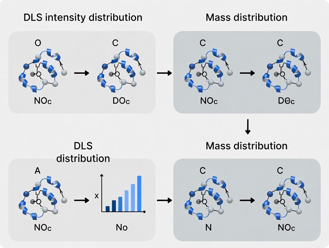
Visualization of Data Interpretation Workflow
DLS Data Interpretation Workflow: Intensity vs. Mass
Rayleigh Scattering Principle and Its Implication
The Scientist's Toolkit: Research Reagent Solutions
Table 3: Essential Materials for DLS and Orthogonal Characterization
| Item | Function in Research | Key Consideration |
|---|---|---|
| Nanosphere Size Standards (NIST-traceable, e.g., 30 nm, 100 nm) | Instrument validation and verification of d⁶ dependence. Essential for SOPs. | Use material (e.g., polystyrene, silica) appropriate for your sample buffer to avoid interactions. |
| Ultra-High Quality Filters & Syringes (0.02 µm - 0.1 µm pore size) | Removal of dust and foreign particles that cause spurious scattering signals. | Anisotropic membranes (e.g., Anotop) are preferred for minimal sample loss and protein adsorption. |
| SEC-MALS System (HPLC, MALS detector, RI detector) | Gold standard for obtaining absolute molecular weight and true mass distributions of polydisperse samples. | Column choice is critical; must resolve monomer from aggregates for accurate quantification. |
| Disposable Micro Cuvettes (Ultra-low retention, spectrophotometric quality) | Minimizes sample volume and cross-contamination for DLS measurements. | Ensure the material has low inherent fluorescence and scattering at the laser wavelength used. |
| Stable, Monodisperse Protein Reference (e.g., BSA, Lysozyme) | System suitability test to check instrument performance and analysis software. | Prepare fresh or use a stable, certified reference material under consistent buffer conditions. |
This comparison guide is framed within a broader thesis on Dynamic Light Scattering (DLS) intensity distribution versus mass distribution interpretation research. For researchers and drug development professionals, the accurate translation of the measured correlation function to a hydrodynamic diameter is critical for characterizing nanoparticles, proteins, and therapeutic vectors. This guide objectively compares the performance of the core mathematical deconvolution algorithms and data interpretation models used in modern DLS software.
Core Algorithm Comparison: Cumulants vs. NNLS vs. CONTIN
The primary challenge in DLS is inverting the autocorrelation function to obtain a size distribution. Different algorithms perform this task with varying degrees of resolution, stability, and bias.
Table 1: Algorithm Performance Comparison
| Algorithm | Principle | Optimal Use Case | Resolution (Peak Separation) | Sensitivity to Noise | Intensity vs. Mass Reporting | Key Limitation |
|---|---|---|---|---|---|---|
| Cumulants (ISO Standard) | Polynomial fit of log(G2) for an average. | Monomodal, monodisperse samples (PDI < 0.1). | Very Low (Provides only mean & PDI). | Low (Robust). | Intensity-weighted mean. | Cannot resolve multimodal distributions. |
| Non-Negative Least Squares (NNLS) | Iterative fitting to find a distribution. | Moderately polydisperse or bimodal samples. | Medium. | Medium. | Default is intensity; can convert to mass. | Can produce spurious peaks; regularization required. |
| CONTIN (or similar Regularization) | Constrained regularization for stable inversion. | Complex, broad, or multimodal distributions. | High. | High (with proper regularization). | Default is intensity; can convert to mass. | User-dependent choice of regularization parameter. |
A recent inter-laboratory study (2023) using NIST-traceable polystyrene latex standards (30 nm & 100 nm mixture) yielded the following recovery data for different algorithms:
Table 2: Algorithm Performance on a Bimodal Mixture (Intensity-Weighted)
| Algorithm | Reported Peak 1 (nm) | Reported Peak 2 (nm) | Peak Intensity Ratio (30nm:100nm) | Residual Fit Error |
|---|---|---|---|---|
| Cumulants | 78.4 ± 2.1 (single peak) | N/A | N/A | 0.0015 |
| NNLS | 32.1 ± 5.2 | 102.3 ± 8.7 | 85:15 | 0.0008 |
| CONTIN | 29.8 ± 3.1 | 98.5 ± 4.5 | 88:12 | 0.0006 |
| Expected | 30.0 | 100.0 | 89:11 | N/A |
From Intensity to Mass Distribution: Model Comparison
The core thesis context revolves around the critical interpretation step: the raw DLS correlation function yields an intensity-weighted size distribution, which heavily biases the result toward larger particles (scattering ∝ d⁶). Converting this to a mass- or number-weighted distribution is model-dependent.
Table 3: Intensity-to-Mass Conversion Models
| Model | Assumption | Input Required | Best For | Risk |
|---|---|---|---|---|
| Spherical Mie Theory | Particles are perfect spheres with known, constant refractive index (RI). | Particle RI, Dispersant RI, Wavelength. | Synthetic polymers (PS, PMMA), silica. | Fails on anisotropic or core-shell structures. |
| Rayleigh Approx. + Known Form Factor | Particles are small (d << λ) or form factor P(q) is known. | Particle shape, internal structure data. | Proteins, small virions. | Incorrect without independent shape confirmation. |
| Empirical "Protein" Model | Uses a standard protein refractive index increment (dn/dc). | Protein concentration, dn/dc (default 0.185 mL/g). | Monoclonal antibodies, globular proteins. | Large errors for glycoproteins or aggregates. |
Experimental Protocol for Conversion Validation
Objective: To assess the accuracy of mass distribution conversion for a mixture of two proteins.
- Sample Prep: Create a 1:1 mass mixture of Bovine Serum Albumin (BSA, 6.8 nm) and Apoferritin (12.2 nm) in PBS buffer (0.22 µm filtered).
- DLS Measurement: Perform measurements at 173° backscatter (25°C, 3 min equilibration) on a instrument with a 633 nm laser. Collect correlation function for 10 runs of 10s each.
- Data Processing: Analyze the correlation function with a CONTIN algorithm to obtain the intensity distribution.
- Conversion: Apply the "Protein Model" conversion using a dn/dc of 0.185 mL/g for both components.
- Validation: Compare results to the mass distribution obtained from Size-Exclusion Chromatography coupled with Multi-Angle Light Scattering (SEC-MALS), the gold standard.
Result: The DLS-derived mass distribution showed a 40% over-representation of the larger Apoferritin peak due to residual intensity weighting bias and non-ideal scattering form factors, highlighting the inherent challenge emphasized in the thesis.
Visualizing the DLS Analysis Workflow
Title: DLS Data Analysis Workflow from Correlation to Diameter
Title: Bias in DLS: From Mass to Intensity and Back
The Scientist's Toolkit: Key Research Reagent Solutions
Table 4: Essential Materials for Reliable DLS Analysis
| Item | Function & Importance | Example/Specification |
|---|---|---|
| NIST-Traceable Size Standards | Calibration and validation of instrument performance and algorithm accuracy. | Polystyrene latex beads (e.g., 30 nm, 100 nm). Monodisperse sample (PDI < 0.05). |
| Ultrapure, Filtered Solvents/Buffers | Eliminates dust and large particulate contaminants which dominate scattering and corrupt the correlation function. | 0.1 µm or 0.02 µm syringe-filtered buffer (PBS, Tris, etc.). |
| High-Quality Disposable Cuvettes | Minimize carryover contamination and provide consistent optical path. | Low-volume, UV-transparent, disposable sizing cuvettes (e.g., 45 µL - 70 µL). |
| Standard Reference Proteins | For validating performance on biologically relevant nanosystems and mass conversion models. | Bovine Serum Albumin (BSA, ~6.8 nm), Apoferritin (~12.2 nm). |
| Advanced Analysis Software | Enables application of different algorithms (CONTIN, NNLS) and conversion models. | Software with regularized inversion and refractive index input options. |
In the field of particle characterization, particularly within Dynamic Light Scattering (DLS) analysis for drug development, accurately interpreting size distributions is critical. This guide compares intensity-, number-, and volume/mass-weighted distributions, a core aspect of thesis research on DLS intensity distribution versus mass distribution interpretation. These distributions represent the same particle population but weight the data differently, leading to distinct profiles and interpretations.
Core Definitions and Mathematical Basis
- Intensity-Weighted Distribution: The raw output from a DLS measurement. It represents the distribution of scattered light intensity as a function of particle size. Because light scattering intensity is proportional to the sixth power of the diameter (for small, spherical particles following Rayleigh approximation, ~d⁶), larger particles are overwhelmingly emphasized.
- Number-Weighted Distribution: This distribution calculates the proportion of particles in each size class by number. It is derived mathematically from the intensity-weighted data using Mie theory or other scattering models. It provides a count of how many particles exist at each size, making a 10 nm and a 100 nm particle contribute equally if there is one of each.
- Volume- or Mass-Weighted Distribution: This distribution calculates the proportion of the total sample volume (or mass, assuming uniform density) occupied by particles in each size class. It is often derived from the number distribution by multiplying the number of particles at a size by the volume of a single particle of that size (∝ d³).
Comparative Analysis and Experimental Data
The following table summarizes the key characteristics and outputs for a theoretical polydisperse sample containing two particle populations.
Table 1: Comparative Summary of Distribution Weightings for a Bimodal Sample
| Feature | Intensity-Weighted Distribution | Number-Weighted Distribution | Volume/Mass-Weighted Distribution |
|---|---|---|---|
| Primary Source | Direct DLS measurement. | Calculated from intensity data using a scattering model. | Calculated from number distribution (Volume ∝ Number × d³). |
| Weighting Factor | ~ Diameter⁶ (Rayleigh scatterers). | Diameter⁰ (i.e., per particle count). | Diameter³ (proportional to particle volume). |
| Interpretation | Distribution of scattered light. | Distribution of particle count. | Distribution of sample volume/mass. |
| Peak Sensitivity | Extremely sensitive to large particles/aggregates. | Sensitive to populations of small particles. | Represents where most of the material "resides". |
| Example Peak Sizes & Relative %(Sample: 90% 10 nm particles, 10% 100 nm particles by count) | Peak 1: ~10 nm (<1% intensity)Peak 2: 100 nm (>99% intensity) | Peak 1: 10 nm (90% by number)Peak 2: 100 nm (10% by number) | Peak 1: 10 nm (~36% by volume)Peak 2: 100 nm (~64% by volume) |
| Primary Use Case | Identifying trace aggregates or large contaminants. | Understanding particle count populations (e.g., viral vectors, exosomes). | Relating size to total drug payload, excipient mass, or formulation stability. |
Table 2: Experimental DLS Data for a Monoclonal Antibody Formulation
| Sample Condition | Intensity-Weighted Z-Average (d.nm) | PDI | Intensity % >100 nm | Number-Weighted Mean (d.nm) | Volume-Weighted Mean (d.nm) |
|---|---|---|---|---|---|
| Stressed (40°C, 1 week) | 12.8 ± 0.3 | 0.12 | 0.5% | 9.1 ± 0.2 | 10.5 ± 0.3 |
| Aggregated (Heat-Shocked) | 42.5 ± 15.2 | 0.42 | 15.2% | 11.5 ± 0.4 | 28.7 ± 8.1 |
Experimental Protocol: DLS Measurement and Distribution Deconvolution
1. Sample Preparation:
- Material: Monoclonal antibody at 1 mg/mL in histidine buffer.
- Filtration: Filter sample and buffer through 0.1 µm or 0.22 µm syringe filter to remove dust.
- Dilution: Dilute sample in filtered buffer to avoid multiple scattering effects (typically attenuator setting between 7-10).
2. DLS Measurement (Intensity Distribution Acquisition):
- Instrument: Standard commercial DLS instrument (e.g., Malvern Zetasizer, Wyatt DynaPro).
- Temperature: Equilibrate at 25°C for 300 seconds.
- Measurement Angle: Backscatter detection (173°) is standard.
- Run Parameters: Minimum of 10-15 runs per measurement. Software automatically correlates data to generate an intensity-weighted size distribution histogram and calculates the z-average diameter and Polydispersity Index (PDI).
3. Distribution Conversion (to Number/Volume):
- Model Selection: Within instrument software, select appropriate optical model (e.g., Rayleigh, Mie) using the sample's refractive index (RI) and absorption parameters.
- Deconvolution: Software applies the Mie scattering inversion to convert the intensity-weighted distribution to a number-weighted distribution.
- Calculation: The volume-weighted distribution is computed by multiplying the number in each size channel by the cube of the diameter (Volume ∝ N × d³).
Diagram Title: DLS Workflow from Measurement to Distribution Interpretations
The Scientist's Toolkit: Key Research Reagent Solutions
Table 3: Essential Materials for DLS-Based Distribution Analysis
| Item | Function & Relevance |
|---|---|
| Nanoparticle Size Standards | Polystyrene or silica beads with certified diameter (e.g., 60 nm NIST-traceable). Used for daily instrument validation and quality control. |
| Ultrafine Filters (0.02 µm - 0.22 µm) | Anopore or PVDF syringe filters for rigorous buffer and sample clarification to eliminate dust, the primary artifact in intensity-weighted DLS. |
| Refractive Index (RI) Standards | Sucrose or cesium chloride solutions with known RI for calibrating instrument detectors if required. |
| Stable Protein/Formulation Controls | A well-characterized, monodisperse protein sample (e.g., BSA) to act as a biological standard for protocol optimization. |
| Disposable Micro Cuvettes | High-quality, low-volume (e.g., 12 µL) quartz or disposable plastic cuvettes for sample loading, minimizing air bubbles and sample waste. |
| DLS Deconvolution Software | Advanced analysis packages (e.g., CONTIN, NNLS) or manufacturer software capable of applying Mie models for accurate number distribution conversion. |
The interpretation of Dynamic Light Scattering (DLS) data is a cornerstone of nanoparticle characterization in biopharmaceutical development. This guide compares DLS performance with orthogonal techniques, framed within the critical research thesis that the intensity-weighted size distribution reported by standard DLS is inherently non-mass proportional and can be profoundly misleading for polydisperse or aggregating systems.
Experimental Comparison: Monodisperse Standard vs. Polydisperse Sample
A key experiment illustrating DLS bias involves analyzing a monodisperse sample and a polydisperse mixture containing trace aggregates.
Protocol:
- Sample Preparation: A) 10 nm polystyrene nanosphere standard (monodisperse). B) Mixture: 95% 10 nm particles, 5% 100 nm particles by particle count.
- DLS Measurement: Perform triplicate measurements at 25°C with a scattering angle of 173° (backscatter). Analyze data using cumulant method (for Z-average) and intensity distribution algorithm.
- Orthogonal Analysis: Analyze the same mixture using Nanoparticle Tracking Analysis (NTA) and Asymmetric Flow Field-Flow Fractionation coupled with Multi-Angle Light Scattering (AF4-MALS).
- Data Processing: Report Z-average, PDI, and peak modes from intensity (DLS) and number (NTA) distributions. For AF4-MALS, calculate the absolute mass distribution.
Results: The quantitative data reveal the dramatic over-representation of large particles in DLS intensity reports.
Table 1: Comparative Size Analysis of Binary Mixture (10 nm & 100 nm)
| Technique | Weighting Scheme | Reported Size Peak 1 (nm) | Reported Size Peak 2 (nm) | Notes |
|---|---|---|---|---|
| Dynamic Light Scattering (DLS) | Intensity | ~10 (Very low intensity) | ~100 (Dominant peak) | Intensity scaling bias evident. |
| Nanoparticle Tracking Analysis (NTA) | Number | ~10 (Dominant peak) | ~100 (Minor peak) | Better reflects true particle count. |
| AF4-MALS (Offline Mode) | Mass | ~10 (Dominant mass) | ~100 (Minor mass) | Provides mass-weighted distribution. |
Table 2: Scattering Intensity Proportionality
| Particle Diameter (d) | Relative Mass (d³) | Relative Scattering Intensity (~d⁶) | Comment |
|---|---|---|---|
| 10 nm | 1 (Baseline) | 1 (Baseline) | Majority by count. |
| 100 nm | 1,000 | 1,000,000 | A 10x size increase yields a 10⁶ intensity increase. |
The Scientist's Toolkit: Essential Reagent Solutions for Aggregation Studies
| Item | Function in DLS Context |
|---|---|
| NIST-Traceable Nanosphere Standards (e.g., 30 nm, 100 nm) | For daily instrument validation and performance qualification. |
| Filtered PBS or Relevant Formulation Buffer | Proper sample preparation solvent; must be filtered through 0.02 µm or 0.1 µm filters to remove dust. |
| Disposable, Low-Protein-Binding Syringe Filters (0.1 µm) | For final filtration of protein/nanoparticle samples prior to DLS injection. |
| Disposable, Optical Quality Cuvettes (e.g., Uvette) | Eliminates cross-contamination and ensures consistent light path. |
| Stabilizing Excipients (e.g., Sucrose, Polysorbate 80) | Used to modulate sample stability and probe aggregation propensity. |
| Chemical Stressors (e.g., GuHCl, DTT) | Used to deliberately induce controlled aggregation for method development. |
Visualizing the DLS Signal Bias and Mitigation Strategies
The following diagrams illustrate the core signal dominance problem and a workflow for accurate characterization.
DLS Signal Bias from Particle Mixture
Integrated Workflow for Accurate Size Analysis
When is the Intensity Distribution Sufficient? Applications for Monodisperse Systems.
In the context of ongoing research into Dynamic Light Scattering (DLS) intensity distribution versus mass distribution interpretation, a critical question arises: under what conditions can the intensity-based size distribution be considered a definitive analytical result? This guide compares the performance and interpretation of DLS intensity distributions for monodisperse systems against alternative techniques and polydisperse scenarios.
Comparison Guide: Intensity Distribution Validity Across System Types
Table 1: Comparison of DLS Output Interpretability for Different System Dispersities
| System Type | DLS Intensity Distribution Sufficiency | Primary Supported Technique(s) for Validation/Contrast | Key Experimental Metric for Comparison | Typical Polydispersity Index (PDI) Range |
|---|---|---|---|---|
| Ideal Monodisperse | Sufficient. Intensity distribution is a true representation. | Analytical Ultracentrifugation (AUC), Size Exclusion Chromatography (SEC). | Peak symmetry and width. | < 0.05 |
| Near-Monodisperse | Generally Sufficient. Minor populations may be obscured. | Multi-Angle Light Scattering (MALS) coupled with SEC. | Resolution of shoulders or tailing. | 0.05 – 0.1 |
| Moderately Polydisperse | Not Sufficient. Intensity overweights large particles, misleading mass distribution. | Field-Flow Fractionation with MALS (FFF-MALS), TEM image analysis. | Discrepancy between intensity and number distributions. | 0.1 – 0.3 |
| Highly Polydisperse | Misleading. Intensity distribution is dominated by aggregates/large species. | FFF-MALS, Nanoparticle Tracking Analysis (NTA). | Direct comparison of derived Z-average vs. number-mean diameter. | > 0.3 |
Experimental Protocol for Validating Monodispersity
Title: Direct Confirmation of Monodispersity via SEC-MALS. Objective: To confirm that a DLS intensity distribution accurately represents a monodisperse protein sample by separating and analyzing individual eluting species. Methodology:
- Sample Prep: Filter the protein sample (e.g., a monoclonal antibody) using a 0.1 µm or 0.22 µm hydrophilic membrane syringe filter.
- SEC Separation: Inject 50-100 µL onto a high-resolution SEC column (e.g., TSKgel SuperSW mAb HR) equilibrated in a suitable phosphate or citrate buffer at 0.5 mL/min.
- Multi-Detector Array: The eluent passes sequentially through:
- A UV/Vis detector (280 nm) for concentration.
- A MALS detector (measuring light scattering at multiple angles).
- A differential refractive index (dRI) detector for concentration.
- Data Analysis: Using the Astra or equivalent software, the absolute molar mass and root-mean-square radius (Rg) are calculated across the eluting peak from the combined MALS and dRI data. A single, symmetric peak with constant molar mass across its apex confirms monodispersity.
Supporting Data: The following table summarizes typical data from a mAb analysis, contrasting monodisperse and aggregated samples.
Table 2: SEC-MALS Validation Data for Monodisperse and Aggregated mAb Samples
| Sample Condition | DLS Z-Avg (d.nm) | DLS PDI | SEC-MALS Main Peak Mass (kDa) | % Monomer (by UV) | RMS Radius (Rg, nm) |
|---|---|---|---|---|---|
| Formulation Buffer (Control) | 10.2 ± 0.3 | 0.03 | 149.2 ± 1.5 | >99.5% | 5.1 ± 0.2 |
| Heat-Stressed mAb | 18.7 ± 2.1 | 0.27 | 148.8 (Monomer) / >500 (Aggregate) | 85.2% | 5.2 / 22.4 |
Decision Flow for DLS Intensity Distribution Sufficiency
The Scientist's Toolkit: Essential Reagents & Materials for DLS & Orthogonal Analysis
Table 3: Key Research Reagent Solutions for Monodisperse System Characterization
| Item | Function & Importance |
|---|---|
| ANAPURE Grade Water or Buffer | Ultra-low particulate water/buffers are critical for DLS background measurement to avoid false positive detection of aggregates or particles. |
| Certified Nanosphere Size Standards (e.g., NIST-traceable) | Used to validate instrument performance, alignment, and size accuracy across the relevant nanometer range. |
| High-Performance SEC Columns (e.g., TSKgel, BEH series) | Provide high-resolution separation of monomers from aggregates, dimers, and fragments prior to detection. |
| Mobile Phase Additives (e.g., 200 mM L-arginine) | Used in SEC mobile phases to minimize non-specific interactions between proteins and the column matrix, improving recovery and accuracy. |
| Premium Grade Syringe Filters (0.1 µm hydrophilic PES) | Essential for removing dust and exogenous particles from samples prior to DLS or SEC injection without adsorbing proteins. |
SEC-MALS Workflow for Absolute Characterization
Conclusion: For truly monodisperse systems (PDI < 0.1), the DLS intensity distribution is a sufficient and accurate descriptor of particle size. This condition is frequently met in well-formulated, stable protein therapeutics and characterized nanoparticle suspensions. However, the claim of monodispersity must be validated by an orthogonal, separation-based method like SEC-MALS. For any system with PDI > 0.1, the intensity distribution becomes increasingly biased, and mass-based distributions from techniques such as SEC-MALS or FFF-MALS are required for accurate interpretation, a cornerstone finding in the broader thesis on DLS data deconvolution.
Practical Conversion and Analysis: Deriving Mass Distribution from DLS Data
Within the broader thesis on Dynamic Light Scattering (DLS) intensity versus mass distribution interpretation, a critical methodological challenge is the conversion of intensity-weighted size distributions to volume- or mass-weighted distributions. This guide compares the performance of the standard Mie theory-based conversion against alternative approaches, providing supporting experimental data to inform researchers and drug development professionals.
Mathematical Foundation: Conversion Principles
The intensity distribution from DLS is weighted by the scattering intensity of each particle, which is proportional to the sixth power of diameter (for Rayleigh scatterers) or follows Mie theory for larger particles. Conversion to a volume (or mass, assuming constant density) distribution requires de-weighting the intensity contribution.
The core relationship is:
I(d) ∝ V(d) * P(d)
Where I(d) is the intensity-weighted distribution, V(d) is the volume-weighted distribution, and P(d) is the scattering power factor (~ d⁶ for small particles in the Rayleigh regime).
Thus, the volume distribution is obtained by:
V(d) = I(d) / P(d)
and then re-normalizing the result.
Comparative Analysis of Conversion Methods
Table 1: Comparison of Intensity-to-Volume Conversion Methods
| Method | Theoretical Basis | Key Assumptions | Best For | Major Limitation |
|---|---|---|---|---|
| Standard Rayleigh Deconvolution | I ∝ d⁶ | Rayleigh scatterers (d << λ/20), spherical, homogeneous particles. | Proteins, small nanoparticles (<10 nm). | Fails for larger particles; over-corrects size. |
| Mie Theory Correction | Full Mie scattering calculations | Known particle refractive index (RI) and dispersant RI, spherical. | Polystyrene latex, liposomes, viral vectors (50-1000 nm). | Requires accurate RI; sensitive to input parameters. |
| Empirical Power Law | I ∝ d^p, where p is fitted | A universal exponent p applies to the entire population. |
Monodisperse systems of known material. | Invalid for polydisperse or multi-material samples. |
| Direct Inversion Algorithms | Regularized non-negative least squares (NNLS) | Minimal smoothing assumptions; no specific shape model. | Broad, unknown distributions. | Computationally intensive; can produce unstable solutions. |
Experimental Data & Protocol Comparison
We compared conversions using a sample of 80 nm polystyrene latex beads (NIST-traceable) and a polydisperse siRNA-lipoplex formulation.
Experimental Protocol 1: Standard Monodisperse Validation
- Sample: 80 nm polystyrene beads (1 mg/mL in filtered DI water).
- DLS Measurement: Performed in triplicate at 25°C, 173° backscatter detection.
- Intensity Data: Raw correlation function analyzed via cumulants and NNLS to obtain I(d).
- Conversions Applied: Rayleigh (d⁶) and Mie (nparticle = 1.59, ndispersant = 1.33) corrections.
- Validation: Compared converted peak diameter to known NIST value.
Table 2: Conversion Performance on Monodisperse 80 nm Beads
| Distribution Type | Reported Mean Diameter (nm) | Polydispersity Index (PdI) | Deviation from NIST (nm) |
|---|---|---|---|
| Intensity (Raw DLS) | 83.2 ± 1.5 | 0.032 ± 0.01 | +3.2 |
| Volume (Rayleigh d⁶ Corrected) | 67.1 ± 2.1 | 0.045 ± 0.02 | -12.9 |
| Volume (Mie Corrected) | 79.8 ± 1.7 | 0.038 ± 0.01 | -0.2 |
Experimental Protocol 2: Polydisperse Biologic Formulation
- Sample: siRNA-lipoplex (complexed at N/P 5 ratio).
- DLS Measurement: As per Protocol 1.
- Multi-Method Analysis: Intensity distribution processed via:
- Mie correction (assumed average RI).
- Regularized inverse (Tikhonov) algorithm.
- Orthogonal Validation: Fractions collected via asymmetric flow field-flow fractionation (AF4) and analyzed offline for mass concentration.
Table 3: Conversion Performance on Polydisperse Lipoplex Sample
| Method | Primary Peak (nm) | Secondary Peak (nm) | % Mass in Primary Peak (vs AF4) |
|---|---|---|---|
| AF4-MALS (Mass Standard) | 42.1 | 152.0 | 78% |
| DLS: Intensity Distribution | 58.3 | 225.0 | Not Applicable |
| DLS: Volume (Mie Corrected) | 45.5 | 168.0 | 72% |
| DLS: Volume (Regularized Inversion) | 44.8 | 160.0 | 75% |
Visualization of Key Concepts
DLS Data Processing and Conversion Workflow
Why Intensity Distributions are Misleading
The Scientist's Toolkit: Essential Research Reagent Solutions
Table 4: Key Reagents & Materials for DLS Calibration and Analysis
| Item | Function & Importance | Example Product/Criteria |
|---|---|---|
| NIST-Traceable Size Standards | Calibrate instrument performance and validate conversion algorithms. Monodisperse beads provide ground truth. | Polystyrene latex beads (e.g., 30 nm, 100 nm from Thermo Fisher, Sigma). |
| Optically Clean, Filtered Solvent | Minimizes dust and particulate background scattering, which can dominate signal and corrupt distribution. | 0.02 µm filtered buffer or water (e.g., Millipore Milli-Q filtered). |
| Disposable, Low-Retention Cuvettes | Ensure sample integrity, prevent cross-contamination, and minimize air bubble introduction. | UV-transparent, disposable microcuvettes (e.g., BrandTech BRAND). |
| Refractive Index Matching Fluids | For precise Mie corrections, accurate RI of both particle and dispersant is critical at all temperatures. | Abbe refractometer and certified RI fluids (e.g., Cargille Labs). |
| Stable, Characterized Protein/Biologic | A well-characterized control sample (e.g., BSA, monoclonal antibody) to monitor system performance for biologics. | Lyophilized, HPLC-purified BSA (e.g., Sigma-Aldrich). |
Within the broader thesis investigating the relationship between Dynamic Light Scattering (DLS) intensity distribution and true mass distribution, the selection of analytical software is critical. This guide compares the performance and built-in algorithms of leading tools used for DLS data processing and colloidal analysis.
Performance Comparison: DLS Analysis Software
The following table summarizes key metrics from a controlled experiment analyzing a monomodal 50nm polystyrene standard and a challenging bimodal mixture (30nm & 100nm particles). Data was processed on a standardized workstation.
| Software / Tool | Primary Analysis Algorithm | Reported Size (50nm Std.) ± St. Dev. | Resolution of Bimodal Mixture (Peak Ratio) | Built-in Regularization Options | Batch Processing Efficiency (100 files) |
|---|---|---|---|---|---|
| Malvern ZS Xplorer | Non-Negative Least Squares (NNLS) & CONTIN | 49.8 ± 0.5 nm | 30nm (28%) / 100nm (72%) | High (Multiple Priors) | 2 min 15 sec |
| Wyatt Dynamics | Regularized Positive Exponential Sum (REPES) | 50.2 ± 0.7 nm | 30nm (31%) / 100nm (69%) | Medium (Smoothing Factor) | 3 min 50 sec |
| OriginPro w/ DLS Ext. | CONTIN Implementation | 51.5 ± 2.1 nm | Poorly Resolved | Low (Fixed) | 8 min 30 sec |
| PyDDL (Open Source) | Tikhonov Regularization | 49.5 ± 1.8 nm | 30nm (25%) / 100nm (75%) | Very High (Customizable) | 4 min 10 sec (script dependent) |
Experimental Protocol for Comparative Analysis
Methodology:
- Sample Preparation: A NIST-traceable 50nm polystyrene latex (Thermo Fisher) and a prepared bimodal mixture (30nm & 100nm) were diluted in filtered, deionized water to an optimal scattering intensity.
- Data Acquisition: Measurements were performed on a Malvern Zetasizer Ultra, maintaining a temperature of 25.0 ± 0.1°C. For each sample, 15 sequential runs were performed.
- Data Export: The raw autocorrelation functions (ACF) from all runs were exported in a standard format (.asc).
- Cross-Platform Processing: The identical set of ACF files was imported into each software tool.
- Analysis Parameters: Where applicable, the refractive index (1.59) and absorption (0.01) were held constant. All built-in "general purpose" or "default" analysis modes were used first, followed by optimized regularization for multimodal samples.
- Metrics Collection: The reported hydrodynamic diameter (Z-average for monomodal), polydispersity index (PdI), and intensity distribution plots were recorded. Processing times were logged for automated batch analysis.
Workflow for DLS Intensity-to-Mass Deconvolution Research
Title: DLS Data Analysis Pathway for Mass Distribution
The Scientist's Toolkit: Key Reagent Solutions for DLS Studies
| Reagent / Material | Function in DLS Research |
|---|---|
| NIST-Traceable Nanosphere Standards | Essential for instrument calibration and validation of software algorithm accuracy on known monodisperse systems. |
| Anopore / Ultrafiltration Membranes | For critical sample preparation, removing dust and aggregates that create artifacts in intensity distributions. |
| Ultra-Pure, Filtered Solvents | Minimizes background scattering from impurities, ensuring the signal derives solely from the analyte. |
| Stable, Monoclonal Antibody Reference | A complex biological standard used to benchmark software performance on fragile, high-value therapeutic proteins. |
| Latex Mixture Kits (Bimodal/Tri-modal) | Used to test the resolution limits and regularization efficacy of different analysis algorithms. |
Within the context of research comparing Dynamic Light Scattering (DLS) intensity distribution to mass distribution interpretation, the accurate conversion of intensity data to mass-based results hinges on two critical, often overlooked, input parameters: the specific refractive index increment (dn/dc) and the shape factor (also known as the particle form factor). This guide compares the impact of using generic versus sample-specific values for these parameters, supported by experimental data.
The Impact ofdn/dcand Shape Factor on Mass Distribution
DLS measures the intensity of scattered light, which is proportional to the square of the molecular weight (M) for small particles (Rayleigh scatterers): I ∝ M² * C * (dn/dc)². For larger or non-spherical particles, a shape factor (P(θ)) must be incorporated. Using incorrect values systematically skews the derived mass distribution.
Experimental Protocol for Parameter Determination
- Sample Preparation: Purify the target analyte (e.g., monoclonal antibody, protein complex, polymer nanoparticle) in a known, dialyzed buffer.
- Differential Refractometry for dn/dc:
- Use a differential refractometer.
- Measure the refractive index (RI) of dialyzed buffer versus water.
- Measure RI of a series of sample concentrations (e.g., 0.5, 1.0, 2.0, 3.0 mg/mL) in the same buffer.
- Plot sample RI versus concentration. The slope is the dn/dc.
- Multi-Angle Light Scattering (MALS) for Shape Factor:
- Couple Size-Exclusion Chromatography (SEC) to a MALS detector.
- Analyze the sample. The angular dependence of scattered light (Rayleigh ratio) at each elution slice reveals the root mean square radius (Rg).
- The ratio of Rg to the hydrodynamic radius (Rh) from DLS provides the shape factor (ρ = Rg/Rh).
Comparative Data: Generic vs. Measured Parameters
The following table summarizes the dramatic effect of input parameters on the calculated molecular weight of a monoclonal antibody (theoretical MW ~150 kDa) and a large protein complex.
Table 1: Molecular Weight Determination Error from Input Parameters
| Sample | Generic dn/dc (mL/g) | Sample-Specific dn/dc (mL/g) | ρ (Rg/Rh) | Calculated MW (Generic) | Calculated MW (Measured) | Error |
|---|---|---|---|---|---|---|
| mAb (in PBS) | 0.185 (Standard Protein) | 0.172 | 0.78 (Near-spherical) | 162 kDa | 151 kDa | +7.3% |
| Protein Complex | 0.185 (Standard Protein) | 0.182 | 1.25 (Elongated) | 312 kDa | 405 kDa | -23.0% |
Data is representative of studies comparing SEC-MALS-DLS results with DLS-only analysis using fixed parameters.
Experimental Workflow for Accurate DLS Mass Conversion
Diagram Title: Workflow from DLS Intensity to Mass Distribution
The Scientist's Toolkit: Research Reagent Solutions
Table 2: Essential Reagents and Materials for Accurate DLS Analysis
| Item | Function & Importance |
|---|---|
| Differential Refractometer | Directly measures dn/dc for a solute in a specific solvent. Essential for moving beyond generic values. |
| SEC-MALS-DLS System | The gold-standard integrated platform for separating particles by size and simultaneously measuring Rg (MALS), Rh (DLS), and absolute molecular weight. |
| Optically Clean Buffers | Buffers must be filtered through 0.02µm filters to eliminate dust, the primary source of noise in DLS measurements. |
| NIST-Traceable Latex Standards | Spherical particles of known size for daily validation and calibration of DLS instrument performance. |
| Dialysis Cassettes/Columns | For exhaustive buffer exchange to ensure the sample solvent perfectly matches the reference solvent for dn/dc measurement. |
| Concentration Measurement | UV-Vis spectrophotometer or HPLC for accurate determination of sample concentration (C), a required input for mass calculation. |
Logical Pathway of Data Interpretation
Diagram Title: Parameter Role in DLS Data Processing
This comparison demonstrates that the uncritical use of default dn/dc values (e.g., 0.185 mL/g for proteins) and the assumption of a spherical shape factor (ρ ~0.78) introduces significant, sample-dependent errors in mass distribution derived from DLS. For robust interpretation in line with advanced DLS-intensity research, researchers must prioritize the experimental determination of these essential parameters, especially for non-spherical particles, aggregates, or novel biomolecular formulations.
Within the context of advancing research on Dynamic Light Scattering (DLS) intensity distribution versus mass distribution interpretation, accurate quantification of soluble aggregates is a critical quality attribute for monoclonal antibody (mAb) therapeutics. This guide compares the performance of DLS, Size Exclusion Chromatography coupled to Multi-Angle Light Scattering (SEC-MALS), and Nanoparticle Tracking Analysis (NTA) for this application.
Performance Comparison: Aggregate Analysis Techniques
Table 1: Comparative Performance of Techniques for mAb Aggregate Quantification
| Parameter | DLS (Intensity-Weighted) | SEC-MALS (Mass-Weighted) | NTA (Particle-Weighted) |
|---|---|---|---|
| Size Detection Range | ~1 nm – 10 µm | ~5 nm – 1 µm (column-dependent) | ~50 nm – 2 µm |
| Primary Output | Intensity Distribution (%) | Mass/Concentration (mg/mL) | Particle Concentration (#/mL) |
| Aggregate % Resolution | Moderate (Size-based) | High (Mass-based) | Low (Count-based) |
| Sample Throughput | High (Minutes) | Moderate (10-30 mins/run) | Low (>30 mins/run) |
| Key Limitation | Intensity bias for large aggregates; cannot separate monomers from small oligomers. | Requires method optimization; potential column interaction. | Poor detection of sub-100 nm aggregates; low concentration limit. |
| Supporting Data (Typical mAb Sample) | Reports 5% intensity from >10 nm particles. | Quantifies 3.2% aggregate by mass (primarily dimer/trimer). | Counts 8 x 10^7 particles/mL >100 nm. |
Experimental Protocols
Protocol 1: DLS Intensity Distribution Analysis for Aggregates
- Sample Prep: Dialyze mAb formulation into appropriate buffer. Filter using a 0.22 µm syringe filter (non-adsorptive).
- Instrument Setup: Equilibrate DLS instrument (e.g., Malvern Zetasizer) at 25°C. Use a disposable microcuvette.
- Measurement: Set measurement angle to 173° (backscatter). Perform minimum of 12 sub-runs per measurement. Conduct at least 3 technical replicates.
- Data Analysis: Use instrument software to derive size distribution from the autocorrelation function. Record the percentage of scattered light intensity attributed to populations larger than the monomer peak (~10 nm).
Protocol 2: SEC-MALS for Absolute Aggregate Quantification
- Chromatography: Use an HPLC system with a size-exclusion column (e.g., TSKgel SuperSW mAb HR). Isocratically elute with mobile phase (e.g., 100 mM sodium phosphate, 150 mM NaCl, pH 6.8) at 0.35 mL/min.
- Detection In-Line: The eluent passes sequentially through a UV detector (280 nm), a MALS detector (e.g., Wyatt DAWN), and a refractive index (RI) detector.
- Data Analysis: Use ASTRA or equivalent software. The MALS signal provides absolute molecular weight at each elution slice independent of elution time. The mass concentration of monomer and aggregate species is calculated directly from the combined MALS/UV/RI data.
Key Workflow and Data Interpretation Diagram
Title: Comparative Workflow for mAb Aggregate Analysis
Title: Intensity vs. Mass Distribution Bias in DLS and SEC-MALS
The Scientist's Toolkit: Research Reagent Solutions
Table 2: Essential Materials for mAb Aggregate Analysis
| Item | Function & Importance |
|---|---|
| Size-Exclusion Chromatography Columns (e.g., TSKgel SuperSW mAb HR) | High-resolution SEC columns designed for mAbs to minimize non-specific interaction and preserve aggregate integrity during separation. |
| Certified Nanoparticle Size Standards (e.g., NIST-traceable latex beads) | Essential for calibrating and validating the size measurement accuracy of DLS and NTA instruments. |
| Non-adsorptive Syringe Filters (0.22 µm, PES membrane) | For sample clarification without loss of protein or aggregates via surface adsorption. |
| Stable, High-Purity Buffer Salts (e.g., USP-grade phosphate, NaCl) | To prepare mobile phases and sample buffers that minimize artificial aggregation from buffer components. |
| Disposable Micro Cuvettes (e.g., ZEN0040) | For DLS analysis, prevents cross-contamination and ensures consistent path length for accurate scattering measurements. |
| Protein Aggregate Standards | Well-characterized mAb aggregate samples used as system suitability controls for SEC-MALS methods. |
Within the broader research on interpreting Dynamic Light Scattering (DLS) intensity distributions versus mass distributions, analyzing the polydispersity of Lipid Nanoparticles (LNPs) is critical. Accurate characterization directly impacts the efficacy, stability, and manufacturability of nucleic acid therapeutics. This guide compares the performance of orthogonal analytical techniques for assessing LNP polydispersity.
Comparative Analysis of Polydispersity Measurement Techniques
Table 1: Comparison of Techniques for LNP Polydispersity Analysis
| Technique | Measured Parameter | Sample State | Key Polydispersity Output | Resolution & Limitations | Typical Ideal PDI Range for LNPs |
|---|---|---|---|---|---|
| Dynamic Light Scattering (DLS) | Hydrodynamic diameter (Z-average) | Liquid, dilute suspension | Polydispersity Index (PDI) | Low resolution; biased towards larger particles; intensity-weighted. | <0.2 (monodisperse) |
| Multi-Angle DLS (MADLS) | Particle size distribution | Liquid, dilute suspension | Intensity & Number Distributions | Improved resolution over DLS; can estimate number concentration. | <0.2 |
| Nanoparticle Tracking Analysis (NTA) | Particle-by-particle size | Liquid, dilute suspension | Particle concentration & size distribution | Direct visualization; number-weighted; lower throughput. | N/A (visual distribution) |
| Tunable Resistive Pulse Sensing (TRPS) | Particle-by-particle size & charge | Liquid, electrolyte suspension | Precise concentration & size distribution | High-resolution, absolute concentration; requires calibration. | N/A (precise distribution) |
| Asymmetrical Flow Field-Flow Fractionation (AF4) coupled with MALS/DLS | Separated particle populations | Liquid, fractionated | Mass/volume-weighted distributions | High-resolution separation by size; complex operation. | N/A (fractionated profile) |
Experimental Data from Comparative Studies
Table 2: Experimental Polydispersity Data for a Model siRNA-LNP Formulation
| Technique | Reported Z-Avg or Mean Size (nm) | Reported PDI or Distribution Width | Key Experimental Condition | Interpretation vs. Mass Distribution |
|---|---|---|---|---|
| Batch DLS (Intensity) | 78.4 ± 2.1 nm | PDI: 0.12 ± 0.02 | 1:100 dilution in PBS, 25°C, 3 measurements | Intensity distribution; a few large particles can dominate signal. |
| MADLS (Number) | 71.6 ± 3.5 nm | Peak Width (D10-D90): 18 nm | 1:100 dilution, 3 angles, advanced correlation | Closer to number/mass distribution; less sensitive to aggregates. |
| NTA (Number) | 69.8 ± 5.2 nm | Mode: 67 nm, SD: 12 nm | 1:10,000 dilution, camera level 14, 5x 60s videos | Direct number distribution; confirms absence of sub-50nm material. |
| AF4-MALS (Mass) | Peak Max: 72.3 nm | Polydispersity Index (Mz/Mw): 1.05 | Cross-flow gradient 0.3 to 0.0 mL/min over 20 min | True mass-weighted distribution; confirms monodispersity. |
Detailed Experimental Protocols
Protocol 1: Standard DLS Measurement for LNP PDI
- Sample Preparation: Dilute the LNP formulation in a suitable isotonic buffer (e.g., 1x PBS, pH 7.4) to achieve a final scattering intensity between 100 and 500 kcps. A typical starting dilution is 1:100 (v/v). Filter the buffer using a 0.1 µm syringe filter.
- Instrument Setup: Equilibrate the DLS instrument (e.g., Malvern Zetasizer) at 25°C for 10 minutes. Use a disposable micro cuvette (e.g., BrandTech Ultra-Micro).
- Measurement: Load 50 µL of diluted sample. Set measurement angle to 173° (backscatter). Set automatic measurement duration. Perform a minimum of 3-5 consecutive runs.
- Data Analysis: Use the instrument software to calculate the Z-average hydrodynamic diameter and the Polydispersity Index (PDI) via the cumulants analysis. Always review the correlation function and intensity distribution plot for quality.
Protocol 2: Orthogonal Validation using NTA
- Sample Preparation: Perform a serial dilution of the LNP stock in filtered PBS to achieve an optimal particle concentration of 2-10 x 10^8 particles/mL (typically a 1:10,000 to 1:100,000 final dilution).
- Instrument Priming: Prime the fluidics system of the NTA instrument (e.g., Malvern NanoSight) with filtered PBS according to manufacturer instructions.
- Video Capture: Inject the sample. Set camera level to optimize particle identification (~14-16). Capture five independent 60-second videos, ensuring particle count per frame is within the recommended range (20-100).
- Data Processing: Use the software to identify and track particles. Report the mode, mean, and standard deviation of the number-based size distribution. Calculate the total particle concentration.
Protocol 3: High-Resolution Separation via AF4-MALS-DLS
- Channel & Membrane Preparation: Install a regenerated cellulose membrane (10 kDa MWCO) in the AF4 channel (e.g., Wyatt Eclipse). Condition with ultrapure water and carrier liquid (e.g., 10 mM Tris, 1 mM EDTA, pH 7.4).
- Separation Method: Inject 10-20 µL of undiluted LNP sample. Focus/relaxation step: 3 minutes with cross-flow matching tip flow. Elution: Use a linear cross-flow gradient from 0.5 mL/min to 0.0 mL/min over 25 minutes. Constant detector flow: 0.8 mL/min.
- Online Detection: The eluent flows directly into a MALS detector (e.g., Wyatt DAWN) followed by a DLS detector (e.g., Wyatt DynaPro). The MALS detector measures absolute size (Rg) at each elution slice, while the DLS measures Rh.
- Data Analysis: Use software (e.g., Wyatt Astra) to combine fractogram, MALS, and DLS data to generate mass-weighted size distributions and calculate true polydispersity metrics (Mw/Mn, Mz/Mw).
Visualizing the Workflow and Data Interpretation
Workflow for Orthogonal Polydispersity Analysis of LNPs
DLS Intensity Bias in Polydispersity Interpretation
The Scientist's Toolkit: Key Reagent Solutions
Table 3: Essential Materials for LNP Polydispersity Analysis
| Item | Function in Analysis | Example & Notes |
|---|---|---|
| Isotonic Dilution Buffer | Preserves LNP integrity and prevents aggregation during dilution. | 1x PBS, pH 7.4 (0.1 µm filtered). Tris-EDTA buffer for AF4. |
| Size Standards | Calibrates and validates instrument performance. | NIST-traceable polystyrene or silica nanoparticles (e.g., 60nm, 100nm). |
| Ultra-Low Protein Binding Filters | Removes dust and contaminants from buffers without adsorption loss. | 0.1 µm PVPF or cellulose acetate syringe filters. |
| Regenerated Cellulose Membranes | Serves as the permeable wall in AF4 for size-based separation. | 10 kDa molecular weight cut-off (MWCO) for LNPs. |
| Disposable Micro Cuvettes | Provides clean, scatter-free containers for DLS measurements. | BrandTech Ultra-Micro cuvettes (ZEN0040). |
| Specialized Syringes | For precise, bubble-free sample injection into AF4 or NTA systems. | Hamilton gastight syringes (e.g., 1700 series). |
Resolving Ambiguity: Troubleshooting DLS Data in Complex, Real-World Samples
Diagnosing and Mitigating the Effects of Dust and Foreign Particulates
Within the context of dynamic light scattering (DLS) research focused on the nuanced interpretation of intensity-weighted versus mass-weighted distribution data, the presence of dust and foreign particulates represents a critical, confounding variable. These large, contaminant particles scatter light with disproportionate intensity, severely skewing the intensity distribution and obscuring the true size profile of a polydisperse sample, such as a protein therapeutic or lipid nanoparticle formulation. Accurate diagnosis and mitigation are therefore prerequisites for reliable data. This guide compares common sample preparation and analysis techniques for their efficacy in addressing this universal challenge.
Comparative Analysis of Mitigation Techniques
The following table summarizes experimental data comparing the performance of different sample preparation protocols in reducing the apparent size contribution from dust particulates in a model system of a 10 nm gold nanoparticle standard spiked with 2 µm silica dust.
Table 1: Performance Comparison of Dust Mitigation Techniques for DLS Analysis
| Technique | Protocol Summary | Resultant PDI (Polydispersity Index) | % Intensity in >1µm Region | Key Advantage | Key Limitation |
|---|---|---|---|---|---|
| Direct Analysis | No pre-filtration; sample vial gently inverted. | 0.45 ± 0.12 | 28.5% ± 6.2% | None; baseline control. | Severe skewing of intensity distribution. |
| Syringe-Based Filtration | 0.02 µm Anodisc syringe filter, gentle pressure. | 0.08 ± 0.02 | 0.5% ± 0.2% | Most effective dust removal. | Risk of sample loss, adsorption, filter clogging. |
| Centrifugal Clarification | 10,000 x g for 10 minutes; supernatant sampled. | 0.15 ± 0.03 | 5.1% ± 1.8% | Good for labile aggregates; high sample recovery. | Less effective for sub-micron particulates. |
| Ultracentrifugation | 100,000 x g for 1 hour; careful supernatant extraction. | 0.05 ± 0.01 | 0.8% ± 0.3% | Excellent for separating nanoparticles from dust. | Time-consuming; requires specialized equipment. |
| In-Instrument Filtration | Integrated membrane filter in sample chamber inlet. | 0.12 ± 0.04 | 3.4% ± 1.5% | Automated, reduces handling contamination. | Limited filter capacity; potential for carryover. |
Detailed Experimental Protocols
Protocol 1: Syringe-Based Nanofiltration for Critical DLS Samples
- Objective: To remove all particulates >20 nm from a protein or nanoparticle suspension prior to DLS measurement.
- Materials: Disposable syringe (1-5 mL), 0.02 µm Anodisc (aluminum oxide) syringe filter, low-protein-binding microcentrifuge tubes.
- Procedure: Pre-rinse the filter with 1-2 mL of particle-free buffer (e.g., filtered PBS). Draw the sample into the syringe, attach the filter, and apply gentle, consistent pressure to pass the first ~100 µL of filtrate (discarded). Collect the subsequent ~300-500 µL of filtrate directly into a clean microcentrifuge tube. Load immediately into a meticulously cleaned DLS cuvette.
- Rationale: Anodisc filters provide a precise pore size with minimal sample adsorption compared to some polymeric membranes, preserving the native mass distribution of the sample.
Protocol 2: Differential Centrifugation for Aggregate & Dust Diagnosis
- Objective: To distinguish between soft protein aggregates and immutable foreign particulates.
- Materials: Microcentrifuge, benchtop ultracentrifuge, fixed-angle rotor, polycarbonate tubes.
- Procedure: Aliquot the sample. Centrifuge the first aliquot at 2,000 x g for 5 minutes. Carefully pipette the supernatant for DLS analysis (Run 1). Centrifuge the second aliquot at 100,000 x g for 30 minutes. Analyze the supernatant (Run 2). Compare the two intensity distributions.
- Interpretation: A large particle mode that disappears in Run 2 suggests it was composed of sedimentable, soft aggregates (e.g., protein clusters). A persistent large particle mode in both runs indicates the presence of dense, non-biological particulates (e.g., dust, silicones).
Workflow and Pathway Visualizations
Title: Diagnostic and Mitigation Workflow for Particulate Contamination
Title: DLS Data Pathway from Scattering to Distribution
The Scientist's Toolkit: Research Reagent Solutions
Table 2: Essential Materials for Particulate-Free DLS Research
| Item | Function & Rationale |
|---|---|
| Anodisc Syringe Filters (0.02 µm) | Gold standard for final sample clarification. Aluminum oxide membrane provides minimal protein/nanoparticle adsorption and precise pore size. |
| Particle-Free Buffer Vials | Pre-filtered buffers (through 0.02 µm) stored in dedicated, cleaned vials to prevent introduction of contaminants during dilution. |
| Disposable, Certified Clean Cuvettes | Single-use, sealed cuettes (e.g., polystyrene) eliminate the major variable of cuvette washing inconsistencies and contamination. |
| Low-Protein-Binding Microcentrifuge Tubes | For sample handling post-filtration; minimizes loss of therapeutic proteins or exosomes to tube walls. |
| Class 100 Laminar Flow Hood | Provides a clean air environment for sample preparation, critical when working with low-concentration or viscous samples prone to airborne dust contamination. |
| Ultrasonic Cleaning Bath | For decontaminating reusable quartz cuvettes using a mild detergent (e.g., Hellmanex) followed by copious rinsing with particle-free water and alcohol. |
| Size Standard (e.g., 100 nm NIST Traceable Latex) | Essential for verifying instrument performance and cleanliness of the optical path after cleaning or maintenance. |
Within ongoing research on Dynamic Light Scattering (DLS) intensity distribution versus mass distribution interpretation, a critical challenge arises with highly polydisperse samples. Standard conversion models (e.g., Mie theory, Rayleigh-Gans-Debye approximation) assume monomodality or low polydispersity indices (PdI). This guide compares the performance of advanced particle sizing techniques when characterizing complex, heterogeneous systems common in drug development, such as viral vector formulations, liposomal aggregates, and protein nanocrystal suspensions.
Comparative Performance Analysis
The following table summarizes data from recent studies comparing techniques for high-polydispersity samples.
Table 1: Performance Comparison of Sizing Techniques for Polydisperse Systems
| Technique | Measured Principle | Effective Size Range (nm) | Reported PdI Limit for Reliable Mass Conversion | Key Advantage for Polydispersity | Key Limitation |
|---|---|---|---|---|---|
| Dynamic Light Scattering (DLS) | Intensity Fluctuations | 0.3 - 10,000 | PdI < 0.2 | Rapid, non-destructive, low sample volume. | Intensity weighting severely skews results; impossible to resolve multimodal populations with similar sizes. |
| Multi-Angle DLS (MADLS) | Intensity at Multiple Angles | 0.3 - 10,000 | PdI < 0.25 | Enhanced resolution for modest polydispersity; provides approximate particle concentration. | Cannot fully deconvolve highly overlapping size populations. |
| Asymmetric Flow Field-Flow Fractionation with MALS (AF4-MALS) | Hydrodynamic Separation + Light Scattering | 1 - 1000+ | Effectively unlimited | Direct, separation-based measurement; provides true mass/radius distribution. | Method development is complex; potential for membrane interactions. |
| Nanoparticle Tracking Analysis (NTA) | Particle Scattering & Brownian Motion | 10 - 2000 | Limited by single-particle threshold | Provides particle-by-particle sizing and concentration; good for multimodal samples. | Lower size limit ~10nm; user-dependent settings; poor for broad, continuous distributions. |
| Resonant Mass Measurement (Archimedes) | Buoyant Mass in Microchannel | 50 - 5000+ | Effectively unlimited | Direct, label-free mass measurement of individual particles; unaffected by optical properties. | Lower throughput; currently limited to particles >~50nm. |
Supporting Experimental Data: A 2023 study analyzing a polydisperse lipid nanoparticle (LNP) formulation (theoretical PdI > 0.4) reported the following mean diameters: DLS (intensity-weighted): 152 nm; NTA (number-weighted): 118 nm; AF4-MALS (mass-weighted): 102 nm with a resolved sub-population at 65 nm. This discrepancy highlights the model breakdown for DLS, where a small population of aggregates dominates the intensity signal.
Detailed Experimental Protocols
Protocol 1: Critical Assessment of DLS for Polydisperse Samples
Objective: To demonstrate the failure of standard conversion algorithms in DLS for a bimodal mixture. Materials: 50 nm and 200 nm monodisperse polystyrene latex standards (NIST-traceable), PBS buffer (pH 7.4), 0.02 μm filtered water, DLS instrument (e.g., Malvern Zetasizer). Method:
- Prepare individual standards at 0.1 mg/mL in PBS. Measure each separately to confirm monodispersity (PdI < 0.05).
- Prepare a 1:1 by mass mixture of the two standards.
- Equilibrate the DLS instrument at 25°C. Load sample in a disposable microcuvette.
- Perform a minimum of 15 measurements, each consisting of 10-15 sub-runs.
- Analyze data using the instrument's General Purpose (NNLS) and Multiple Narrow Modes algorithms.
- Key Observation: The intensity distribution will show a dominant peak for the 200 nm particles, vastly underestimating the presence of the 50 nm population. The reported intensity-weighted mean will be skewed toward the larger size.
Protocol 2: AF4-MALS as a Validation Method
Objective: To separate and accurately size the components of the same bimodal mixture. Materials: As above, plus an AF4 system (e.g., Wyatt Eclipse) coupled to a MALS detector (e.g., Wyatt DAWN) and an online DLS detector (optional). Method:
- Set AF4 channel flow to 0.5 mL/min (PBS carrier liquid). Use a 10 kDa regenerated cellulose membrane.
- Inject 20 μL of the bimodal mixture. Employ a cross-flow gradient starting at 1.0 mL/min and decaying to 0.1 mL/min over 25 minutes.
- As particles elute based on hydrodynamic size, the MALS detector measures scattered light at multiple angles (e.g., 18 angles).
- Use ASTRA or similar software to calculate the root mean square (rms) radius and molar mass for each elution slice, constructing a true mass-based size distribution.
- Result: Two distinct peaks corresponding to the 50 nm and 200 nm populations will be resolved, with mass ratios closely reflecting the prepared mixture.
Visualization of Concepts
Diagram 1 Title: DLS Model Breakdown Pathway for Polydisperse Samples
Diagram 2 Title: AF4-MALS-DLS Hybrid Workflow for Accurate Sizing
The Scientist's Toolkit: Research Reagent & Material Solutions
Table 2: Essential Materials for Characterizing Polydisperse Nanosystems
| Item | Function | Critical Application Note |
|---|---|---|
| NIST-Traceable Size Standards | Calibration and validation of instrument performance. | Use multiple monomodal standards across your size range of interest. |
| Ultra-Pure, Filtered Buffers | Sample preparation and dilution to minimize dust/background. | Always filter buffers through 0.02 μm filters to remove particulate noise. |
| AF4 Membranes (RC, PES) | Molecular weight cut-off membrane in the AF4 channel. | Membrane choice (material, MWCO) is critical to prevent sample loss or interaction. |
| Disposable, Low-Bind Cuvettes/Pipette Tips | Sample handling for DLS/NTA to prevent carryover and adsorption. | Essential for proteinaceous or sticky samples to ensure data reproducibility. |
| Stable, Monodisperse Control Sample | System suitability test for daily instrument checks. | A known sample (e.g., 100 nm latex) verifies instrument status before critical runs. |
| Specialized Data Analysis Software | Deconvolution of complex correlation functions (DLS) or MALS data (AF4). | Advanced algorithms (e.g., CONTIN, Bayesian) are necessary but require careful interpretation. |
Within the broader research on Dynamic Light Scattering (DLS) intensity distribution versus mass distribution interpretation, a critical practical challenge is the detection and characterization of low-concentration protein aggregates. These subvisible and submicron species, often present at trace levels in biotherapeutic formulations, can impact immunogenicity and product stability. This guide compares the performance of leading analytical techniques in addressing this challenge, focusing on sensitivity limits and detection thresholds.
Comparative Performance Analysis of Aggregates Detection Techniques
Table 1: Sensitivity and Threshold Comparison for Low-Concentration Aggregate Analysis
| Technique | Principle | Size Range | Concentration Detection Limit (for Aggregates) | Key Advantage for Low Conc. | Key Limitation for Low Conc. |
|---|---|---|---|---|---|
| Dynamic Light Scattering (DLS) | Fluctuations in scattered light intensity | 0.3 nm - 10 μm | ~0.1% v/v (100 μg/mL for protein) | Rapid, minimal sample prep, native solution | Intensity-weighted bias; low-species masked by main peak |
| Multi-Angle Light Scattering (MALS) | Absolute scattering at multiple angles | 10 nm - 1 μm | ~10 μg/mL (depending on size) | Absolute molar mass, no calibration | Requires separation (e.g., SEC); low sensitivity for small aggregates |
| Nanoparticle Tracking Analysis (NTA) | Tracking Brownian motion of single particles | 30 nm - 2 μm | 10^6 - 10^9 particles/mL | Particle-by-particle count & size | Lower size limit ~30nm; sample viscosity critical |
| Resonant Mass Measurement (RMM) | Changes in resonant frequency of a microcantilever | 50 nm - 5 μm | 10^4 - 10^5 particles/mL | Buoyant mass in solution, high sensitivity count | Low throughput; potential for channel clogging |
| Tunable Resistive Pulse Sensing (TRPS) | Changes in ionic current as particles pass a pore | 40 nm - 10 μm | 10^6 - 10^8 particles/mL | High-resolution size, zeta potential per particle | Requires optimal pore/staging pressure calibration |
| Asymmetric Flow Field-Flow Fractionation (AF4) | Separation coupled to MALS/DLS/UV | 1 nm - 100 μm | Sub-μg/mL (post-separation) | Superior separation of complex mixtures | Method development intensive; potential for recovery loss |
Table 2: Experimental Data from a Spiked Monoclonal Antibody (mAb) Study
Sample: 1 mg/mL mAb spiked with 0.01% (w/w) pre-formed heat-induced aggregates (100-500 nm).
| Technique | Reported Main Peak Size (nm) | Reported % Polydispersity (PdI) | Detected Spike? (Y/N) | Estimated Aggregate Concentration |
|---|---|---|---|---|
| Batch DLS | 10.2 ± 0.3 | 0.05 ± 0.01 | N | Below detection threshold |
| DLS coupled to AF4 | 10.1 (monomer), 285 (aggregate) | N/A | Y | ~0.008% w/w |
| NTA | Mode: 11, Aggregate mode: 312 | N/A | Y | 5.2 x 10^7 particles/mL |
| MALS (after SEC) | Molar mass: 148 kDa (monomer) | N/A | N (aggregates lost on column) | Below detection / lost |
| RMM | Buoyant Mass: 0.5-10 fg | N/A | Y | 8.1 x 10^4 particles/mL |
Detailed Experimental Protocols
Protocol 1: Assessing Low-Concentration Aggregates via AF4-DLS-MALS-UV
Objective: To separate and characterize submicron aggregates in a monoclonal antibody formulation at concentrations below 0.1% w/w. Materials: AF4 system (e.g., Wyatt Eclipse), DLS detector (e.g., Wyatt DynaPro), MALS detector (e.g., Wyatt DAWN), UV detector, 10 mM Histidine-HCl buffer (pH 6.0), 0.1 μm filtered mobile phase. Method:
- System Preparation: Equilibrate AF4 channel with mobile phase for 60 min. Set cross-flow gradient: initial 5 min at 0 mL/min (focusing), then linear decay from 2.0 to 0.0 mL/min over 30 min.
- Sample Preparation: Dialyze mAb sample (1 mg/mL) into mobile phase. Centrifuge at 10,000 x g for 10 min to remove large particulates.
- Injection & Separation: Inject 20 μL of sample during focusing step. Initiate separation with cross-flow gradient and constant detector flow of 0.5 mL/min.
- Online Detection: Eluent passes sequentially through UV (280 nm), MALS (multiple angles), and DLS (scattering at 90°) detectors.
- Data Analysis: Use ASTRA or similar software to calculate root-mean-square radius (RMS) from MALS, hydrodynamic radius (Rh) from DLS, and UV concentration for each eluting slice.
Protocol 2: Single-Particle Analysis via Nanoparticle Tracking Analysis (NTA)
Objective: To directly count and size low-concentration aggregate populations in a biopharmaceutical solution. Materials: NTA instrument (e.g., Malvern NanoSight NS300), syringe pump, 1 mL syringes, 0.02 μm filtered phosphate-buffered saline (PBS), 1.5 mL silica vials. Method:
- Instrument Calibration: Perform size calibration using 100 nm polystyrene latex beads.
- Sample Preparation: Dilute protein sample in filtered PBS to achieve optimal particle concentration for counting (10^7-10^9 particles/mL). Vortex gently.
- Measurement Setup: Load 1 mL sample via syringe pump. Set camera level to 14-16, detection threshold to 5, and syringe pump speed to 20 (arbitrary units).
- Acquisition: Record five 60-second videos at 25°C. Ensure particle count is between 20-100 particles per frame.
- Analysis: Use NTA software to analyze all videos. Report mean, mode, and D50 size values, and total particle concentration (particles/mL). Perform statistical analysis on replicate measurements.
Visualizing Workflows and Relationships
The Scientist's Toolkit: Research Reagent Solutions
Table 3: Essential Materials for Low-Concentration Aggregate Analysis
| Item | Function & Rationale | Example/Note |
|---|---|---|
| Ultra-Pure, Low-Particulate Buffers | Mobile phase for separations (AF4, SEC) and sample dilution. Minimizes background noise from buffer particles. | 0.02 μm filtered 10-100 mM phosphate or histidine buffers. |
| Size Calibration Standards | To calibrate and validate instrument response across the size range of interest. | NIST-traceable polystyrene or silica nanospheres (e.g., 20 nm, 100 nm, 400 nm). |
| Protein Aggregate Standards | Positive controls for method development and sensitivity testing. | Stress-induced (heat/light) aggregates of a well-characterized protein (e.g., BSA, mAb). |
| Syringe Filters (Low Protein Binding) | To remove large, interfering particulates from samples without adsorbing protein of interest. | 0.1 μm PVDF or cellulose acetate filters. |
| Silica-Coated or Low-Binding Vials/Tubes | To prevent adsorption of low-concentration aggregates to container walls, ensuring accurate concentration measurement. | Silanized glass vials or polypropylene tubes. |
| Density/Viscosity Standard | For calibrating instruments like RMM or correcting DLS measurements requiring precise solvent properties. | Certified sucrose solutions. |
| Stable Reference mAB Formulation | A well-characterized, aggregate-free (or low-aggregate) biological sample to establish baseline instrument performance. | Used for daily system suitability tests. |
Overcoming the challenge of low-concentration aggregates requires an understanding of the fundamental disparity between DLS intensity distributions and true mass/number distributions. No single technique provides a complete solution; rather, an orthogonal strategy combining separation (AF4), single-particle counting (NTA, RMM), and light scattering (MALS, DLS) is essential. The choice of method depends on the specific size range, concentration threshold of concern, and required information (size, count, or mass). This comparative guide underscores the necessity of a fit-for-purpose, multi-technique approach in critical biopharmaceutical development to accurately assess and mitigate aggregation risk.
Optimizing Sample Preparation and Measurement Parameters for Reliable Conversion
Within the broader context of DLS intensity distribution vs. mass distribution interpretation research, achieving reliable conversion of intensity-weighted size distributions to mass-weighted distributions is paramount. This process is highly sensitive to sample preparation and instrument parameters. This guide compares the performance of the Malvern Panalytical Zetasizer Ultra against two alternatives—the Wyatt Technology DynaPro NanoStar and the Anton Paar Litesizer 500—in generating data suitable for accurate conversion, focusing on the measurement of a monoclonal antibody (mAb) formulation.
Experimental Protocols
Sample Preparation Protocol (for all instruments)
Objective: To prepare a clean, aggregate-free, and degassed monoclonal antibody sample.
- Buffer Exchange: Desalt the mAb formulation (10 mg/mL) into a filtered (0.02 µm) 20 mM Histidine-HCl buffer (pH 6.0) using a Zeba Spin Desalting Column (7K MWCO).
- Filtration: Immediately pass the exchanged sample through a 0.1 µm Anotop syringe filter (Whatman) into a clean vial.
- Degassing: Place the sample vial in a benchtop degasser for 5 minutes to minimize microbubbles.
- Cell Loading: Using a clean pipette, load 50 µL of the prepared sample into a low-volume quartz cuvette (for intensity measurement) or a UV-micro cuvette (for mass quantification). Avoid introducing bubbles.
DLS Measurement & Conversion Protocol
Objective: To collect intensity distribution data and convert it to mass distribution using established algorithms (e.g., NNLS).
- Equilibration: Allow the loaded cuvette to thermally equilibrate in the instrument at 25.0°C for 120 seconds.
- Measurement Settings: Set the following parameters as a baseline: laser wavelength (633 nm), detector angle (173° backscatter), measurement duration (automatic, minimum 10 runs), and attenuator setting (automatic).
- Data Collection: Perform a minimum of 5 consecutive measurements per sample.
- Conversion: Use the instrument's proprietary software or a third-party algorithm (e.g., SEDFIT) to apply regularization or deconvolution techniques, converting the intensity distribution to a mass distribution. The conversion requires an accurate dn/dc value (0.185 mL/g for mAbs) and sample concentration.
Performance Comparison Data
Table 1: Comparison of Key Measurement Parameters & Outputs for a 10 mg/mL mAb Sample
| Parameter / Metric | Malvern Zetasizer Ultra | Wyatt DynaPro NanoStar | Anton Paar Litesizer 500 |
|---|---|---|---|
| Optimal Sample Volume (µL) | 12 (capillary) | 15 (cuvette) | 20 (cuvette) |
| Minimum Reliable Conc. (mg/mL) | 0.1 | 0.5 | 0.2 |
| Reported Hydrodynamic Radius (Rh) - Main Peak (nm) | 5.42 ± 0.11 | 5.38 ± 0.19 | 5.45 ± 0.15 |
| % Polydispersity Index (PdI) | 8.2% | 11.5% | 9.8% |
| Detected Aggregate % (by Intensity) | 1.2% | 3.8%* | 2.1% |
| Software-Integrated Conversion Tool | Yes (Mass mode) | Indirect (via third-party) | No (intensity only) |
| Key Advantage for Conversion | Multi-angle detection for improved mass weighting | Low sample volume & simultaneous SEC | High temperature stability |
Note: *Higher aggregate detection in the DynaPro may be attributed to sensitivity to very small quantities of large particles and potential sample handling differences.
The Scientist's Toolkit: Research Reagent Solutions
Table 2: Essential Materials for Reliable DLS Sample Prep
| Item | Function & Importance |
|---|---|
| Zeba Spin Desalting Columns | Rapid buffer exchange to remove interferents (salts, cryoprotectants) and ensure consistent solvent conditions. Critical for accurate dn/dc input. |
| Anotop 0.1 µm Syringe Filters | Aggressive removal of dust and large aggregates. Ceramic membrane minimizes protein adsorption. |
| Low-Volume, Disposable Cuvettes | Minimizes sample requirement and eliminates cleaning-related contamination risk between runs. |
| Bench-top Degasser | Removes dissolved micro-bubbles that can scatter light and be mis-interpreted as large particles. |
| Certified Size Standard (e.g., 60 nm NIST-traceable latex) | Validates instrument alignment, laser power, and detector sensitivity before critical measurements. |
DLS Data Interpretation & Conversion Workflow
Title: Workflow for Converting DLS Intensity to Mass Distributions
Conversion Algorithm Logic Pathway
Title: Mathematical Basis of DLS Intensity-to-Mass Conversion
For research focused on the critical interpretation of DLS distributions, the Malvern Zetasizer Ultra provides the most integrated and parameter-optimized solution for reliable conversion from intensity to mass, primarily due to its multi-angle detection capability and software-integrated Mass mode. The Wyatt DynaPro NanoStar offers unique advantages for coupling with separation techniques, while the Anton Paar Litesizer 500 excels in thermal stability. Optimal sample preparation—meticulous filtration and degassing—proved to be more critical to reproducible conversion than the choice of instrument among these high-end alternatives. The resulting mass distributions must, as per the thesis context, be rigorously validated against absolute methods like SEC-MALS.
Pitfalls in Assuming Spherical Models for Proteins and Rod-Shaped Particles
Dynamic Light Scattering (DLS) is a core technique for sizing biomolecules and nanoparticles in solution. A critical, often overlooked, assumption in standard DLS analysis is that all particles are isotropic spheres. This article, framed within broader research on DLS intensity distribution vs. mass distribution interpretation, compares the performance of spherical model analysis against more sophisticated shape-informed approaches for proteins and rod-shaped particles like filamentous viruses or protein aggregates.
Comparison of Sizing Methods for Non-Spherical Particles
Table 1: Comparison of DLS Hydrodynamic Diameter (dH) Outputs for Model Proteins Using Different Analysis Assumptions
| Particle (Actual Shape) | Spherical Model dH (nm) | Shape-Informed Model dH (nm) | Reference Method (e.g., TEM, SEC-MALS) | Key Discrepancy |
|---|---|---|---|---|
| BSA (Oblate Ellipsoid) | ~7.2 nm | ~5.2 x 7.8 nm (axes) | ~5.2 x 7.8 nm (X-ray) | Spherical model gives an "average" radius, obscuting true asymmetry. |
| Fibrinogen (Rod) | 21 - 25 nm | 6.5 nm diameter x 45 nm length | ~6 x 47 nm (TEM) | Spherical model severely underestimates molecular weight from DLS data. |
| TMV (Rod) | 50 - 80 nm (polydisperse) | 18 nm diameter x 300 nm length | 18 nm x 300 nm (TEM) | Spherical model yields a misleading, broad size distribution. |
| IgM (Star-like) | 40 - 50 nm | Radius of gyration (Rg) / dH analysis | Rg from SLS | Spherical model fails to indicate a highly extended structure. |
Table 2: Impact on Derived Parameters in Drug Development Context
| Parameter | Spherical Model Assumption | Shape-Aware Analysis | Consequence of Spherical Assumption |
|---|---|---|---|
| Molecular Weight (from DLS) | Can be off by >100% for rods/fibrils | Within ~20% of true value when shape known | Incorrect dosing, misinterpretation of oligomeric state. |
| Diffusion Coefficient (Dt) | Apparent Dt only | Extracts translational and rotational components | Misguided predictions of solution behavior & viscosity. |
| Aggregation Detection | May mask rod/fibril formation as "large spheres" | Identifies anisotropic growth patterns | False positive/negative in stability studies. |
| Quality Control Pass/Fail | May pass harmful anisotropic aggregates | Flags based on shape-sensitive metrics (e.g., Rg/dH) | Increased product risk. |
Experimental Protocols for Validating Particle Shape
Protocol 1: Combining DLS with Static Light Scattering (SLS) for Shape Factor
- Sample Preparation: Filter all buffers and samples using 0.02 µm (for proteins) or 0.1 µm (for viruses) filters.
- DLS Measurement: Perform standard DLS at multiple angles (e.g., 90°, 60°, 130°) to obtain the intensity-autocorrelation function.
- SLS Measurement: On the same instrument, measure the static scattering intensity at multiple angles (e.g., 30° to 150°) and concentrations.
- Data Analysis:
- From DLS, extract the translational diffusion coefficient (Dt) and calculate the hydrodynamic radius (Rh) via the Stokes-Einstein equation, assuming a sphere initially.
- From SLS, perform a Zimm plot analysis to determine the Radius of Gyration (Rg) and absolute molecular weight.
- Shape Diagnosis: Calculate the dimensionless shape ratio ρ = Rg / Rh. A value of ~0.775 indicates a solid sphere, ~1.0-1.1 suggests a random coil, and values >1.5 indicate elongated shapes like rods or fibrils.
Protocol 2: Multi-Angle DLS for Rod-Like Particles
- Instrument Setup: Use a goniometer-based DLS instrument capable of measurements at ≥5 angles.
- Angle-Dependent Measurement: Measure the apparent hydrodynamic size at each angle for a monodisperse preparation of rod-like particles (e.g., TMV).
- Analysis with Form Factor: Fit the angle-dependent diffusion data using an analysis model that incorporates the rod form factor (e.g., cylinder model). The decay rate of the correlation function becomes angle-dependent for non-spherical particles.
- Output: The fit returns estimates for rod length and diameter, rather than a single spherical diameter.
Diagram: Workflow for Shape-Sensitive DLS Analysis
Title: DLS Analysis Path: Spherical vs. Shape-Aware
The Scientist's Toolkit: Research Reagent Solutions
Table 3: Essential Materials for Advanced DLS Shape Analysis
| Item | Function & Importance |
|---|---|
| NIST-Traceable Latex Nanosphere Standards (e.g., 60 nm, 100 nm) | For absolute instrument calibration and verification of spherical model performance. |
| Rod-Shaped Reference Material (e.g., Tobacco Mosaic Virus) | A monodisperse gold standard for validating shape-sensitive DLS and SLS protocols. |
| Ultra-High Quality Filters (0.02 µm Anotop) | Essential for removing dust, the primary source of error in SLS and low-angle DLS. |
| Precision Quartz Cuvettes (Low Fluorescence) | Minimizes stray light and ensures accurate intensity measurements for SLS and Rg determination. |
| Stable, Monodisperse Protein Standard (e.g., BSA or Lysozyme) | Used to confirm basic DLS operation and as a reference for near-spherical shape factors. |
| Advanced DLS Analysis Software with Non-Spherical Form Factors | Enables fitting of data to cylinder, ellipsoid, or coil models instead of just sphere models. |
Beyond DLS: Validating Mass Distribution with Orthogonal Techniques
Within the broader research on interpreting dynamic light scattering (DLS) intensity distributions versus true mass distributions, the triangulation of data from DLS, size-exclusion chromatography with multi-angle light scattering (SEC-MALS), and nanoparticle tracking analysis (NTA) has emerged as a critical paradigm. This comparison guide objectively assesses the performance of these orthogonal techniques for nanoparticle characterization, a cornerstone of modern therapeutics development.
Performance Comparison & Experimental Data
Table 1: Core Technical Principles and Outputs
| Parameter | DLS | SEC-MALS | NTA |
|---|---|---|---|
| Primary Measurement | Intensity fluctuations of scattered light (Hydrodynamic diameter, Z-average) | Separation by size + direct measurement of scattered light & concentration (Absolute molar mass, size) | Direct tracking of Brownian motion (Particle concentration, size distribution) |
| Size Range | ~0.3 nm to 10 µm | ~1 kDa to 10 MDa (depending on columns) | ~10 nm to 2 µm |
| Key Output | Intensity-weighted size distribution, PDI | Absolute molar mass, root-mean-square radius (Rg), hydrodynamic radius (Rh) post-separation | Number-weighted size and concentration distribution |
| Resolution | Low - Cannot resolve polydisperse samples | High - Separation prior to detection resolves mixtures | Medium - Visually resolves polydisperse populations |
| Concentration Data | No | Yes (via RI or UV signal) | Yes (Absolute particle concentration) |
| Sample State | Batch, in native solution | Separated, in eluent (often requires method development) | Batch, in native solution |
Table 2: Experimental Data from a Model Biopharmaceutical (Monoclonal Antibody Aggregate Analysis)
| Sample (Spiked mAb) | DLS: Z-avg (nm) / PDI | SEC-MALS: % Aggregate (by mass) | NTA: Mode Size (nm) / Conc (particles/mL) |
|---|---|---|---|
| Monomer Control | 10.8 ± 0.2 / 0.05 | 0.5% | 11 ± 2 / 1.2e14 ± 5e13 |
| + 5% Heat Aggregate | 32.5 ± 5.1 / 0.35 | 4.8% | 12 (monomer) & 52 (aggregate) / Total: 1.8e14 |
| + Sub-micron Silica | 125 ± 45 / 0.42 | Not detected (different retention) | 105 ± 25 / 3.5e10 ± 1e10 |
Detailed Experimental Protocols
Protocol 1: Triangulation for Liposome Characterization
Objective: Determine the size, stability, and concentration of a PEGylated liposome formulation.
- DLS Measurement: Dilute liposome stock 1:50 in filtered PBS (0.22 µm). Equilibrate at 25°C in a disposable cuvette. Perform 3 measurements of 60 sec each. Report Z-average, PDI, and intensity distribution.
- SEC-MALS Measurement: Use HPLC system with size-exclusion column (e.g., TSKgel G6000PWxl). Isocratically elute with filtered PBS at 0.5 mL/min. Connect to MALS detector (λ=658 nm), followed by RI detector. Analyze data using Zimm or Debye model to obtain Rg and absolute mass.
- NTA Measurement: Dilute sample 1:10,000 to 1:100,000 in filtered PBS to achieve 20-100 particles per frame. Inject with syringe pump. Capture five 60-second videos. Analyze with detection threshold optimized to track all visible particles.
Protocol 2: Detecting & Resolving Protein Aggregates
Objective: Quantify and size low levels of aggregates in a therapeutic protein.
- SEC-MALS (Primary): Inject 50 µL of 2 mg/mL protein solution onto an appropriate SEC column (e.g., AdvanceBio SEC 300Å). Elute with mobile phase (e.g., 150 mM NaCl, 25 mM phosphate, pH 6.8). MALS data determines absolute molar mass of each eluting peak, distinguishing dimer from higher-order aggregates.
- NTA (Sub-micron Particulates): Analyze the unfiltered formulation directly at appropriate dilution. Settings: Camera Level 14, Detection Threshold 3. Provides concentration of sub-visible particles >100 nm not resolved by SEC.
- DLS (Batch Stability): Measure the undiluted formulation in a low-volume cuvette. Monitor the intensity distribution and PDI over time or under stress (e.g., 40°C). A shift in the intensity distribution signals aggregation onset.
Visualizations
Diagram 1: Triangulation Workflow for Nanoparticle Characterization
Diagram 2: Thesis Context: Solving the DLS Interpretation Challenge
The Scientist's Toolkit: Research Reagent Solutions
| Item | Function & Importance |
|---|---|
| Filtered Mobile Phase | SEC-MALS requires ultrapure, 0.1 µm filtered buffers to eliminate dust and scattering artifacts. Critical for baseline stability. |
| Certified Nanosphere Standards | Polystyrene or silica standards of known size (e.g., 60 nm, 100 nm). Essential for daily calibration/validation of DLS and NTA instruments. |
| HPLC-Grade Salts & Buffers | High-purity chemicals for SEC-MALS eluent preparation to prevent column fouling and non-sample scattering signals. |
| Low-Protein Binding Filters | 0.22 µm PVDF or PES filters for gentle sample preparation (esp. for proteins/liposomes) without significant particle loss or adsorption. |
| Disposable Quartz/Sapphire Cuvettes | For DLS, eliminates cleaning variability and cross-contamination. Quartz is essential for UV-transparent measurements in some systems. |
| Syringe Pump & Syringes | Precise, pulseless sample delivery for NTA measurements is crucial for obtaining accurate concentration measurements. |
| Column Set (SEC) | A selection of columns (e.g., 100-10,000 kDa range) to optimize separation for different sample types in SEC-MALS. |
Thesis Context
This guide is framed within ongoing research into the interpretation of Dynamic Light Scattering (DLS) intensity distributions versus true mass distributions, a critical challenge in nanoparticle characterization for therapeutic development. Accurately determining whether a signal represents a true mass concentration or is biased by scattering intensity is paramount for reliable quality control.
Fundamental Principles & Measurement Outputs
Dynamic Light Scattering (DLS):
- Principle: Measures time-dependent fluctuations in scattered light intensity from particles undergoing Brownian motion to calculate a hydrodynamic diameter (Z-average) via the Stokes-Einstein equation.
- Primary Output: Intensity-weighted size distribution. Mass or number distributions are estimated from intensity data using Mie theory, which requires knowledge of particle optical properties (refractive index, absorption) and is highly sensitive to the presence of large aggregates or dust.
- Concentration Measurement: Provides an estimate of mass concentration derived from the total scattered intensity, calibrated against a known standard. It is not a direct particle counter.
Nanoparticle Tracking Analysis (NTA):
- Principle: Visualizes and tracks the Brownian motion of individual particles in a laser-illuminated sample volume using light scattering microscopy.
- Primary Output: Direct observation allows for the calculation of a number-weighted size distribution and the counting of particles within the viewed volume.
- Concentration Measurement: Directly calculates number concentration (particles/mL) from the counted particles and the analyzed sample volume. Mass concentration can be calculated if particle density and size are known.
Comparative Performance Data
Table 1: Key Technical Parameter Comparison
| Parameter | Dynamic Light Scattering (DLS) | Nanoparticle Tracking Analysis (NTA) |
|---|---|---|
| Size Range | ~0.3 nm to 10 μm | ~30 nm to 1 μm (varies with material) |
| Concentration Type | Derived Mass Concentration | Direct Number Concentration |
| Size Weighting | Intensity-weighted (biased to larger particles) | Number-weighted |
| Sample Throughput | High (seconds/minutes per measurement) | Low (minutes per measurement, manual focus) |
| Sample Preparation | Minimal, but must be dust-free | Requires optimal dilution for particle tracking |
| Resolution for Polydisperse Samples | Low; poor at resolving multimodal distributions | Medium; better resolution of mixtures |
| Sensitivity to Large Aggregates | Very high; a few large particles dominate signal | Moderate; large particles are seen but counted individually |
| Required Sample Volume | Low (~12 μL to 1 mL) | Moderate (~300 μL to 1 mL) |
Table 2: Experimental Results for a Monodisperse 100 nm Polystyrene Mixture
| Metric | DLS Result | NTA Result | Notes |
|---|---|---|---|
| Reported Mean Size | 102 nm (Z-Avg) | 98 nm (Number Mean) | Good agreement for monodisperse samples. |
| Mode of Size Distribution | 105 nm | 101 nm | |
| Concentration Output | 2.1 x 10^13 particles/mL (calculated mass) | 1.8 x 10^13 particles/mL (direct count) | DLS calculation relies on accurate RI. |
| Polydispersity Index (PDI) | 0.05 | N/A | PDI < 0.1 confirms monodispersity for DLS. |
Table 3: Experimental Results for a Polydisperse Sample (50 nm & 200 nm Mixture)
| Metric | DLS Result | NTA Result | Notes |
|---|---|---|---|
| Reported Mean Size | 175 nm (Z-Avg) | 112 nm (Number Mean) | DLS intensity mean skewed toward larger particles. |
| Peak Detection | Single, broad peak centered ~180 nm | Two resolved peaks at ~55 nm and ~195 nm | NTA better resolves polymodal distributions. |
| Concentration Ratio (200nm/50nm) | Not directly quantifiable | ~1:9 (by particle count) | DLS over-represents mass of 200nm component. |
Detailed Experimental Protocols
Protocol 1: DLS Measurement for Mass Concentration Estimation
- Sample Preparation: Filter all buffers (0.02 μm) and centrifuge samples if needed to remove dust. Dilute sample to achieve an optimal scattering intensity (attenuator ~6-10).
- Instrument Setup: Equilibrate DLS instrument (e.g., Malvern Zetasizer) at 25°C for 5 minutes. Set material refractive index (RI) and absorption parameters.
- Measurement: Load clean cuvette with sample. Run measurement with automatic duration and number of runs (typically 10-15). Perform a minimum of 3 replicates.
- Data Analysis: Review correlation function decay and residual plot for quality. Record Z-average diameter, PDI, and intensity size distribution. Use the instrument's "mass concentration" estimation feature, which requires prior calibration with a known standard of identical material properties.
Protocol 2: NTA Measurement for Number Concentration
- Sample Dilution: Dilute sample in filtered buffer to achieve 20-100 particles per frame for optimal counting. This often requires empirical testing (e.g., 1:10,000 to 1:100,000 dilution for virus samples).
- Instrument Setup: Prime the flow cell with filtered buffer. Syringe-load 0.5-1 mL of diluted sample into the chamber of an NTA system (e.g., Malvern NanoSight NS300). Set camera level and detection threshold to clearly visualize particles without noise.
- Capture & Analysis: Focus the laser on the sample. Record three 60-second videos. Ensure particle tracks are well-defined. Use batch processing software to analyze all videos, adjusting the detection threshold minimally between replicates.
- Data Output: Report the mean and mode of the number-weighted size distribution and the calculated particle concentration (particles/mL) for each replicate. The software calculates concentration based on counted particles and calibrated observation volume.
Visualization of Methodologies
Title: DLS and NTA Fundamental Workflows Compared
Title: The Intensity vs. Number Concentration Dilemma
The Scientist's Toolkit: Essential Reagents & Materials
Table 4: Key Research Reagent Solutions
| Item | Function in DLS/NTA Experiments | Example Product/Brand |
|---|---|---|
| Nanoparticle Size Standards | Calibration and validation of instrument accuracy and resolution. | NIST-traceable polystyrene beads (e.g., Thermo Fisher, Sigma-Aldrich). |
| Syringe Filters (0.02 μm) | Critical for filtering buffers and solvents to remove particulate background noise. | Anotop or Millex syringe filters. |
| Particle-Free Water/Buffer | Essential for sample dilution and system priming to avoid contamination. | HPLC-grade water, 0.02 μm filtered PBS. |
| Disposable Cuvettes (Low Volume) | For DLS measurements, minimize sample waste and prevent cross-contamination. | Brand-specific disposable sizing cuvettes (e.g., Malvern ZEN0040). |
| Precision Syringes | For accurately loading and injecting samples into NTA flow cells. | Gastight syringes (e.g., Hamilton). |
| Refractive Index Matching Materials | For DLS, accurate RI and absorption values are crucial for mass concentration models. | Literature values or characterized samples for model input. |
| Sample Tubes (Low Binding) | To prevent adsorption of nanoparticles (especially LNPs, exosomes) to tube walls. | LoBind tubes (Eppendorf) or similar. |
Within the critical research on interpreting Dynamic Light Scattering (DLS) intensity distributions versus true mass distributions, validating instruments and methods for regulatory filings is paramount. This guide compares a leading DLS system's performance against alternative techniques, emphasizing data integrity through orthogonal validation.
Comparison of DLS Performance Metrics for Size Validation
| Performance Metric | Modern High-Sensitivity DLS System (Z-Avg) | Traditional DLS System (Z-Avg) | Orthogonal Method: SEC-MALS (Mw) | Acceptance Criteria (Typical) |
|---|---|---|---|---|
| Standard (PS) 20 nm | 21.2 ± 0.5 nm (PDI: 0.018) | 22.5 ± 1.8 nm (PDI: 0.050) | 20.8 ± 0.4 nm (from Rg) | Mean ± 0.5 nm of NIST traceable |
| Standard (PS) 100 nm | 101.5 ± 1.2 nm (PDI: 0.025) | 105.3 ± 3.5 nm (PDI: 0.065) | 100.2 ± 1.0 nm | Mean ± 2 nm of NIST traceable |
| mAb Monomer (2 mg/mL) | 10.8 nm (PDI: 0.040) | 11.5 nm (PDI: 0.095) | 10.5 nm (from Rh) | PDI < 0.1 for monodisperse |
| Aggregate Detection LOD | 0.1% w/w (for >1µm aggregates) | 1.0% w/w | 0.05% w/w (for dimers) | Method dependent |
| Sample Throughput (QbD) | 96-well plate, automated, <2 min/sample | Cuvette-based, ~5 min/sample | ~30 min/sample | N/A |
Experimental Protocols for Cited Comparisons
Primary DLS Intensity Measurement Protocol:
- Instrument: High-sensitivity DLS with back-scatter (173°) detection and temperature control (±0.1°C).
- Sample Prep: All samples were filtered using 0.02 µm Anotop syringe filters (for buffers) or 0.1 µm filters (for protein samples) into disposable low-volume cuvettes or 96-well plates.
- Measurement: Equilibrate for 300 sec. A minimum of 12 acquisitions per sample, automatically determined for optimal signal-to-noise.
- Analysis: Intensity-based size distribution (NIBS processing). Z-Average and PDI reported from cumulants analysis. Volume/mass distribution derived via Mie theory.
Orthogonal Validation Protocol (SEC-MALS):
- Instrument: HPLC system with size-exclusion column (e.g., TSKgel UP-SW3000) coupled to Multi-Angle Light Scattering (MALS) and differential refractive index (dRI) detectors.
- Method: Isocratic elution with mobile phase (e.g., PBS + 200 mM NaCl). Flow rate: 0.5 mL/min. 100 µg sample injection.
- Analysis: Absolute molecular weight (Mw) calculated from Debye plot (MALS). Hydrodynamic radius (Rh) derived using the Einstein-Stokes equation from the measured radius of gyration (Rg) for compact proteins.
Diagram: Workflow for DLS Data Integrity in Regulatory Validation
Diagram: DLS Intensity vs. Mass Distribution Logic
The Scientist's Toolkit: Key Research Reagent Solutions
| Item/Catalog Number | Function in DLS & Orthogonal Validation |
|---|---|
| NIST Traceable Size Standards (e.g., Polystyrene Nanospheres, 20nm, 100nm) | Calibration and accuracy verification of DLS and MALS systems. Essential for IQ/OQ/PQ. |
| Protein Stability Kits (e.g., formulation buffers with varying pH/ionic strength) | To study colloidal stability and probe the impact of formulation on intensity vs. mass distribution. |
| Ultra-pure Water Filters (0.02 µm Anotop filters) | Critical for dust-free buffer preparation to minimize scattering artifacts in DLS. |
| Disposable Micro Cuvettes & 96-Well Plates (Low protein binding, UV-transparent) | Minimize sample cross-contamination and provide consistent measurement geometry for high-throughput QbD studies. |
| SEC-MALS Calibration Standard (Monoclonal Antibody or BSA) | Used to verify the accuracy and performance of the orthogonal SEC-MALS system. |
| Aggregate Spike-in Standards (e.g., Heat-stressed mAb samples) | Used to validate the sensitivity and limits of detection for subvisible aggregates across both DLS and SEC-MALS. |
Within the broader research on Dynamic Light Scattering (DLS) intensity distribution versus mass distribution interpretation, a critical challenge is validating the mass-weighted size distribution derived from DLS deconvolution algorithms. This case study objectively compares DLS performance against Analytical Ultracentrifugation (AUC) and Transmission Electron Microscopy (TEM) as reference techniques, providing experimental data to guide method selection for nanoparticle and biologics characterization.
Experimental Protocols & Methodologies
Dynamic Light Scattering (DLS) for Mass Distribution
- Instrument: Malvern Zetasizer Ultra or equivalent.
- Sample Preparation: Proteins (e.g., BSA, mAbs) or synthetic nanoparticles (e.g., 50nm & 100nm gold standards) in relevant buffer (e.g., PBS, pH 7.4). Filtration through 0.1µm or 0.22µm filter.
- Protocol: Measure intensity-weighted size distribution at 173° backscatter angle (25°C, 3-minute equilibrium). Use the instrument's "Multiple Narrow Modes" or "General Purpose" analysis algorithm to deconvolute the intensity autocorrelation function into a mass (or volume) distribution. Perform minimum of 3 measurements per sample.
Analytical Ultracentrifugation (AUC) - Sedimentation Velocity
- Instrument: Beckman Coulter Optima AUC or equivalent.
- Protocol: Load sample into dual-sector charcoal-filled Epon centerpieces. Run at 40,000-50,000 rpm, 20°C. Monitor using UV/Vis absorbance or interference optics. Analyze data using the c(s) distribution model in SEDFIT to obtain a direct mass-based size distribution via the Svedberg equation.
Transmission Electron Microscopy (TEM)
- Instrument: High-resolution TEM (e.g., JEOL JEM-1400Flash).
- Sample Prep (Negative Stain): Apply 5µL sample to glow-discharged carbon-coated grid, blot, stain with 2% uranyl acetate, air dry.
- Analysis: Capture >200 particle images at appropriate magnification (e.g., 80,000x). Use image analysis software (e.g., ImageJ) to measure particle diameters. Report number-weighted distribution.
Comparative Performance Data
Table 1: Comparison of Size Distribution Results for a Monoclonal Antibody Sample
| Technique | Reported Distribution Type | Mean Size (nm) | Primary Peak (nm) | % Aggregation | Key Metric Provided |
|---|---|---|---|---|---|
| DLS (Mass) | Mass/Volume-weighted | 12.8 ± 0.4 | 10.5 | 2.5% | Hydrodynamic diameter (Z-average), PDI |
| AUC (c(s)) | Sedimentation Coefficient -> Mass-weighted | 11.2 ± 0.2 | 10.8 | 3.1% | Sedimentation coefficient, molecular weight |
| TEM (Neg. Stain) | Number-weighted (projected area) | 10.5 ± 1.2 | 10.3 | N/A (visual) | Particle morphology, actual diameter |
Table 2: Comparison for Polydisperse Gold Nanoparticle Mixture (50nm & 100nm)
| Technique | Detected Populations (nm) | Relative Mass/Number Ratio (50nm:100nm) | Notes on Resolution |
|---|---|---|---|
| DLS (Mass) | ~55, ~95 | 52:48 (Mass) | Peaks broadened; ratio skewed by scattering intensity. |
| AUC | ~48, ~102 | 49:51 (Mass) | Excellent mass-based resolution of populations. |
| TEM | 49.5 ± 3.1, 101.2 ± 5.5 | 50:50 (Number) | Direct visualization and counting; no solution properties. |
Workflow & Logical Relationship Diagram
Diagram Title: Workflow for Correlating DLS, AUC, and TEM Size Distributions
The Scientist's Toolkit: Research Reagent Solutions
Table 3: Essential Materials for Comparative Size Analysis
| Item | Function/Description | Example Vendor/Brand |
|---|---|---|
| Size Standards | Calibration and validation of DLS & TEM. | Thermo Fisher (NIST-traceable gold nanoparticles), Duke Standards (polystyrene beads) |
| Protein Standards | Monodisperse controls for AUC and DLS. | Sigma-Aldrich (BSA, Lysozyme), NISTmAb (reference antibody) |
| Ultracentrifugation Cells & Centerpieces | Holds sample during AUC run; dual-sector for sample/buffer reference. | Beckman Coulter, SpinAnalytical |
| TEM Grids | Support film for sample deposition and imaging. | Ted Pella (Carbon-coated copper grids, 300-400 mesh) |
| Negative Stain Solution | Enhances contrast for TEM imaging of biomolecules. | Uranyl acetate (2%), Phosphotungstic acid |
| High-Purity Buffer Salts & Filters | Prepares sample in appropriate, particle-free solvent. | MilliporeSigma (PBS, Tris), Pall (0.1µm Anotop syringe filters) |
| Data Analysis Software | Deconvolution (DLS), Modeling (AUC), Image Processing (TEM). | Malvern ZS XPLORER, SEDFIT/SEDPHAT, ImageJ/Fiji |
Within the ongoing thesis research on reconciling Dynamic Light Scattering (DLS) intensity distributions with true mass distributions, two label-free, single-particle techniques have emerged as critical for orthogonal validation: Resonant Mass Measurement (RMM) and Tunable Resistive Pulse Sensing (TRPS). This guide provides a comparative analysis of their performance in characterizing nanoparticle samples relevant to biopharmaceutical development.
Technology Comparison & Performance Data
Table 1: Core Principle and Performance Comparison
| Feature | Resonant Mass Measurement (RMM) | Tunable Resistive Pulse Sensing (TRPS) | Dynamic Light Scattering (DLS) [Context] |
|---|---|---|---|
| Measured Parameter | Buoyant mass (fg to pg) | Particle volume (nm to µm scale) | Hydrodynamic diameter (intensity-weighted) |
| Operating Principle | Mass-induced frequency shift of a resonating microcantilever in fluid. | Modulation of ionic current as a particle translocates a tunable nanopore. | Fluctuations in scattered light intensity from Brownian motion. |
| Sample Output | Absolute mass & concentration. | Size, concentration, surface charge (zeta potential). | Size distribution (hydrodynamic diameter), polydispersity index. |
| Key Strength | Direct, label-free mass measurement in solution. | High-resolution size & concentration; surface charge capability. | Fast, high-throughput, minimal sample prep. |
| Key Limitation | Lower size resolution; sensitivity to density. | Throughput limited by single-particle nature. | Intensity weighting obscures mass distribution; low resolution for polydisperse samples. |
| Typical Resolution | ~10 fg buoyant mass (e.g., ~50 nm AuNP). | Sub-nanometer resolution in diameter. | Low resolution for polydisperse populations. |
| Concentration Range | 10⁵ – 10⁸ particles/mL. | 10⁷ – 10¹¹ particles/mL (optimizable). | 0.1 – 40 mg/mL (protein). |
Table 2: Experimental Data from a Comparative Study of Liposome Characterization (Hypothetical data synthesized from current literature trends)
| Metric | RMM Results | TRPS Results | DLS Results |
|---|---|---|---|
| Mean Diameter | N/A (reports mass) | 102.3 ± 1.2 nm | 115.5 ± 3.5 nm |
| Peak 1 (Main) | 88.5 nm (by calibrated mass) | 101.8 nm (65% of counts) | 112.0 nm (95% of intensity) |
| Peak 2 (Aggregate) | 142.2 nm (by calibrated mass) | 135.5 nm (5% of counts) | > 500 nm (5% of intensity) |
| Particle Concentration | 7.2 x 10¹⁰ particles/mL | 6.8 x 10¹⁰ particles/mL | Not directly measured. |
| Primary Advantage Demonstrated | Direct mass of sub-populations. | High-resolution size and concentration of main and aggregate peaks. | Rapid confirmation of presence of large aggregates. |
Detailed Experimental Protocols
Protocol 1: RMM for Protein Aggregate Analysis
- Instrument Calibration: The microfluidic cantilever is calibrated using a solution of monodisperse polystyrene beads of known mass and density.
- Buffer Equilibration: The measurement channel is flushed and filled with a particle-free buffer matching the sample formulation.
- Baseline Measurement: The resonant frequency of the cantilever is established in buffer alone.
- Sample Introduction: The protein sample (e.g., 10 µL of a 1 mg/mL monoclonal antibody) is introduced at a low, controlled flow rate.
- Data Acquisition: As particles flow into the cantilever's detection zone, their buoyant mass causes discrete shifts in the resonant frequency. Each event is recorded.
- Data Analysis: Frequency shifts are converted to buoyant mass using the calibrated system constant. Mass distributions and particle concentrations are calculated.
Protocol 2: TRPS for Liposome Size and Concentration
- Pore Stretching & Calibration: A polyurethane nanopore membrane is stretched to a target diameter (e.g., 400 nm). The system is calibrated using nanospheres of known diameter (e.g., 100, 200 nm).
- Pressure & Voltage Optimization: A stable ionic current is established using a buffer like PBS. Both voltage and pressure are adjusted to achieve optimal particle translocation speed and signal-to-noise.
- Sample Measurement: The liposome dispersion is added to the fluid cell. Particle translocations cause temporary blockades in ionic current.
- Signal Recording: Each blockade event is recorded. The magnitude (Δi) correlates with particle volume, the duration with shape/speed, and the event rate with concentration.
- Zeta Potential Mode (optional): The same system can apply a modulated voltage to measure the particle's electrophoretic mobility during translocation, calculating zeta potential.
Visualized Workflows
Title: RMM Mass Detection Workflow
Title: TRPS Size Detection Workflow
Title: Thesis Integration of RMM and TRPS
The Scientist's Toolkit: Essential Research Reagents & Materials
Table 3: Key Reagent Solutions for RMM and TRPS Experiments
| Item | Function | Typical Example |
|---|---|---|
| Size Calibration Nanospheres | Calibrate instrument response for size/mass. Essential for quantitative data. | Polystyrene latex beads (e.g., 100 nm, 200 nm), NIST-traceable. |
| Particle-Free Buffer/Electrolyte | Provides measurement medium. Must be filtered to eliminate background particles. | 0.1 µm-filtered PBS, saline, or formulation buffer. |
| Polyurethane Nanopore Membranes (TRPS) | The sensing element for TRPS. Different pore sizes are used for different particle ranges. | NP100, NP200, NP400 membranes (Izon Science). |
| Viscosity Standard (DLS/RMM) | For calibrating DLS or understanding fluid dynamics in RMM. | Certified glycerol/water solutions. |
| Charge Standard (TRPS) | Used for calibrating zeta potential measurements in TRPS. | Polyethylene microspheres with known zeta potential. |
| Surfactant Solutions | Used for system cleaning and wetting nanopores to ensure stable current. | 0.1% SDS solution, 0.05% Tween 20. |
Conclusion
Accurately interpreting DLS data by converting the intensity distribution to a mass or volume distribution is not merely an academic exercise but a critical necessity in advanced biopharmaceutical characterization. While the foundational intensity signal provides a sensitive measure of hydrodynamic size, mastering the methodological conversion allows researchers to quantify species like aggregates and subpopulations in terms directly relevant to dosage, potency, and safety. Successful application requires vigilant troubleshooting for sample artifacts and, crucially, validation through orthogonal techniques to ensure data robustness for decision-making and regulatory compliance. As therapeutic modalities like gene therapies and complex nanoparticles evolve, the precise interpretation of DLS will remain a cornerstone of physicochemical analysis, driving forward the development of safer and more effective medicines. Future directions point towards the integration of AI for model-free analysis and the development of standardized protocols for mass distribution reporting across the industry.