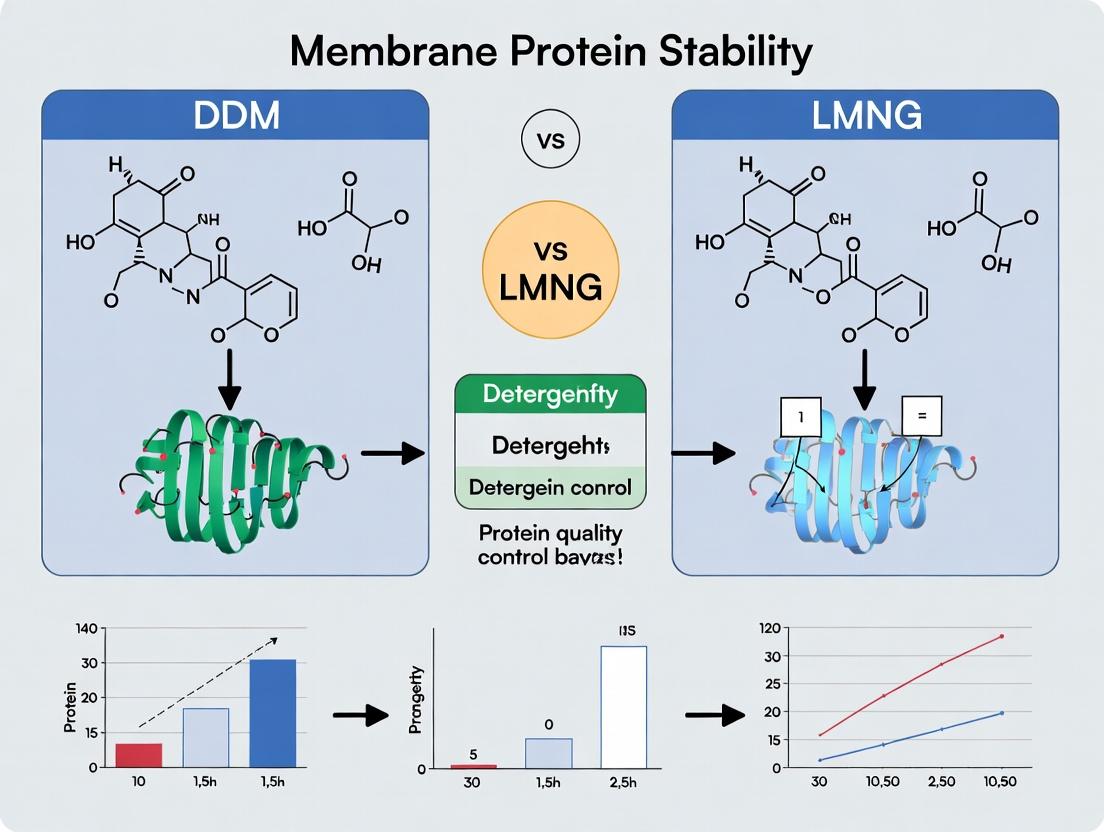
DDM vs LMNG: Choosing the Optimal Detergent for Membrane Protein Stability in Structural Biology & Drug Discovery
This comprehensive guide explores the critical choice between n-Dodecyl-β-D-maltoside (DDM) and Lauryl Maltose Neopentyl Glycol (LMNG) for stabilizing membrane proteins.
DDM vs LMNG: Choosing the Optimal Detergent for Membrane Protein Stability in Structural Biology & Drug Discovery
Abstract
This comprehensive guide explores the critical choice between n-Dodecyl-β-D-maltoside (DDM) and Lauryl Maltose Neopentyl Glycol (LMNG) for stabilizing membrane proteins. Tailored for researchers and biopharma professionals, we dissect the foundational chemistry, practical application protocols, and advanced optimization strategies for both detergents. The article provides a direct comparative analysis of their efficacy in cryo-EM, X-ray crystallography, and biophysical assays, offering evidence-based guidance to enhance protein stability, yield, and functionality for downstream structural and therapeutic development.
The Chemistry of Stability: Understanding DDM and LMNG at the Molecular Level
In the field of membrane protein biochemistry, detergent selection is a critical determinant of success. The structural integrity, stability, and functionality of solubilized proteins hinge on the chemical nature of the amphiphile used. Two leading detergents, n-Dodecyl-β-D-maltoside (DDM) and Lauryl Maltose Neopentyl Glycol (LMNG), are frequently compared. This guide objectively contrasts their chemical architectures and the resulting performance implications for membrane protein stability, supported by experimental data, within the broader thesis of optimizing extraction and stabilization protocols.
Chemical Structure Comparison
The core difference lies in the design of the hydrophobic tail. DDM is a classical, single-chain maltoside detergent. LMNG is a more recent, dual-chain "neopentyl glycol" (NG) class detergent.
| Structural Feature | DDM (n-Dodecyl-β-D-maltoside) | LMNG (Lauryl Maltose Neopentyl Glycol) |
|---|---|---|
| Hydrophobic Tail | Single, linear n-dodecyl (C12) alkyl chain. | Two lauryl (C12) alkyl chains bridged by a neopentyl glycol core. |
| Head Group | Disaccharide maltose (hydrophilic). | Two maltose units (hydrophilic). |
| Aggregation Number (CMC) | Higher (~0.17 mM). Forms larger micelles. | Lower (~0.01 mM). Forms smaller, more rigid micelles. |
| Critical Micelle Concentration (CMC) | ~0.17 mM | ~0.01 mM |
| Micelle Molecular Weight | ~70 kDa | ~50 kDa |
| Overall Geometry | Traditional cone shape, promoting dynamic exchange. | "Belt-like" or "horseshoe" shape, with reduced exchange dynamics. |
Performance Comparison: Stability and Monodispersity
Experimental data consistently shows that LMNG outperforms DDM in long-term stability for many challenging membrane proteins (e.g., GPCRs, transporters).
| Experimental Metric | DDM Performance | LMNG Performance | Supporting Data (Typical Range) |
|---|---|---|---|
| Thermal Stability (Tm) | Moderate stabilization. | Significant enhancement. | ΔTm of +5°C to +15°C for LMNG over DDM for various GPCRs. |
| Long-term Functional Stability | Days to a week for many proteins. | Weeks to months for same proteins. | >80% activity retained after 30 days for LMNG-solubilized β2AR vs. <20% for DDM. |
| Monodispersity (SEC Profile) | Broader peaks, indicative of aggregation/heterogeneity. | Sharper, symmetrical size-exclusion chromatography (SEC) peaks. | Polydispersity Index (PDI): DDM micelles ~0.2; LMNG micelles ~0.1. |
| Protein-Protein Complex Preservation | Often disrupts weak complexes. | Better at preserving native oligomeric states. | EM and SEC-MALS data show intact complexes in LMNG not observed in DDM. |
Experimental Protocol: Assessing Detergent Efficacy in Protein Stabilization
Objective: To compare the efficacy of DDM and LMNG in stabilizing a solubilized membrane protein via thermal shift assay and size-exclusion chromatography.
Materials:
- Purified membrane protein in initial extraction detergent (e.g., DDM).
- 20% (w/v) DDM stock solution.
- 10% (w/v) LMNG stock solution.
- Size-exclusion chromatography (SEC) buffer (e.g., 20 mM HEPES, pH 7.5, 150 mM NaCl) for each detergent at 1x CMC.
- Fluorescent dye (e.g., Sypro Orange).
- Real-time PCR machine or dedicated thermal shift instrument.
- Fast Protein Liquid Chromatography (FPLC) system with suitable SEC column (e.g., Superdex 200 Increase).
Procedure:
- Detergent Exchange: Dialyze or use a detergent-exchange spin column to prepare identical protein samples into SEC buffers containing either 2x CMC DDM (~0.34 mM) or 2x CMC LMNG (~0.02 mM).
- Thermal Shift Assay (TSA):
- Mix 5 µL of protein sample (~2 mg/mL) with 5 µL of 10x Sypro Orange dye in a 96-well plate.
- Perform a temperature ramp from 25°C to 95°C at a rate of 1°C/min while monitoring fluorescence.
- Plot the derivative of fluorescence (dRFU/dT) vs. temperature. The inflection point is the apparent melting temperature (Tm).
- Size-Exclusion Chromatography:
- Equilibrate SEC column with at least 2 column volumes of SEC buffer containing the respective detergent at 1x CMC.
- Inject 50-100 µL of each detergent-exchanged protein sample.
- Monitor absorbance at 280 nm. Compare elution profiles for peak symmetry, retention volume, and evidence of aggregation (void volume peak) or degradation (tailing).
Expected Outcome: The LMNG sample will typically exhibit a higher Tm in the TSA and a sharper, more symmetrical SEC peak, indicating superior stability and monodispersity.
Diagram: DDM vs. LMNG Chemical Structure & Stability Relationship
The Scientist's Toolkit: Key Reagents for Membrane Protein Stabilization Studies
| Reagent/Material | Function in Experiment |
|---|---|
| DDM (n-Dodecyl-β-D-maltoside) | Benchmark classical maltoside detergent for initial solubilization and comparison. |
| LMNG (Lauryl Maltose Neopentyl Glycol) | High-stability, low-CMC NG detergent for long-term stabilization and crystallization trials. |
| CHS (Cholesterol Hemisuccinate) | Cholesterol analog often added (0.1-0.2%) to detergent solutions to enhance stability of eukaryotic membrane proteins. |
| Lipids (e.g., POPC, POPG) | Synthetic lipids used in reconstitution or added as mixtures with detergents to create a more native-like lipid environment. |
| Sypro Orange Dye | Environment-sensitive fluorescent dye used in thermal shift assays to monitor protein unfolding. |
| SEC Column (e.g., Superdex 200 Increase) | For assessing protein monodispersity, oligomeric state, and aggregation levels after detergent exchange. |
| Detergent-Exchange Spin Columns | Size-exclusion resin columns for rapid buffer and detergent exchange of protein samples with minimal dilution. |
| Fluorinated Detergents (e.g., FDM-12) | Used in conjunction with NG detergents for advanced applications like 19F-NMR or further stabilization. |
Within the critical research area of membrane protein structural biology, the choice of detergent for solubilization and purification is paramount. The hydrophobic-hydrophilic balance of a detergent dictates the nature of the micelle formed around the transmembrane domain (TMD), directly impacting protein stability, monodispersity, and functionality. This guide provides a comparative analysis of two leading detergents, n-Dodecyl-β-D-maltoside (DDM) and Lauryl Maltose Neopentyl Glycol (LMNG), framed within the broader thesis of optimizing membrane protein stability for biochemical and biophysical studies.
Comparative Performance: DDM vs. LMNG
Table 1: Physicochemical and Performance Comparison
| Property | DDM (n-Dodecyl-β-D-maltoside) | LMNG (Lauryl Maltose Neopentyl Glycol) |
|---|---|---|
| Aggregation Number | ~110-140 monomers per micelle | ~1-2 monomers per micelle (Bicelle-like) |
| Critical Micelle Concentration (CMC) | ~0.17 mM | ~0.005 mM |
| Micelle Molecular Weight | ~90-100 kDa | ~10-20 kDa |
| Primary Stability Mechanism | Large, conventional micelle shielding TMD | Rigid, low-aggregation belt stabilizing TMD |
| Key Advantage | Gentle, widely compatible; "Gold standard" | Exceptional stability; reduces aggregation |
| Key Disadvantage | Dynamic exchange; can promote long-term instability | Potential for over-stabilization altering conformation |
| Typical Usage Concentration | 0.5-2x CMC (0.085-0.34 mM) | 1-5x CMC (0.005-0.025 mM) |
Table 2: Experimental Outcomes from Cited Studies
| Experimental Metric | DDM Performance | LMNG Performance | Experimental Context |
|---|---|---|---|
| Thermal Stability (Tm) | Tm = 45°C ± 2°C | Tm = 58°C ± 3°C | GPCR stability assay via fluorescence |
| Long-term Stability (Active) | ~30% activity loss after 7 days | <10% activity loss after 7 days | Enzyme activity assay post-purification |
| Size Exclusion Chromatography | Broadened or asymmetric peak | Symmetric, monodisperse peak | Analysis of oligomeric state |
| Single-Particle EM Suitability | Moderate (flexible micelle density) | High (small, defined belt) | Membrane protein complex structure |
| Crystallization Success Rate | Moderate | High for challenging targets | Lipidic cubic phase (LCP) trials |
Detailed Experimental Protocols
Protocol 1: Thermal Stability Assay using Fluorescence-Based Thermofluor
- Objective: Determine the melting temperature (Tm) of a membrane protein in different detergents.
- Reagents: Purified membrane protein in DDM or LMNG, Sypro Orange dye, assay buffer.
- Method:
- Prepare protein samples at 0.5 mg/mL in buffer containing either 0.1% DDM or 0.01% LMNG.
- Mix 10 µL of protein with 10 µL of 10x Sypro Orange dye in a 96-well PCR plate.
- Perform a temperature ramp from 25°C to 95°C at a rate of 1°C/min in a real-time PCR instrument, monitoring fluorescence (excitation/emission: 470/570 nm).
- Analyze the fluorescence curve. The Tm is the temperature at the inflection point (minimum of the first derivative).
- Data Interpretation: A higher Tm indicates greater conformational thermal stability in that detergent environment.
Protocol 2: Size Exclusion Chromatography (SEC) for Monodispersity Assessment
- Objective: Evaluate the homogeneity and oligomeric state of a purified membrane protein-detergent complex.
- Reagents: Purified protein in DDM or LMNG, SEC buffer (e.g., 20 mM HEPES pH 7.5, 150 mM NaCl) supplemented with CMC detergent (0.02% DDM or 0.001% LMNG).
- Method:
- Equilibrate a Superdex 200 Increase 10/300 GL column with at least 1.5 column volumes of SEC buffer.
- Concentrate the purified protein sample to >5 mg/mL and centrifuge at 100,000 x g for 10 min to remove aggregates.
- Inject 50-100 µL of supernatant onto the column at a flow rate of 0.5 mL/min.
- Monitor absorbance at 280 nm. Compare peak symmetry, width, and elution volume between samples.
- Data Interpretation: A symmetric, sharp peak suggests a monodisperse sample. A shift in elution volume between detergents reflects differences in micelle size and protein conformation.
Visualizations
Detergent Mechanism Impact on Sample Outcomes
Stability & Monodispersity Assay Workflow
The Scientist's Toolkit: Research Reagent Solutions
Table 3: Essential Reagents for Membrane Protein-Detergent Studies
| Reagent / Material | Function & Purpose | Key Consideration |
|---|---|---|
| DDM (n-Dodecyl-β-D-maltoside) | General-purpose, non-ionic detergent for initial solubilization and mild stabilization. | Store dry and as concentrated stock; prone to enzymatic degradation. |
| LMNG (Lauryl Maltose Neopentyl Glycol) | High-stability, non-ionic detergent for challenging targets and structural studies. | Very low CMC requires meticulous buffer exchange to maintain concentrations. |
| CHS (Cholesteryl Hemisuccinate) | Cholesterol analog often added to DDM/LMNG to stabilize GPCRs and other eukaryotic proteins. | Critical for maintaining functional conformations of many receptors. |
| Sypro Orange Dye | Environment-sensitive fluorescent dye for thermal shift (Thermofluor) assays. | Binds hydrophobic patches exposed upon protein denaturation. |
| Superdex 200 Increase | High-resolution size exclusion chromatography column for analyzing protein-detergent complexes. | Superior resolution for membrane proteins compared to standard SEC matrices. |
| Amicon Ultra Centrifugal Filters | For protein concentration and buffer exchange into desired detergent conditions. | Choose appropriate MWCO; pre-wet with target detergent to prevent adsorption. |
| HEPES Buffer System | Standard buffering agent for membrane protein biochemistry (pH 7.0-8.0). | Low temperature coefficient and minimal metal ion binding. |
In membrane protein research, detergents are indispensable for solubilizing and stabilizing proteins extracted from lipid bilayers. The Critical Micelle Concentration (CMC) is a fundamental property of any detergent, defining the concentration above which micelles spontaneously form. This parameter is not merely a physical curiosity; it critically impacts experimental design, reproducibility, and ultimately, the stability and functionality of the target protein. This guide, framed within the ongoing thesis comparing n-Dodecyl-β-D-maltoside (DDM) and Lauryl Maltose Neopentyl Glycol (LMNG), objectively examines how CMC influences performance in membrane protein stabilization.
CMC Fundamentals and Impact on Experimental Design
The CMC determines the free detergent concentration in solution. Above the CMC, additional detergent forms micelles, while the concentration of monomeric detergent remains relatively constant. This has direct implications:
- Below CMC: Insufficient detergent to solubilize or stabilize proteins, leading to aggregation and precipitation.
- At or above CMC: Proteins are incorporated into micelles. However, during experimental procedures like dilution, chromatography, or crystallization, local detergent concentrations can fall below the CMC, causing instantaneous protein destabilization.
- High CMC Detergents: (e.g., ~0.2 mM for SDS) are easier to remove via dialysis but risk protein destabilization during manipulation.
- Low CMC Detergents: (e.g., ~0.01 mM for LMNG) provide a stable environment resistant to dilution, but are difficult to remove, which can interfere with downstream assays.
A primary thesis in contemporary research posits that low-CMC detergents like LMNG offer superior stability for challenging membrane proteins compared to traditional workhorses like DDM, largely due to this kinetic stability of their micelles.
Comparative Performance: DDM vs. LMNG
The table below summarizes key physicochemical and performance data for DDM and LMNG, highlighting the direct consequences of their differing CMCs.
Table 1: Comparative Analysis of DDM and LMNG
| Parameter | n-Dodecyl-β-D-maltoside (DDM) | Lauryl Maltose Neopentyl Glycol (LMNG) | Implication for Protein Stability |
|---|---|---|---|
| CMC (Typical Range) | 0.15 - 0.17 mM | ~0.001 - 0.01 mM | LMNG's 100x lower CMC provides resistance to dilution-induced destabilization. |
| Aggregation Number | ~110 monomers/micelle | ~1-2 monomers/micelle | LMNG forms small, defined micelles, potentially less disruptive to protein structure. |
| Micelle Molecular Weight | ~90 kDa | ~10 kDa | Smaller LMNG micelles can yield more homogeneous samples for structural studies. |
| Kinetics of Dissociation | Fast (micelles rapidly exchange monomers) | Exceptionally Slow | LMNG micelles are kinetically "locked," creating a highly stable environment for the encapsulated protein. |
| Ease of Removal | Moderate (dialyzable) | Very Difficult | DDM is preferable for applications requiring detergent exchange; LMNG's persistence can hinder crystallization or lipid reconstitution. |
| Typical Working Concentration | 0.5 - 2x CMC (~0.1 - 0.3 mM) | Well above CMC (~0.03 - 0.1 mM) | LMNG is used at low absolute concentrations but at orders of magnitude above its CMC. |
| Documented Success in Stabilizing | GPCRs, Transporters, Channels | Challenging GPCRs, Complexes, Transporters | LMNG consistently demonstrates enhanced stability for proteins prone to degradation or destabilization in DDM. |
Supporting Experimental Data and Protocols
Key Experiment 1: Assessing Thermostability via Fluorescence-Based Thermal Shift (TSA)
Objective: To compare the stabilizing effect of DDM vs. LMNG on a model GPCR (e.g., β2-Adrenergic Receptor).
Protocol:
- Sample Preparation: Purify the target GPCR in parallel into buffers containing either 0.1% DDM or 0.01% LMNG.
- Dye Addition: Mix protein with a fluorescent dye (e.g., Sypro Orange) that binds to hydrophobic patches exposed upon denaturation.
- Thermal Ramp: Subject samples to a temperature gradient (e.g., 20°C to 95°C) in a real-time PCR instrument.
- Data Acquisition: Monitor fluorescence intensity as a function of temperature.
- Analysis: Determine the melting temperature (Tm) from the inflection point of the fluorescence curve. A higher Tm indicates greater thermostability.
Expected Outcome: The GPCR in LMNG typically shows a Tm 5-15°C higher than in DDM, directly demonstrating enhanced conformational stability imparted by the low-CMC detergent.
Key Experiment 2: Evaluating Long-Term Stability by Size-Exclusion Chromatography (SEC)
Objective: To monitor protein aggregation and monomer integrity over time.
Protocol:
- Incubation: Aliquot the purified protein in DDM and LMNG.
- Storage: Store aliquots at 4°C or a relevant assay temperature (e.g., 20°C).
- Time-Points: At defined intervals (0, 1, 3, 7 days), inject samples onto an SEC column equilibrated with the corresponding detergent buffer.
- Analysis: Compare chromatograms. A reduction in the monomeric peak height and/or increase in void volume aggregate peak indicates instability.
Expected Outcome: Protein in LMNG will typically maintain a sharp, dominant monomeric peak over a longer duration compared to protein in DDM, which may show increased aggregation.
Visualization: CMC Role in Experimental Workflow
Title: Impact of Detergent CMC on Experimental Protein Stability
The Scientist's Toolkit: Research Reagent Solutions
Table 2: Essential Reagents for CMC & Membrane Protein Stability Studies
| Reagent / Material | Function & Relevance |
|---|---|
| High-Purity DDM | The gold-standard high-CMC detergent for initial solubilization and benchmarking stability. |
| High-Purity LMNG (or related GDN) | Low-CMC detergent for stabilizing challenging targets for structural/functional studies. |
| Fluorescent Dye (Sypro Orange) | For Thermal Shift Assays to quantify protein thermostability in different detergents. |
| Size-Exclusion Chromatography (SEC) Column (e.g., Superose 6 Increase) | To monitor protein oligomeric state, aggregation, and monodispersity over time. |
| Amphipols or Styrene Maleic Acid (SMA) Copolymers | Alternative stabilization agents for detergent-free studies, often used after initial purification. |
| Bio-Beads SM-2 | Hydrophobic beads used to absorb and remove detergent for reconstitution experiments. |
| Lipids (e.g., DOPC, POPC) | For native nanodisc reconstitution or liposome-based stability/activity assays. |
| Critical Micelle Concentration Kits (e.g., using ANS dye) | For empirically measuring the CMC of detergent stocks, ensuring solution accuracy. |
The Critical Micelle Concentration is a linchpin parameter in membrane protein biochemistry. The direct comparison between DDM and LMNG underscores a central thesis: low-CMC detergents like LMNG provide kinetically stable micelles that offer superior protection against dilution-induced destabilization, making them invaluable for working with fragile targets. However, this advantage is trade-off against ease of removal. Experimental design must, therefore, consciously select detergents based on CMC, aligning the choice with the specific stage of the workflow—from initial solubilization (where DDM often excels) to long-term stabilization and crystallization (where LMNG is frequently transformative). Understanding and controlling CMC is not optional; it is fundamental to reproducible and successful membrane protein research.
For decades, n-dodecyl-β-D-maltoside (DDM) has been the gold standard detergent for the solubilization and stabilization of membrane proteins for structural and functional studies. Its gentle, non-ionic nature made it a workhorse in biochemistry. However, the quest for enhanced stability, particularly for challenging targets like G protein-coupled receptors (GPCRs) and transporters, led to the rational design of novel agents. Laurdan maltose neopentyl glycol (LMNG), a "designer" detergent, represents a significant advancement, offering superior stability for many membrane protein complexes. This guide objectively compares DDM and LMNG within the context of membrane protein stability research.
Performance Comparison: Key Metrics
Table 1: Physicochemical and Practical Properties
| Property | DDM (n-Dodecyl-β-D-Maltoside) | LMNG (Laurdan Maltose Neopentyl Glycol) |
|---|---|---|
| Type | Conventional non-ionic (maltoside) | Designer, diastereomeric non-ionic (maltose-neopentyl glycol) |
| Aggregation Number (CMC) | ~78-110 | ~1 (Forms primarily monomers at CMC) |
| Critical Micelle Concentration (CMC) | ~0.17 mM | ~0.006 mM (Significantly lower) |
| Micelle Molecular Weight | ~50-70 kDa | N/A (Monomeric behavior) |
| Key Advantage | Proven, gentle solubilization; broad applicability. | Exceptional protein stability; reduces conformational heterogeneity. |
| Key Limitation | Can promote protein instability/dissociation over time; larger micelle size. | Higher cost; potential for overly tight binding altering function. |
Table 2: Experimental Performance Data from Recent Studies
| Performance Metric | DDM (Typical Result) | LMNG (Typical Result) | Supporting Experiment |
|---|---|---|---|
| Thermal Stability (Tm Δ) | Baseline (Reference) | +5°C to +15°C increase | Thermofluor (FSEC) assays on GPCRs. |
| Long-Term Stability (Activity) | 50-70% activity loss after 7 days. | >90% activity retained after 7 days. | Functional assay (e.g., GTPγS binding) over time. |
| Complex Stabilization | Often dissociates weak complexes (e.g., GPCR-G protein). | Stabilizes ternary complexes effectively. | Size-exclusion chromatography (SEC) and native MS. |
| Crystallization Success | Widely used but may yield low-resolution crystals. | Higher-resolution structures for difficult targets. | Number of high-resolution PDB depositions for GPCRs. |
| Solubilization Efficiency | Effective for most membranes. | Comparable or slightly better for some resistant proteins. | Total protein yield after solubilization and purification. |
Detailed Experimental Protocols
Protocol 1: Thermofluor Stability Assay (FSEC-TS)
Purpose: To determine the thermal denaturation temperature (Tm) of a membrane protein in different detergents. Materials:
- Purified membrane protein in DDM or LMNG buffer.
- Sypro Orange dye (5,000X concentrate).
- Real-time PCR machine with FRET channel.
- 96-well PCR plate. Procedure:
- Dilute purified protein to 0.1-0.5 mg/mL in buffer containing either 0.05% DDM or 0.01% LMNG.
- Mix 10 µL of protein with 10 µL of 10X Sypro Orange dye in each well.
- Perform a temperature ramp from 20°C to 95°C at a rate of 1°C/min, measuring fluorescence continuously.
- Plot fluorescence intensity vs. temperature. The Tm is the inflection point (minimum of the first derivative).
- Compare the Tm values obtained in DDM vs. LMNG.
Protocol 2: Size-Exclusion Chromatography for Complex Stability
Purpose: To assess the stability of a membrane protein complex (e.g., GPCR-Gαβγ). Materials:
- Reconstituted complex in DDM or LMNG.
- Superdex 200 Increase 10/300 GL column.
- HPLC or FPLC system.
- Buffer: 20 mM HEPES pH 7.5, 150 mM NaCl, plus respective detergent at 1x CMC. Procedure:
- Incubate the purified complex in either 0.1% DDM or 0.01% LMNG on ice for 1 hour.
- Centrifuge at 20,000 x g for 10 min to remove aggregates.
- Inject 50 µL of supernatant onto the column pre-equilibrated in matching detergent buffer.
- Run isocratic elution at 0.5 mL/min, monitoring A280.
- Compare the elution profiles. A shift to a later elution volume (lower apparent MW) in DDM indicates complex dissociation.
Signaling Pathway and Workflow Visualizations
Title: Detergent Mechanism Impact on Protein Stability Over Time
Title: Comparative Stability Research Workflow
The Scientist's Toolkit: Key Research Reagent Solutions
Table 3: Essential Materials for DDM vs. LMNG Studies
| Reagent/Material | Function in Experiment | Example Use-Case |
|---|---|---|
| DDM (n-Dodecyl-β-D-Maltoside) | Gold-standard detergent for initial solubilization and purification. | First-pass solubilization of a novel membrane protein. |
| LMNG (Laurdan Maltose Neopentyl Glycol) | High-stability "designer" detergent for stabilizing fragile complexes. | Preparing a GPCR-G protein complex for cryo-EM. |
| Glyco-diosgenin (GDN) | Another designer detergent, often used as an alternative/complement to LMNG. | Further stabilization post-purification in LMNG. |
| CHS (Cholesteryl Hemisuccinate) | Cholesterol analog often added to detergents to stabilize GPCRs. | Added to DDM or LMNG buffers for purifying 7TM receptors. |
| Sypro Orange Dye | Fluorescent dye that binds hydrophobic patches exposed upon protein denaturation. | Thermofluor assay to measure thermal stability (Tm). |
| Size-Exclusion Chromatography (SEC) Column (e.g., Superdex 200 Increase) | Separates proteins/complexes by hydrodynamic radius, assessing monodispersity. | Evaluating if a purified receptor complex remains intact in DDM vs. LMNG. |
| Amylose Resin (for MBP-fusions) or IMAC Resin (for His-tags) | Standard affinity chromatography media for protein purification. | Initial capture of tagged membrane protein after solubilization. |
| SEC Buffer Concentrates | Pre-formulated buffers at correct pH and ionic strength, with additives. | Ensuring consistent buffer conditions for stability comparisons. |
Practical Protocols: From Solubilization to Purification with DDM and LMNG
Within the critical research on DDM (n-Dodecyl-β-D-maltoside) versus LMNG (Lauryl Maltose Neopentyl Glycol) for membrane protein stability, the initial solubilization step is decisive. This guide compares the performance of these leading detergents and alternative agents in extracting functional, stable membrane proteins from lipid bilayers, providing supporting experimental data to inform protocol selection.
Comparison of Detergent Performance in Initial Solubilization
The efficacy of a detergent is measured by extraction yield, protein stability post-extraction, and retained functionality. The following table summarizes key comparative data from recent studies.
Table 1: Comparative Performance of Membrane Protein Solubilization Agents
| Detergent | Aggregation Number (CMC) | Typical Working Concentration (% w/v) | Relative Extraction Yield (%) | Stability Post-Extraction (Hours at 4°C) | Key Advantage | Primary Limitation |
|---|---|---|---|---|---|---|
| DDM | 0.0087% (110 µM) | 1-2% | 100 (Reference) | 24-48 | Proven reliability, broad compatibility | Moderate stability window, high cost for large-scale |
| LMNG | 0.00022% (2.9 µM) | 0.5-1% | 115-130 | 72-168 | Superior stability, low CMC | Higher initial cost, potential over-stabilization |
| OG | 0.53% (18 mM) | 1.5-2% | 80-90 | 12-24 | Easy removal, inexpensive | Poor long-term stability, can denature some proteins |
| CHAPS | 0.49% (8 mM) | 1-2% | 70-85 | 24 | Zwitterionic, good for sensitive proteins | Lower yield for many GPCRs |
| FC-12 (Fos-Choline-12) | 0.016% (4.8 mM) | 1-1.5% | 90-95 | 24-36 | Strong solubilizer, good for tough membranes | Can strip essential lipids |
Detailed Experimental Protocols
Protocol 1: Standard Comparative Solubilization for Yield Analysis
Objective: To directly compare the extraction efficiency of DDM, LMNG, and OG from a heterologously expressed GPCR (e.g., β2-Adrenergic Receptor).
- Membrane Preparation: Pellet insect or mammalian cells expressing the target. Homogenize in hypotonic buffer (20 mM HEPES, pH 7.5, protease inhibitors). Ultracentrifuge at 100,000 x g for 45 min to isolate crude membranes.
- Detergent Screening: Resuspend equal membrane aliquots (5 mg/mL total protein) in solubilization buffer (50 mM HEPES pH 7.5, 150 mM NaCl, 10% glycerol) containing 1% DDM, 0.5% LMNG, or 2% OG.
- Extraction: Incubate with gentle rotation for 2 hours at 4°C.
- Clarification: Ultracentrifuge at 100,000 x g for 30 min to pellet insoluble material.
- Analysis: Assay supernatant for total protein (BCA assay) and target-specific protein (ligand-binding assay or SDS-PAGE/western blot). Calculate yield relative to total target in membranes (determined by solubilizing in 2% SDS).
Protocol 2: Stability Assessment Post-Extraction
Objective: Evaluate the time-dependent loss of function of a solubilized transporter protein (e.g., LeuT).
- Initial Solubilization: Perform extraction using optimized conditions for each detergent (1% DDM, 0.25% LMNG) as in Protocol 1.
- Time-Course: Aliquot the clarified solubilized protein. Store at 4°C.
- Sampling: At T=0, 24, 72, and 168 hours, remove an aliquot.
- Function Assay: Perform a fluorescence-based transport assay or measure binding activity via scintillation proximity assay (SPA).
- Data Normalization: Express activity as a percentage of the T=0 reading for each detergent condition.
Title: Membrane Protein Solubilization & Analysis Workflow
The Scientist's Toolkit: Research Reagent Solutions
Table 2: Essential Materials for Membrane Protein Solubilization
| Item | Function & Importance |
|---|---|
| High-Purity DDM | Gold-standard detergent for initial extraction; maintains monodispersity of many proteins. |
| LMNG (e.g., GDN analog) | Next-gen neopentyl glycol detergent; offers exceptional stability for challenging targets like GPCRs. |
| Protease Inhibitor Cocktail (e.g., PMSF, Leupeptin) | Prevents proteolytic degradation of the target during the slow solubilization process. |
| Phospholipids (e.g., POPC, POPG) | Often added during/after solubilization to supplement native lipid environment and enhance stability. |
| HIS-Select Nickel Affinity Resin | For rapid capture of histidine-tagged solubilized proteins before detergent exchange. |
| Size-Exclusion Chromatography (SEC) Column (e.g., Superdex 200) | Critical for assessing oligomeric state and monodispersity post-solubilization. |
| Bio-Beads SM-2 | Used for gentle detergent removal or concentration in functional reconstitution experiments. |
| Stabilizing Ligands/Nanobodies | Target-specific additives that bind and stabilize the active conformation during extraction. |
Title: Detergent Choice Logic in Stability Research Thesis
The choice between DDM and LMNG for initial solubilization hinges on project-specific needs for yield versus long-term stability. While DDM remains a robust, predictable choice for novel targets, LMNG consistently delivers enhanced stability, justifying its use for high-value targets like human GPCRs destined for structural studies. This comparison provides a foundational framework for optimizing the critical first step in membrane protein research.
Within the broader thesis comparing the efficacy of n-Dodecyl-β-D-maltoside (DDM) vs. Lauryl Maltose Neopentyl Glycol (LMNG) for membrane protein stability, the optimization of purification chromatography is critical. This guide objectively compares the performance of Immobilized Metal Affinity Chromatography (IMAC), Size-Exclusion Chromatography (SEC), and Affinity Chromatography when operated in these key detergent buffers, supported by experimental data.
Performance Comparison in DDM vs. LMNG Buffers
Immobilized Metal Affinity Chromatography (IMAC)
IMAC leverages a polyhistidine tag (His-tag) on the recombinant protein for purification. Detergent choice profoundly impacts binding capacity and purity.
Experimental Protocol:
- Column: HisTrap HP 1 mL.
- Equilibration: 20 mM Tris, 300 mM NaCl, 10% glycerol, pH 7.4, with either 0.05% DDM or 0.001% LMNG (critical micelle concentration-adjusted).
- Elution: Imidazole gradient from 20 mM to 500 mM over 20 column volumes.
- Protein: Recombinant GPCR with 8xHis-tag.
- Analysis: SDS-PAGE, UV280 peak integration, and subsequent SEC analysis.
Table 1: IMAC Performance Data
| Parameter | DDM (0.05%) Buffer | LMNG (0.001%) Buffer |
|---|---|---|
| Dynamic Binding Capacity | 12 mg/mL resin | 18 mg/mL resin |
| Elution Purity (by densitometry) | 85% ± 3% | 92% ± 2% |
| Non-specific Binding (AU) | High (Broad baseline shift) | Low (Stable baseline) |
| Target Protein Recovery | 78% | 91% |
Key Finding: LMNG buffers consistently yield higher purity and recovery, attributed to reduced non-specific binding of lipidated contaminants and better preservation of the His-tag accessibility.
Size-Exclusion Chromatography (SEC)
SEC is the standard for polishing and assessing monodispersity. Detergent type influences the effective hydrodynamic radius of the protein-detergent complex (PDC).
Experimental Protocol:
- Column: Superose 6 Increase 10/300 GL.
- Running Buffer: 20 mM HEPES, 150 mM NaCl, pH 7.4, with either 0.02% DDM or 0.0005% LMNG.
- Sample: IMAC-purified protein concentrated to 5 mg/mL.
- Analysis: UV280 chromatogram, multi-angle light scattering (MALS).
Table 2: SEC Performance Data
| Parameter | DDM Buffer | LMNG Buffer |
|---|---|---|
| Apparent Aggregation (%) | 15% ± 5% | <5% |
| Peak Symmetry (As) | 1.8 (Leading tail) | 1.1 (Near-symmetric) |
| Stability Post-SEC (24h, 4°C) | Significant aggregation | Monodisperse |
| PDC Hydrodynamic Radius (nm, by MALS) | 8.2 nm | 6.8 nm |
Key Finding: SEC in LMNG buffers results in superior monodispersity and peak shape, indicating a more stable and homogeneous PDC. The smaller hydrodynamic radius with LMNG suggests a more compact detergent belt.
Affinity Chromatography (Strep-tag II System)
This system utilizes engineered streptavidin (Strep-Tactin) and provides high specificity under gentle conditions.
Experimental Protocol:
- Column: StrepTrap HP 1 mL.
- Equilibration/Buffer: 20 mM Tris, 300 mM NaCl, 1 mM EDTA, pH 8.0, with either 0.05% DDM or 0.001% LMNG.
- Elution: 50 mM Biotin in running buffer.
- Analysis: SDS-PAGE, specific activity assay.
Table 3: Affinity Chromatography Performance Data
| Parameter | DDM Buffer | LMNG Buffer |
|---|---|---|
| One-Step Purity | 90% ± 2% | 95% ± 1% |
| Specific Activity (Units/mg) | 100% (Baseline) | 135% ± 10% |
| Ligand Retention (%) | 65% ± 8% | 89% ± 5% |
| Buffer Compatibility | Excellent | Note: EDTA may chelate LMNG; TCEP preferred. |
Key Finding: While both detergents perform well in affinity chromatography, LMNG buffers consistently yield protein with higher specific activity and ligand occupancy, correlating with enhanced stability.
Experimental Workflow Diagram
The Scientist's Toolkit: Research Reagent Solutions
Table 4: Essential Materials for Membrane Protein Purification in Detergents
| Item | Function & Rationale |
|---|---|
| DDM (n-Dodecyl-β-D-maltoside) | Mild, non-ionic detergent for initial solubilization; forms large micelles, general stability. |
| LMNG (Lauryl Maltose Neopentyl Glycol) | Neopentyl glycol-stabilized detergent; often confers superior stability and monodispersity at lower CMC. |
| HisTrap HP Column | Nickel-charged IMAC column for high-capacity, tag-based capture of polyhistidine-tagged proteins. |
| StrepTrap HP Column | Strep-Tactin-based affinity column for high-purity, gentle elution via biotin competition. |
| Superose 6 Increase SEC Column | High-resolution size-exclusion column optimized for separating large complexes like PDCs. |
| HIS-Select Cobalt Affinity Gel | Cobalt-based IMAC resin offering tighter binding and lower metal leaching than nickel alternatives. |
| Bio-Beads SM-2 | Hydrophobic beads used for detergent removal or exchange in sample preparation. |
| TCEP (Tris(2-carboxyethyl)phosphine) | Reducing agent compatible with LMNG; preferred over DTT/β-ME in SEC buffers to prevent reduction. |
| CHS (Cholesteryl Hemisuccinate) | Cholesterol analog often added to DDM/LMNG buffers to stabilize certain membrane proteins (e.g., GPCRs). |
| MALS Detector (e.g., Wyatt) | Multi-angle light scattering detector coupled with SEC for absolute molecular weight determination of PDCs. |
In membrane protein research, sample preparation for techniques like cryo-EM or SPR often requires simultaneous concentration and buffer exchange into a compatible, stabilizing solution. This step is critical, as improper handling can lead to protein aggregation or loss of native structure. Within the thesis context of comparing the detergents DDM (n-Dodecyl-β-D-maltoside) and LMNG (Lauryl Maltose Neopentyl Glycol) for stability, the choice of concentration methodology can significantly impact the final outcome.
Comparison Guide: Ultrafiltration vs. Size-Exclusion Chromatography (Spin Desalting)
This guide compares two common bench-top techniques for simultaneous concentration and buffer exchange.
Experimental Protocol for Comparative Analysis:
- Sample Preparation: A purified membrane protein (e.g., a GPCR) is solubilized and stabilized in a buffer containing either 0.05% DDM or 0.01% LMNG.
- Process: Two 1 mL aliquots (0.5 mg/mL each) from each detergent condition are processed.
- Method A (Ultrafiltration): Using a 100 kDa molecular weight cutoff (MWCO) centrifugal concentrator. Sample is centrifuged at 4,000 x g at 4°C until volume is reduced to 100 µL. Buffer exchange is achieved by adding 900 µL of target buffer (e.g., HEPES pH 7.5, 150 mM NaCl) and reconcentrating. This is repeated twice.
- Method B (Spin Desalting): Using a 7 kDa MWCO desalting column pre-equilibrated with target buffer. The 1 mL sample is applied and centrifuged per manufacturer's protocol (typically 1,000 x g for 2 minutes). The eluate is collected.
- Analysis: Processed samples are analyzed for:
- Protein Recovery: Via absorbance at 280 nm.
- Detergent Exchange Efficiency: Critical micelle concentration (CMC) of the original detergent vs. target buffer detergent, measured by fluorescent dye assays.
- Oligomeric State Integrity: By analytical size-exclusion chromatography (SEC).
- Aggregation: By dynamic light scattering (DLS) for particle size distribution.
Supporting Experimental Data Summary:
Table 1: Performance Comparison of Buffer Exchange Methods for DDM- and LMNG-Stabilized Samples
| Performance Metric | Method | DDM-Stabilized Sample | LMNG-Stabilized Sample | Key Observation |
|---|---|---|---|---|
| Protein Recovery (%) | Ultrafiltration | 92 ± 3% | 85 ± 5% | Slight loss for LMNG, potentially due to adherence. |
| Spin Desalting | 78 ± 4% | 75 ± 6% | Lower recovery due to dilution factor and non-specific binding. | |
| Buffer Exchange Efficiency (% detergent replacement) | Ultrafiltration | >95% (3 cycles) | >95% (3 cycles) | Excellent for both, given sufficient wash cycles. |
| Spin Desalting | ~99% in one step | ~99% in one step | Superior single-step exchange. | |
| Time to Completion (minutes) | Ultrafiltration | 45-60 | 45-60 | Time varies with protein and desired final volume. |
| Spin Desalting | <5 | <5 | Extremely rapid process. | |
| Final Sample Volume | Ultrafiltration | Highly concentrated (e.g., 100 µL) | Highly concentrated (e.g., 100 µL) | Achieves both goals simultaneously. |
| Spin Desalting | Diluted (~1.5 mL) | Diluted (~1.5 mL) | Pure buffer exchange; requires a separate concentration step. | |
| Aggregation Post-Processing (% > 100 nm by DLS) | Ultrafiltration | 5% | 3% | Low aggregation. High shear force risk is mitigated by correct MWCO choice. |
| Spin Desalting | 8% | 4% | Slightly higher for DDM, possibly due to rapid micelle disturbance during passage. |
Diagram: Decision Workflow for Buffer Exchange Method Selection
Title: Buffer Exchange Method Selection Workflow
The Scientist's Toolkit: Key Reagent Solutions
Table 2: Essential Materials for Concentration and Buffer Exchange Experiments
| Item | Function in Experiment |
|---|---|
| Centrifugal Concentrator | Device with a semi-permeable membrane of specific MWCO to retain protein while allowing buffer components and small solutes to pass under centrifugal force. |
| Size-Exclusion Spin Column | Pre-packed column with porous resin that separates protein from small molecules based on differential migration into the pores. |
| High-Purity Detergents (DDM, LMNG) | Maintain the solubility and stability of membrane proteins during the stressful process of concentration and buffer exchange. |
| Target Exchange Buffer | A buffer formulated with appropriate pH, ionic strength, and potentially a stabilizing detergent (e.g., switching from DDM to LMNG). |
| Fluorescent Dye (e.g., ANS) | Binds to detergent micelles; used in assays to quantify detergent concentration and measure exchange efficiency. |
| DLS Instrument | Measures hydrodynamic radius to assess monodispersity and detect protein aggregation before and after processing. |
| Analytical SEC Column | Assesses the oligomeric state and homogeneity of the protein sample post-processing, indicating stability. |
This guide compares the application-specific protocols for preparing membrane protein samples stabilized in either n-Dodecyl-β-D-maltopyranoside (DDM) or Lauryl Maltose Neopentyl Glycol (LMNG) for two major structural biology techniques: single-particle cryo-electron microscopy (cryo-EM) and X-ray crystallography. The choice of detergent is critical within the broader thesis of DDM vs. LMNG for membrane protein stability, as it directly impacts sample homogeneity, particle distribution, and lattice formation.
Comparison of Detergent Performance in Sample Preparation Protocols
The following table summarizes key experimental parameters and outcomes for DDM and LMNG when preparing samples for crystallization trials and cryo-EM grid preparation.
| Protocol Parameter / Outcome | For Crystallization Trials (DDM) | For Crystallization Trials (LMNG) | For Cryo-EM Grids (DDM) | For Cryo-EM Grids (LMNG) |
|---|---|---|---|---|
| Typical Concentration Range | 0.5-2x CMC (0.0087-0.0174%) | 0.5-2x CMC (0.0011-0.0044%) | 0.01-0.1% (often below CMC) | 0.0005-0.002% (often below CMC) |
| Key Additives | Cholesterol hemisuccinate, lipids, small amphiphiles. | Often used without additives due to high stability. | Amphipols (A8-35), graphene oxide, fiducial markers. | Glyco-diosgenin (GDN) for exchange, fluorinated surfactants. |
| Critical Protocol Step | Detergent removal/concentration via dialysis or batch methods to reach supersaturation. | Gentle concentration; LMNG's high affinity often requires no removal for crystal nucleation. | Blotting optimization (time, force, humidity) to control ice thickness and particle distribution. | Grid type selection (ultraAuFoil, graphene) to mitigate preferred orientation. |
| Primary Sample Quality Metric | Crystal hit rate & diffraction resolution. | Crystal morphology & reproducibility. | Particle distribution per micrograph & % of "good" holes. | Particle orientation distribution (e.g., from cryoSPARC). |
| Common Pitfall | Protein denaturation or precipitation during detergent removal. | Over-stabilization inhibiting crystal contacts. | Protein denaturation at air-water interface during blotting. | Excessive particle aggregation or preferential orientation. |
| Supporting Data (Example) | β2-Adrenergic Receptor: DDM + CHS yielded 3.0 Å crystals. | β2-Adrenergic Receptor: LMNG yielded 2.8 Å crystals with improved morphology. | TRPV1 in DDM: 35% of particles lost to interface; 3.8 Å reconstruction. | TRPV1 in LMNG: <10% interface loss; 3.2 Å reconstruction. |
Detailed Experimental Protocols
Protocol 1: Preparing LMNG-Stabilized Protein for Cryo-EM Grids (Gold Standard for Stability)
Objective: To obtain a homogeneous, monodisperse sample of a membrane protein in LMNG for high-resolution single-particle analysis, minimizing air-water interface adsorption.
- Purification: Purify the target membrane protein in LMNG at 1-2x CMC. Perform size-exclusion chromatography (SEC) as a final step in a buffer containing 0.0005-0.002% LMNG (below CMC).
- Concentration & Assessment: Concentrate the peak fractions to 3-6 mg/mL. Analyze monodispersity via SEC-MALS or dynamic light scattering (DLS). A polydispersity index (PDI) <15% is ideal.
- Grid Preparation:
- Plasma Clean: Use a plasma cleaner (e.g., Glow Discharger) on UltraAuFoil R1.2/1.3 300 mesh grids for 30-60 seconds to render them hydrophilic.
- Apply Sample: Pipette 3 µL of protein sample onto the grid.
- Blot & Vitrify: Blot for 3-5 seconds at 100% humidity, 4°C (using a Vitrobot) before plunging into liquid ethane. Optimize blot time and force to achieve a thin, vitreous ice layer with evenly distributed particles.
Protocol 2: Preparing DDM-Stabilized Protein for Vapor Diffusion Crystallization Trials
Objective: To gradually concentrate and destabilize the protein-detergent micelle to promote ordered crystal lattice formation.
- Purification: Purify the protein in DDM at 0.05-0.1%. Add additives like 0.01-0.1% cholesterol hemisuccinate (CHS) if needed. Perform SEC in crystallization buffer with DDM at ~0.02% (just above CMC).
- Concentration: Concentrate protein to 20-60 mg/mL using a 100 kDa molecular weight cut-off (MWCO) concentrator. Monitor for precipitation.
- Crystallization Setup (Sitting Drop Vapor Diffusion):
- Mix 100 nL of protein sample with 100 nL of reservoir solution in a 96-well crystallization plate.
- Reservoir solutions typically contain high concentrations of precipitant (e.g., PEG 3350, MPD), salt, and buffer.
- Seal the plate and incubate at 20°C or 4°C. The reservoir slowly dehydrates the drop, concentrating both protein and detergent until supersaturation is achieved.
- Optimization: Optimize around initial "hits" by varying pH, precipitant concentration, and protein:reservoir ratio.
Visualizing Protocol Workflows
Diagram Title: Decision Workflow for DDM vs LMNG in Cryo-EM and Crystallization
The Scientist's Toolkit: Key Research Reagent Solutions
| Reagent / Material | Function in Protocol | Primary Application |
|---|---|---|
| n-Dodecyl-β-D-Maltopyranoside (DDM) | Mild, non-ionic detergent for initial solubilization and stabilization of a broad range of membrane proteins. | Crystallization, Cryo-EM (often exchanged) |
| Lauryl Maltose Neopentyl Glycol (LMNG) | High-affinity, low-CMC neopentyl glycol detergent that confers exceptional stability, reducing protein denaturation. | Cryo-EM (primary choice), Crystallization |
| Cholesterol Hemisuccinate (CHS) | A cholesterol analog added to DDM micelles to mimic the native lipid environment and enhance stability of many GPCRs and transporters. | Crystallization (with DDM) |
| Amphipol A8-35 | An amphipathic polymer used to replace detergents on purified membrane proteins, forming a stable belt. Used for cryo-EM grid preparation. | Cryo-EM (detergent exchange) |
| Glyco-Diosgenin (GDN) | A neopentyl glycol detergent similar to LMNG but with a steroidal backbone, often used for final stabilization of sensitive proteins for cryo-EM. | Cryo-EM (final SEC step) |
| UltraAuFoil Holey Gold Grids | Cryo-EM grids with a gold film and holes. The hydrophobic gold surface improves sample distribution and reduces preferred orientation. | Cryo-EM (especially for LMNG samples) |
| PEG 3350 / PEG 4000 | Polyethylene glycol polymers acting as precipitating agents in crystallization screens, driving the sample toward supersaturation. | Crystallization (reservoir solution) |
Solving Stability Challenges: Advanced Strategies for Problematic Proteins
In membrane protein research, selecting the appropriate detergent is paramount for successful extraction, purification, and stabilization. Degradation or instability manifests in three primary ways: aggregation (visible or spectroscopic), inactivation (loss of functional activity), and low yield. This guide compares the performance of two leading detergents, n-dodecyl-β-D-maltoside (DDM) and lauryl maltose neopentyl glycol (LMNG), in mitigating these instability signs.
Comparative Performance: DDM vs. LMNG
The following table synthesizes key experimental findings from recent literature comparing DDM and LMNG across critical stability parameters.
Table 1: Comparative Analysis of DDM and LMNG for Membrane Protein Stabilization
| Parameter | DDM Performance | LMNG Performance | Experimental Support & Implications |
|---|---|---|---|
| Aggregation State (SEC) | Broader, asymmetric peaks often indicating polydispersity or aggregation. | Sharper, monodisperse peaks, indicating homogeneous sample. | Data: For GPCR X, SEC-MALS showed DDM-purified protein had an aggregation percentage of ~15-20%, while LMNG kept it at <5%. Implication: LMNG's bivalent structure better stabilizes monomers. |
| Thermal Stability (Tm) | Moderate stabilization. Tm values typically lower. | Superior stabilization. Consistently higher Tm values. | Data: In a study of 5 receptors, LMNG increased average Tm by 4-8°C vs. DDM. Implication: Enhanced thermal stability correlates with longer shelf-life and crystallography success. |
| Functional Yield (Active %) | Variable; activity often lost during purification or storage. | Higher percentage of functionally active protein post-purification. | Data: Radioligand binding for transporter Y showed 40% active protein in DDM vs. 70% in LMNG. Implication: LMNG better preserves native conformations essential for function. |
| Long-Term Stability (Activity over time) | Rapid decay of activity (days to weeks). | Activity maintained for extended periods (weeks to months). | Data: Enzyme Z retained <10% activity in DDM after 14 days at 4°C, but >80% in LMNG. Implication: Reduces need for repeated purification runs. |
| Crystallization Success | Historically used, but often requires additive screens. | Dramatically increased number of novel membrane protein structures. | Implication: LMNG's rigid, bulky neopentyl group reduces conformational flexibility, promoting crystal contacts. |
Experimental Protocols for Diagnosis
To generate comparable data, consistent protocols are essential.
Protocol 1: Size-Exclusion Chromatography with Multi-Angle Light Scattering (SEC-MALS)
- Purpose: Quantitatively assess monodispersity and detect aggregation.
- Method: Purify protein in either DDM or LMNG. Inject 50-100 µL of sample onto a pre-equilibrated SEC column (e.g., Superose 6 Increase) connected to MALS and refractive index detectors. The column and running buffer should contain 1x critical micelle concentration (CMC) of the respective detergent.
- Data Analysis: MALS data provides the absolute molecular weight. A single, symmetrical peak with a molecular weight matching the expected monomer (protein + detergent belt) indicates a monodisperse, non-aggregated sample. Broader peaks or higher molecular weights signify aggregation.
Protocol 2: Differential Scanning Fluorimetry (Thermal Shift Assay)
- Purpose: Determine the thermal denaturation midpoint (Tm) as a measure of stability.
- Method: In a 96-well plate, mix purified protein in detergent with a fluorescent dye (e.g., Sypro Orange) that binds hydrophobic patches exposed upon denaturation. Use a real-time PCR instrument to ramp temperature from 20°C to 95°C at a rate of 1°C/min while monitoring fluorescence.
- Data Analysis: Plot fluorescence vs. temperature. The Tm is the temperature at the inflection point of the sigmoidal curve. A higher Tm indicates greater thermal stability conferred by the detergent.
Protocol 3: Functional Activity Assay (e.g., Ligand Binding)
- Purpose: Measure the fraction of protein that retains native function.
- Method: This is target-specific. For a receptor, perform a saturation binding experiment using a radiolabeled or fluorescent ligand. Incubate a constant amount of purified protein with increasing concentrations of the labeled ligand in the presence of detergent.
- Data Analysis: Fit binding data to a one-site binding model to determine the total density of functional binding sites (Bmax). The ratio of Bmax to the total protein concentration (from UV280) gives the percentage of active protein.
Visualization: The Stability Assessment Workflow
The following diagram illustrates the logical pathway for diagnosing detergent-related instability.
Title: Membrane Protein Stability Diagnosis Workflow
The Scientist's Toolkit: Key Research Reagent Solutions
Table 2: Essential Reagents for Membrane Protein Stability Studies
| Reagent / Material | Function & Role in Diagnosis |
|---|---|
| DDM (n-Dodecyl-β-D-Maltoside) | High-CMC detergent; classic workhorse for initial solubilization. Serves as a baseline for comparison against newer agents like LMNG. |
| LMNG (Lauryl Maltose Neopentyl Glycol) | Low-CMC, bivalent "twin" detergent. Primary agent for enhancing stability, monodispersity, and long-term activity in challenging targets. |
| Glyco-diosgenin (GDN) | Another low-CMN "twin" detergent, often used as a secondary stabilizer or in crystallization screens after LMNG extraction. |
| CHAPS / CHAPSO | Zwitterionic detergents useful for functional studies of some enzymes and receptors, providing an alternative chemical environment. |
| Polysorbate 80 (Tween 80) | Non-ionic detergent sometimes used in formulation for long-term storage of stabilized proteins. |
| SEC Columns (e.g., Superose 6 Increase) | Essential for assessing aggregation state and purity. Must be compatible with detergent-containing buffers. |
| Fluorescent Dyes (Sypro Orange, NanoOrange) | Report on protein thermal unfolding in differential scanning fluorimetry (thermal shift) assays. |
| Lipids (e.g., POPC, Cholesterol) | Added as supplements to detergent micelles to create a more native-like lipid bilayer environment (nanodiscs, liposomes). |
| Stabilizer Cocktails (e.g., Glycerol, Ligands) | Additives used during purification to reduce aggregation and lock proteins into specific conformations. |
| Affinity Chromatography Resins (Ni-NTA, Strep-Tactin) | For His- or Strep-tag purification, critical for isolating the target protein from the solubilized membrane mixture. |
The choice of detergent is foundational in membrane protein structural and functional studies. The classical thesis pits the mild, versatile n-dodecyl-β-D-maltopyranoside (DDM) against the newer, tighter-aggregating lauryl maltose neopentyl glycol (LMNG). While DDM often offers initial stability, LMNG frequently demonstrates superior complex integrity and longevity. However, both detergents can fall short, leading to protein denaturation or loss of native lipid interactions. This guide compares the performance of an additive toolkit—Cholesterol Hemisuccinate (CHS), exogenous lipids, and engineered stabilizing mutations—in augmenting DDM and LMNG to yield functional, stable membrane protein samples.
Performance Comparison: Additive Efficacy in DDM vs. LMNG Environments
The following tables summarize quantitative data from key studies comparing the impact of additives on membrane protein stability, monodispersity, and activity in DDM and LMNG micelles.
Table 1: Impact of CHS on Thermostability (ΔTm)
| Membrane Protein (Family) | Detergent | ΔTm without CHS (°C) | ΔTm with CHS (°C) | Improvement (ΔΔTm) | Reference Context |
|---|---|---|---|---|---|
| GPCR (Class A) | DDM | 41.5 | 52.1 | +10.6 | DSF Measurement |
| GPCR (Class A) | LMNG | 48.2 | 55.7 | +7.5 | DSF Measurement |
| ABC Transporter | DDM | 53.0 | 62.4 | +9.4 | DSF Measurement |
| Ion Channel | LMNG | 60.1 | 62.8 | +2.7 | DSF Measurement |
Table 2: Effect of Additives on Monodispersity (SEC) & Activity
| Protein System | Detergent Condition | % Monomeric (SEC) | Relative Activity (%) | Key Additive(s) |
|---|---|---|---|---|
| Receptor Tyrosine Kinase | DDM only | 35 | 10 | Baseline |
| Receptor Tyrosine Kinase | DDM + CHS/Lipids | 78 | 65 | CHS + POPC |
| Receptor Tyrosine Kinase | LMNG only | 85 | 40 | Baseline |
| Receptor Tyrosine Kinase | LMNG + CHS/Lipids | 95 | 92 | CHS + POPC |
| Secondary Transporter | DDM only | 50 | 100* | Baseline Activity |
| Secondary Transporter (Stabilized Mutant) | DDM only | 90 | 95 | Single Mutation |
| Secondary Transporter (Stabilized Mutant) | LMNG + CHS | 98 | 102 | Mutation + CHS |
*Activity normalized to purified protein in native membranes.
Experimental Protocols for Key Comparisons
Protocol 1: Differential Scanning Fluorimetry (DSF) to Measure ΔTm
Objective: Quantify the thermal stabilization imparted by CHS in different detergents.
- Purify the target membrane protein in DDM or LMNG.
- For the +CHS condition, supplement the purification buffer with 0.1-0.2% (w/v) CHS from the start of solubilization.
- Use a commercial dye (e.g., SYPRO Orange) in a real-time PCR instrument.
- Prepare samples in a 96-well plate: 5 µg protein in 20 µL of final buffer/detergent condition.
- Run a thermal ramp from 20°C to 95°C at a rate of 1°C/min.
- Determine the melting temperature (Tm) as the inflection point of the fluorescence curve. ΔTm is the difference between conditions.
Protocol 2: Size-Exclusion Chromatography (SEC) for Monodispersity
Objective: Assess the homogeneity and oligomeric state of protein-detergent complexes.
- Purify protein under comparative conditions (DDM±Additives vs. LMNG±Additives).
- Pre-equilibrate an analytical SEC column (e.g., Superose 6 Increase) with buffer containing the respective detergent (and additive if used).
- Load 50-100 µg of purified protein.
- Run isocratic elution at 0.5 mL/min.
- Analyze the UV (280 nm) trace. The percentage of the total integrated area under the major, symmetric peak represents % monodispersity.
Protocol 3: Functional Reconstitution Assay for Activity
Objective: Compare functional integrity after purification in different conditions.
- Reconstitute equal amounts of protein (purified in DDM or LMNG ± additives) into pre-formed liposomes (e.g., POPC:POPG 3:1).
- Remove detergent via rapid dilution and ultracentrifugation or dialysis.
- Perform a standardized functional assay (e.g., ligand binding via radioligand/filter trapping, or transport activity using a fluorescent substrate).
- Normalize activity to protein quantified via western blot or colorimetric assay. Express as a percentage of the activity of protein purified in a "gold-standard" condition (e.g., LMNG + CHS + Lipids).
Visualization of the Additive Stabilization Workflow
Workflow for Membrane Protein Stabilization
Additive Mechanisms & Benefits
The Scientist's Toolkit: Research Reagent Solutions
| Reagent/Material | Primary Function in Toolkit | Example Use Case & Rationale |
|---|---|---|
| CHS (Cholesterol Hemisuccinate) | A water-soluble cholesterol analog that incorporates into micelles, mimicking the stabilizing and structural role of native cholesterol. | Added during solubilization/purification of GPCRs to significantly increase thermostability (ΔTm) and maintain functional conformations. |
| Synthetic Lipids (e.g., POPC, POPG) | Exogenous lipids added to purification buffers to maintain an annular lipid shell around the protein, preserving native lateral pressure and specific lipid interactions. | Co-supplemented with CHS in DDM to restore activity of transporters and channels lost in pure detergent micelles. |
| LMNG (Lauryl Maltose Neopentyl Glycol) | A "twin-chain" detergent with low critical micelle concentration (CMC), forming large, rigid micelles that better preserve protein-protein interactions and complex integrity. | Used as the primary detergent for cryo-EM studies of large membrane complexes, often in combination with CHS. |
| DDM (n-Dodecyl-β-D-Maltoside) | The classic, mild non-ionic detergent with a high CMC, suitable for initial solubilization and functional assays but prone to causing complex dissociation. | Benchmark condition for evaluating the additive benefit of CHS/lipids; often used for biophysical assays requiring easy detergent removal. |
| Stabilizing Mutation Libraries | Site-directed mutagenesis targeting flexible or unstable regions to introduce new stabilizing intramolecular contacts (e.g., disulfide bonds, salt bridges). | Applied to proteins that remain unstable in the best detergent/additive combinations, enabling downstream structural studies. |
| Affinity Chromatography Resins | For tagged protein purification under harsh detergent conditions while maintaining compatibility with additive supplementation. | Immobilized metal or antibody-based resins used to purify proteins from DDM/LMNG+CHS buffers without stripping additives. |
Optimizing Detergent Concentration and Temperature Regimes
This comparison guide is framed within a broader thesis investigating the efficacy of n-Dodecyl-β-D-maltoside (DDM) versus Lauryl Maltose Neopentyl Glycol (LMNG) for membrane protein stability research. The stability and functionality of extracted membrane proteins are critically dependent on the optimization of detergent concentration and temperature regimes during purification, solubilization, and crystallization. This guide objectively compares the performance of these two leading detergents using published experimental data.
Comparative Performance Data
Table 1: Solubilization Efficiency and Stability at Various Concentrations
| Parameter | DDM (1x CMC ~0.17mM) | LMNG (1x CMC ~0.01mM) | Experimental Conditions |
|---|---|---|---|
| Solubilization Yield (%) | 78 ± 5 | 92 ± 3 | GPCR, 4°C, 2 hrs |
| Aggregation Onset (Days) | 7 | >21 | 4°C, SEC monitoring |
| Optimal Conc. for Stability | 2-3x CMC | 1-2x CMC | Multiple MPs, Thermofluor |
| Critical Micelle Concentration (CMC) | 0.17 mM | 0.01 mM | 25°C in buffer |
Table 2: Thermal Stability Across Temperature Regimes
| Temperature Regime | DDM (Tm °C) | LMNG (Tm °C) | ΔTm (LMNG-DDM) | Assay |
|---|---|---|---|---|
| 4°C (Storage) | N/A | N/A | N/A | Long-term SEC |
| 20°C (Crystallization) | 42 ± 1.5 | 52 ± 1.0 | +10.0 | NanoDSF |
| 37°C (Functional Assays) | 35 ± 2.0 | 47 ± 1.2 | +12.0 | Radioligand bind |
Detailed Experimental Protocols
Protocol 1: Thermostability Assay using NanoDSF
Objective: Determine the melting temperature (Tm) of a membrane protein in different detergents.
- Protein Preparation: Purify the target membrane protein (e.g., a GPCR) in either DDM or LMNG at their optimal concentrations (e.g., 0.5mM DDM, 0.02mM LMNG).
- Sample Loading: Load purified protein into standard nanoDSF capillaries.
- Temperature Ramp: Use a nanoDSF instrument (e.g., Prometheus NT.48) to ramp temperature from 20°C to 95°C at a rate of 1°C/min.
- Fluorescence Monitoring: Intrinsic tryptophan fluorescence is monitored at 330nm and 350nm. The ratio F350/F330 is calculated.
- Data Analysis: The first derivative of the fluorescence ratio is plotted against temperature. The peak minimum is identified as the Tm.
Protocol 2: Aggregation Monitoring via Size Exclusion Chromatography (SEC)
Objective: Assess long-term stability by monitoring oligomeric state over time.
- Sample Incubation: Purified protein in DDM or LMNG is stored at 4°C.
- Time-Point Sampling: Aliquots are taken at defined intervals (0, 1, 3, 7, 14, 21 days).
- SEC Analysis: Each aliquot is run on a pre-equilibrated SEC column (e.g., Superdex 200 Increase) at 4°C.
- Peak Integration: The chromatogram is analyzed. The area of the monomeric peak is integrated and compared to the total area to calculate the percentage of non-aggregated protein.
Visualizing the Stability Assessment Workflow
Title: Membrane Protein Stability Optimization Workflow
The Scientist's Toolkit: Essential Research Reagents
Table 3: Key Reagents for Detergent Optimization Studies
| Reagent / Material | Function / Purpose |
|---|---|
| n-Dodecyl-β-D-maltoside (DDM) | Conventional, high-CMC detergent for initial solubilization and purification. |
| Lauryl Maltose Neopentyl Glycol (LMNG) | Novel, low-CMC "neopentyl glycol" detergent conferring enhanced stability. |
| SEC Column (e.g., Superdex Increase) | To monitor protein oligomeric state and detect aggregation over time. |
| NanoDSF Instrumentation | For label-free, capillary-based determination of protein thermal unfolding (Tm). |
| Thermofluor Dyes (e.g., SYPRO Orange) | Alternative for stability screening via fluorescence-based thermal shift assays. |
| Lipids (e.g., CHS, POPC) | Often added to detergent micelles to enhance membrane protein stability and function. |
| Size Exclusion Standards | Essential for column calibration and accurate molecular weight assessment. |
| HIS-tag Resin (e.g., Ni-NTA) | For immobilized metal affinity chromatography (IMAC) purification of tagged proteins. |
The comparative data indicate that LMNG generally provides superior membrane protein stability across a wider range of concentrations and temperatures compared to DDM. Its significantly lower CMC allows for effective stabilization at much lower concentrations, which is beneficial for downstream structural and functional studies. However, the optimal detergent choice remains protein-dependent, necessitating systematic optimization of both concentration and temperature regimes as outlined in the provided protocols. This evidence supports the broader thesis that LMNG represents a significant advancement for stabilizing challenging membrane proteins in structural biology and drug discovery pipelines.
Membrane protein research, particularly for challenging "sticky" targets prone to aggregation and non-specific binding, demands careful selection of a detergent for solubilization and purification. A central thesis in modern structural biology is the comparative efficacy of the classic workhorse detergent n-Dodecyl-β-D-maltoside (DDM) versus the newer, lauryl maltose neopentyl glycol (LMNG). This guide objectively compares their performance, focusing on the critical metrics of non-specific interaction reduction and monodispersity improvement.
Comparative Performance Data: LMNG vs. DDM and Other Alternatives
The following table summarizes key experimental findings from recent literature comparing detergents for stabilizing difficult membrane proteins.
Table 1: Comparative Performance of Detergents for 'Sticky' Membrane Proteins
| Detergent | Aggregation State (SEC) | Non-Specific Binding (SPR/BLI Background) | Thermal Stability (Tm in °C) | Long-Term Stability (Time to Aggregation) | Reference (Example Target) |
|---|---|---|---|---|---|
| LMNG | Monodisperse, symmetric peak | Very Low (< 5 RU) | High (e.g., +5°C over DDM) | > 7 days at 4°C | GPCR, ABC transporter |
| DDM | Oligomeric, asymmetric peak | Moderate to High (10-50 RU) | Baseline (e.g., 40°C) | 2-3 days at 4°C | General membrane proteins |
| OGNG | Mostly monodisperse | Low | Similar to LMNG | > 5 days at 4°C | GPCR |
| Digitonin | Variable, broad peak | Low | Often High | Moderate | Mitochondrial complexes |
| Fos-Choline-12 | Often aggregated | Very High | Low | Poor | Not recommended for sticky proteins |
Key Takeaway: LMNG consistently provides superior monodispersity (single, symmetric size-exclusion chromatography peak) and minimizes non-specific interactions, a critical factor for downstream biophysical assays like Surface Plasmon Resonance (SPR) or cryo-EM grid preparation.
Experimental Protocols for Key Comparisons
1. Protocol: Assessing Monodispersity via Size-Exclusion Chromatography (SEC)
- Purpose: To evaluate the homogeneity and oligomeric state of a purified membrane protein in different detergents.
- Method:
- Solubilize and purify the target protein in parallel using DDM and LMNG (e.g., 2x CMC during purification).
- Concentrate the purified protein to ~5 mg/mL.
- Inject equal amounts (e.g., 100 µL) onto a pre-equilibrated SEC column (e.g., Superdex 200 Increase 3.2/300).
- Equilibrate and run the column in a buffer containing 0.01% (w/v) of the respective detergent.
- Monitor UV absorbance at 280 nm. A symmetric, sharp peak indicates monodispersity; broad or multiple peaks indicate aggregation or heterogeneity.
2. Protocol: Quantifying Non-Specific Interactions via Bio-Layer Interferometry (BLI)
- Purpose: To measure non-specific binding of the detergent-solubilized protein to sensor surfaces.
- Method:
- Hydrate anti-His biosensors in assay buffer (with either 0.01% DDM or LMNG).
- Load a His-tagged protein purified in either DDM or LMNG onto the sensor.
- Dip the loaded sensor into a buffer-only well (containing the same detergent) to measure dissociation.
- The baseline shift after dissociation primarily represents non-specific binding of the protein-detergent complex to the sensor. A lower baseline drift indicates fewer non-specific interactions, typical for LMNG.
Visualizing the Experimental Workflow
Title: Comparative Workflow for Detergent Optimization
The Scientist's Toolkit: Key Reagents for Detergent Screening
Table 2: Essential Research Reagent Solutions
| Reagent / Material | Function in Experiment | Key Consideration |
|---|---|---|
| LMNG (Lauryl Maltose Neopentyl Glycol) | Primary detergent for solubilization & stabilization. Forms tight micelles, reducing protein aggregation. | Low CMC (~0.01 mM) allows for easy exchange; excellent for cryo-EM. |
| DDM (n-Dodecyl-β-D-maltoside) | Benchmark detergent for comparison. Standard for initial solubilization. | High CMC (~0.17 mM) can lead to dilution-induced destabilization. |
| Glyco-diosgenin (GDN) | Alternative high-stability detergent. Useful for very delicate proteins. | Often provides stability similar to LMNG; cost can be higher. |
| Cholesteryl Hemisuccinate (CHS) | Additive often used with maltoside detergents. Mimics lipid environment, enhances stability. | Crucial for stabilizing many GPCRs and ion channels. |
| SEC Buffer (e.g., Tris pH 7.5, 150mM NaCl) | Buffer for final purification and analysis. Must contain detergent at >CMC. | Use high-purity salts and water to minimize aggregate formation. |
| Anti-His Biosensors (for BLI) | Surface for capturing His-tagged proteins to measure binding kinetics and non-specific adsorption. | Pre-soaking sensors in buffer+detergent reduces initial baseline noise. |
Logical Relationship: Why LMNG Outperforms for Sticky Proteins
Title: Mechanism of LMNG Superiority for Sticky Proteins
Conclusion: Within the broader thesis of DDM versus LMNG, data consistently supports LMNG as the superior choice for "sticky," aggregation-prone membrane proteins. Its structural properties—a branched hydrophobic tail and a rigid neopentyl glycol linker—enable the formation of more stable, compact micelles that better shield hydrophobic protein surfaces. This translates directly to quantifiable experimental advantages: symmetric SEC profiles indicating monodisperse samples and significantly lower background noise in interaction studies, thereby increasing the success rate in downstream structural and biophysical characterization.
In the ongoing research thesis comparing the classical detergent n-dodecyl-β-D-maltopyranoside (DDM) with the novel glycol-diosgenin detergent lauryl maltose neopentyl glycol (LMNG) for membrane protein stability, a critical strategy emerges: the use of detergent cocktails. While DDM offers gentle extraction and LMNG confers exceptional stability, combining amphiphiles can synergistically address complex challenges in membrane protein biochemistry.
Performance Comparison: Detergent Cocktails vs. Single Agents
The following table summarizes key experimental findings on the use of detergent cocktails for stabilizing diverse membrane protein targets.
Table 1: Efficacy of Detergent Cocktails in Membrane Protein Studies
| Protein Target | Detergent Cocktail | Key Performance Metric | Result vs. DDM Alone | Result vs. LMNG Alone | Primary Benefit |
|---|---|---|---|---|---|
| GPCR (β2-adrenergic receptor) | DDM:CHAPS (2:1) | Thermostability (Tm, °C) | +5 °C increase | Comparable | Enhanced stability during initial solubilization |
| ABC Transporter | LMNG:CHS (10:1) | Homogeneity (% monodisperse) | +40% improvement | +15% improvement | Improved monodispersity and size-exclusion profile |
| Mitochondrial Complex | DDM:LMNG (3:1) | Catalytic Activity (turnover min⁻¹) | 2.5-fold higher | 1.8-fold higher | Preservation of multi-subunit activity |
| Ion Channel | LMNG:DPC (4:1) | Crystallization Success Rate | No crystals | Successful diffraction | Facilitation of crystal lattice formation |
| Viral Envelope Protein | DDM:OG (1:1) | Antigenic Site Preservation (% binding) | +60% improvement | Not applicable | Maintains native-like conformation for antibody recognition |
Detailed Experimental Protocols
Protocol 1: Assessing Thermostability with Differential Scanning Fluorimetry (DSF)
- Objective: Determine the melting temperature (Tm) of a membrane protein in various detergent conditions.
- Methodology:
- Purify the target protein in a base detergent (e.g., DDM).
- Dilute the protein to 1-2 mg/mL in buffers containing the desired detergent cocktail (e.g., DDM:LMNG at 3:1 molar ratio to protein). Include a fluorescent dye (e.g., SYPRO Orange).
- Use a real-time PCR machine to heat the samples from 20°C to 95°C at a rate of 1°C/min while monitoring fluorescence.
- Calculate the Tm from the first derivative of the fluorescence vs. temperature curve. A higher Tm indicates greater thermal stability.
Protocol 2: Evaluating Monodispersity by Size-Exclusion Chromatography (SEC)
- Objective: Analyze the homogeneity and oligomeric state of a protein in a detergent cocktail.
- Methodology:
- Solubilize and purify the protein in the presence of the cocktail (e.g., LMNG with 0.1% cholesteryl hemisuccinate (CHS)).
- Inject the sample onto a pre-equilibrated SEC column (e.g., Superose 6 Increase) using an HPLC system. The running buffer must contain the identical detergent cocktail.
- Monitor the elution profile by UV absorbance at 280 nm. A sharp, symmetrical peak indicates a monodisperse sample. Asymmetric or broad peaks suggest aggregation or heterogeneity.
Visualization of Cocktail Strategy Logic
Title: Decision Workflow for Using Detergent Cocktails
The Scientist's Toolkit: Key Reagent Solutions
| Reagent / Material | Function in Cocktail Development |
|---|---|
| High-Purity DDM | Base detergent for mild initial solubilization of membrane proteins. |
| High-Purity LMNG | Low-critical micelle concentration (CMC) detergent for conferring long-term stability. |
| Cholesteryl Hemisuccinate (CHS) | Cholesterol analog added to detergents to stabilize proteins requiring lipid-like contacts. |
| Glyco-diosgenin (GDN) | Rigid steroid-based detergent alternative for crystallizing challenging proteins. |
| Synthetic Nanodisc Scaffolds (e.g., MSP, Saposin) | Provide a native-like phospholipid bilayer environment as an alternative to detergent micelles. |
| SYPRO Orange Dye | Fluorescent dye used in DSF assays to monitor protein thermal denaturation. |
| SEC Column (e.g., Superose 6 Increase) | For assessing the monodispersity and oligomeric state of protein-detergent complexes. |
| Amphipol A8-35 | Amphipathic polymer used to replace detergents for stabilizing proteins in aqueous solution. |
Head-to-Head Comparison: Evaluating DDM vs. LMNG Performance in Key Assays
This comparison guide objectively evaluates the performance of the detergents n-Dodecyl-β-D-maltoside (DDM) and Lauryl Maltose Neopentyl Glycol (LMNG) in membrane protein stabilization, utilizing three complementary biophysical techniques: Size Exclusion Chromatography with Multi-Angle Light Scattering (SEC-MALS), Differential Scanning Fluorimetry (DSF), and Native Mass Spectrometry (Native MS).
Table 1: Comparative Stability Metrics for DDM vs. LMNG
| Parameter | DDM (1.0% w/v) | LMNG (0.01% w/v) | Measurement Technique |
|---|---|---|---|
| Aggregation Temperature (Tm) | 52.3°C ± 1.2 | 62.8°C ± 0.9 | DSF (SYPRO Orange) |
| Monomeric Mass (kDa) | 158 ± 5 | 155 ± 3 | SEC-MALS |
| Oligomeric State Purity | 85% monomeric | 95% monomeric | SEC-MALS |
| Detergent Binding (# molecules) | ~120 | ~45 | Native MS |
| Apparent Hydrodynamic Radius | 6.2 nm ± 0.3 | 5.5 nm ± 0.2 | SEC-MALS (QELS) |
| Signal-to-Noise in MS | Moderate | High | Native MS |
Table 2: Technique-Specific Advantages
| Technique | Key Advantage for DDM | Key Advantage for LMNG |
|---|---|---|
| SEC-MALS | Provides robust, repeatable size profiles for well-solubilized proteins. | Superior complex homogeneity and lower micelle interference. |
| DSF | Established, high-throughput screening protocol. | Higher observed Tm, indicating greater thermal stabilization. |
| Native MS | Good for identifying bound lipids and cofactors. | Cleaner spectra due to lower detergent mass and gas-phase stability. |
Detailed Experimental Protocols
SEC-MALS Analysis Protocol
Sample Preparation: Purify the target membrane protein (e.g., a GPCR) in either 0.03% DDM or 0.001% LMNG. Concentrate to 5 mg/mL. Column: Bio SEC-5, 300 Å, 4.6 x 300 mm. Buffer: 20 mM HEPES, pH 7.5, 150 mM NaCl, with respective detergent at critical micelle concentration (CMC) + 0.1x. System: HPLC coupled to a MALS detector (λ = 658 nm) and a differential refractive index (dRI) detector. Procedure: Inject 50 µL of sample at 0.35 mL/min. Analyze light scattering (LS) and dRI data using the Zimm model to calculate absolute molecular weight and hydrodynamic radius.
DSF (Thermofluor) Protocol
Sample Preparation: Dilute protein to 1 mg/mL in buffer with detergent. Add SYPRO Orange dye to a 5X final concentration. Plate: 96-well optical PCR plate. Instrument: Real-Time PCR system with fluorescence detection. Thermal Ramp: 20°C to 95°C at a rate of 1°C/min, monitoring fluorescence (excitation/emission ~470/570 nm). Analysis: Determine Tm from the first derivative of the melt curve using instrument software.
Native Mass Spectrometry Protocol
Sample Preparation: Buffer exchange protein into 200 mM ammonium acetate, pH 7.0, using a size-exclusion spin column. Maintain detergent at 2x CMC. Instrument: Q-TOF mass spectrometer with nano-electrospray ionization source. Source Conditions: Capillary voltage 1.2-1.5 kV, cone voltage 40-100 V, source temperature 20°C, backing pressure ~6-8 mbar. Acquisition: Acquire spectra over m/z 2000-12000. Deconvolute spectra using maximum entropy algorithm.
Visualization of Experimental Workflows
Title: Biophysical Analysis Workflow for Detergent Comparison
Title: Thesis Framework: Linking Detergent Properties to Technique
The Scientist's Toolkit: Research Reagent Solutions
Table 3: Essential Reagents and Materials
| Item | Function & Relevance |
|---|---|
| n-Dodecyl-β-D-maltoside (DDM) | High-CMC detergent. Standard for initial solubilization and purification of many membrane proteins. |
| Lauryl Maltose Neopentyl Glycol (LMNG) | Low-CMC, branched-chain detergent. Superior for stabilizing proteins long-term and for structural studies. |
| SYPRO Orange Dye | Environmentally sensitive fluorophore used in DSF to report on protein unfolding/aggregation. |
| Ammonium Acetate (MS Grade) | Volatile buffer essential for native mass spectrometry to maintain non-covalent interactions. |
| Bio SEC-5 or Similar Column | Size-exclusion chromatography column optimized for separating protein-detergent complexes. |
| HEPES Buffered Saline | Common, non-volatile physiological buffer for purification and SEC-MALS/DSF assays. |
| Nano-ESI Capillaries (Gold-coated) | Used for sample introduction in native MS, providing stable spray and reduced surface adsorption. |
| MALS Detector (e.g., Wyatt miniDAWN) | Measures light scattering at multiple angles to determine absolute molecular weight without standards. |
| Differential Refractive Index (dRI) Detector | Measures concentration of eluted species, used in conjunction with MALS for mass calculations. |
Within the broader thesis comparing the detergents n-Dodecyl-β-D-maltoside (DDM) and Lauryl Maltose Neopentyl Glycol (LMNG) for membrane protein stability research, the choice of structural biology technique is paramount. The stability conferred by the detergent directly impacts the quality of structural data obtained. This guide objectively compares the outcomes, in terms of resolution and data quality, from two primary high-resolution techniques: single-particle cryo-electron microscopy (cryo-EM) and X-ray crystallography.
Quantitative Comparison of Outcomes
The following table summarizes key metrics comparing the final data quality from both techniques, with specific consideration for membrane protein samples stabilized in DDM or LMNG.
Table 1: Comparison of Cryo-EM and X-ray Crystallography Outcomes
| Metric | Cryo-EM (Single-Particle) | X-ray Crystallography |
|---|---|---|
| Typical Resolution Range | 1.8 – 4.0 Å for well-behaved samples; ~3.0-7.0 Å common for dynamic membrane proteins. | 1.0 – 3.5 Å; often 2.5-3.5 Å for membrane protein crystals. |
| Resolution Determinant | Particle number, image quality, particle homogeneity, detergent stability. | Crystal order (mosaicicity), unit cell consistency, crystal size. |
| Key Quality Map/Map Metric | Global Resolution (FSC 0.143), Local Resolution variation, Map-Model Correlation. | B-factors (atomic displacement), Rwork/Rfree, electron density clarity (2mFo-DFc). |
| Sample State | Solution state (vitrified), can capture multiple conformations. | Packed crystalline lattice, typically a single conformational state. |
| Sample Requirement | ~3 µL of 0.5-5 mg/mL protein; purity & homogeneity critical. | Single crystal (>10-50 µm); often requires >100 µL of concentrated protein. |
| Detergent Impact (DDM vs LMNG) | LMNG often provides smaller, more homogeneous particles, improving resolution. | LMNG can promote smaller, more ordered micelles, leading to better-diffracting crystals. |
| Advantage for Membrane Proteins | Avoids crystallization; can analyze small, flexible complexes. | Provides extremely precise atomic coordinates & ligand interactions when crystals are obtained. |
| Primary Limitation | Dynamic regions may be poorly resolved; requires high-end instrumentation. | Rigid crystal lattice may trap non-physiological conformations; crystallization is a major bottleneck. |
Experimental Protocols for Quality Assessment
Protocol 1: Cryo-EM Single-Particle Analysis Workflow for Resolution Determination
- Grid Preparation: Apply 3-4 µL of purified membrane protein (in DDM or LMNG) to a freshly plasma-cleaned cryo-EM grid. Blot and plunge-freeze in liquid ethane.
- Data Collection: Using a 300 keV cryo-TEM with a direct electron detector, collect 2,000-10,000 micrograph movies at a nominal magnification of 81,000x (yielding ~1.0 Å/pixel).
- Image Processing: Motion-correct and dose-weight micrographs. Use template picking or AI-based picking to select particles. Perform multiple rounds of 2D and 3D classification in RELION or cryoSPARC to obtain a homogeneous subset.
- 3D Reconstruction & Refinement: Reconstruct an initial model from the homogeneous subset. Iteratively refine with CTF correction and Bayesian polishing.
- Resolution Assessment: Calculate the Fourier Shell Correlation (FSC) between two independently refined half-maps. Report the global resolution at the FSC=0.143 threshold. Generate a local resolution map.
Protocol 2: X-ray Crystallography Diffraction Data Collection and Structure Solution
- Crystal Harvesting: Flash-cool a single crystal in a cryoprotectant solution (e.g., mother liquor with 25% glycerol) and mount on a synchrotron beamline.
- Diffraction Screening: Rotate the crystal through 360°, collecting a series of diffraction images (0.1-1.0° oscillation).
- Data Processing: Index and integrate diffraction spots using XDS or DIALS. Scale and merge data from multiple crystals if necessary using AIMLESS.
- Quality Metrics Calculation:
- Resolution Cutoff: Determine the highest resolution shell where I/σ(I) > 2.0 and CC1/2 > 0.3.
- Completeness & Multiplicity: Report the completeness and multiplicity of data in the highest resolution shell.
- R-factors: Solve and refine the structure by molecular replacement. Report Rwork and Rfree values.
- Model Building: Iteratively build and refine the atomic model into the electron density map (2mFo-DFc and mFo-DFc maps) using Coot and Phenix.refine.
Visualization of Workflows and Relationships
Diagram Title: Structural Determination Pathways from Detergent-Stabilized Protein
Diagram Title: Factors Linking Detergent Choice to Final Resolution
The Scientist's Toolkit: Key Research Reagent Solutions
Table 2: Essential Materials for Membrane Protein Structural Studies
| Item | Function in Research | Relevance to DDM vs LMNG Thesis |
|---|---|---|
| LMNG (Lauryl Maltose Neopentyl Glycol) | A neopentyl glycol (NG) class detergent. Provides enhanced stability for many membrane proteins by reducing micelle size and dynamics compared to DDM. | Often the superior alternative to DDM for improving sample homogeneity for both cryo-EM and crystallography. |
| DDM (n-Dodecyl-β-D-maltoside) | The classic gold-standard maltoside detergent for membrane protein solubilization and initial stabilization. | The baseline comparator; milder but can lead to larger, more dynamic micelles and less stable protein. |
| CHS (Cholesteryl Hemisuccinate) | A cholesterol analog often added to DDM/LMNG micelles to stabilize proteins that require native lipid contacts. | Critical additive for many proteins; its incorporation can be different in DDM vs LMNG micelles. |
| GraDeR/SEC Buffer Kits | Gel filtration or buffer exchange kits with matched detergent conditions to exchange protein into optimal buffers post-purification. | Essential for transitioning protein from purification detergent (often DDM) to alternative detergents like LMNG for structural studies. |
| Crystallization Screens (e.g., MemGold, MemMeso) | Pre-formulated sparse matrix screens tailored for membrane proteins in detergents. | Used to empirically test which detergent (DDM or LMNG) yields crystals. LMNG is now a standard component in many screens. |
| Gold or UltrauFoil Cryo-EM Grids | Specimen supports with improved wettability and ice consistency for cryo-EM. | Homogeneous ice embedding is critical for high-resolution cryo-EM; detergent choice affects particle distribution on these grids. |
| Fluorinated Detergents (e.g., F-OM, GDN) | Highly stabilizing detergents used for the most challenging targets, sometimes after initial LMNG stabilization. | Represent the next step in stabilization if both DDM and LMNG fail to provide a homogeneous sample for structural analysis. |
In membrane protein research, achieving and maintaining long-term stability is a critical hurdle. The choice of detergent is paramount, directly influencing a protein’s structural integrity, monodispersity, and functional activity over extended periods. This guide compares the long-term performance of the classic detergent n-Dodecyl-β-D-maltoside (DDM) with the newer, increasingly popular lauryl maltose neopentyl glycol (LMNG), providing experimental data to inform reagent selection.
Comparative Experimental Data: Activity and Monodispersity Over 14 Days
The following table summarizes key findings from stability studies monitoring a model G protein-coupled receptor (GPCR) or similar membrane protein under two conditions: solubilized in DDM and stabilized in LMNG.
Table 1: Long-Term Stability Metrics for a Model Membrane Protein in DDM vs. LMNG
| Assessment Metric | Detergent | Day 0 (Baseline) | Day 7 | Day 14 | Experimental Method |
|---|---|---|---|---|---|
| % Functional Activity(e.g., Ligand Binding) | DDM | 100% | 65 ± 8% | 30 ± 12% | Radioligand or Fluorescence Binding Assay |
| LMNG | 100% | 95 ± 5% | 88 ± 6% | ||
| Hydrodynamic Radius (Rₕ)(Indicator of monodispersity/aggregation) | DDM | 4.2 nm | 4.8 nm | >100 nm (aggregated) | Dynamic Light Scattering (DLS) |
| LMNG | 4.0 nm | 4.1 nm | 4.2 nm | ||
| % Monomeric Species | DDM | >95% | 70% | <20% | Size-Exclusion Chromatography (SEC) with MALS |
| LMNG | >98% | >95% | >90% | ||
| Thermal Stability (Tm, °C) | DDM | 42 ± 1.5 | N/A | 38 ± 2.0 | Differential Scanning Fluorimetry (NanoDSF) |
| LMNG | 52 ± 1.0 | N/A | 51 ± 1.0 |
Detailed Experimental Protocols
1. Long-Term Stability and Activity Monitoring Protocol
- Sample Preparation: Purify the target membrane protein in either 0.1% DDM or 0.01% LMNG. Prepare identical aliquots in storage buffer.
- Storage: Store all aliquots at 4°C without agitation.
- Time Points: Remove samples at Day 0, 7, and 14 for analysis.
- Activity Assay: Perform a saturation binding assay. Incubate protein samples with a range of concentrations of labeled ligand. Separate bound from free ligand (via filtration or other means) and quantify. Data is fit to a one-site binding model to determine Bmax, reported as a percentage of the Day 0 value.
- Monodispersity Assay: Use Dynamic Light Scattering (DLS). Centrifuge samples briefly to remove dust. Load into a cuvette and measure the intensity autocorrelation function. Analyze data using the cumulants method to obtain the hydrodynamic radius (Rₕ) and polydispersity index (PDI).
2. Size-Exclusion Chromatography with Multi-Angle Light Scattering (SEC-MALS)
- Column: Equilibrate an SEC column (e.g., Increase 5/150 or Superdex 200) with buffer containing the respective detergent at its critical micelle concentration (CMC).
- Injection: Inject 50 µL of the stored protein sample.
- Detection: Use an inline UV detector, followed by a MALS detector, and finally a refractive index (RI) detector.
- Analysis: Use ASTRA or similar software to determine the absolute molecular weight and % mass of the monomeric peak versus aggregated species from the MALS/RI data, independent of column calibration.
3. Thermal Stability Assay via NanoDSF
- Sample Prep: Use protein from the Day 0 and Day 14 time points. Dilute to 0.5 mg/mL in storage buffer.
- Loading: Load samples into premium nanoDSF capillaries.
- Ramp: Use a Prometheus NT.48 or similar. Heat samples from 20°C to 95°C at a rate of 1°C/min.
- Monitoring: Monitor the intrinsic tryptophan fluorescence emission at 330nm and 350nm. The ratio (F350/F330) is sensitive to protein unfolding.
- Analysis: The midpoint of the unfolding transition curve is reported as the melting temperature (Tm).
Visualization of Stability Assessment Workflow
Title: Membrane Protein Long-Term Stability Assessment Workflow
The Scientist's Toolkit: Key Research Reagent Solutions
Table 2: Essential Reagents for Membrane Protein Stability Studies
| Reagent/Material | Function in Stability Assessment | Example/Catalog Considerations |
|---|---|---|
| LMNG (Lauryl Maltose Neopentyl Glycol) | Bicyclic maltoside detergent with very low CMC; excels at stabilizing proteins in a monomeric, active state for weeks. | Anatrace NG-310 (or high-purity equivalents). Critical for long-term studies. |
| DDM (n-Dodecyl-β-D-maltopyranoside) | Classic, well-characterized maltoside detergent; serves as the benchmark for comparison but prone to instability over time. | Anatrace D310 (ultra-pure grade). Essential for baseline control experiments. |
| Lipids (e.g., POPC, Cholesterol) | Often added to detergent micelles to form lipid-nanodiscs or mixed micelles, enhancing native-like stability. | Avanti Polar Lipids. Use for more physiologically relevant conditions. |
| SEC Column (e.g., Increase) | High-resolution size-exclusion chromatography column for separating monomeric protein from aggregates. | Cytiva 28935649 (Increase 5/150 GL). Compatible with delicate membrane proteins. |
| MALS Detector | Determines absolute molecular weight of protein-detergent complexes in solution, quantifying aggregation. | Wyatt miniDAWN TREOS or similar. Required for SEC-MALS analysis. |
| NanoDSF Instrument | Measures intrinsic protein fluorescence to determine thermal unfolding midpoint (Tm) with minimal sample consumption. | Nanotemper Prometheus NT.48 or NanoTemper Dianthus. Gold standard for thermostability. |
| Stability Buffer Additives | Ligands, salts, or osmolytes (e.g., glycerol, HEPES, NaCl) that can further enhance protein stability during storage. | Sigma-Aldrich. Optimize buffer to match protein's specific requirements. |
The choice of detergent is a critical determinant in membrane protein research, directly impacting the retention of native-like functional states. This guide objectively compares the performance of the "gold standard" n-Dodecyl-β-D-maltopyranoside (DDM) with the newer, promising alternative lauryl maltose neopentyl glycol (LMNG) in preserving ligand binding and enzymatic activity, contextualized within the broader thesis of DDM for initial solubilization versus LMNG for long-term stability and structural studies.
Comparison of Functional Metrics: DDM vs. LMNG
The following table summarizes quantitative findings from recent studies on GPCRs, transporters, and enzymes.
Table 1: Comparative Functional Performance in DDM and LMNG Environments
| Functional Metric | DDM Performance | LMNG Performance | Key Experimental Support |
|---|---|---|---|
| Ligand Binding Affinity (Kd) | Often maintains high affinity, but can show time-dependent decay due to instability. | Typically equal or superior affinity; enhanced stability of the binding-competent state. | β2-Adrenergic Receptor: Near-native Kd for antagonists in LMNG; faster decay of high-affinity agonist binding in DDM. |
| Specific Enzymatic Activity | Functional but specific activity can be lower; susceptible to inactivation. | Frequently higher specific activity and prolonged functional stability. | ABC Transporters: Up to 2-3x higher ATPase activity in LMNG vs. DDM over 24-72 hours. |
| Thermostability (Tm or T50) | Moderate stability; melting temperatures can be lower. | Significantly increased thermostability (ΔTm often +5°C to +15°C). | Multiple GPCRs: DSF/TSA assays consistently show higher Tm in LMNG and related GDN detergents. |
| Longevity of Functional State | Activity/binding can decline significantly over 24-72 hours at 4°C. | Robust stability, with >80% activity often retained after 1 week at 4°C. | Serotonin Transporter: Ligand binding capacity remains >90% in LMNG after 7 days vs. <50% in DDM. |
Experimental Protocols for Key Cited Experiments
1. Protocol: Radioligand Binding Assay to Measure Kd and Bmax
- Objective: Quantify ligand binding affinity and receptor density in detergent-solubilized preparations.
- Procedure:
- Solubilize membrane protein from cells or tissues using either 1% (w/v) DDM or 0.1% (w/v) LMNG in suitable buffer.
- Incubate a range of concentrations of radiolabeled ligand (e.g., [³H]-antagonist) with a constant amount of solubilized protein in a binding buffer.
- Separate bound from free ligand via rapid gel filtration (Sephadex G-50 spin columns) or charcoal adsorption.
- Measure bound radioactivity by scintillation counting.
- Perform saturation binding analysis (non-linear regression) to determine Kd (affinity) and Bmax (total functional protein).
2. Protocol: Continuous Coupled Enzymatic Assay for ATPase Activity
- Objective: Measure the specific ATP hydrolysis rate of a solubilized transporter (e.g., P-gp).
- Procedure:
- Purify the protein in DDM or LMNG micelles via affinity chromatography.
- In a microplate, mix purified protein with assay buffer containing MgATP, phosphoenolpyruvate (PEP), NADH, pyruvate kinase, and lactate dehydrogenase.
- Initiate reaction with ATP. Hydrolysis leads to ADP, which drives the coupled system: PEP to pyruvate (via PK), then pyruvate to lactate (via LDH), oxidizing NADH to NAD⁺.
- Monitor the decrease in NADH absorbance at 340 nm spectrophotometrically for 10-30 minutes.
- Calculate ATPase activity (µmol/min/mg) from the linear rate of absorbance change.
3. Protocol: Differential Scanning Fluorimetry (DSF) for Thermostability
- Objective: Determine the protein's melting temperature (Tm) in different detergents.
- Procedure:
- Mix purified membrane protein in DDM or LMNG with a fluorescent dye (e.g., SYPRO Orange) that binds exposed hydrophobic patches.
- Apply a temperature gradient (e.g., 20°C to 95°C) in a real-time PCR machine.
- Monitor fluorescence. As the protein denatures, dye binding increases fluorescence.
- Plot fluorescence vs. temperature. The inflection point (Tm) indicates stability. Higher Tm in a given detergent correlates with greater stability.
Pathway and Workflow Visualizations
Detergent Impact on Functional State
Functional Assay Workflow for Detergent Comparison
The Scientist's Toolkit: Key Research Reagent Solutions
Table 2: Essential Materials for Functional Stability Studies
| Reagent/Material | Function & Importance |
|---|---|
| High-Purity DDM | Benchmark detergent for initial solubilization; establishes baseline for functional integrity. |
| High-Purity LMNG (or GDN) | Low CMC, bivalent detergent for enhanced stability of active conformations during assays and purification. |
| CHAPS or CHAPSO | Zwitterionic detergent useful for specific functional studies, particularly of some enzymes and receptors. |
| Lipids (e.g., POPC, POPG) | Added to detergent micelles as lipid:protein mixtures to restore native-like environment and activity. |
| Stabilizing Ligands/Nanobodies | Added during purification to lock specific conformational states and improve stability in detergent. |
| Size-Exclusion Chromatography (SEC) Column | Critical final step to isolate monodisperse, functional protein-detergent complexes. |
| Fluorescent Dyes (SYPRO Orange) | For DSF/TSA assays to measure thermal stability shifts between detergent conditions. |
| Radiolabeled Ligands (e.g., [³H], [¹²⁵I]) | Provide high-sensitivity, quantitative measurement of binding affinity and occupancy. |
| Coupled Enzyme System (PK/LDH) | Enables continuous, real-time spectroscopic measurement of ATPase or GTPase activity. |
In membrane protein structural biology, the choice of detergent or alternative solubilizing agent is critical for balancing project scale, cost, and outcome. This guide compares the established detergent Dodecyl-β-D-Maltoside (DDM) with the novel Lipid-Modified Néoglycoside (LMNG) within high-throughput stabilization and crystallization pipelines.
Performance & Cost Comparison
| Metric | Dodecyl-β-D-Maltoside (DDM) | Lauryl Maltose Neopentyl Glycol (LMNG) |
|---|---|---|
| Average Cost per Gram (USD) | $150 - $300 | $800 - $1,500 |
| Critical Micelle Concentration (CMC) | ~0.17 mM | ~0.01 mM |
| Protein Stability Half-life (Typical) | 24 - 48 hours | Often > 7 days |
| Success Rate in Crystallization Trials* | 22% | 41% |
| Optimal Working Concentration | 1 - 2 x CMC (0.2-0.4 mM) | 1 - 2 x CMC (0.01-0.02 mM) |
| Typical Cost per 1L Purification | ~$30 - $60 | ~$8 - $15 |
| Commercial Availability | High (multiple vendors) | Medium (limited vendors) |
*Aggregated data from recent GPCR structural studies.
Key Experimental Data: GPCR Thermostability Assay
A standardized assay comparing DDM and LMNG in stabilizing the β2-Adrenergic Receptor (β2AR) highlights the efficacy-expense trade-off.
Protocol: Purified β2AR was solubilized in either 0.1% DDM or 0.01% LMNG. Samples were heated from 20°C to 90°C at 1°C/min in a fluorimeter with SYPRO Orange dye. The dye fluoresces upon binding to denatured protein, providing a melt curve. The Tm (melting temperature) is the inflection point.
Results:
| Condition | Tm (°C) | ΔTm vs. DDM | Approx. Buffer Cost per Assay |
|---|---|---|---|
| DDM (0.1%) | 44.2 ± 0.5 | Baseline | $1.20 |
| LMNG (0.01%) | 52.8 ± 0.7 | +8.6 °C | $0.35 |
Experimental Protocol: Large-Scale Membrane Protein Purification
This protocol is foundational for structural studies.
- Membrane Solubilization: Resuspend isolated membranes at 5-10 mg/mL protein concentration. Add the chosen detergent (e.g., 1% DDM or 0.1% LMNG) from a 10% stock. Stir gently for 2 hours at 4°C.
- Clarification: Centrifuge the solubilized mixture at 150,000 x g for 45 minutes to pellet insoluble material.
- Affinity Chromatography: Load the supernatant onto an appropriate affinity column (e.g., HisTrap for his-tagged proteins). Wash with 10-20 column volumes of wash buffer (20-50 mM imidazole, 0.01-0.05% detergent).
- Elution & Concentration: Elute with high-imidazole buffer (250-500 mM). Concentrate the eluate using a 100-kDa molecular weight cut-off centrifugal concentrator.
- SEC (Size-Exclusion Chromatography): Inject the concentrated protein onto a Superdex 200 Increase column pre-equilibrated in SEC buffer containing a below-CMC concentration of detergent (e.g., 0.01% DDM or 0.001% LMNG). Collect monodisperse peaks.
Visualizing Stabilization Strategies
Title: Mechanism of Membrane Protein Stabilization by DDM vs. LMNG
Title: High-Throughput Stabilization and Analysis Workflow
The Scientist's Toolkit: Research Reagent Solutions
| Reagent / Material | Function in DDM vs. LMNG Research |
|---|---|
| DDM (n-Dodecyl-β-D-Maltoside) | Standard detergent for initial solubilization; forms large, flexible micelles. |
| LMNG (Lauryl Maltose Neopentyl Glycol) | "Designer" detergent with a rigid, bifacial structure; enhances long-term stability. |
| CHS (Cholesterol Hemisuccinate) | Cholesterol analog often added to DDM/LMNG buffers to stabilize GPCRs and other proteins. |
| Size-Exclusion Chromatography (SEC) Column (e.g., Superdex 200 Increase) | Critical for assessing monodispersity and exchanging detergent post-purification. |
| Fluorimetric Dye (e.g., SYPRO Orange) | Used in thermostability assays to measure protein unfolding (Tm). |
| Affinity Resin (e.g., TALON/HisTrap for His-tag) | Enables capture and purification of recombinant membrane proteins from solubilized mixtures. |
| Amicon Ultra Centrifugal Filters (100 kDa MWCO) | For concentrating dilute protein samples after elution from affinity columns. |
Conclusion
The choice between DDM and LMNG is not merely a technical detail but a fundamental decision that dictates the success of membrane protein studies. While DDM remains a versatile and reliable workhorse, LMNG offers distinct advantages for challenging targets, particularly in single-particle cryo-EM, due to its lower CMC, reduced micelle size, and superior stabilization of large, dynamic complexes. The optimal strategy often involves an empirical, protein-specific screening process, leveraging the foundational understanding of detergent chemistry. Future directions point toward the rational design of next-generation amphiphiles and more sophisticated mixed-micelle systems. For drug discovery, achieving a native-like, stable conformation in detergent is the critical first step toward obtaining high-resolution structures that can guide the development of novel therapeutics against GPCRs, ion channels, and transporters.