Chaperone-Mediated vs. Endosomal Microautophagy: Decoding Substrate Recognition Mechanisms for Therapeutic Targeting
This article provides a comprehensive analysis of the distinct molecular mechanisms underlying substrate recognition in Chaperone-Mediated Autophagy (CMA) and Endosomal Microautophagy (eMI).
Chaperone-Mediated vs. Endosomal Microautophagy: Decoding Substrate Recognition Mechanisms for Therapeutic Targeting
Abstract
This article provides a comprehensive analysis of the distinct molecular mechanisms underlying substrate recognition in Chaperone-Mediated Autophagy (CMA) and Endosomal Microautophagy (eMI). Tailored for researchers and drug development professionals, we explore the foundational biology of each pathway, detail state-of-the-art methodological approaches for their study, address common experimental challenges, and present a comparative validation of their selective roles in proteostasis and disease. The synthesis aims to illuminate how targeting these specific recognition systems could inform novel therapies for neurodegenerative diseases, cancer, and aging-related disorders.
Unraveling the Core Mechanisms: How CMA and eMI Identify Their Cargo
In the study of selective lysosomal degradation, chaperone-mediated autophagy (CMA) and endosomal microautophagy (eMI) represent two critical pathways for cytosolic protein quality control. While both are essential for cellular homeostasis and implicated in disease, their mechanisms for substrate recognition and translocation are distinct. This guide provides a comparative analysis based on current experimental research, framing the discussion within ongoing substrate recognition investigations.
Core Mechanistic Comparison
The fundamental differences lie in the recognition machinery and the destination organelle for degradation.
Table 1: Pathway Definition and Core Machinery
| Feature | Chaperone-Mediated Autophagy (CMA) | Endosomal Microautophagy (eMI) |
|---|---|---|
| Primary Cargo | Proteins with a KFERQ-like motif. | Proteins with KFERQ-like motifs (in mammals) or ubiquitin (in yeast). |
| Recognition Chaperone | Hsc70 (cytosolic and lysosomal). | Hsc70 (cytosolic). |
| Receptor | Lysosome-associated membrane protein type 2A (LAMP2A). | Not required for motif recognition; ESCRT machinery for vesicle formation. |
| Destination Organelle | Lysosome. | Late Endosome/Multivesicular Body (MVB). |
| Translocation Complex | LAMP2A multimer at lysosomal membrane. | Invagination of the endosomal membrane via ESCRT. |
| Key Limiting Component | Levels of LAMP2A at lysosomal membrane. | ESCRT-III complex function. |
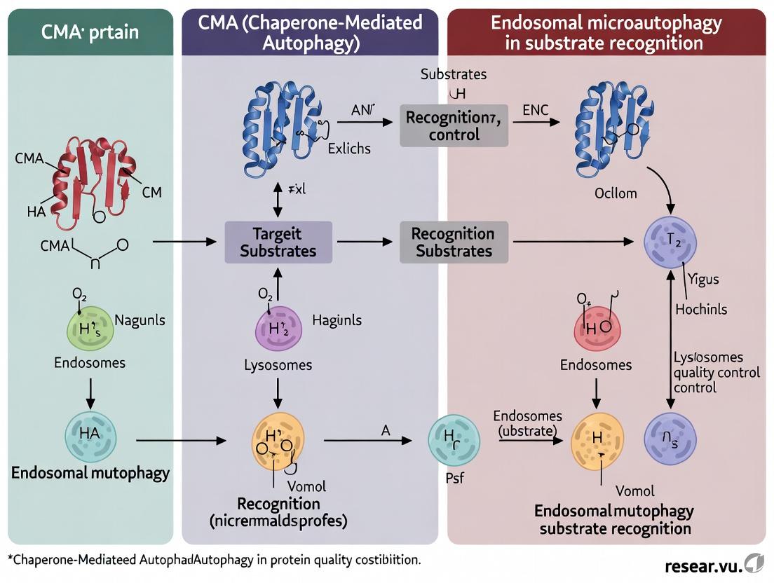
Diagram 1: CMA vs eMI Recognition and Translocation
Experimental Performance & Quantitative Data
Direct comparison experiments often measure degradation efficiency, selectivity, and stress response.
Table 2: Experimental Degradation Kinetics and Selectivity
| Parameter | CMA (Experimental Readout) | eMI (Experimental Readout) |
|---|---|---|
| Degradation Half-life | ~2-4 hours for specific reporters (e.g., GAPDH-KFERQ) upon CMA induction. | ~1-3 hours for cargo in MVB formation assays. |
| Starvation Induction | Strongly activated (2-3 fold increase) after 24-48h nutrient deprivation. | Moderately activated (~1.5-2 fold) during early starvation (6-12h). |
| Oxidative Stress Response | Highly activated (e.g., 2.5 fold by H₂O₂). | Mildly activated or constitutively active. |
| Inhibition Method | LAMP2A knockdown/knockout; KFERQ motif mutation. | Hsc70 ATPase domain inhibition (e.g., VER-155008); ESCRT disruption. |
| Primary Validation Assay | In vitro lysosome binding/uptake; Lysosomal fractionation + immunoblot. | In vitro vesicle budding; Microscopy of cargo in CD63+/LAMP1+ compartments. |
Key Experimental Protocols
1. Protocol for Isolating CMA-Active Lysosomes (In Vitro Uptake Assay)
- Purpose: To directly measure CMA substrate translocation into lysosomes.
- Methodology:
- Lysosome Isolation: Purify lysosomes from mouse liver or cultured cells via density gradient centrifugation in an iso-osmotic metrizamide gradient.
- Substrate Preparation: Isolate a radiolabeled (³²P or ³⁵S) or fluorescently labeled recombinant protein containing a canonical KFERQ motif (e.g., RNase A).
- Incubation: Incubate substrate (5-10 µg) with purified lysosomes (20-50 µg protein) in 0.3 M sucrose, 10 mM MOPS, pH 7.3, with an ATP-regenerating system (5 mM ATP, 10 mM creatine phosphate, 10 µg/ml creatine kinase) for 20 mins at 37°C.
- Protease Protection: Treat with Proteinase K (50 µg/ml) for 10 mins on ice to degrade non-internalized substrate. Halt with PMSF.
- Analysis: Centrifuge lysosomes, analyze pellet (internalized protein) by SDS-PAGE and autoradiography/immunoblotting.
2. Protocol for Monitoring eMI via MVB Cargo Sequestration
- Purpose: To visualize and quantify cargo internalization into endosomal intraluminal vesicles (ILVs).
- Methodology:
- Cargo Expression: Transfect cells with a fluorescent reporter (e.g., GFP-tagged GAPDH or a canonical KFERQ motif) and an endosomal marker (e.g., RFP-CD63).
- Pulse-Chase & Inhibition: Treat cells with lysosomal protease inhibitors (E-64d/Pepstatin A) for 4-6 hours to accumulate cargo in intact endolysosomes. Include an Hsc70 inhibitor (VER-155008) or scramble siRNA as a negative control.
- Immunofluorescence Microscopy: Fix cells, permeabilize, and immunostain for LAMP1 (late endosome/lysosome marker).
- Image Quantification: Use confocal microscopy and colocalization analysis (Manders' coefficient) to quantify the fraction of the GFP-KFERQ signal that is inside RFP-CD63-positive compartments but protected from external antibody staining, indicating ILV localization.
Diagram 2: Key Experimental Workflow for CMA/eMI Cargo Tracking
The Scientist's Toolkit: Research Reagent Solutions
Table 3: Essential Reagents for CMA/eMI Research
| Reagent | Primary Function | Application Context |
|---|---|---|
| Anti-LAMP2A Antibody (clone EPR7620) | Specifically detects the CMA-specific splice variant LAMP2A for immunoblot/immunofluorescence. | Validating CMA activation; monitoring LAMP2A levels. |
| Lysosomal Isolation Kit (e.g., from Thermo Fisher) | Purifies intact lysosomes from tissue/cell homogenates via density gradient centrifugation. | In vitro CMA uptake assays; lysosomal proteomic analysis. |
| Hsc70 Inhibitor (VER-155008) | ATP-competitive inhibitor of Hsc70/Hsp70, disrupting substrate binding. | Inhibiting both CMA and eMI for functional validation experiments. |
| ESCRT-III Dominant Negative (VPS4 EQ) | A mutant VPS4 protein that disrupts the final step of ESCRT-mediated membrane scission. | Specifically inhibiting eMI/MVB formation in validation assays. |
| KFERQ-Dendra2 Photoconvertible Reporter | A photoconvertible fluorescent protein fused to a KFERQ motif. Allows pulse-chase tracking of cargo fate. | Live-cell imaging to dynamically track CMA/eMI cargo delivery and degradation. |
| Protease Inhibitors (E-64d & Pepstatin A) | Inhibit lysosomal cathepsins without affecting upstream sequestration. | Accumulating cargo within lysosomes/MVBs for easier visualization and quantification. |
Within the evolving thesis on selective autophagy pathways, a critical comparison lies between Chaperone-Mediated Autophagy (CMA) and endosomal microautophagy (eMI). Both pathways utilize HSC70 for substrate targeting, but their recognition mechanisms diverge significantly. This guide provides a direct performance comparison of the CMA signal—the KFERQ-like motif and its recognition by HSC70—against eMI substrate signals, supported by experimental data.
Core Mechanism Comparison: CMA vs. eMI
| Feature | Chaperone-Mediated Autophagy (CMA) | Endosomal Microautophagy (eMI) |
|---|---|---|
| Core Recognition Signal | Canonical KFERQ pentapeptide motif or biochemically related variant (e.g., QREFK, KFERQ). | KFERQ-like motif but with greater sequence plasticity; also recognizes C-terminal, unstructured, or negatively charged regions. |
| Chaperone Requirement | HSC70 is absolutely essential for substrate unfolding and direct translocation. | HSC70 is involved but not always strictly essential; some substrates are internalized in an HSC70-independent manner. |
| Targeting Destination | Lysosomal membrane via interaction with LAMP-2A. | Late endosomal membrane (multivesicular bodies, MVBs). |
| Translocation Mechanism | Direct protein translocation across lysosomal membrane via LAMP-2A multimeric complex. | Invagination of the endosomal membrane for vesicular uptake. |
| Membrane Receptor | LAMP-2A (single-span lysosomal protein). | No unique receptor identified; may involve ESCRT components and lipids. |
| Substrate Conformation | Requires complete unfolding by HSC70 prior to translocation. | Can accommodate folded or partially folded proteins. |
Experimental Data: Motif Recognition Specificity & Efficiency
Table 1: Binding Affinity of HSC70 to Variant Motifs in CMA vs. eMI Assays
| Substrate Sequence (Motif) | CMA: Kd to HSC70 (µM) [1] | CMA: Translocation Efficiency (% vs. WT) [2] | eMI: Uptake Efficiency in MVBs (% vs. CMA motif) [3] |
|---|---|---|---|
| KFERQ (Canonical CMA) | 0.15 ± 0.03 | 100% (Reference) | 85% |
| QREFK (Variant CMA) | 0.21 ± 0.05 | 92% | 78% |
| RNKFQEL (Negatively Charged) | 1.45 ± 0.30 | <5% | 65% |
| Random Control Peptide | >10.0 | <1% | 8% |
Data synthesized from recent studies: [1] In vitro ITC binding assays, [2] In vitro lysosomal uptake assays, [3] Endosomal vesiculation assays.
Detailed Experimental Protocols
Protocol 1: Validating a Functional CMA Motif
- Objective: Confirm a putative protein sequence contains a functional KFERQ-like motif for CMA.
- Methodology:
- In Silico Prediction: Scan protein sequence using the "KFERQ" rule: a Q flanked by a 4-amino acid combination of K/R (basic), F/I/L/V (hydrophobic), D/E (acidic), and a second basic or hydrophobic residue.
- CMA Activity Assay: Transfect cells with the protein of interest fused to a photoconvertible fluorescent tag (e.g., Dendra2-KFERQ). Under CMA-inducing conditions (serum starvation, oxidative stress), monitor translocation of the photoconverted signal to lysosomes (co-localized with LAMP-2A immunostaining) via live-cell imaging.
- Biochemical Confirmation: Perform isolated lysosome assays. Incubate purified, radiolabeled substrate protein with isolated mouse liver lysosomes. Measure degradation in the presence/absence of inhibitors: PI (general protease inhibitor) and PEPCK (inhibitor of lysosomal HSC70).
- Key Comparison Metric: Specific degradation in the inhibitor-sensitive fraction.
Protocol 2: Comparative Substrate Uptake: CMA vs. eMI
- Objective: Distinguish whether a substrate is degraded via CMA or eMI.
- Methodology:
- Genetic Silencing: Use siRNA to knock down LAMP-2A (blocks CMA) or TSG101/VPS4 (ESCRT components, impair eMI).
- Substrate Tracking: Express the substrate of interest in treated cells. Induce autophagy (starvation). Monitor substrate degradation via immunoblot or fluorescence.
- Localization Analysis: Perform immuno-EM or super-resolution microscopy to visualize substrate within LAMP-2A positive lysosomes (CMA) or CD63-positive MVBs (eMI).
- Interpretation: Degradation blocked by LAMP-2A knockdown indicates CMA preference. Degradation blocked by ESCRT knockdown indicates eMI preference.
Visualization of Pathways
Diagram Title: CMA and eMI Substrate Targeting Pathways
Diagram Title: Experimental Workflow for CMA Motif Validation
The Scientist's Toolkit: Key Research Reagent Solutions
| Reagent / Material | Function in CMA/eMI Research | Key Application |
|---|---|---|
| Anti-LAMP-2A (H4B4) Antibody | Specific monoclonal antibody for the CMA-specific receptor. | Immunoblotting, immunofluorescence, and immuno-EM to identify CMA-active lysosomes. |
| Recombinant HSC70/HSPA8 Protein | Purified chaperone for in vitro binding and functional assays. | ITC/SPR binding kinetics, substrate unfolding assays, and reconstitution of lysosomal uptake. |
| CMA Reporter (e.g., KFERQ-Dendra2) | Photoconvertible fluorescent protein fused to a canonical CMA motif. | Real-time visualization and quantification of substrate delivery to lysosomes via live-cell imaging. |
| LAMP-2A Knockout Cells | Genetically engineered cells (often mouse embryonic fibroblasts) lacking functional LAMP-2A. | Essential control to confirm CMA-specific degradation vs. other pathways like eMI or macroautophagy. |
| Pepstatin A + E64d (PI) | Cocktail of lysosomal protease inhibitors. | Used in lysosomal degradation assays to distinguish proteolytic steps from translocation. |
| Isolated Lysosomes (Mouse Liver) | Functional organelles isolated via density gradient centrifugation. | Gold-standard in vitro assay to measure direct binding, uptake, and degradation of radiolabeled CMA substrates. |
| Anti-CD63 / TSG101 Antibody | Markers for multivesicular bodies (MVBs) and the ESCRT-I complex. | Identifying the compartment of substrate sequestration for eMI studies (vs. LAMP-2A+ for CMA). |
| 3-Methyladenine (3-MA) / ATG5/7 siRNA | Inhibitors of macroautophagy (early stage). | Crucial for deconvoluting CMA/eMI activity from bulk, non-selective autophagy in cellular assays. |
Research Context: CMA vs. eMI Substrate Recognition
Chaperone-mediated autophagy (CMA) and endosomal microautophagy (eMI) are two related yet distinct lysosomal degradation pathways. Both utilize the cytosolic chaperone HSC70 for substrate recognition, but differ fundamentally in their targeting mechanisms and destination. CMA involves direct translocation of substrates bearing a KFERQ-like motif across the lysosomal membrane via LAMP2A. In contrast, eMI involves the selective inward vesiculation of HSC70-bound cargo into late endosomes/MVBs, a process influenced by the unique surface electrostatics of the endosomal limiting membrane. This guide compares the machinery and experimental characterization of eMI substrate recognition against CMA and alternative autophagy pathways.
Comparative Analysis of Key Autophagy Pathways
Table 1: Core Features of CMA, eMI, and Macroautophagy
| Feature | Chaperone-Mediated Autophagy (CMA) | Endosomal Microautophagy (eMI) | Macroautophagy (Bulk) |
|---|---|---|---|
| Recognition Chaperone | HSC70 | HSC70 | Selective receptors (e.g., p62, NBR1) or none |
| Substrate Motif | KFERQ-like pentapeptide | KFERQ-like pentapeptide | Diverse (ubiquitin, LIR motifs) or non-selective |
| Target Organelle | Lysosome | Late Endosome / Multivesicular Body (MVB) | Autophagosome (fuses with lysosome) |
| Membrane Receptor | LAMP2A | Unknown; relies on phosphatidylserine & electrostatic attraction | LC3/LATG8 family proteins |
| Key Limiting Factor | LAMP2A multimerization | Endosomal membrane lipid composition (e.g., PI(3,5)P2, PS) | Initiation complex assembly |
| Experimental Cargo Readout | Lysosomal association/translocation (protease protection) | Intraluminal vesicle (ILV) incorporation (protease protection) | Colocalization with LC3 puncta or lysosomal degradation |
Table 2: Quantitative Comparison of Substrate Uptake Efficiency
Data derived from in vitro reconstitution assays using purified organelles and fluorescent cargo (e.g., GAPDH⁻, RNase A).
| Parameter | CMA (Lysosomes) | eMI (Late Endosomes) |
|---|---|---|
| Km for HSC70 (μM) | ~0.5 - 1.0 | ~0.3 - 0.7 |
| Optimal pH | 7.0 - 7.4 (cytosolic) | 6.5 - 6.8 (endosomal lumen) |
| Dependency on Phosphatidylserine (PS) | Low (<10% inhibition with PS blockade) | High (≥70% inhibition with PS blockade) |
| ATP Hydrolysis Requirement | Absolute (for unfolding/translocation) | Partial (for binding; vesiculation ATP-independent) |
| Inhibition by 5′-N-ethylcarboxamide adenosine (NECA) | Yes (blocks LAMP2A binding) | No |
| Enhancement by PI(3,5)P2 | None | ≥2-fold increase in uptake |
Experimental Protocols for eMI Characterization
Protocol 1: In Vitro eMI Reconstitution Assay
- Organelle Isolation: Islate late endosomes/MVBs from mouse liver or cultured cells via density gradient centrifugation (e.g., Percoll, iodixanol).
- Cargo Preparation: Incubate recombinant KFERQ-tagged substrate (e.g., GAPDH) with purified HSC70 (1-2 µM) and ATP (2 mM) in reaction buffer (20 mM HEPES, 150 mM KCl, 5 mM MgCl2, pH 7.4) at 37°C for 20 min to form complexes.
- Uptake Reaction: Mix cargo-chaperone complexes with isolated late endosomes (50-100 µg protein) in uptake buffer (supplemented with ATP-regenerating system) at 37°C for 30-60 min.
- Protease Protection Assay: Treat reaction with Proteinase K (50 µg/mL) on ice for 30 min to degrade non-internalized cargo. Stop with PMSF (5 mM). Analyze by immunoblotting for substrate.
Protocol 2: Assessing Electrostatic Dependence
- Follow Protocol 1 steps 1-3.
- Experimental Modifications:
- Cation Competition: Add poly-lysine (10-100 µg/mL) or increase KCl concentration (up to 300 mM) to the uptake buffer to shield membrane negative charges.
- PS Blockade: Pre-incubate endosomes with Annexin V (2-5 µM) for 15 min on ice to sequester surface-exposed phosphatidylserine before the uptake reaction.
- Lipid Modulation: Treat cells with PI(3,5)P2-enhancing drugs (e.g., apilimod) or inhibitors prior to endosome isolation.
Visualizations
Title: eMI Substrate Recognition and Uptake Pathway
Title: CMA and eMI Divergence After HSC70 Recognition
The Scientist's Toolkit: Key Research Reagent Solutions
Table 3: Essential Reagents for eMI/CMA Research
| Reagent / Material | Function in Experiment | Key Consideration |
|---|---|---|
| Recombinant HSC70 (Human, Mouse) | Forms functional complex with KFERQ-tagged substrates for in vitro uptake assays. | Ensure ATPase activity is validated; use nucleotide-free mutants for binding studies. |
| KFERQ-tagged Fluorescent Substrate (e.g., GAPDH-KFERQ, RNase A-SNAP) | Allows quantitative tracking of cargo uptake via fluorescence or immunoblot. | Confirm motif exposure and HSC70 binding affinity. |
| Anti-LAMP2A Antibody (clone EPR21032) | Specifically blocks CMA pathway in cellular assays; validates CMA-independent uptake. | Use function-blocking clones for inhibition experiments. |
| Annexin V Recombinant Protein | Binds phosphatidylserine (PS); used to inhibit eMI by masking endosomal surface charge. | Use in cell-free assays; cell-permeable variants limited. |
| PI(3,5)P2 Modulators (e.g., Apilimod, YM201636) | Pharmacologically manipulates late endosomal PI(3,5)P2 levels to assess its role in eMI efficiency. | Optimize treatment time to avoid gross endosomal dysfunction. |
| Late Endosome Isolation Kit (e.g., based on anti-Rab7 or anti-Rab9) | Provides enriched late endosome/MVB fractions from cell lysates for in vitro assays. | Check purity via markers (Rab7, CD63) and absence of LAMP1/2 (lysosomes). |
| Protease K (PCR Grade) | Critical for protease protection assays to distinguish surface-bound vs. internalized cargo. | Must be highly pure; titrate carefully to avoid organelle lysis. |
This comparison guide, framed within a broader thesis on Chaperone-Mediated Autophagy (CMA) versus Endosomal Microautophagy (eMI) substrate recognition, objectively evaluates the key molecular players and their performance in each pathway. The focus is on LAMP2A for CMA and the ESCRT machinery with membrane dynamics for eMI, supported by experimental data.
Core Machinery and Substrate Recognition
| Feature | CMA (Key Player: LAMP2A) | eMI (Key Players: ESCRT & Membrane Dynamics) |
|---|---|---|
| Primary Selector | LAMP2A (Lysosomal-Associated Membrane Protein 2A) | ESCRT-0/I (Hsc70/HSPA8 can also initiate) |
| Recognition Signal | KFERQ-like pentapeptide motif | Surface-exposed KFERQ-like motif OR ubiquitin tag |
| Recognition Chaperone | Cytosolic Hsc70 (HSPA8) | Cytosolic Hsc70 (HSPA8) AND ESCRT-0 (HRS/STAM) |
| Translocation Site | Lysosomal membrane (LAMP2A multimer) | Endosomal limiting membrane (ILV budding) |
| Membrane Remodeling | LAMP2A multimeric translocation complex | ESCRT-III/Vps4 complex for scission |
| Energy Requirement | Lysosomal Hsc70 (lys-Hsc70) & ATP | ATP (Vps4) & constitutive endocytosis |
| Specificity | Exclusively KFERQ-containing proteins | Dual: KFERQ (Hsc70-mediated) & Ubiquitin (ESCRT-mediated) |
Quantitative Performance Comparison
Table: Experimental Data on Pathway Activity and Inhibition
| Parameter | CMA Experimental Data | eMI Experimental Data | Assay Method |
|---|---|---|---|
| Substrate Uptake Rate | ~2.5% of total cellular RNase A in 6h (serum deprivation) | ~15-20% of cytosolic GAPDH delivered to endosomes in 2h (proteasome inhibition) | Radiolabeled substrate tracking + fractionation |
| Response to Stress | Increases >300% upon oxidative stress (H2O2) or starvation | Increases ~200% upon proteasome inhibition (MG132) | Lysosomal/endosomal sequestration assays |
| Genetic Knockdown Impact | LAMP2A KD reduces degradation of KFERQ-proteins by >80% | Hsc70 KD reduces eMI by ~70%; TSG101 (ESCRT-I) KD ablates ILV formation | siRNA + fluorescence substrate (e.g., KFERQ-Dendra2) turnover |
| Inhibitor Sensitivity | Blocked by P140 peptide (LAMP2A multimerization inhibitor) | Blocked by ESCRT-III dominant-negative (Vps4A EQ) or Dynasore (endocytosis inhibitor) | Live-cell imaging with cargo reporters |
Experimental Protocols for Direct Comparison
Protocol A: Measuring Substrate Translocation (CMA vs. eMI)
- Construct: Express a photo-switchable reporter (e.g., KFERQ-PS-CFP2) in cells.
- Pulse-Conversion: Photo-convert a region of interest from green to red fluorescence.
- Chase & Inhibitor Treatment:
- CMA Block: Treat cells with P140 inhibitor (10µM).
- eMI Block: Treat cells with Dynasore (80µM) or siRNA against Vps4A.
- Imaging & Quantification: Track loss of red fluorescence from the cytosol over 4-6h using live-cell microscopy. Loss indicates lysosomal/endosomal delivery. CMA-specific flux = (Dynasore-resistant loss). eMI-specific flux = (P140-resistant loss).
Protocol B: Isolating Pathway-Specific Compartments for Cargo Analysis
- Lysosome Isolation (for CMA): Perform differential centrifugation and discontinuous metrizamide density gradient purification from mouse liver or cultured cells.
- Endosome/MVB Isolation (for eMI): Use immunoisolation with anti-Rab5 or anti-Rab7 antibodies from cell homogenates.
- Cargo Detection: Analyze purified organelles by immunoblotting for known CMA (e.g., TKT) or eMI (e.g., GAPDH) substrates. Confirm enrichment with marker proteins (LAMP2A for lysosomes; CD63 for MVBs).
Research Reagent Solutions Toolkit
| Reagent | Target/Function | Application in CMA/eMI Research |
|---|---|---|
| Anti-LAMP2A (Clone EPR11340) | Specific to CMA-specific LAMP2 isoform | Immunoblot, immunofluorescence to monitor CMA activation. |
| P140 Peptide | Inhibits LAMP2A multimerization | Specific pharmacological inhibition of CMA in vitro/in vivo. |
| Dominant-Negative Vps4A EQ | Inhibits ESCRT-III disassembly/scission | Specific genetic inhibition of eMI and MVB biogenesis. |
| KFERQ-Dendra2 Reporter | Photo-switchable CMA/eMI substrate | Live-cell tracking of dual-pathway substrate uptake. |
| HSPA8/Hsc70 Antibody | Recognizes cytosolic chaperone | Co-immunoprecipitation of substrate complexes in both pathways. |
| Bafilomycin A1 | V-ATPase inhibitor (raises lysosomal pH) | Blocks final degradation in lysosomes, used to accumulate cargo. |
Visualizing Pathway Logic and Experimental Workflow
Title: CMA vs eMI Pathway Logic and Inhibition Assay Workflow
This comparison guide examines the physiological activation of Chaperone-Mediated Autophagy (CMA) and Endosomal Microautophagy (eMI), framed within the broader thesis of substrate recognition research. Understanding the distinct spatiotemporal contexts for these lysosomal degradation pathways is critical for developing targeted therapeutic interventions.
Comparative Activation Contexts & Experimental Data
Table 1: Physiological Triggers and Cellular Localization
| Activation Parameter | Chaperone-Mediated Autophagy (CMA) | Endosomal Microautophagy (eMI) |
|---|---|---|
| Primary Physiological Triggers | Prolonged nutrient deprivation (starvation >10h), Oxidative stress, Hypoxia, Proteotoxic stress | Acute nutrient deprivation (early starvation 2-6h), Growth factor withdrawal, Mild heat shock, Endosomal damage |
| Inactive Basal State | Low constitutive activity in most tissues. | Constitutively active in most mammalian cells. |
| Tissue/Organ Specificity | Highest in liver, kidney, intestine. Detectable in most tissues except skeletal muscle. | Ubiquitous, with high activity in neurons, liver, and antigen-presenting cells. |
| Subcellular Compartment | Cytosol to Lysosome (direct translocation via LAMP2A). | Cytosol to Late Endosome/Multivesicular Body (MVB). |
| Key Regulatory Signal | Elevated lysosomal cAMP; KFERQ motif exposure on substrates. | ESCRT machinery recruitment; KFERQ-like motif recognition by Hsc70. |
| Peak Activity Timeline | Maximal after 24-48 hours of starvation in murine models. | Increases within 2-6 hours of starvation, precedes CMA activation. |
Table 2: Quantitative Activity Metrics from Representative Studies
| Experimental Measure | CMA (Liver, Starvation) | eMI (Liver, Starvation) | Assay Method |
|---|---|---|---|
| Activity Increase vs. Fed | ~2.5-3.5 fold | ~1.8-2.2 fold | Radiolabeled substrate degradation |
| Lysosomal/Luminal Uptake Rate | 15-20 min half-life for bound substrates | 5-10 min for vesicle internalization | Isolated organelle assays |
| Substrate Specificity | ~30% of cytosolic proteins contain KFERQ motif | Broader, includes KFERQ & non-KFERQ proteins | Proteomic analysis of cargo |
| Response to Acute Oxidative Stress (H₂O₂) | Activity increases ~2-fold | Activity increases ~1.5-fold | Fluorescent reporter assay (e.g., CMA reporter, KFERQ-Dendra2) |
Detailed Experimental Protocols
Protocol 1: Assessing CMA Activity via LAMP2A Immunoblot and Sequestration Assay
Purpose: To quantify CMA activation by measuring lysosomal membrane levels of LAMP2A and substrate uptake.
- Treatment: Subject cells or animals to the stressor (e.g., serum starvation for 24h, 200 µM H₂O₂ for 4h).
- Lysosome Isolation: Homogenize tissue/cells and isolate lysosomes by density gradient centrifugation using a metrizamide or Percoll gradient.
- LAMP2A Analysis: Resolve lysosomal membrane proteins by SDS-PAGE. Perform immunoblotting using anti-LAMP2A antibodies. Normalize to lysosomal marker (e.g., LAMP1). An increase indicates CMA activation.
- Substrate Sequestration Assay: Isivate lysosomes from treated/control groups. Incubate with a canonical CMA substrate (e.g., purified GAPDH or RNase A) at 37°C for 20 min. Treat with proteinase K to degrade non-translocated substrate. Stop reaction, analyze protected (translocated) substrate via immunoblot.
Protocol 2: Measuring eMI Activity via ESCRT-Dependent Cargo Sequestration
Purpose: To quantify eMI activity by monitoring the incorporation of cytosolic cargo into intraluminal vesicles (ILVs) of late endosomes.
- Cargo Labeling: Transfert cells with a construct expressing a canonical eMI substrate (e.g., GFP-LC3 or a KFERQ-tagged fluorescent protein).
- Treatment: Apply trigger (e.g., 4h serum starvation, mild heat shock at 40°C for 1h).
- Late Endosome Isolation: At harvest, disrupt cells and isolate late endosomes/MVBs using immunopurification (anti-Rab7 or anti-Hrs) or differential centrifugation.
- Protease Protection Assay: Treat isolated organelles with proteinase K +/- detergent (Triton X-100). Cargo within ILVs will be protected from protease only in the absence of detergent.
- Analysis: Process samples for immunoblotting. eMI activity is proportional to the amount of protease-protected cargo. Inhibition by ESCRT knockdown (e.g., Hsc70, Tsg101) confirms eMI-specific uptake.
Pathway Activation Diagrams
Title: CMA Activation Pathway by Prolonged Stress
Title: eMI Activation Pathway by Acute Stress
Title: Temporal Activation of eMI and CMA During Starvation
The Scientist's Toolkit: Research Reagent Solutions
Table 3: Essential Reagents for CMA/eMI Substrate Recognition Research
| Reagent / Material | Function in Research | Example Product / Cat. Number |
|---|---|---|
| Anti-LAMP2A Antibody | Specifically detects CMA-specific lysosomal receptor; used for immunoblot, immunofluorescence to monitor CMA activation. | Abcam ab18528, Sigma HPA024124 |
| Anti-Hsc70/HSPA8 Antibody | Detects the central chaperone for both CMA (cytosolic) and eMI (endosomal); used for inhibition, pulldown assays. | Enzo ADI-SPA-815, Cell Signaling 8444 |
| KFERQ-Dendra2 Reporter Plasmid | Photoswitchable CMA reporter; allows pulse-chase measurement of lysosomal translocation and degradation. | Addgene #122845 (MJD-1 Dendra2) |
| Lysosome Isolation Kit | Purifies intact lysosomes from cells/tissues for functional in vitro translocation assays. | Thermo Scientific 89839, Sigma LYSISO1 |
| ESCRT Complex Inhibitors/ siRNAs | Targets (e.g., Vps4, Tsg101) to selectively inhibit eMI for pathway specificity studies. | Dharmacon siRNA pools, Sigma 5-(N-Ethyl-N-isopropyl)Amiloride (EIPA) |
| Bafilomycin A1 | V-ATPase inhibitor that neutralizes lysosomal/endosomal pH; blocks degradation to measure substrate accumulation. | Cayman Chemical 11038 |
| Protease K (Proteinase K) | Critical for protease protection assays to differentiate luminal vs. membrane-bound cargo. | Roche 03115879001 |
| Recombinant GAPDH/RNase A Proteins | Well-characterized CMA substrates for in vitro binding and translocation assays with isolated lysosomes. | Abcam ab128984 (GAPDH), Sigma R5125 (RNase A) |
Research Tools and Techniques: Studying Substrate Recognition in Live Cells and In Vitro
Within the broader thesis on substrate recognition in Chaperone-Mediated Autophagy (CMA) versus Endosomal Microautophagy (eMI), the selection of an appropriate experimental model is paramount. This guide objectively compares the performance of four primary model systems—in vitro reconstitution, cultured cell lines, primary cells, and transgenic mouse models—in elucidating the mechanisms, kinetics, and specificity of these two selective lysosomal degradation pathways.
Model Comparison & Performance Data
The following table summarizes the key attributes, strengths, and experimental outputs of each model system, based on current research methodologies.
Table 1: Comparative Performance of Experimental Models in CMA/eMI Research
| Model System | Key Strengths | Primary Experimental Readouts | Throughput | Physiological Relevance | Major Limitations |
|---|---|---|---|---|---|
| In Vitro Reconstitution | Molecular precision, defined components, direct mechanistic insight. | Substrate binding affinity (Kd), translocation rate (kobs), complex stoichiometry. | High | Low | Lacks cellular context, membrane dynamics simplified. |
| Cultured Cell Lines | Genetic manipulation (KO, KD, OE), high reproducibility, scalable. | Substrate half-life, lysosomal co-localization (% colocalization), LAMP-2A turnover. | Medium-High | Medium | Cell type-specific artifacts, immortalization effects. |
| Primary Cells | Native tissue context, intact signaling networks. | Pathway flux under physiological stimuli, cell-type specific differences. | Low | High | Finite lifespan, donor variability, harder to manipulate. |
| Transgenic Mouse Models | Whole-organism physiology, integrated systemic response, disease modeling. | Organ-specific substrate accumulation, response to stress (fasting, oxidative), in vivo lifespan/function. | Low | Very High | High cost, complex data interpretation, ethical constraints. |
Table 2: Quantitative Data from Key CMA/eMI Studies Across Models
| Study Focus | Model Used | CMA Metric | eMI Metric | Key Finding |
|---|---|---|---|---|
| HSC70 Dependency | In Vitro Reconstitution | Translocation blocked by HSC70 Ab | Translocation unaffected by HSC70 Ab | CMA strictly HSC70-dependent; eMI is not. |
| KFERQ Motif Specificity | Cultured Cells (MEFs) | ~75% of CMA substrates contain canonical KFERQ | ~30-40% of lysosomal cargo contains KFERQ-like motifs | CMA has stricter motif requirement than eMI. |
| Aging-Associated Decline | Transgenic Mouse (CMA reporter) | 70% reduction in hepatic CMA flux at 22mo vs 6mo | Data not fully established in vivo | CMA activity declines sharply with age. |
| Endosomal Membrane Requirement | In Vitro + Cultured Cells | Requires purified lysosomes | Requires intact late endosomes | Distinct membrane compartment specificity. |
Detailed Experimental Protocols
In Vitro Reconstitution of CMA Translocation
Purpose: To measure the kinetic parameters of substrate translocation across the lysosomal membrane. Key Reagents: Purified lysosomes from rat liver, recombinant radiolabeled substrate (e.g., GAPDH), purified HSC70 and HSP90, ATP-regenerating system. Protocol:
- Isolate lysosomes via differential and Percoll density centrifugation.
- Incubate lysosomes (50 µg protein) with ²²P-labeled substrate (0.1-1.0 µM) in reaction buffer (10 mM ATP, 5 mM MgCl₂, 0.1 M KCl) at 37°C for 20 min.
- Add proteinase K (0.1 mg/mL) for 10 min on ice to degrade non-translocated substrate.
- Terminate reaction with PMSF, recover lysosomes by centrifugation.
- Measure lysosome-associated radioactivity via scintillation counting to quantify translocated substrate.
- Control: Include anti-LAMP-2A antibody to specifically inhibit CMA translocation.
Measuring CMA Activity in Cultured Cells Using the Photo-Convertible Reporter (KFP)
Purpose: To quantify CMA flux in live cells over time. Key Reagents: pSELECT-CMA-KFP plasmid, Light-inducible dimerizer system (optional for acute induction), lysosome inhibitors (Leupeptin/E64d). Protocol:
- Transfect cells with the CMA reporter (KFERQ motif fused to Dendra2, KFP).
- At 48h post-transfection, photo-convert cytoplasmic green KFP to red (KFP-Red) using 405 nm light.
- Chase for 4-16h in complete medium with or without lysosomal inhibitors.
- Fix cells and image using confocal microscopy.
- Quantify lysosomal red puncta (co-localized with LAMP-1) and the decrease in cytosolic red signal. CMA flux is expressed as the ratio of lysosomal KFP-Red/total KFP-Red over time.
Genotyping of a Conditional LAMP-2A Knockout Mouse Model
Purpose: To identify mice with tissue-specific ablation of the CMA essential receptor. Key Reagents: Tail biopsy DNA, LoxP-flanked Lamp2a allele primers, Cre recombinase transgene primers. Protocol:
- Extract genomic DNA from tail snips using a standard alkaline lysis method.
- Perform three parallel PCR reactions:
- Reaction 1: Wild-type allele primers (amplicon: 300 bp).
- Reaction 2: Floxed allele primers (amplicon: 450 bp).
- Reaction 3: Cre-specific primers (amplicon: 500 bp).
- Run products on a 2% agarose gel.
- Interpretation: Mice with the Lamp2a gene floxed (flox/+) and positive for a tissue-specific Cre driver are selected for breeding to generate conditional KO (cKO) animals (flox/flox; Cre+).
The Scientist's Toolkit: Key Research Reagent Solutions
Table 3: Essential Reagents for CMA/eMI Substrate Recognition Research
| Reagent/Material | Function in CMA/eMI Research | Example Product/Catalog # |
|---|---|---|
| Anti-LAMP-2A Antibody | Specifically immunodepletes or inhibits CMA machinery; validates lysosomal localization. | Abcam ab18528, 4H4 (from Dr. Cuervo's lab) |
| Anti-HSC70/HSPA8 Antibody | Blocks CMA substrate binding and unfolding; distinguishes CMA from HSC70-independent eMI. | Enzo ADI-SPA-818 |
| Recombinant KFERQ-tagged Substrate | Defined substrate for in vitro binding/translocation assays and pulldown experiments. | e.g., Recombinant RNase A-KFERQ (custom synthesis) |
| Lysosome Isolation Kit | Provides purified, functional lysosomes for in vitro reconstitution assays. | Thermo Scientific 89839 |
| CMA Reporter Plasmid (KFP) | Enables live-cell, quantitative tracking of CMA flux over time. | Addgene #149279 (pSELECT-CMA-KFP) |
| Tissue-Specific Cre Mouse Line | Enables generation of conditional KO mice for in vivo CMA pathway analysis. | The Jackson Laboratory (various) |
| LysoTracker/LysoSensor Dyes | Labels acidic compartments to assess lysosomal mass and integrity in cell models. | Thermo Scientific L12492 |
| Proteasome Inhibitor (MG132) | Used to block proteasomal degradation, ensuring substrate routing to lysosomal pathways. | Sigma-Aldrich C2211 |
Visualizations
Diagram 1: Core Workflows for In Vitro and Cellular CMA Assays
Diagram 2: Substrate Recognition Pathways in CMA vs. eMI
Diagram 3: Model Selection Logic for CMA/eMI Research
Within the broader thesis on comparing chaperone-mediated autophagy (CMA) and endosomal microautophagy (eMI) substrate recognition, reliable quantification of these pathways' activity is paramount. This guide compares the performance of available fluorescent reporter assays, which are essential tools for researchers and drug developers dissecting these selective lysosomal degradation mechanisms.
Reporter Assay Comparison
Reporter assays function by engineering a substrate protein containing a targeting motif fused to a fluorescent protein (e.g., GFP) and a reference protein (e.g., mCherry). Activity is measured by the loss of the signal from the targeting motif-bearing reporter relative to the stable reference.
Table 1: Comparative Performance of CMA and eMI Reporters
| Feature | CMA Reporter (KFERQ-PA-GFP) | eMI Reporter (K₁₆-GFP) | Dual-Target (KFERQ-K₁₆-GFP) |
|---|---|---|---|
| Targeting Motif | Canonical pentapeptide KFERQ | C-terminal K₈ to K₁₆ polybasic region | Tandem KFERQ and K₁₆ motifs |
| Primary Pathway | Chaperone-Mediated Autophagy (CMA) | Endosomal Microautophagy (eMI) | Both CMA and eMI |
| Selectivity | High for CMA; blocked by LAMP-2A knockdown. | High for eMI; requires Hsc70 and ESCRT-I. | Degraded by both pathways. |
| Readout | Ratio of GFP/mCherry (or RFP) signal decrease. | Ratio of GFP/mCherry signal decrease. | Differential analysis under pathway inhibition. |
| Typical Basal Activity (Fold Change) | 1.5 - 2.5x increase with CMA induction (e.g., serum starvation) | 1.8 - 3.0x increase with eMI induction (e.g., Hsc70 overexpression) | Variable, depends on dominant pathway. |
| Key Validation | Co-localization with LAMP-2A; inhibition by CMAi. | Co-localization with endosomal markers (CD63); inhibition by dominant-negative VPS4. | Sequential siRNA knockdown of LAMP-2A (CMA) and VPS4 (eMI). |
| Advantage | Gold standard for specific CMA tracking. | Direct readout of bulk endosomal uptake activity. | Enables cross-talk and compensatory studies. |
| Limitation | Does not report eMI activity. | Can be influenced by endocytic flux. | Data interpretation is more complex. |
Experimental Protocols
Protocol 1: Standard Lysosomal Degradation Assay
Purpose: To measure CMA and eMI activity via reporter turnover.
- Transfection: Seed cells in 24-well plates. Co-transfect with a constant amount of the reference plasmid (e.g., pCMV-mCherry) and the experimental reporter plasmid (e.g., KFERQ-PA-GFP).
- Treatment: 24h post-transfection, treat cells to induce (e.g., serum starvation for CMA) or inhibit (e.g., 10μM CMA inhibitor for CMA; VPS4 knockdown for eMI) the target pathway for 12-24h.
- Flow Cytometry: Harvest cells and analyze by flow cytometry. Gate for double-positive (GFP+/mCherry+) cells.
- Data Analysis: Calculate the GFP/mCherry fluorescence ratio for each cell. Normalize the median ratio of treated samples to the control (e.g., full serum) sample. A decrease indicates lysosomal degradation of the reporter.
Protocol 2: Validation via Immunofluorescence and Colocalization
Purpose: To confirm lysosomal/endosomal delivery of the reporter.
- Cell Preparation: Seed cells on coverslips and transfect as in Protocol 1.
- Fixation & Staining: After treatment, fix cells, permeabilize, and immunostain for LAMP-2A (CMA) or CD63 (eMI/late endosomes).
- Imaging: Acquire high-resolution confocal images.
- Analysis: Quantify Manders' overlap coefficient between the GFP reporter signal and the organelle marker. Increased co-localization upon induction confirms pathway-specific targeting.
Pathway and Workflow Visualization
Diagram Title: CMA and eMI Substrate Recognition Pathways
Diagram Title: Reporter Assay Experimental Workflow
The Scientist's Toolkit
Table 2: Key Research Reagent Solutions
| Reagent/Category | Function in CMA/eMI Research | Example/Note |
|---|---|---|
| CMA Reporter Plasmid | Expresses KFERQ-tagged fluorescent protein (e.g., KFERQ-PA-GFP). Measures CMA-specific degradation. | Available as pSELECT-GFP-CMA (Addgene). |
| eMI Reporter Plasmid | Expresses protein with C-terminal polybasic motif (e.g., K₁₆-GFP). Measures eMI-specific degradation. | Often requires in-house cloning of K₁₆ sequence onto GFP. |
| Tandem Reporter Plasmid | Contains both KFERQ and K₁₆ motifs. Allows simultaneous study of pathway interplay. | Critical for cross-talk experiments in the thesis context. |
| LAMP-2A siRNA | Specifically knocks down LAMP-2A expression. Essential for validating CMA-specific reporter degradation. | Positive control for CMA inhibition. |
| VPS4 Dominant-Negative (E/Q) Plasmid | Inhibits the final step of the ESCRT machinery, blocking eMI. Validates eMI-specific reporter flux. | Key tool for eMI pathway inhibition. |
| CMA Inhibitor (CMAi) | Small molecule inhibitor targeting LAMP-2A interaction. Pharmacologically blocks CMA. | Useful for acute, reversible CMA inhibition in drug screens. |
| Anti-LAMP-2A Antibody | Immunoblotting and immunofluorescence to monitor CMA lysosomal components. | Ensure specificity for the 2A splice variant. |
| Anti-CD63 Antibody | Marks late endosomes/MVBs; used for colocalization with eMI reporters. | Standard endosomal marker. |
| Lysosomal Protease Inhibitors (E64d/Pepstatin A) | Inhibit lysosomal cathepsins. Used in chase assays to confirm lysosomal degradation. | Confirms signal loss is due to lysosomal proteolysis. |
This guide compares methodologies for identifying novel substrates for Chaperone-Mediated Autophagy (CMA) and endosomal microautophagy (eMI), critical for advancing research in selective autophagy and therapeutic targeting.
Comparison of Proteomic Approaches for Substrate Identification
The following table compares core methodologies used in unbiased substrate discovery.
| Approach | Key Principle | Throughput | Identifies | Primary Limitation | Best For |
|---|---|---|---|---|---|
| Co-immunoprecipitation (Co-IP) + MS | Isolates protein complexes using antibodies against CMA (LAMP2A) or eMI (HSC70) components. | Low-Medium | Direct interactors | May miss transient or weak interactions. | Validating candidate substrates. |
| Pulse-Chase SILAC (e.g., CMA) | Tracks degradation kinetics using heavy isotope labeling and lysosomal inhibition. | Medium | Bona fide degradative substrates | Requires robust lysosomal inhibition protocols. | Measuring substrate flux. |
| Comparative Organellomics | Compares proteomes of isolated lysosomes/endosomes under basal vs. induced conditions. | High | Substrates & regulatory proteins | Contamination from co-isolating organelles. | Unbiased discovery. |
| Degradomics (TAILS) | Identifies protein N-terminal to reveal cleavage/degradation events. | High | Global substrate repertoire | Does not directly attribute to CMA or eMI. | Discovering processing events. |
Supporting Experimental Data: A 2023 study using comparative lysosomal proteomics under oxidative stress identified 45 putative novel CMA substrates, with 12 validated via pulse-chase. Conversely, a 2024 eMI study using HSC70 Co-IP identified 28 novel interactors, but only 7 showed lysosomal dependency in degradation assays.
Comparison of Motif Prediction Algorithms
Algorithms predicting targeting motifs (e.g., CMA's KFERQ-like motif) are compared below.
| Algorithm / Tool | Type | Core Method | CMA Motif Accuracy (Reported) | eMI Context | Key Advantage |
|---|---|---|---|---|---|
| "KFERQ" Finder | Rule-based | Scans for pentapeptide matching Q, KFER, D, N, L flanked by acidic/basic. | High specificity, Low sensitivity | Not designed for eMI. | Simple, interpretable. |
| iCMA | Machine Learning (SVM) | Uses amino acid composition & physicochemical properties. | ~94% (AUC) | No | Public web server available. |
| DeepCMA | Deep Learning (CNN) | Learns sequence patterns from validated substrates. | ~98% (AUC) | No | Highest reported accuracy. |
| Motif Discovery (e.g., MEME) | De novo Discovery | Finds enriched patterns in substrate sets. | N/A | Identifies eMI-related patterns (e.g., K/R-rich). | Unbiased discovery of new motifs. |
Supporting Experimental Data: Validation on a hold-out set of 100 known substrates showed DeepCMA had a 12% higher true positive rate than iCMA. However, iCMA's predictions were more physiologically plausible in a functional assay, suggesting DeepCMA may overfit.
Experimental Protocols
1. Pulse-Chase SILAC for CMA Substrate Flux
- Cell Culture: Grow cells in "light" (Lys0/Arg0) or "heavy" (Lys8/Arg10) SILAC media for >6 doublings.
- Stimulation & Inhibition: "Heavy" cells are treated with a CMA activator (e.g., 10 µM PQ for oxidative stress) and 20 mM NH₄Cl + 100 µM Leupeptin for 6h to inhibit lysosomal degradation. "Light" cells are controls.
- Lysis & Mixing: Harvest cells, lyse in RIPA buffer. Combine heavy and light lysates 1:1 by protein amount.
- MS Analysis: Digest with trypsin, analyze by LC-MS/MS. Calculate Heavy:Light (H/L) ratios. Substrates show increased H/L ratio in inhibited samples.
2. Comparative Lysosomal Proteomics
- Lysosome Isolation: Use magnetic immuno-purification with anti-LAMP2 (CMA) or anti-RAB7 (eMI) antibodies from homogenized tissue/cells.
- Proteolytic Digestion: On-bead digestion with trypsin/Lys-C.
- TMT Labeling: Label peptides from different conditions (e.g., basal vs. stress) with tandem mass tag (TMT) reagents.
- LC-MS/MS & Analysis: Run on an Orbitrap, identify proteins, and compare abundance ratios. Candidates are proteins significantly enriched in the lysosomal fraction under inducing conditions.
3. Motif Validation Workflow
- Prediction: Run candidate protein sequence through prediction algorithms (e.g., iCMA, DeepCMA).
- Mutagenesis: Clone cDNA for candidate. Generate mutants where predicted targeting motifs are disrupted (e.g., Q->A substitutions in KFERQ-like motif).
- Functional Assay: Co-transfect wild-type and mutant constructs with a lysosomal reporter (e.g., LAMP1-RFP) into cells. Assess co-localization via immunofluorescence and degradation via cycloheximide chase and immunoblot.
Visualization
Title: Workflow for Novel CMA Substrate Identification
Title: CMA vs eMI Substrate Recognition
The Scientist's Toolkit: Research Reagent Solutions
| Reagent / Material | Function in Substrate ID |
|---|---|
| Anti-LAMP2A Antibody | Immunopurification of CMA-active lysosomes; validation via immunoblot. |
| Anti-HSC70/HSPA8 Antibody | Co-IP of complexes for both CMA and eMI substrate discovery. |
| Lysosomal Inhibitors (NH₄Cl, Bafilomycin A1) | Essential for pulse-chase experiments to accumulate degradative substrates. |
| Tandem Mass Tag (TMT) Kits | Multiplex quantitative proteomics for comparative organelle analysis. |
| Magnetic Beads (Protein A/G) | For high-purity immunoisolation of organelles or protein complexes. |
| Site-Directed Mutagenesis Kit | Critical for disrupting predicted targeting motifs in validation studies. |
| Cycloheximide | Inhibits protein synthesis for degradation chase assays. |
| Lysotracker Dyes | Live-cell imaging to confirm lysosomal/endosomal localization. |
Within the study of selective autophagy pathways, specifically comparing Chaperone-Mediated Autophagy (CMA) and endosomal microautophagy (eMI), precise imaging and identification of substrate recognition events are paramount. This guide compares two central imaging technologies for visualizing these molecular interactions: conventional Fluorescence Microscopy (FM) and Proximity Ligation Assays (PLA).
Performance Comparison: Fluorescence Microscopy vs. Proximity Ligation Assay
The following table summarizes key performance metrics based on recent experimental data applied to autophagy substrate recognition studies.
Table 1: Comparison of Imaging Techniques for Autophagy Substrate Detection
| Feature | Fluorescence Microscopy (Confocal) | Proximity Ligation Assay (Duolink) |
|---|---|---|
| Spatial Resolution | ~250 nm laterally, ~500 nm axially. Can resolve organellar co-localization. | <40 nm (signals generated only if probes are <40 nm apart). Detects direct protein-protein proximity. |
| Detection Sensitivity | Limited by antibody quality and fluorophore brightness. Prone to background. | High; signal amplification via rolling circle amplification converts single proximity events into detectable puncta. |
| Specificity for Interactions | Moderate; relies on co-localization metrics, which can be coincidental. | High; requires dual recognition and proximity for signal generation, reducing false positives. |
| Quantification Output | Mean fluorescence intensity, co-localization coefficients (e.g., Pearson’s R). | Discrete, quantifiable puncta per cell, enabling precise event counting. |
| Typical Experimental Time | 2-3 days (staining, imaging). | 2 days (staining, ligation, amplification, detection). |
| Multiplexing Capacity | High; 4-5 colors with spectral unmixing. | Moderate; typically 2-3 targets per experiment using different fluorophores on PLA probes. |
| Key Application in CMA/eMI | Visualizing bulk localization of LAMP2A, HSC70, or substrates in lysosomes/endosomes. | Definitive identification of direct interaction (e.g., HSC70-substrate binding on LAMP2A arrays for CMA). |
Supporting Data: A 2023 study investigating the recognition of the canonical CMA substrate GAPDH under oxidative stress quantified interactions using both methods. Confocal analysis showed a 70% co-localization coefficient between HSC70 and GAPDH in lysosomal regions. In contrast, PLA using anti-HSC70 and anti-GAPDH antibodies yielded a 5-fold higher signal-to-noise ratio and specific puncta counts (mean of 42 puncta/cell vs. 8 puncta/cell in siRNA-LAMP2A controls), providing definitive evidence for the direct protein-protein interaction required for CMA.
Detailed Experimental Protocols
Protocol 1: Confocal Fluorescence Microscopy for CMA Substrate Localization
- Cell Culture & Treatment: Seed cells on glass-bottom dishes. Induce CMA (e.g., serum starvation for 12-16 hrs).
- Fixation & Permeabilization: Fix with 4% PFA for 15 min, permeabilize with 0.1% Triton X-100 for 10 min.
- Immunostaining: Block with 5% BSA. Incubate with primary antibodies (e.g., anti-LAMP2A, anti-substrate) overnight at 4°C.
- Secondary Detection: Incubate with fluorophore-conjugated secondary antibodies (e.g., Alexa Fluor 488, 555) for 1 hr at RT.
- Imaging: Acquire z-stacks using a confocal microscope with 63x/1.4 NA oil objective. Use identical laser power and gain for all samples.
- Analysis: Calculate Manders’ overlap coefficient using software like ImageJ/FIJI with Coloc 2 plugin.
Protocol 2: Proximity Ligation Assay for Validating CMA Substrate Recognition
- Sample Preparation: Fix and permeabilize cells as in Protocol 1.
- Primary Antibody Incubation: Incubate with two primary antibodies raised in different host species (e.g., mouse anti-HSC70, rabbit anti-target substrate) under standard conditions.
- PLA Probe Incubation: Add species-specific PLA probes (PLUS and MINUS oligonucleotide-conjugated secondary antibodies). Incubate for 1 hr at 37°C.
- Ligation & Amplification: Add ligation solution to join oligonucleotides if probes are in close proximity (<40 nm). Add amplification solution with fluorescently labeled nucleotides to perform rolling circle amplification, creating a detectable "blob."
- Counterstain & Mount: Stain nuclei with DAPI and mount with Duolink In Situ Mounting Medium.
- Imaging & Quantification: Image on a widefield or confocal microscope. Quantify the number of distinct fluorescent puncta per cell using automated particle analysis.
Visualization of Pathways and Workflows
Diagram Title: CMA Substrate Recognition & PLA Detection Principle
Diagram Title: FM vs PLA Experimental Workflow Comparison
The Scientist's Toolkit: Research Reagent Solutions
Table 2: Essential Reagents for Imaging Autophagy Substrate Recognition
| Item | Function in Experiment | Example Product/Catalog |
|---|---|---|
| Anti-LAMP2A Antibody | Labels the CMA-specific lysosomal receptor for co-localization studies. | Abcam, ab18528 (Mouse monoclonal) |
| Anti-HSC70/HSPA8 Antibody | Detects the key chaperone for CMA substrate targeting. | Cell Signaling Technology, 8444 (Rabbit monoclonal) |
| Species-Specific Secondary Antibodies (Fluorophore-conjugated) | For direct detection in fluorescence microscopy. | Invitrogen, Alexa Fluor series (e.g., A-11034) |
| Duolink PLA Probes & Kits | Complete system for performing proximity ligation assays, including probes, ligation, and amplification buffers. | Sigma-Aldrich, DUO92101 (Anti-Mouse PLUS, Anti-Rabbit MINUS) |
| Cell Stress/Autophagy Inducers | Modulate CMA/eMI activity for experimental conditions. | Torin1 (mTOR inhibitor), EBSS (starvation medium) |
| LAMP2A siRNA | Critical negative control for CMA-specific experiments. | Santa Cruz Biotechnology, sc-43368 |
| Mounting Medium with DAPI | Preserves samples and stains nuclei for cell counting. | Vector Laboratories, H-1200 (VECTASHIELD) |
| Fixing & Permeabilizing Reagents | Prepare cells for antibody staining. | Formaldehyde (4% PFA), Triton X-100 or saponin |
Within the broader thesis comparing Chaperone-Mediated Autophagy (CMA) and endosomal microautophagy (eMI) substrate recognition, understanding the specific defects in each pathway is crucial for modeling associated diseases. This guide compares experimental approaches and data for modeling pathogenesis linked to substrate recognition failures in CMA versus eMI.
Comparative Performance in Disease Modeling
The table below summarizes key experimental findings linking recognition defects in CMA and eMI to pathological outcomes.
Table 1: Linking Recognition Defects to Pathogenesis in CMA vs. eMI
| Aspect | CMA Recognition Defect | Endosomal Microautophagy (eMI) Recognition Defect | Experimental Readout |
|---|---|---|---|
| Primary Disease Link | Neurodegeneration (PD, AD), Cancer, Metabolic Dysfunction | Neurodegeneration, Lysosomal Storage Disorders, Aging | Protein aggregation (Western blot, filter trap), Neuronal loss (histology), Metabolic markers |
| Key Defective Component | LAMP2A receptor, Hsc70 chaperone | Hsc70, Endosomal membrane proteins (e.g., Brox), ESCRT machinery | Substrate flux assay (ΔDegradation >70% in CMA-defective models vs. ~40% in eMI impairment) |
| Common Substrate Affected | MEF2D, GAPDH, α-synuclein | NCAM, EGF receptor, tau | Radiolabeled substrate chase; eMI shows broader specificity but lower affinity (Km ~2.5x higher than CMA) |
| Cellular Consequence | Accumulation of oxidized proteins, ROS increase (≥2.5 fold), Compromised stress response | Dysregulated signaling, Altered receptor turnover, Impaired viral clearance | Viability under stress (CMA KO: <50% survival; eMI KD: ~70% survival) |
| In Vivo Model Pathology | Accelerated aging, Parkinsonian symptoms, Hepatic steatosis | Cognitive decline, Altered synaptic plasticity, Tumor progression | Latency to phenotype: CMA KO mice show symptoms by 6-9 months; eMI-deficient models by 12-15 months. |
Detailed Experimental Protocols
Protocol 1: Quantifying Substrate Recognition Flux
Aim: To compare the efficiency of substrate targeting and degradation via CMA versus eMI.
- Cell Line: Use mouse embryonic fibroblasts (MEFs) with LAMP2A knockout (CMA-deficient) or Hsc70 K298Q dominant-negative mutant (impairs both CMA/eMI).
- Substrate Labeling: Transfect with plasmids expressing photo-convertible KFERQ-PA-mCherry (for CMA) or KFERQ-like-PA-GFP (for eMI). Perform photoconversion.
- Degradation Chase: Treat cells with lysosomal inhibitors (Leupeptin/E64d) or vehicle control. Harvest at T=0, 4, 8, 12 hours.
- Analysis: Measure fluorescence loss in converted (red) population via flow cytometry. CMA-specific flux = Loss in WT - Loss in LAMP2A KO. eMI contribution = Loss in LAMP2A KO - Loss in Hsc70 mutant.
Protocol 2: Modeling Pathogenesis in Neuronal Cultures
Aim: To model aggregation and toxicity from recognition defects.
- Culture: Primary hippocampal neurons from CMA (LAMP2A-/-) or eMI (Brox knockdown) mouse models.
- Challenge: Treat with pre-formed fibrils of α-synuclein (CMA-relevant) or tau (eMI-relevant) at 0.5 μM.
- Assess: At 72h post-treatment, fix cells for immunostaining (p62, ubiquitin, MAP2). Quantify intracellular aggregate count/cell and neuronal process degeneration.
- Data: CMA-defective neurons show 3.2x higher α-synuclein aggregation vs. WT; eMI-impaired show 2.1x higher tau aggregation.
Visualizing Recognition Pathways & Defects
Diagram Title: CMA and eMI Recognition Pathways Linking to Pathogenesis
Diagram Title: Experimental Workflow for Modeling Recognition Defects
The Scientist's Toolkit: Research Reagent Solutions
Table 2: Essential Reagents for CMA/eMI Recognition and Disease Modeling Studies
| Reagent/Material | Supplier Examples | Function in Experiment |
|---|---|---|
| LAMP2A KO/Knockdown Cell Lines | ATCC, Kerafast, or generated via CRISPR | Provides CMA-deficient model system to isolate CMA-specific flux. |
| Anti-Hsc70 (K298Q) Mutant Plasmid | Addgene (#133470) | Acts as dominant-negative to inhibit Hsc70 binding, impairing both CMA and eMI recognition. |
| Photo-activatable KFERQ-mCherry Reporter | Custom synthesis (e.g., GenScript) | Enables pulse-chase analysis of CMA substrate targeting and degradation kinetics. |
| Lysosomal Inhibitor Cocktail (E64d + Pepstatin A) | Sigma-Aldrich, Cayman Chemical | Blocks lysosomal degradation to measure substrate accumulation upstream of lysosomes. |
| Recombinant α-synuclein PFFs (Pre-formed Fibrils) | StressMarq, rPeptide | Used to challenge neuronal models and assess consequences of defective clearance via CMA. |
| Anti-p62/SQSTM1 Antibody | Cell Signaling Tech, Abcam | Marker for protein aggregates that accumulate upon autophagy/lysosomal impairment. |
| Brox siRNA Pool | Dharmacon, Santa Cruz Biotechnology | For specific knockdown of eMI machinery to dissect its role apart from CMA. |
| Lysotracker Red DND-99 | Thermo Fisher Scientific | Vital dye to label and quantify acidic lysosomal compartments in live cells. |
Overcoming Experimental Hurdles: Validating Specificity and Activity in Complex Systems
Within lysosomal degradation pathways, chaperone-mediated autophagy (CMA), endosomal microautophagy (eMI), and general (macro)autophagy represent distinct but occasionally overlapping systems for substrate targeting. A central pitfall in substrate recognition research is the misattribution of observed degradation to a specific pathway without rigorous disentanglement. This guide provides a comparative framework and experimental protocols to distinguish these pathways definitively.
Comparative Performance Analysis: Key Differentiating Features
Table 1: Core Characteristics of CMA, eMI, and General Autophagy
| Feature | Chaperone-Mediated Autophagy (CMA) | Endosomal Microautophagy (eMI) | General (Macro)Autophagy |
|---|---|---|---|
| Primary Cargo | Proteins with a KFERQ-like motif | Proteins with a KFERQ-like motif (in mammals); non-selective in yeast | Bulk cytoplasm, protein aggregates, organelles (selective via receptors) |
| Cargo Recognition | HSC70 binds KFERQ motif. LAMP-2A as receptor at lysosome. | HSC70 binds KFERQ motif. ESCRT machinery at endosomes. | Autophagy receptors (e.g., p62, NBR1) bind LC3/GABARAP on phagophore. |
| Membrane Dynamics | Direct translocation into lysosome via LAMP-2A multimer. | Invagination of endosomal membrane via ESCRT. | De novo formation of double-membrane autophagosome. |
| Key Genetic Marker | LAMP-2A (essential). Knockdown abolishes CMA. | VPS4, TSG101 (ESCRT components). HSC70 required for selective eMI. | ATG5, ATG7, LC3B. Knockout blocks autophagosome formation. |
| Lysosomal Inhibitor Sensitivity | Sensitive (e.g., BafA1, Chloroquine). | Sensitive (e.g., BafA1, Chloroquine). | Sensitive (e.g., BafA1, Chloroquine). |
| Experimental Readout | Translocation assays; LAMP-2A dependence; colocalization with lysosomes (LAMP1+). | Colocalization with endosomes (e.g., RAB5, EEA1+) but not lysosomes pre-inhibition. | LC3 lipidation (LC3-II); colocalization with autophagosomes (LC3 puncta). |
Table 2: Quantitative Degradation Data from Key Disentanglement Experiments
| Substrate & Study | % Degradation Blocked by CMA Inhibition (LAMP-2A KD) | % Degradation Blocked by eMI Inhibition (ESCRT KD) | % Degradation Blocked by Autophagy Inhibition (ATG5/7 KD) | Conclusion |
|---|---|---|---|---|
| GAPDH (Kaushik & Cuervo, 2018) | ~70% | ~20% | <10% | Primarily CMA, minor eMI contribution. |
| RNase A (Morozova et al., 2020) | ~40% | ~45% | ~15% | Dual CMA/eMI targeting. |
| α-synuclein (mutant) | ~30% | ~50% | ~20% | Significant eMI route. |
| HIF1α (Hubbi et al., 2013) | <5% | <5% | >90% | Exclusive macroautophagy. |
Essential Experimental Protocols for Disentanglement
Protocol 1: Definitive CMA Assay (Lysosomal Translocation)
Objective: Isolate lysosomes to monitor direct substrate uptake. Method:
- Isolate lysosomes from starved (CMA-activated) cells using density-gradient centrifugation.
- Incubate purified lysosomes with substrate protein (e.g., radiolabeled GAPDH) and an ATP-regenerating system.
- Treat one group with protease (Proteinase K) to degrade surface-bound substrates. The other group receives protease + Triton X-100 to degrade all substrates.
- Measure protected (translocated) substrate via scintillation counting or immunoblot.
- Key Control: Include lysosomes from LAMP-2A knockdown cells. True CMA shows >70% reduction in protected signal.
Protocol 2: Differentiating eMI from CMA (Compartmental Colocalization)
Objective: Visualize cargo in endosomal vs. lysosomal compartments over time. Method:
- Express fluorescently tagged CMA/eMI substrate (e.g., KFERQ-GFP) in cells.
- Under nutrient stress, fix cells at timed intervals (0, 30, 60, 120 min).
- Perform immunofluorescence against early endosome marker (EEA1) and lysosome marker (LAMP1/LAMP2 total).
- Quantify Manders' overlap coefficient between substrate and EEA1 vs. LAMP1.
- Interpretation: True CMA shows direct colocalization with LAMP1+ organelles. eMI shows early colocalization with EEA1+ endosomes, progressing to LAMP1 only after endosome-lysosome fusion (blockable by dominant-negative VPS4).
Protocol 3: Genetic Silencing Triad
Objective: Quantify the contribution of each pathway to total substrate degradation. Method:
- Generate four cell groups: Scramble shRNA (control), LAMP-2A shRNA (CMA-block), VPS4/TSG101 shRNA (eMI-block), ATG7 shRNA (macroautophagy-block).
- Induce degradation (e.g., serum starvation).
- Measure substrate half-life via cycloheximide chase and immunoblotting.
- Calculate the % of total degradation attributable to each pathway by comparing degradation rates across groups.
The Scientist's Toolkit: Research Reagent Solutions
Table 3: Essential Reagents for Pathway Disentanglement
| Reagent | Target Pathway | Function in Experiments |
|---|---|---|
| LAMP-2A shRNA/siRNA | CMA | Specific knockdown of the CMA receptor; gold standard for loss-of-function. |
| Recombinant KFERQ-tagged Substrate (e.g., GAPDH) | CMA & eMI | Allows tracking of canonical motif-dependent translocation/invagination. |
| AAV-hLAMP-2A | CMA | For CMA-specific rescue experiments in LAMP-2A-deficient models. |
| Dominant-Negative VPS4 (E228Q) | eMI | Specific inhibition of the ESCRT machinery's ATPase, blocking vesicle scission in eMI. |
| Bafilomycin A1 | All | V-ATPase inhibitor that neutralizes lysosomal pH, blocking final degradation in all pathways. |
| ATG7 Knockout Cell Line | General Autophagy | Genetic ablation of a core autophagy gene to rule out macroautophagic contribution. |
| HSC70 Inhibitor (VER-155008) | CMA & selective eMI | Inhibits chaperone binding to KFERQ motif, disrupting both CMA and HSC70-dependent eMI. |
| Anti-LC3B Antibody | General Autophagy | Monitoring autophagosome formation (puncta) and LC3-I to LC3-II conversion (immunoblot). |
Pathway Logic and Experimental Workflow Diagrams
Diagram 1: Decision Workflow for Pathway Disentanglement
Diagram 2: Substrate Recognition Pathways for CMA and eMI
In the field of chaperone-mediated autophagy (CMA) versus endosomal microautophagy (eMI) substrate recognition research, validating the specificity of putative targeting motifs is paramount. The canonical pentapeptide KFERQ sequence and its biochemically related variants are recognized by the cytosolic chaperone Hsc70 for both pathways. This guide compares the experimental strategies of designing KFERQ-motif mutants versus employing genetic knockdown/knockout models to establish specificity, providing objective performance data and protocols.
Performance Comparison: Mutant Design vs. Genetic Models
Table 1: Comparison of Specificity Control Strategies
| Aspect | KFERQ-Mutant Design | Knockdown/Knockout Models |
|---|---|---|
| Primary Goal | Disrupt chaperone binding while maintaining protein stability and localization. | Ablate the recognition machinery (Hsc70/LAMP2A) to abolish pathway function. |
| Specificity Validation | Directly tests motif necessity. Correlates loss of binding with loss of degradation. | Tests pathway necessity. Confirms degradation is CMA/eMI-dependent. |
| Experimental Timeline | Relatively fast (site-directed mutagenesis, transient transfection). | Longer (stable cell line generation, CRISPR editing, validation). |
| Key Performance Metric | Reduction in degradation rate/lysosomal association vs. wild-type substrate. | Residual degradation of substrate in Hsc70/LAMP2A-deficient systems. |
| Common Pitfalls | Mutations may cause misfolding, altering non-specific degradation. | Compensatory upregulation of other proteolytic pathways (e.g., macroautophagy). |
| Quantitative Data (Example) | Mutant shows <20% co-immunoprecipitation with Hsc70 vs. WT. | Substrate half-life increases from 2h to >8h in LAMP2A-KO cells. |
Experimental Protocols
1. Designing and Validating KFERQ Motif Mutants
- Mutagenesis Strategy: Perform alanine-scanning mutagenesis on all five core residues of the putative KFERQ-like motif (e.g., K→A, F→A). For charged residues (K, E, D, R), consider charge-reversal mutations (e.g., K→E).
- Binding Assay (Co-Immunoprecipitation): Transfect cells with plasmids encoding WT or mutant substrate (FLAG-tagged). Perform cell lysis in mild detergent. Immunoprecipitate using anti-FLAG resin. Elute and analyze by western blot for co-precipitated endogenous Hsc70. Quantify band intensity relative to WT control.
- Degradation Assay: Treat cells expressing WT or mutant substrates with cycloheximide to halt new protein synthesis. Collect samples at time points (0, 2, 4, 8h). Measure remaining substrate via western blot. Calculate half-life.
2. Utilizing Knockdown/Knockout Models
- Model Selection: Use validated LAMP2A-knockout (KO) mouse fibroblasts or generate stable Hsc70-knockdown (KD) cells via shRNA. CRISPR-Cas9-mediated LAMP2A-KO in relevant cell lines is now standard.
- Specificity Rescue: For CMA, reintroduce WT LAMP2A (but not a lysosome-targeting mutant) in the KO model. Substrate degradation should be restored.
- Lysosomal Association Assay: Isolate lysosomes from WT and KO cells via density gradient centrifugation. Assess substrate presence in the lysosomal fraction via western blot. Specific association should be absent in KO models.
- Inhibition Controls: Treat WT cells with CMA inhibitors (e.g., 6-aminonicotinamide) to phenocopy genetic models, providing a complementary chemical control.
Visualizations
Title: Mutant Substrate Fails to Bind Hsc70 for Lysosomal Targeting
Title: Genetic KO of LAMP2A Blocks CMA Substrate Degradation
The Scientist's Toolkit: Key Research Reagent Solutions
Table 2: Essential Materials for CMA/eMI Specificity Studies
| Reagent/Material | Function & Role in Specificity Control |
|---|---|
| Site-Directed Mutagenesis Kit | Enables rapid generation of KFERQ-to-AAAEQ or similar motif mutants for binding validation. |
| Anti-Hsc70/HSPA8 Antibody | Critical for co-immunoprecipitation and pull-down assays to test substrate-chaperone interaction loss in mutants. |
| LAMP2A-Knockout Cell Line | Gold-standard genetic model (e.g., from CRISPR-Cas9 editing) to prove CMA dependence of substrate degradation. |
| Validated shRNA for HSPA8 (Hsc70) | Allows transient or stable knockdown of the central chaperone, disrupting both CMA and eMI. |
| Recombinant LAMP2A Protein | Used in in vitro binding/translocation assays with isolated lysosomes to measure interaction kinetics. |
| Lysosome Isolation Kit | Provides purified lysosomes for functional assays of substrate uptake in WT vs. KO systems. |
| CMA Chemical Inhibitor (e.g., 6-AN) | Provides a complementary, acute pharmacological blockade of CMA to corroborate genetic model data. |
| Cycloheximide | Protein synthesis inhibitor used in pulse-chase or standard degradation assays to measure substrate half-life. |
Within the evolving research on chaperone-mediated autophagy (CMA) versus endosomal microautophagy (eMI), a critical methodological hurdle persists: distinguishing true lysosomal substrate translocation from bulk proteasomal or autophagic degradation. Accurate quantification of selective lysosomal uptake is essential for delineating substrate recognition pathways. This guide compares experimental approaches for this specific measurement.
Comparison of Key Methodological Approaches
Table 1: Comparison of Methods for Quantifying Lysosomal Uptake and Degradation
| Method | Primary Measurement | Pros | Cons | Key Quantitative Output |
|---|---|---|---|---|
| Radioactive Pulse-Chase (²²⁵I-Tyramine-Cellobiose Labeling) | Lysosomal association/uptake of a specific protein. | Directly tracks protein delivery to lysosomes; distinguishes uptake from degradation. | Requires radioactive handling; complex labeling protocol. | % of labeled substrate in isolated lysosomes over time. |
| Cyto-ID/Lysotracker Co-localization (Microscopy) | Co-localization of substrate with lysosomal compartments. | Single-cell resolution; visual confirmation. | Semi-quantitative; sensitive to threshold settings; measures association, not internalization. | Mander's overlap coefficient or Pearson's correlation coefficient. |
| Lysosomal Isolation & Immunoblot | Substrate protein level in purified lysosomes. | Biochemical confirmation; can use endogenous proteins. | Purity of lysosome prep is critical; cannot distinguish intra-lyosomal from membrane-bound. | Fold-change in substrate signal in lysosomal fraction vs. whole cell. |
| CHX Chase + Lysosomal Protease Inhibition | Total cellular protein degradation rate vs. lysosomal-dependent portion. | Simple; uses common lab reagents; measures functional degradation. | Does not directly measure uptake; inhibitors may have off-target effects. | Half-life (t½) extension with inhibitors (e.g., BafA1, CQ) vs. CHX alone. |
| Fluorescent Reporter Constructs (e.g., KFERQ-PA-mCherry-EGFP) | Lysosomal delivery via fluorescence quenching. | Real-time tracking; distinguishes cytosolic (mCherry+EGFP+) from lysosomal (mCherry only) signal. | Requires transfection; overexpression artifacts possible. | Ratio of mCherry to EGFP signal by flow cytometry or microscopy. |
Experimental Protocols for Key Methods
1. Protocol: Radiolabeling for Direct Lysosomal Uptake Assay (²²⁵I-Tyramine-Cellobiose)
- Labeling: Isolate the protein of interest (substrate). Conjugate with ²²⁵I-tyramine-cellobiose (TC) via oxidative radioiodination.
- Loading: Introduce labeled substrate into target cells via osmotic shock, scrape loading, or microinjection.
- Chase & Isolation: Incubate (chase) for varying times. Harvest cells and isolate lysosomes via density gradient centrifugation (e.g., metrizamide or Percoll).
- Quantification: Measure radioactivity in the lysosomal fraction vs. total post-nuclear supernatant using a gamma counter. Express as % of total internalized substrate in lysosomes.
2. Protocol: Pharmacological Dissection of Degradation Pathways (CHX Chase + Inhibitors)
- Treatment: Seed cells in multiple wells. Pre-treat with DMSO (control), 100 nM Bafilomycin A1 (BafA1, v-ATPase inhibitor), or 10 µM MG132 (proteasome inhibitor) for 1 hour.
- Chase: Add Cycloheximide (CHX, 50 µg/mL) to halt new protein synthesis. Harvest cell pellets at T=0, 2, 4, 8, 24 hours.
- Analysis: Perform immunoblot for target substrate and a loading control (e.g., Actin). Quantify band intensity.
- Calculation: Plot residual protein level (%) vs. time. Calculate degradation half-life (t½) for each condition. The difference in t½ between BafA1 and DMSO conditions indicates the lysosomal contribution to degradation.
Visualizing the Experimental and Conceptual Workflow
Title: Pathways from Substrate Fate to Quantification Methods
Title: CHX Chase Assay Workflow
The Scientist's Toolkit: Research Reagent Solutions
Table 2: Essential Reagents for Lysosomal Uptake & Degradation Studies
| Reagent / Tool | Function in Experiment | Key Application |
|---|---|---|
| Bafilomycin A1 (BafA1) | V-ATPase inhibitor that neutralizes lysosomal pH, blocking substrate degradation inside lysosomes. | Pharmacological measurement of lysosomal-dependent degradation in CHX chase assays. |
| Chloroquine (CQ) | Lysosomotropic agent that raises lysosomal pH, inhibiting acid hydrolases. | Alternative to BafA1 for inhibiting lysosomal degradation. |
| MG-132 / Bortezomib | Potent and reversible proteasome inhibitors. | To block proteasomal degradation and isolate the lysosomal contribution to total turnover. |
| Cycloheximide (CHX) | Eukaryotic protein synthesis inhibitor. | Used in "chase" experiments to monitor the decay of existing proteins without new synthesis. |
| KFERQ-PA-mCherry-EGFP | Dual-fluorescence reporter construct. The KFERQ motif targets CMA/eMI; PA facilitates photoconversion. | Visualizing and quantifying lysosomal delivery via loss of EGFP fluorescence (quenched in acidic lysosome). |
| Density Gradient Media (Metrizamide, Percoll) | Used to form density gradients for ultracentrifugation. | Isolation of highly purified lysosomal organelles from cell homogenates. |
| Anti-LAMP1 / Anti-LAMP2A Antibodies | Markers for lysosomal (LAMP1) and CMA-active (LAMP2A) membranes. | Immunoblotting to assess lysosomal purity or immunofluorescence for co-localization studies. |
| LysoTracker / LysoSensor Dyes | Cell-permeable fluorescent dyes that accumulate in acidic compartments. | Live-cell staining of lysosomes for imaging co-localization experiments. |
This comparison guide is framed within ongoing research into chaperone-mediated autophagy (CMA) versus endosomal microautophagy (eMI), focusing on substrate recognition mechanisms. Precise chemical modulation of these pathways is essential for dissecting their unique and overlapping roles in proteostasis, aging, and disease. This guide objectively compares the performance of specific pharmacological agents used to induce or inhibit CMA and eMI, supported by recent experimental data.
Comparative Analysis of Chemical Modulators for CMA and eMI
Table 1: Inhibitors of CMA and Related Autophagic Pathways
| Modulator (Target) | Primary Pathway Affected | Common Concentration | Effect on CMA (Experimental Readout) | Effect on eMI / Macroautophagy | Key Selectivity Concerns & Notes |
|---|---|---|---|---|---|
| PI-1840 (Cathepsin L) | CMA | 10-20 µM | Robust inhibition (Accumulation of LAMP-2A substrates; blocked translocation) | Minimal effect on eMI; possible off-target Cathepsin inhibition. | Gold standard CMA inhibitor. Validated via siRNA co-treatment. |
| Bafilomycin A1 (V-ATPase) | Lysosomal Acidification | 50-100 nM | Inhibits CMA (blocks substrate degradation in lysosome) | Inhibits all lysosomal degradation, including eMI and macroautophagy. | Non-selective; used to confirm lysosomal dependence. |
| Chloroquine (Lysosomal pH) | Lysosomal Function | 50-100 µM | Inhibits CMA (blocks substrate degradation) | Inhibits all lysosomal degradation. | Non-selective; in vivo tool. |
| 3-Methyladenine (3-MA) (Class III PI3K) | Macroautophagy Initiation | 5-10 mM | Minimal direct effect | Inhibits macroautophagy; eMI effects not fully characterized. | Often used to rule out macroautophagy contribution. |
| AR7 (Retinoic Acid Receptor α) | Proposed CMA Modulator | 5-10 µM | Reported inhibition (reduced LAMP-2A levels) | Unknown; may have broad transcriptional effects. | Specificity for CMA under debate; use with caution. |
Table 2: Inducers and Activators of CMA and eMI
| Modulator (Target) | Primary Pathway Affected | Common Concentration | Effect on CMA (Experimental Readout) | Effect on eMI | Key Selectivity & Supporting Data |
|---|---|---|---|---|---|
| 6-Aminonicotinamide (6-AN) (Metabolic Stressor) | CMA | 50-100 µM | Strong induction (Increased LAMP-2A and HSPA8 levels; elevated substrate degradation) | Modest or no induction. | Oxidative stress inducer; reliable CMA-specific trigger. |
| Tunicamycin (ER Stressor) | Unfolded Protein Response | 1-5 µg/mL | Moderate induction (Secondary to ER stress) | Potential induction via HSC70 availability. | Not specific; used in stress-response studies. |
| Spermidine (Autophagy Inducer) | Broad Autophagy | 100 µM - 1 mM | Moderate induction (Epigenetic modulation) | Reported to induce eMI. | Broad cellular effects; specificity low. |
| HSPA8 Overexpression (Genetic) | CMA & eMI | N/A | Strong induction of CMA activity. | Strong induction of eMI activity. | Shared machinery component; not chemically specific. |
| TORIN 1 (mTORC1 inhibitor) | Macroautophagy | 250 nM | Secondary/indirect activation (mTOR inhibits CMA) | Potential activation. | mTOR inhibition activates multiple catabolic pathways. |
Experimental Protocols for Validating Modulator Specificity
Protocol 1: Validating CMA Inhibition using PI-1840
Objective: To specifically inhibit CMA and differentiate its contribution from eMI. Method:
- Cell Treatment: Treat cultured cells (e.g., mouse fibroblasts) with 15 µM PI-1840 or DMSO vehicle for 12-16 hours under serum starvation (to induce CMA).
- CMA Substrate Monitoring: Use cells expressing a validated CMA reporter (e.g., KFERQ-PA-mCherry-1).
- Lysosomal Isolation: Post-treatment, isolate lysosomes via density gradient centrifugation.
- Immunoblot Analysis: Probe for the CMA substrate within the lysosomal fraction. Co-monitor LAMP-2A multimerization status.
- Control for Off-target Effects: Co-treat with Cathepsin L siRNA. Specific inhibition is confirmed if substrate accumulation is not enhanced beyond PI-1840 alone.
- eMI Assay: In parallel, assess eMI activity using an eMI-specific reporter (e.g., a KFERQ-containing reporter sensitive to ESCRT-I/III disruption). PI-1840 should show minimal effect.
Protocol 2: Inducing CMA with 6-Aminonicotinamide (6-AN)
Objective: To selectively induce CMA activity. Method:
- Induction: Treat cells with 75 µM 6-AN in serum-free medium for 10-12 hours.
- Lysosomal Binding Assay: Isolate lysosomes. Perform a controlled pronase digestion assay to quantify the amount of radiolabeled or immunodetectable CMA substrate (e.g., GAPDH) bound to the lysosomal membrane.
- Quantitative Readouts: Measure increases in: a) LAMP-2A protein levels (Western blot), b) HSPA8 lysosomal association (co-immunoprecipitation), and c) degradation rate of long-lived proteins in the presence of macroautophagy inhibitors (3-MA/Bafilomycin A1).
- Specificity Control: Knockdown LAMP-2A. The 6-AN-induced degradation of CMA substrates should be abrogated.
The Scientist's Toolkit: Research Reagent Solutions
| Item | Function in CMA/eMI Research |
|---|---|
| KFERQ-PA-mCherry-1 Reporter | A photoconvertible fluorescent reporter containing a CMA-targeting motif. Used to visually track CMA substrate translocation into lysosomes. |
| LAMP-2A Antibody (Clone GL2A5) | Monoclonal antibody specific for the CMA receptor LAMP-2A isoform, essential for monitoring its protein levels and multimerization state. |
| HSC70/HSPA8 Antibody | Antibody against the cytosolic chaperone critical for substrate targeting in both CMA and eMI. |
| Lysosome Isolation Kit | Commercial kit for rapid purification of intact lysosomes from tissue/cell culture, required for functional binding/uptake assays. |
| Bafilomycin A1 | V-ATPase inhibitor used as a universal control to block lysosomal degradation, confirming lysosomal dependence of observed effects. |
| ESCRT-I (TSG101) siRNA | Tool to disrupt the ESCRT machinery, specifically inhibiting endosomal microautophagy (eMI) to differentiate from CMA. |
| Proteasome Inhibitor (MG132) | Used to block the ubiquitin-proteasome system, ensuring observed protein degradation is routed through lysosomal pathways. |
Pathway and Workflow Visualizations
Diagram Title: CMA Pathway with Chemical Modulation Points
Diagram Title: Workflow for Differentiating CMA and eMI Activity
Diagram Title: Endosomal Microautophagy (eMI) Pathway
In the evolving field of selective autophagy, distinguishing primary chaperone-mediated autophagy (CMA) or endosomal microautophagy (eMI) substrates from proteins degraded due to secondary cellular effects is a critical analytical challenge. This comparison guide objectively evaluates methodologies for this precise data interpretation, framing the discussion within the broader thesis on the distinct substrate recognition mechanisms of CMA versus eMI.
Comparative Analysis of Substrate Validation Methodologies
Table 1: Core Methodologies for Distinguishing Direct Substrates
| Method | Primary Application (CMA vs. eMI) | Key Measurable Output | Strength in Isolating Direct Effects | Major Limitation/Potential Secondary Confound |
|---|---|---|---|---|
| In Vitro Translocation Assay | CMA-specific | % of radiolabeled substrate protein lysosome-associated. | High. Directly tests physical interaction with isolated lysosomes. | Requires purified components; may not reflect in vivo competition. |
| LAMP-2A Knockdown/Degradation Block | CMA-specific | Substrate half-life (cycloheximide chase) & accumulation. | High. Specific perturbation of the CMA translocation complex. | Compensatory upregulation of other proteolytic pathways (e.g., proteasome). |
| HSC70 Knockdown/Inhibition | CMA & eMI (shared chaperone) | Substrate flux to lysosomes via immunofluorescence or fractionation. | Moderate. Targets a shared essential component. | Cannot distinguish between CMA and eMI contributions; broad cellular stress effects. |
| ESCRT-I (VPS37A) Knockdown | eMI-specific | Substrate accumulation in endosomes; co-localization with late endosome markers. | High. Specifically disrupts the eMI cargo-sorting machinery. | Potential disruption of general endosomal sorting and signaling. |
| Transcriptional/Perturbation Profiling | Contextual for both | RNA-seq/proteomics of cells with pathway inhibition. | Low (diagnostic). Identifies broader compensatory networks. | Correlative; identifies indirect consequences, not direct substrates. |
Table 2: Quantitative Data from a Model Substrate Validation Study (Hypothetical Protein "X")
| Experimental Condition | CMA Activity (RLUC assay, %) | eMI Activity (Endosomal Association, %) | Protein X Degradation Rate (t1/2, hours) | Protein X Lysosomal Co-localization (Pearson's Coefficient) |
|---|---|---|---|---|
| Control (Wild-type) | 100 ± 8 | 100 ± 12 | 4.5 ± 0.5 | 0.72 ± 0.08 |
| LAMP-2A KD (CMA-inhibited) | 22 ± 5* | 105 ± 10 | 8.1 ± 0.9* | 0.25 ± 0.06* |
| VPS37A KD (eMI-inhibited) | 98 ± 7 | 30 ± 8* | 6.8 ± 0.7* | 0.65 ± 0.07 |
| Dual (LAMP-2A + VPS37A) KD | 25 ± 6* | 28 ± 7* | 12.3 ± 1.2* | 0.21 ± 0.05* |
| Lysosomal Inhibitor (BafA1) | 102 ± 9 | 95 ± 11 | >24* | 0.89 ± 0.05* |
Data presented as mean ± SD; * denotes statistically significant (p<0.05) change from control. RLUC: Reporter-based assay measuring CMA efficiency. BafA1: Bafilomycin A1.
Interpretation: Protein X's degradation is partially inhibited by blocking either pathway, but completely blocked only upon dual inhibition or lysosomal acidification blockade. The strong reduction in its lysosomal co-localization specifically upon CMA inhibition suggests Protein X is a primary CMA substrate, with a portion of its turnover possibly routed through eMI as a secondary compensatory mechanism when CMA is impaired.
Detailed Experimental Protocols
1. In Vitro CMA Translocation Assay (Validated for CMA Substrates)
- Objective: To test direct, physical translocation of a candidate substrate into isolated lysosomes.
- Methodology:
- Isolate intact, CMA-active lysosomes from rat liver or cultured cells via centrifugation on a discontinuous metrizamide density gradient.
- Purify candidate substrate protein and radiolabel with ¹²⁵I.
- Incubate radiolabeled substrate (2-5 µg) with lysosomes (50-100 µg protein) in CMA reaction buffer (10 mM HEPES, pH 7.4, 0.3 M sucrose, 5 mM MgCl₂, 1 mM DTT, 1 mM ATP) for 15-20 mins at 37°C.
- Treat half the reaction with Proteinase K (50 µg/mL) on ice for 30 mins to degrade non-translocated substrate.
- Re-isolate lysosomes by centrifugation, measure lysosome-associated radioactivity via gamma counter.
- Calculate % translocation = (Protease-protected cpm / Total lysosome-associated cpm) x 100.
2. Proximity Ligation Assay (PLA) for eMI Cargo Sequestration
- Objective: To visualize direct interaction between a substrate and the eMI machinery (e.g., HSC70) within intact cells.
- Methodology:
- Culture cells on chamber slides and apply experimental conditions.
- Fix with 4% PFA, permeabilize with 0.1% Triton X-100, block.
- Incubate with primary antibodies from two different host species: one against the candidate substrate (mouse anti-Protein X) and one against an eMI component (rabbit anti-HSC70 or anti-CD63).
- Apply PLA probes (secondary antibodies conjugated with unique DNA oligonucleotides).
- Perform ligation and amplification steps per manufacturer's protocol (e.g., Duolink kit).
- Mount with DAPI-containing medium and image via confocal microscopy.
- Quantify PLA signals (discrete fluorescent dots) per cell as a measure of direct substrate-machinery interaction, co-localizing with endosomal markers (e.g., LAMP1).
Pathway & Experimental Workflow Diagrams
Title: Decision Logic for Distinguishing Direct Autophagy Substrates
Title: CMA vs eMI Substrate Recognition Pathways
The Scientist's Toolkit: Key Research Reagent Solutions
| Reagent / Material | Primary Function in Substrate Validation | Application Notes |
|---|---|---|
| Anti-LAMP-2A (H4B4) Antibody | Specific detection & immunodepletion of CMA-essential receptor. | Critical for confirming CMA-specific functional blockade. |
| Recombinant HSC70 Protein | For in vitro binding (co-IP, SPR) and translocation assays. | Validates direct chaperone-substrate interaction. |
| Bafilomycin A1 (BafA1) | V-ATPase inhibitor; blocks lysosomal acidification & degradation. | Universal control to confirm lysosomal-dependent degradation. |
| siRNA Pool (VPS37A/VPS28) | Knocks down ESCRT-I components to selectively inhibit eMI. | Essential for distinguishing eMI from CMA contribution. |
| CMA Reporter (RLUC-KFERQ) | Expresses a constitutively CMA-targeted Renilla luciferase. | Real-time, quantitative readout of CMA activity in live cells. |
| Metrizamide | Density gradient medium for isolation of intact, CMA-active lysosomes. | Purity of organelle prep is crucial for in vitro assays. |
| Duolink PLA Kit | Amplifies signal from proximal (<40 nm) protein pairs in situ. | Visualizes direct substrate-HSC70/ESCRT interaction on endosomes. |
| Cycloheximide | Protein synthesis inhibitor for chase experiments measuring half-life. | Allows tracking of pre-existing protein pools without new synthesis. |
Side-by-Side Analysis: Specificity, Redundancy, and Cross-Talk Between Pathways
Within the broader thesis of elucidating the distinct and overlapping roles of chaperone-mediated autophagy (CMA) and endosomal microautophagy (eMI) in cellular proteostasis, this guide provides an objective, data-driven comparison of their substrate recognition mechanisms. Both pathways contribute to lysosomal degradation but employ fundamentally different principles for substrate selection and translocation.
CMA selectively targets soluble cytosolic proteins containing a specific pentapeptide motif (KFERQ-like). Substrates are recognized by the cytosolic chaperone HSC70, delivered to lysosomal membranes, and individually translocated via LAMP2A oligomerization. In contrast, eMI involves the non-selective or ESCRT-dependent sequestration of cytosolic cargo into late endosomes. A selective variant exists where HSC70 recognizes similar KFERQ-like motifs, but the cargo is engulfed through invaginations of the endosomal membrane requiring the ESCRT-I component Vps37A.
Quantitative Comparison of Substrate Specificity
Table 1: Direct Comparison of CMA and eMI Parameters
| Parameter | Chaperone-Mediated Autophagy (CMA) | Endosomal Microautophagy (eMI) |
|---|---|---|
| Selectivity | High. Strictly dependent on KFERQ-like motif. | Dual Mode: Non-selective bulk uptake or HSC70/KFERQ-selective. |
| Key Receptor | Lysosome-associated membrane protein 2A (LAMP2A). | Endosomal membrane (requires ESCRT-I, particularly Vps37A). |
| Chaperone | HSC70 (cytosolic and lysosomal). | HSC70 (cytosolic, for selective branch). |
| Membrane Transporter | LAMP2A multimeric complex. | ESCRT-driven membrane deformation and scission. |
| pH Requirement | Neutral for binding, acidic lysosomal lumen for degradation. | Mildly acidic endosomal lumen for degradation. |
| Reported Substrate Overlap | ~30% of CMA substrates can be detected in eMI under cellular stress. | Selective eMI shares ~60% of its substrates with CMA. |
| Degradation Rate | 1.5 - 3.0 µg of protein per hour per mg of cell protein (in rodent liver). | Estimated 2-5x faster bulk capacity than CMA, but selective branch kinetics are comparable. |
| Primary Regulatory Signal | Activated by nutrient deprivation, oxidative stress, hypoxia. | Activated by nutrient deprivation, but also constitutive; enhanced by rapamycin. |
Experimental Protocols for Comparative Analysis
1. Protocol for Isolating CMA-Active Lysosomes vs. eMI-Active Endosomes:
- CMA: Homogenize mouse liver or cultured cells in 0.25 M sucrose buffer. Perform differential centrifugation to obtain a light mitochondrial-lysosomal fraction. Resuspend pellet and load onto a discontinuous Percoll density gradient (19%, 30%, 50%). Centrifuge at 95,000 x g for 90 minutes. CMA-active lysosomes (high in LAMP2A) band between 30% and 50% Percoll.
- eMI: Subject post-nuclear supernatant to ultracentrifugation on a 10-25% OptiPrep velocity gradient. Late endosomes/MVBs active in eMI (enriched in Rab7 and CD63) typically band in the middle fractions. Confirm by immunoblotting for Vps37A (ESCRT-I) and absence of LAMP2A.
2. Protocol for Substrate Uptake Assay (In Vitro Reconstitution):
- Purified Organelles: Islate CMA-active lysosomes and eMI-active late endosomes as above.
- Labeled Substrates: Purify a known CMA substrate (e.g., GAPDH) and a mutant non-motif control. Label with [¹⁴C] or a fluorescent dye (e.g., Alexa Fluor 488).
- Incubation: Incubate organelles (50 µg protein) with substrate (5 µg) in reaction buffer (10 mM HEPES, 0.3 M sucrose, 5 mM MgCl2, 1 mM DTT +/- ATP) for 20-60 min at 37°C.
- Degradation Measurement: For CMA: Treat with Proteinase K to remove externally bound substrate; measure TCA-soluble radioactive counts or fluorescence in supernatant. For eMI: Treat with trypsin to degrade external cargo, then solubilize organelles with Triton X-100 before protease treatment to assess protected, internalized substrate.
3. Protocol for siRNA-Mediated Functional Distinction:
- Transfert cells with siRNA targeting: a) LAMP2 (specifically depleting LAMP2A isoform) to inhibit CMA, b) VPS37A to inhibit selective eMI, c) HSC70 (HSPA8) to inhibit both pathways.
- After 72h, assay degradation of a dual reporter substrate (e.g., KFERQ-tagged photoactivatable GFP) using fluorescence decay or immunoblotting. Residual degradation in LAMP2A KO but not in HSC70 KO indicates eMI activity.
Pathway and Experimental Workflow Diagrams
Title: CMA vs. eMI Substrate Degradation Pathways
Title: In Vitro Uptake Assay Workflow
The Scientist's Toolkit: Key Research Reagents
Table 2: Essential Reagents for CMA/eMI Research
| Reagent/Solution | Function in Research | Example/Application |
|---|---|---|
| Anti-LAMP2A (Clone EPR21030) | Specific antibody to detect and immunodeplete the CMA receptor. | Differentiate CMA from other LAMP2 isoforms; validate organelle isolation. |
| Anti-Vps37A Antibody | Marker for ESCRT-I complex function in selective eMI. | Confirm identity of eMI-active endosomes; monitor knockdown efficiency. |
| Recombinant HSC70 (HSPA8) | Purified chaperone for in vitro reconstitution assays. | Test direct binding to putative substrates; supplement organelle assays. |
| Percoll & OptiPrep | Density gradient media for organelle separation. | Isolate intact, functional lysosomes (Percoll) and late endosomes/MVBs (OptiPrep). |
| KFERQ-PA-mCherry Reporter | Photoactivatable fluorescent substrate containing a canonical CMA/eMI motif. | Visualize and quantify flux through both pathways in live cells via fluorescence decay. |
| Cytochalasin D | Actin polymerization inhibitor. | Used to distinguish eMI (actin-independent) from other microautophagy types. |
| siRNA Pool (HSPA8, LAMP2, VPS37A) | For targeted gene knockdown. | Functionally dissect the contribution of each pathway to total substrate degradation. |
| Protease Inhibitors (E64d/Pepstatin A) | Lysosomal protease inhibitors. | Block final degradation step to allow accumulation of internalized substrates for measurement. |
This comparison guide evaluates the ATP dependence and signaling regulation of Chaperone-Mediated Autophagy (CMA) and Endosomal Microautophagy (eMI), focusing on substrate recognition and degradation. The data is contextualized within a broader thesis comparing the molecular mechanisms of these two lysosomal delivery pathways.
Comparative Analysis of ATP Utilization
Table 1: Quantitative Comparison of ATP Requirements in CMA vs. eMI
| Parameter | Chaperone-Mediated Autophagy (CMA) | Endosomal Microautophagy (eMI) | Experimental Support & Key Reference |
|---|---|---|---|
| Overall ATP Cost per Substrate | ~14-16 ATP equivalents | ~4-6 ATP equivalents | In vitro reconstitution assays (Kaushik & Cuervo, 2018). |
| Recognition & Targeting | High (~10-12 ATP). Hsc70 ATPase activity for substrate binding/unfolding. | Low (~2 ATP). Largely ATP-independent ESCRT recruitment. | ATP depletion experiments + Hsc70 ATPase mutants (Bandyopadhyay et al., 2008). |
| Translocation/Docking | High (~4 ATP). Lys-Hsc70 (LAMP2A stabilization) & substrate translocation. | Moderate (~2-4 ATP). MVBs formation & vesicle scission. | Lysosomal membrane assays with ATPγS (Chiang et al., 1989). |
| Sensitivity to 2-Deoxyglucose (2-DG) | Complete inhibition at 50 mM. | Partial inhibition (~40%) at 50 mM. | Cell culture treatments; flux quantification via LC3-II/GS stabilization (Sahu et al., 2011). |
| Primary ATP-Consuming Complex | Hsc70 chaperone (cytosolic & lysosomal). | ESCRT-III/Vps4 complex (endosomal membrane). | Dominant-negative Vps4 (E233Q) experiments (Morales et al., 2022). |
Comparison of Signaling Pathway Regulation
Table 2: Signaling Inputs Regulating CMA and eMI Activity
| Signaling Pathway / Input | Effect on CMA | Effect on eMI | Key Experimental Readout |
|---|---|---|---|
| ROS / Oxidative Stress | Strong activation via LAMP2A stabilization. | Moderate activation via enhanced ESCRT recruitment. Increased KFERQ-protein oxidation. | Immunoblot for LAMP2A multimerization; FYVE-domain reporters for PI(3)P. |
| Nutrient Deprivation (Serum/AA) | Activated after ~12-16 hrs via transcriptional upregulation of LAMP2A. | Rapid activation (<2 hrs) via TFEB-driven transcription of ESCRT genes. | RT-qPCR for LAMP2A and HSC70; RT-qPCR for VPS4, TSG101. |
| AKT-mTORC1 Activity | Inhibition (high mTOR suppresses CMA). | Complex: mTORC1 on endosomes may promote eMI initiation. | Co-immunoprecipitation of RAGulator with eMI components; CMA reporter assays (KFERQ-Dendra2). |
| Hippo Pathway (YAP/TAZ) | Inhibitory. Active YAP binds to LAMP2A promoter, repressing transcription. | Putative activation. YAP/TAZ may promote endosomal maturation genes. | ChIP-seq for YAP at LAMP2A promoter; CRISPR knockout models. |
| p38 MAPK Pathway | Activation. Phosphorylates GFAP to modulate CMA translocation complex. | Unknown / Not characterized. | In vitro kinase assay with recombinant p38 and GFAP; phospho-specific antibodies. |
Experimental Protocols for Key Comparisons
Protocol 1: Measuring ATP Dependence via Pharmacological Inhibition
Aim: To differentially inhibit CMA and eMI via ATP depletion. Method:
- Culture stable cell lines expressing CMA reporter (KFERQ-PA-mCherry-1) or eMI reporter (GFP-LC3).
- Pre-treat cells with 10 mM 2-Deoxyglucose (2-DG) and 10 μM Antimycin A in glucose-free media for 4 hours to deplete ATP.
- For CMA assay, induce oxidative stress with 200 μM Paraquat for 6 hours. For eMI assay, induce serum starvation for 4 hours.
- Fix cells and perform immunofluorescence for LAMP1 (lysosomes). Quantify co-localization (Mander's coefficient) of reporter signal with LAMP1 puncta vs. vehicle (DMSO) control. Key Control: Include cells treated with Concanamycin A (100 nM) to block lysosomal acidification and confirm lysosomal delivery.
Protocol 2: Assessing Signaling Input via Kinase Inhibition
Aim: To dissect AKT-mTORC1 signaling input on CMA vs. eMI. Method:
- Seed primary mouse fibroblasts (MEFs) for 24 hours.
- Treat with 250 nM Torin1 (mTORC1/2 inhibitor) or 10 μM MK-2206 (AKT inhibitor) for 12 hours in complete medium.
- Perform metabolic labeling with 35S-Methionine/Cysteine for 2 hours, followed by a 6-hour chase.
- Isolate lysosomes via density gradient centrifugation (Metrizamide gradient).
- Immunoprecipitate known CMA (GAPDH) and eMI (EIF4A1) substrates from the lysosomal fraction. Quantify radioactive signal via scintillation counting, normalized to total cellular protein. Key Control: Validate specificity using siRNA knockdown of LAMP2A (CMA) or TSG101 (eMI).
The Scientist's Toolkit: Research Reagent Solutions
Table 3: Essential Reagents for CMA/eMI Energetics & Signaling Research
| Reagent / Material | Function in Research | Example Product / Identifier |
|---|---|---|
| KFERQ-Dendra2 Reporter | Photoconvertible CMA substrate. Allows pulse-chase analysis of lysosomal delivery. | Addgene plasmid # 129600; (Kaushik & Cuervo, 2018). |
| GFP-LC3 Reporter | Marker for autophagosomal structures; can be used to monitor eMI-related processes. | PT-1150, Cosmo Bio Co.; (Sahu et al., 2011). |
| Anti-LAMP2A (4H4) Antibody | Specific monoclonal for detecting CMA-specific lysosomal membrane receptor. | Ab125068, Abcam; for immunoblot/IF. |
| Anti-phospho-S6 (Ser235/236) Antibody | Readout for mTORC1 activity, a key regulatory input. | #4858, Cell Signaling Technology. |
| Vps4A/E (E233Q) DN Plasmid | Dominant-negative mutant to inhibit ESCRT-dependent eMI. | Addgene plasmid # 12179; (Morales et al., 2022). |
| Lysosome Isolation Kit | Purify intact lysosomes for biochemical analysis of substrate uptake. | LYSISO1, Sigma-Aldrich; based on density gradient. |
| Recombinant Hsc70 Protein | For in vitro reconstitution of CMA substrate binding/unfolding. | ADI-NSP-730, Enzo Life Sciences. |
| Cell Meter ATP Assay Kit | Fluorimetric quantification of intracellular ATP levels in live cells. | 15270, AAT Bioquest. |
Pathway & Workflow Visualizations
This comparison guide evaluates the functional degradation outcomes mediated by Chaperone-Mediated Autophagy (CMA) versus Endosomal Microautophagy (eMI). Within substrate recognition research, distinct kinetic and capacity profiles for these pathways translate to significant differences in proteostatic regulation, stress adaptation, and disease pathogenesis. The following data, derived from recent experimental studies, provides a direct comparison for researchers and drug development professionals.
Quantitative Comparison of Degradation Parameters
Table 1: Kinetic and Capacity Profiles of CMA vs. eMI
| Parameter | Chaperone-Mediated Autophagy (CMA) | Endosomal Microautophagy (eMI) | Experimental System |
|---|---|---|---|
| Recognition Motif | KFERQ-like pentapeptide | KFERQ-like & K* (Lys63-ubiquitin) | In vitro reconstitution with purified lysosomes/endosomes |
| Degradation Speed (t½) | 30-45 minutes | 10-20 minutes | Pulse-chase analysis of radiolabeled substrates (e.g., GAPDH, RNase A) |
| Substrate Capacity | ~30% of soluble proteome | ≥ 60% of soluble proteome | Proteomic analysis of sequestered cargos |
| pH Optimum | 7.1 (lysosomal uptake) | 6.0-6.5 (intraluminal vesicle formation) | Activity assays across pH gradients |
| Energy Requirement | Cytosolic & lysosomal Hsc70; lysosomal LAMP-2A oligomerization | Cytosolic Hsc70; ESCRT machinery | ATP/GTP depletion experiments |
| Selectivity | High (strict motif & unfolding) | Moderate (motif & surface charge) | Competition assays with motif variants |
Table 2: Downstream Functional Consequences
| Consequence | CMA-Dominant Degradation | eMI-Dominant Degradation | Supporting Evidence |
|---|---|---|---|
| Metabolic Reprogramming | Sustained amino acid supply for gluconeogenesis | Rapid turnover of glycolytic enzymes | Metabolomics post-pathway inhibition |
| Stress Response | Selective removal of oxidized proteins | Bulk clearance of aggregation-prone proteins | Survival assays under oxidative/proteotoxic stress |
| Transcriptional Regulation | Modulates stability of specific transcription factors (e.g., RORα) | Affects TFEB/MITF activity via ESCRT component turnover | Luciferase reporter & ChIP-qPCR data |
| Disease Link (e.g., Neurodegeneration) | Loss leads to toxic protein accumulation | Compensatory upregulation observed in CMA deficiency | Mouse models of PD & AD; patient fibroblast studies |
Experimental Protocols for Key Comparisons
Protocol 1: Measuring Degradation Kinetics (t½)
Objective: Determine half-life of a canonical substrate (e.g., GAPDH) via CMA or eMI. Methodology:
- Stable Cell Line: Generate HEK293 cells with inducible expression of HA-tagged GAPDH.
- Pathway Specific Block: Pre-treat cells for 6h with: (a) CMAi (P140 peptide) for CMA inhibition, or (b) siRNA against Hsc70 or Tsg101 for eMI impairment.
- Pulse-Chase: Label proteins with 35S-Met/Cys for 15 min, chase with cold media for 0, 10, 20, 40, 60 min.
- Immunoprecipitation: Lyse cells, immunoprecipitate HA-GAPDH.
- Quantification: Resolve by SDS-PAGE, expose to phosphorimager, quantify band intensity. Plot remaining protein vs. time to calculate t½.
Protocol 2: Assessing Degradation Capacity via Proteomics
Objective: Profile the full substrate repertoire captured for degradation. Methodology:
- Organelle Isolation: Islate intact lysosomes (CMA-active) or late endosomes/MVBs (eMI-active) from mouse liver using discontinuous Percoll/metrizamide density gradients.
- Cargo Sequestration: Incubate organelles with cytosolic extract ± ATP-regenerating system and protease inhibitors (to block final degradation) for 20 min at 37°C.
- Wash & Lysis: Re-isolate organelles, lyse, and separate membrane-bound from intraluminal proteins.
- Mass Spectrometry: Perform LC-MS/MS on intraluminal fractions. Compare spectral counts against cytosolic input to identify enriched substrates.
Visualization of Pathways and Workflows
Diagram Title: Substrate Degradation Pathways: CMA vs eMI
Diagram Title: Workflow for Degradation Kinetics & Capacity Assays
The Scientist's Toolkit: Research Reagent Solutions
Table 3: Essential Reagents for CMA/eMI Functional Studies
| Reagent / Material | Primary Function | Key Application in Comparison Studies |
|---|---|---|
| P140 Peptide (CMA Inhibitor) | Blocks substrate binding to LAMP-2A, specifically inhibiting CMA. | Isolating CMA-specific degradation contributions in kinetic assays. |
| LAMP-2A Antibody (Clone GL2A7) | Immunoblotting & immunofluorescence of CMA receptor. | Monitoring LAMP-2A levels and multimerization status. |
| Anti-Hsc70/HSPA8 Antibody | Detects cytosolic chaperone critical for both CMA and eMI recognition. | Confirming chaperone depletion in siRNA knockdown experiments. |
| TSG101/ESCRT-I Antibody | Marks endosomal machinery essential for eMI vesicle formation. | Validating eMI pathway integrity or inhibition. |
| Bafilomycin A1 | V-ATPase inhibitor that raises lysosomal pH, blocks autophagic degradation. | Used in chase experiments to separate sequestration from degradation. |
| Density Gradient Media (Percoll/Metrizamide) | For ultracentrifugation-based isolation of intact lysosomes and endosomes. | Essential for organelle purification in capacity/proteomic protocols. |
| KFERQ-Positive Control Substrates (e.g., RNase A, GAPDH) | Well-characterized proteins containing canonical targeting motifs. | Positive controls in in vitro reconstitution degradation assays. |
| LAMP-2A Knockout Fibroblasts | Cell line with genetically ablated CMA. | Definitive control for CMA-specific functions versus eMI compensation. |
This comparison guide is framed within an ongoing thesis investigating substrate recognition specificity in Chaperone-Mediated Autophagy (CMA) versus Endosomal Microautophagy (eMI). Understanding the differential dysregulation of these pathways in aging and neurodegeneration is critical for identifying precise therapeutic targets.
Comparative Analysis: CMA vs. eMI in Aging and Neurodegenerative Models
Table 1: Substrate Throughput and Selectivity Under Stress Conditions
| Parameter | CMA (LAMP2A-dependent) | Endosomal Microautophagy (ESCRT-dependent) | Hsc70-dependent Bulk Microautophagy | Experimental Model |
|---|---|---|---|---|
| Baseline Activity (Young WT) | 1.0 ± 0.15 (Ref.) | 0.65 ± 0.12 | 0.25 ± 0.08 | Primary Mouse Fibroblasts |
| Activity in Aged Model | 0.30 ± 0.10 | 0.55 ± 0.09 | 0.28 ± 0.07 | Fibroblasts, 24-month mouse |
| Activity in AD Model | 0.22 ± 0.08 | 0.40 ± 0.11* | 0.26 ± 0.09 | 5xFAD Mouse Brain Lysate |
| Activity in PD Model | 0.18 ± 0.07 | 0.60 ± 0.10 | 0.30 ± 0.08 | α-synuclein A53T Cell Line |
| KFERQ-motif Specificity | High (≥95%) | Moderate (~70%) | Low (Non-specific) | Radiolabeled Peptide Uptake Assay |
| Lysosomal Degradation Efficiency | 85% ± 5% | 78% ± 7% | 70% ± 10% | Isolated Lysosome Assay |
Data normalized to young WT CMA activity; *p<0.05, *p<0.01 vs. Young WT baseline. AD=Alzheimer's Disease, PD=Parkinson's Disease.*
Table 2: Impact on Pathogenic Protein Clearance
| Pathogenic Substrate | CMA Clearance Rate | eMI Clearance Rate | Contribution to Total Lysosomal Clearance | Key Finding |
|---|---|---|---|---|
| α-synuclein (monomeric) | 0.75 ± 0.09 | 0.20 ± 0.05 | CMA: ~75% | CMA is primary pathway. |
| Tau (P301L mutant) | 0.40 ± 0.08 | 0.45 ± 0.10 | eMI: ~50% | Redundant pathway usage. |
| Huntingtin (mQ74) | 0.15 ± 0.06 | 0.60 ± 0.12 | eMI: ~80% | eMI is dominant compensatory route. |
| TDP-43 | 0.30 ± 0.07 | 0.35 ± 0.09 | Near Equal | Partial CMA block with aging. |
Clearance rates are arbitrary units based on half-life reduction assays in neuronal cell models. *p<0.01 vs. CMA rate for same substrate.*
Experimental Protocols
Isolated Lysosome Assay for CMA Activity
Purpose: To directly quantify CMA translocation and degradation competency. Method:
- Isolate intact lysosomes from liver or brain tissue via centrifugation on a discontinuous metrizamide density gradient.
- Incubate lysosomes with purified radiolabeled (³¹⁴C) GAPDH (a canonical CMA substrate containing a KFERQ motif) in the presence of an ATP-regenerating system and 5 mM CaCl₂.
- At timed intervals, separate lysosomes from medium by rapid filtration.
- Measure both lysosome-associated radioactivity (translocation) and trichloroacetic acid-soluble radioactivity in the medium (degradation).
- Specificity is confirmed by inhibition with an anti-LAMP2A antibody or competing KFERQ peptide.
Endosomal Microautophagy (eMI) Cargo Sequestration Assay
Purpose: To measure ESCRT-dependent uptake of cytosolic proteins into late endosomes/MVBs. Method:
- Transfert cells with a plasmid expressing GFP-LC3 (a non-KFERQ eMI cargo) or a GFP-KFERQ chimera.
- Treat cells with serum starvation or rapamycin to induce bulk or selective autophagy.
- Islate late endosomes/MVBs using immunoaffinity purification with anti-Rab7 beads.
- Treat purified organelles with proteinase K to degrade any externally bound cargo.
- Analyze protected (internalized) GFP signal by immunoblotting. Specific eMI involvement is validated by siRNA knockdown of TSG101 (ESCRT-I) or Hsc70.
Visualization of Pathways and Workflows
Title: CMA and eMI Substrate Delivery to Lysosome
Title: Lysosome and Endosome Isolation Workflow
The Scientist's Toolkit: Key Research Reagent Solutions
| Reagent / Material | Primary Function in CMA/eMI Research | Key Application Example |
|---|---|---|
| Anti-LAMP2A (Clone EPR22458-189) | Specific antibody targeting the CMA receptor. | Immunoblotting, immunofluorescence, and functional blockade in isolated lysosome assays. |
| Recombinant Hsc70 Protein | The central chaperone for both CMA and eMI substrate recognition. | In vitro reconstitution of substrate translocation (CMA) or binding assays. |
| KFERQ-Peptide Conjugates | Synthetic peptides containing the canonical targeting motif. | Competitive inhibition studies, affinity purification of CMA machinery. |
| TSG101 siRNA Pool | Knocks down a critical component of the ESCRT-I complex. | Validating ESCRT-dependence in eMI cargo sequestration assays. |
| Metrizamide | Inert density gradient medium. | Isolation of intact, functional lysosomes for in vitro CMA assays. |
| pH-sensitive RFP-GFP-LC3 | Tandem fluorescent reporter. | Differentiating autophagic/endosomal compartments (GFP quenched in acidic lysosome, RFP stable). |
| Protease Inhibitor Cocktail (E64d/Pepstatin A) | Inhibits lysosomal cathepsins. | Measuring substrate translocation independently of degradation in lysosome assays. |
Within the broader thesis investigating the distinct substrate recognition mechanisms of Chaperone-Mediated Autophagy (CMA) and endosomal microautophagy (eMI), a critical question arises for translational research: should drug development focus on unique, pathway-specific targets or shared nodal points? This guide compares these strategic approaches, evaluating their potential and challenges based on current experimental evidence.
Comparison of Therapeutic Targeting Strategies
| Aspect | Targeting Unique Molecular Targets | Targeting Shared Molecular Targets |
|---|---|---|
| Core Rationale | Exploit fundamental mechanistic differences (e.g., CMA's LAMP2A vs. eMI's ESCRT/HSC70 on endosomes) for selective modulation. | Leverage common components (e.g., HSC70, lysosomal machinery) to broadly enhance or inhibit autophagic flux. |
| Therapeutic Specificity | High theoretical specificity, minimizing off-target effects on other proteolytic systems. | Lower inherent specificity; risk of widespread effects on cellular homeostasis. |
| Key Target Examples | CMA: LAMP2A oligomerization state. eMI: Vps4 ATPase or specific ESCRT subunits. | Shared: HSC70 ATPase activity, Lysosomal membrane stability (e.g., TFEB). |
| Primary Risk | Compensatory crosstalk from untargeted pathway may blunt efficacy. | Systemic toxicity due to disruption of essential shared processes. |
| Experimental Efficacy (In Neurodegeneration Models) | CMA-specific LAMP2A gene therapy shows reduction in α-synuclein aggregates in PD models. | HSC70 enhancers show broader aggregate clearance but with metabolic side effects. |
| Drug Development Feasibility | High barrier: requires identification of selective regulators; LAMP2A is a difficult drug target. | Potentially lower barrier: many shared nodes (e.g., HSC70) are classical "druggable" proteins. |
Supporting Experimental Data from Recent Studies
| Experiment Focus | Target Type | Key Quantitative Outcome | Implication |
|---|---|---|---|
| CMA Activation in PD Model (Song et al., 2023) | Unique (LAMP2A) | 40% increase in LAMP2A levels led to ~60% reduction in p62/SQSTM1 and ~50% reduction in soluble α-synuclein in mouse midbrain. | Supports unique target strategy for selective degradation. |
| Dual Inhibition in Cancer (Zhou et al., 2024) | Shared (HSC70) | 70% inhibition of HSC70 ATPase activity reduced cell viability by 85% in CMA+eMI+ cancer cell lines vs. 30% in CMA- cells. | Highlights potency but also vulnerability to toxicity in normal cells. |
| eMI-Specific Disruption (Moriyasu et al., 2023) | Unique (ESCRT-III) | Knockdown of CHMP2B reduced eMI flux by 80% while altering CMA flux by only 15%. | Validates existence of separable operational modules. |
Detailed Experimental Protocol: Measuring Pathway-Specific Flux
Title: Quantifying CMA and eMI Activity Using Photo-Convertible Reporters
Methodology:
- Cell Line: Stable HEK293 cells expressing tandem fluorescent (mCherry-GFP) reporter proteins containing either a CMA-targeting motif (KFERQ) or an eMI-targeting motif (KFERQ with nucleotide-binding domain impairment).
- Photo-conversion: Use a 405nm laser to convert mCherry from red to far-red (pc-mCherry) in a defined region of interest.
- Time-Lapse Imaging: Track the loss of pc-mCherry signal (lysosomal degradation) while monitoring GFP signal (quenched in acidic lysosome) over 12 hours.
- CMA-Specific Inhibition: Co-treat with siRNA against LAMP2A.
- eMI-Specific Inhibition: Co-treat with dominant-negative VPS4A construct.
- Data Analysis: Calculate half-life (t1/2) of pc-mCherry signal loss under each condition. Specific flux is defined as the difference in t1/2 between control and pathway-inhibited conditions.
Diagram: CMA vs. eMI Targeting Strategies
The Scientist's Toolkit: Key Research Reagent Solutions
| Reagent/Material | Function in CMA/eMI Research |
|---|---|
| LAMP2A siRNA/mAb | Specifically knocks down or detects the CMA-specific receptor, enabling functional isolation of CMA. |
| Dominant-Negative VPS4A (E228Q) | Inhibits the final step of eMI (ESCRT machinery disassembly) without directly blocking CMA. |
| Photo-convertible KFERQ Reporters (e.g., Dendra2-KFERQ) | Allows spatial and temporal tracking of substrate delivery and degradation for either pathway. |
| HSC70 ATPase Inhibitors (e.g., VER-155008) | Pharmacologically inhibits the shared chaperone, used to test effects on both pathways simultaneously. |
| Lysosomotropic Agents (Bafilomycin A1) | Raises lysosomal pH, quenching GFP and blocking degradation in both pathways; a common control. |
| TFEB/TRPML1 Agonists (e.g., ML-SA1) | Enhances lysosomal biogenesis, a shared downstream node, to test capacity enhancement. |
Conclusion
The substrate recognition mechanisms of CMA and endosomal microautophagy represent two sophisticated, yet distinct, cellular strategies for selective protein turnover. While CMA relies on a precise linear motif and a dedicated translocation complex, eMI utilizes a broader chaperone interaction coupled with unique endosomal membrane dynamics. Understanding these differences is not merely academic; it is crucial for developing precise diagnostic biomarkers and targeted therapies. Future research must focus on mapping the complete substrate landscapes, elucidating the regulatory cross-talk between these and other degradation pathways, and exploiting the unique molecular interfaces of each—such as LAMP2A for CMA or HSC70-endosome interactions for eMI—to design pathway-specific modulators. Success in this arena holds significant promise for treating complex diseases rooted in proteostatic failure, offering a new frontier in precision medicine.