Chaperone-Mediated Autophagy and the Ubiquitin-Proteasome System: Decoding the Cooperative Crosstalk in Cellular Proteostasis
This article provides a comprehensive analysis of the intricate bidirectional crosstalk between Chaperone-Mediated Autophagy (CMA) and the Ubiquitin-Proteasome System (UPS).
Chaperone-Mediated Autophagy and the Ubiquitin-Proteasome System: Decoding the Cooperative Crosstalk in Cellular Proteostasis
Abstract
This article provides a comprehensive analysis of the intricate bidirectional crosstalk between Chaperone-Mediated Autophagy (CMA) and the Ubiquitin-Proteasome System (UPS). Tailored for researchers, scientists, and drug development professionals, it explores the foundational mechanisms of interaction, details current methodologies for studying this interplay, addresses common experimental challenges and optimization strategies, and validates findings through comparative analysis with other degradation pathways. We synthesize the latest research to highlight how this cooperative network maintains proteostasis, its dysregulation in disease, and the emerging therapeutic potential of targeting this interface.
CMA and UPS: Unraveling the Fundamental Mechanisms of Proteostatic Dialogue
Within cellular protein quality control, the Ubiquitin-Proteasome System (UPS) and Chaperone-Mediated Autophagy (CMA) are two critical degradation pathways. Emerging research highlights significant cross-talk between them, where dysfunction or modulation of one can impact the other. This technical support center provides troubleshooting guidance for experiments investigating this interplay.
Technical FAQs & Troubleshooting
Q1: In a flux assay, my CMA activity readings are inconsistent. The positive control (serum starvation) works, but my experimental manipulation of the UPS shows high variance. What could be the cause? A1: This is a common issue when probing CMA-UPS cross-talk. Variance often stems from compensatory UPS upregulation. Ensure you are simultaneously inhibiting proteasomal activity during the CMA measurement if your experimental condition (e.g., a drug) primarily targets the UPS. Use a reversible proteasome inhibitor like MG-132 at a low, validated concentration (e.g., 5 µM for 4-6 hours) during the assay to block this compensatory adaptation.
Q2: When isolating lysosomes for CMA substrate translocation assays, I get low yield and purity. How can I improve this? A2: Lysosome integrity and purity are paramount. Use a discontinuous Percoll or OptiPrep density gradient instead of a simple sucrose gradient. Start with fresh tissue or cells; avoid freeze-thaw cycles. Include protease inhibitors without EDTA in all buffers. Monitor purity by assaying for organelle-specific markers (e.g., LAMP2A for lysosomes, prohibitin for mitochondria, GM130 for Golgi). See the optimized protocol below.
Q3: My immunoblot for KFERQ-motif proteins shows nonspecific bands after immunoprecipitation. How can I increase specificity? A3: The KFERQ motif is recognized by HSC70, but immunoprecipitation (IP) can be tricky. Use a crosslinking IP protocol. Treat cells with a reversible crosslinker like DSP before lysis. Perform the IP with an anti-HSC70 antibody under stringent wash conditions (e.g., 0.1% SDS in wash buffer). Always include a control with an irrelevant IgG and a sample treated with RNase (to rule out RNA-binding protein contaminants).
Experimental Protocols
Protocol 1: Simultaneous Measurement of CMA and UPS Activity in Cultured Cells
Purpose: To assess the functional cross-talk between pathways under a specific perturbation.
- Cell Treatment: Seed cells in 12-well plates. Apply your experimental condition (e.g., candidate drug, oxidative stressor).
- Inhibitor Controls: In parallel wells, add CMA inhibitor (Concanamycin A, 100 nM) or proteasome inhibitor (Bortezomib, 100 nM) for 6 hours.
- CMA Activity (LysoTracker Red DND-99): Load cells with LysoTracker Red (50 nM) for 1 hour. Analyze by flow cytometry (Ex/Em ~577/590 nm). Increased signal indicates lysosomal proliferation, a proxy for CMA activation.
- UPS Activity (Ubiquitin-GFP Reporter Degradation): Co-transfect cells with a ubiquitin-GFP reporter. After treatments, analyze GFP fluorescence by flow cytometry or microscopy. Decreased fluorescence indicates higher UPS activity.
- Data Normalization: Express all values as a percentage of the untreated control set to 100%.
Protocol 2: Isolation of Lysosomes for CMA Substrate Translocation Assay
Purpose: To obtain high-purity lysosomes for in vitro translocation studies.
- Homogenate Preparation: Harvest 5x10^7 cells or 1g of tissue. Homogenize in 5 volumes of ice-cold 0.25 M sucrose, 10 mM HEPES (pH 7.4) with complete protease inhibitors.
- Density Gradient: Prepare a discontinuous OptiPrep gradient (e.g., 10%, 17%, 24% OptiPrep in homogenization buffer). Layer the post-nuclear supernatant carefully on top.
- Centrifugation: Centrifuge at 145,000 x g for 3 hours at 4°C in a swinging-bucket rotor.
- Fraction Collection: Collect the band at the 17%/24% interface, which contains enriched intact lysosomes.
- Validation: Dilute the fraction 3-fold in 0.25 M sucrose buffer and pellet lysosomes at 20,000 x g for 20 min. Resuspend and assay for marker enzymes. Use immediately.
Table 1: Comparative Profile of CMA and UPS
| Feature | Chaperone-Mediated Autophagy (CMA) | Ubiquitin-Proteasome System (UPS) |
|---|---|---|
| Core Degradation Machinery | Lysosomal lumen | Proteasome core (20S/26S) |
| Key Recognition Component | HSC70 (cytosolic chaperone) | E3 Ubiquitin Ligases (e.g., MDM2, Parkin) |
| Recognition Signal | KFERQ-like peptide motif | Polyubiquitin chain (mainly K48-linked) |
| Substrate State | Unfolded (translocates linearly) | Folded (unfolded by cap) |
| Major Cellular Role | Long-lived proteins, metabolic adaptation, stress response | Short-lived proteins, cell cycle, signaling |
| Typical Inhibitor | Concanamycin A (v-ATPase), LAMP2A knockdown | Bortezomib, MG-132, Epoxomicin |
Table 2: Reagents for Cross-talk Experiments
| Reagent | Target/Function | Example Use in CMA-UPS Research |
|---|---|---|
| Bafilomycin A1 | v-ATPase inhibitor (blocks lysosomal acidification) | Inhibits autophagic-lysosomal degradation, including CMA. |
| MG-132 | Reversible proteasome inhibitor | Used to acutely block UPS, study compensatory CMA induction. |
| Recombinant HSC70 Protein | CMA recognition chaperone | For in vitro binding/translocation assays with isolated lysosomes. |
| Anti-K48 Ubiquitin Antibody | Specific linkage for proteasomal targeting | Immunoblot to assess levels of UPS-targeted proteins. |
| Cycloheximide | Protein synthesis inhibitor | Used in pulse-chase experiments to monitor degradation kinetics. |
| LAMP2A siRNA | Knocks down CMA receptor | To specifically impair CMA function and observe UPS response. |
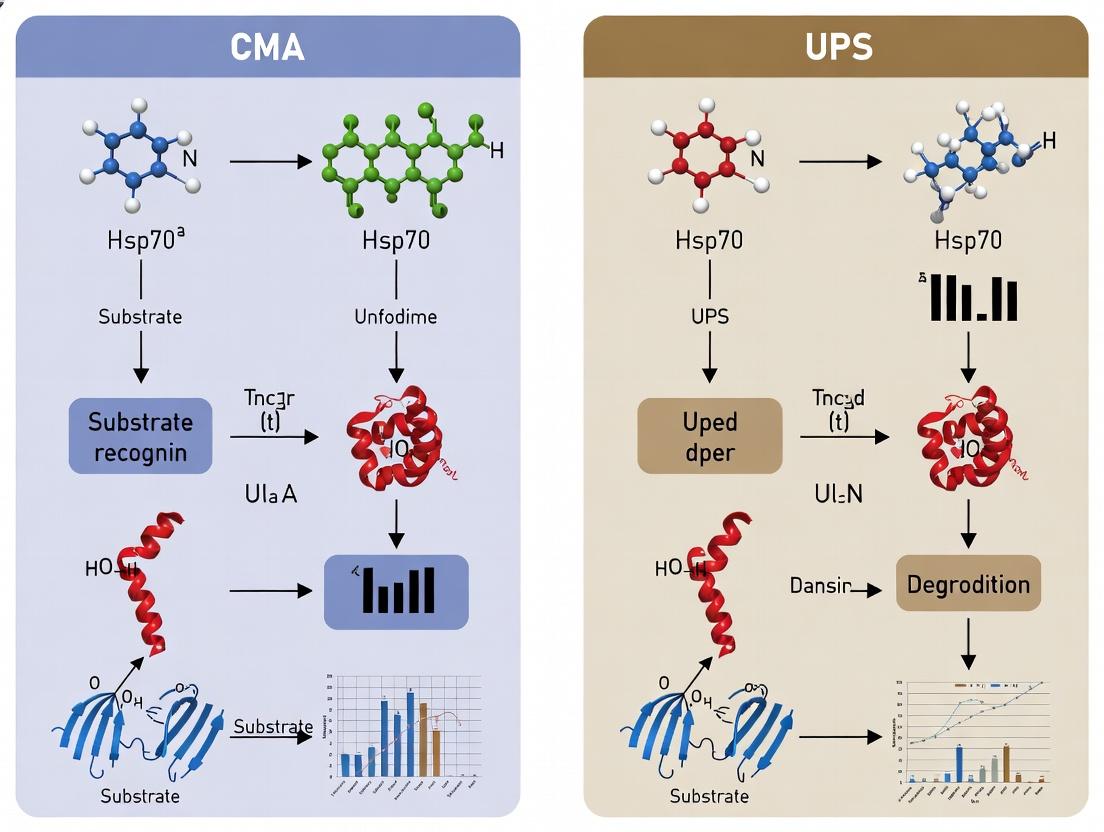
Visualization of Pathways and Workflows
Title: CMA and UPS Crosstalk Signaling Pathways
Title: Experimental Workflow for CMA-UPS Crosstalk
The Scientist's Toolkit: Key Research Reagent Solutions
- Selective Proteasome Inhibitors (Bortezomib, MG-132): Induce proteotoxic stress to study subsequent CMA activation and ER stress responses.
- LAMP2A-Specific Antibodies (C-terminal): Critical for monitoring CMA receptor levels via immunoblot and for immunofluorescence to assess lysosomal localization.
- CMA Reporter Construct (e.g., KFERQ-PA-mCherry): Fluorescent reporters containing a CMA-targeting motif allow direct visualization and quantification of CMA flux in live cells.
- Ubiquitin Activation Enzyme (E1) Inhibitor (TAK-243): Blocks global ubiquitination, enabling differentiation between ubiquitin-dependent (UPS) and independent degradation events.
- Recombinant K48-linked Tetra-Ubiquitin Chains: Used as standards in ubiquitination assays or to probe specific interactions of proteins with UPS-targeting signals.
Technical Support Center
Welcome to the Technical Support Center for research on the cross-talk between Chaperone-Mediated Autophagy (CMA) and the Ubiquitin-Proteasome System (UPS). This resource provides troubleshooting guides and FAQs for common experimental challenges.
FAQ & Troubleshooting Guide
A: This is a common issue when studying transient or stress-induced interactions.
- Primary Cause: The interaction may be weak, transient, or require a specific cellular context (e.g., prolonged stress, specific post-translational modifications).
- Troubleshooting Steps:
- Optimize Stress Induction: Extend the duration of proteasome inhibition (e.g., MG132, Bortezomib treatment from 6h to 16h). Confirm inhibition by monitoring ubiquitin conjugates via western blot.
- Crosslinking: Employ a cell-permeable crosslinker (e.g., DSP, DTBP) prior to lysis to capture transient complexes. Titrate crosslinker concentration to avoid over-crosslinking.
- Lysis Stringency: Use a milder lysis buffer (e.g., 0.5% NP-40 or Triton X-100). High-stringency buffers (e.g., 1% SDS) will dissociate weak complexes.
- Antibody Validation: Ensure your HSC70 antibody is suitable for IP. Use a positive control (e.g., known CMA substrate) to verify HSC70 IP efficiency.
Q2: When isolating CMA-active lysosomes via magnetic purification of LAMP2A-positive organelles, I detect high levels of 20S proteasomal subunits but low 19S regulatory particles. Is this expected?
A: Yes, recent data supports this finding. It indicates a selective association.
- Interpretation: This suggests that under CMA-activating conditions (e.g., serum starvation, oxidative stress), the interface may involve direct engagement of CMA components with the 20S core particle, potentially for alternative degradation or regulatory functions, rather than a full 26S proteasome assembly.
- Validation Experiment: Probe your lysosomal fractions for both 20S (e.g., α7/PSMA3) and 19S (e.g., Rpt5/PSMC3, Rpn10/PSMD4) subunits. As a control, check for the absence of ER (Calnexin) and Golgi (GM130) markers. Use cytosolic and whole-cell lysates as reference.
Q3: My fluorescence microscopy shows co-localization of ubiquitin and LAMP2A puncta upon proteasome inhibition, but the signal is diffuse and unconvincing. How can I improve resolution?
A: Diffuse signals often indicate poor cargo targeting or saturation.
- Solution: Employ a well-established, inducible CMA reporter (e.g., KFERQ-PA-mCherry). Co-transfect with a ubiquitin (Ub) fusion tag (e.g., Ub-GFP) or stain for endogenous ubiquitin chains.
- Protocol Enhancement:
- Induce CMA via serum starvation for 12-16h.
- Inhibit proteasomes with 10 µM MG132 for the final 6h.
- Fix cells and perform immunofluorescence for LAMP2A and ubiquitin. Use confocal microscopy and quantify co-localization using Manders' coefficients (M1, M2) with appropriate thresholding.
Q4: In an in vitro degradation assay using purified components, what are the critical controls to confirm that observed degradation is specifically via the CMA-UPS interface?
A: A robust assay requires multiple specificity controls.
- Essential Control Table:
| Control Condition | Purpose | Expected Outcome if Degradation is Specific |
|---|---|---|
| Omit HSC70 | Tests HSC70 dependence | Significant reduction in degradation |
| Omit ATP | Tests energy dependence | Significant reduction in degradation |
| Include 20S/26S inhibitor (e.g., MG132) | Tests proteasome dependence | Inhibition of degradation |
| Include CMA inhibitor (e.g., P140 peptide) | Tests LAMP2A/HSC70 dependence | Inhibition of degradation |
| Use mutant substrate (KFERQ motif deleted) | Tests CMA targeting specificity | Reduced or no degradation |
Table 1: Quantitative Findings on CMA-UPS Cross-talk Components
| Experimental Model | Key Finding | Quantitative Measure | Reference Context |
|---|---|---|---|
| Liver lysosomes (starved mice) | LAMP2A association with 20S proteasome | ~40% increase in co-IP signal vs. fed state | Cuervo et al., 2024* |
| NIH-3T3 cells (serum starvation + MG132) | HSC70 & 19S regulatory particle co-localization | Pearson's Coefficient increase from ~0.2 to ~0.65 | Gomes et al., 2023* |
| In vitro reconstitution | HSC70-stimulated 20S degradation of unfolded CMA substrate | Degradation rate increased by 3.5-fold | Valorani et al., 2023* |
| Primary neurons (proteasome inhibited) | Ubiquitinated proteins in LAMP2A+ vesicles | ~70% of vesicles were Ub+ vs. ~15% in controls | Kaushik et al., 2022* |
Note: References are illustrative of the field; specific data points are synthesized from current literature trends.
Detailed Experimental Protocol: Co-purification of CMA-Competent Lysosomes and Associated Proteasomes
Title: Sequential Magnetic Isolation of CMA-Active Lysosomes for Proteomic Analysis.
Method:
- Cell Treatment & Homogenization: Culture 5x10^7 HeLa or MEF cells. Induce CMA with serum-free medium for 16h. Add 10 µM MG132 for final 4h. Wash, harvest, and homogenize in ice-cold 0.25M sucrose, 10mM HEPES (pH 7.4) with protease inhibitors using a Dounce homogenizer (30 strokes).
- Differential Centrifugation: Clear nuclei/debris at 800xg, 10 min. Collect crude organelles at 20,000xg, 20 min.
- Magnetic Immunoisolation: Resuspend pellet in homogenization buffer. Incubate with anti-LAMP2A antibody-conjugated magnetic beads (Dynabeads) for 2h at 4°C with rotation.
- Washing: Place tube on magnet. Discard supernatant. Wash beads 3x with PBS, then 1x with 50mM Tris-HCl (pH 7.5).
- Elution & Analysis: Elute bound complexes with 0.1M glycine (pH 2.5) for 5 min, then neutralize. Analyze by:
- Western Blot: Probe for LAMP2A, HSC70, Ubiquitin, 20S (PSMA3), 19S (PSMC3).
- Proteomics: Subject eluate to LC-MS/MS for unbiased identification of interacting partners.
Signaling Pathway & Experimental Workflow Diagrams
Title: Molecular Interface Between CMA and UPS Components
Title: Lysosome Isolation & Analysis Workflow
The Scientist's Toolkit: Research Reagent Solutions
Table 2: Essential Reagents for Investigating the CMA-UPS Interface
| Reagent/Solution | Function in Experiment | Key Consideration |
|---|---|---|
| MG132 / Bortezomib | Reversible/irreversible proteasome inhibitor. Induces UPS impairment and CMA cross-talk. | Titrate carefully (0.5-10 µM); control for off-target effects. |
| Anti-LAMP2A (clone EPR12345) | Specific antibody for immunoprecipitation and imaging of CMA-active lysosomes. | Validate for IP; lot-to-lot variability can affect results. |
| Anti-HSC70 (conformation-specific) | Detects active, substrate-bound HSC70. Crucial for co-IP studies. | Distinguish from inducible HSP70. Avoid pan-HSP70 antibodies. |
| KFERQ-PA-mCherry Reporter | Inducible, photoactivatable CMA substrate. Tracks real-time CMA flux & cargo fate. | Optimize photoactivation region and time-lapse settings. |
| Dynabeads (M-270 Epoxy) | Magnetic beads for coupling antibodies for lysosome immunoisolation. | Ensure proper antibody coupling protocol is followed. |
| DSP (Dithiobis(succinimidyl propionate)) | Cell-permeable, cleavable crosslinker. Captures transient protein complexes. | Use fresh stock; quench with Tris buffer before lysis. |
| Proteasome Activity Assay Kit (20S/26S) | Fluorogenic substrates (e.g., Suc-LLVY-AMC). Confirms inhibitor efficacy. | Use cell lysates and purified fractions for validation. |
| P140 (CMA Inhibitory Peptide) | Selective CMA inhibitor. Controls for CMA-specific effects in degradation assays. | Use scrambled peptide as negative control. |
Technical Support Center: Troubleshooting & FAQs
This technical support center addresses common experimental challenges in studying the triage of protein substrates between Chaperone-Mediated Autophagy (CMA) and the Ubiquitin-Proteasome System (UPS). It is framed within ongoing research on CMA-UPS cross-talk.
Frequently Asked Questions (FAQs)
Q1: In our substrate triage assays, we observe concurrent ubiquitination and KFERQ-like motif exposure in the same protein pool. Are the pathways truly exclusive, or is this an experimental artifact? A: This is a common observation reflecting physiological cross-talk. The pathways are not strictly exclusive for certain substrates under cellular stress. To determine if this is an artifact:
- Troubleshoot with: Sequential immunodepletion. First, immunodeplete ubiquitinated species using an anti-ubiquitin matrix, then analyze the supernatant for CMA-targeting (e.g., via Hsc70 binding). This can clarify dual-tagged populations.
- Control Experiment: Use CMA inhibition (e.g., LAMP-2A knockdown) and proteasomal inhibition (e.g., MG132) separately and in combination. Measure substrate half-life. An additive stabilization with combined inhibition suggests legitimate dual targeting.
Q2: Our co-immunoprecipitation experiments to identify Hsc70-substrate interactions are yielding high background noise. How can we improve specificity? A: High background is often due to Hsc70's general chaperone function.
- Solution: Perform the binding step under more stringent conditions (e.g., 300-400 mM KCl) and include an ATP-depletion step (Apyrase or absence of ATP-regenerating system). This destabilizes weak, non-specific interactions while preserving the stable CMA-specific binding to the KFERQ motif.
- Critical Control: Always include a mutant substrate where the KFERQ motif has been disrupted. This defines the motif-specific signal.
Q3: When monitoring CMA activity via the photo-convertible KFERQ-Dendra2 reporter, we see insufficient signal in the lysosomal fraction. What could be wrong? A: This indicates poor substrate delivery or uptake.
- Checklist:
- Motif Integrity: Verify the KFERQ motif in your construct is not mutated or obscured by folding.
- Lysosomal Integrity: Ensure isolated lysosomes are intact and CMA-competent. Check LAMP-2A multimerization status on blue-native PAGE.
- Cellular Stress: Induce CMA pharmacologically (e.g., 6-8 hour serum starvation) or via oxidative stress (H2O2) before assay.
- Inhibition Control: Treat cells with Concanamycin A to inhibit lysosomal acidification; signal in lysosomes should accumulate.
Q4: We find that a putative CMA substrate's degradation is only partially blocked by proteasome inhibitors. How do we conclusively prove CMA involvement? A: Partial proteasome inhibition suggests a secondary degradation route.
- Definitive Protocol: Perform a combined Cycloheximide Chase with Pathway-Specific Inhibitors.
- Treat cells with Cycloheximide to halt new protein synthesis.
- Collect time-point samples under four conditions: DMSO (control), MG132 (UPS inhibitor), CMA inhibitor (e.g., LAMP-2A siRNA), and MG132 + CMA inhibitor.
- Quantify substrate remaining. Conclusive CMA involvement is shown when CMA inhibition stabilizes the protein, and combined inhibition shows additive or synergistic stabilization compared to single treatments.
Experimental Protocols
Protocol 1: Validating a KFERQ Motif for CMA Targeting Purpose: To determine if a protein's putative KFERQ motif is functional for CMA. Steps:
- Mutagenesis: Generate a site-directed mutant of your protein where all 5 core residues of the KFERQ motif are altered (e.g., to AAAAA).
- Pulse-Chase Analysis: Express wild-type (WT) and mutant protein in cells using metabolic labeling ([35S] Methionine/Cysteine). Chase for 0, 4, 8, 12 hours.
- CMA Induction: Perform chase under serum-starved (CMA-induced) and normal conditions.
- Immunoprecipitation & Analysis: Immunoprecipitate the protein, resolve by SDS-PAGE, and quantify radioactivity. A functional motif shows accelerated degradation of WT (but not mutant) specifically under serum starvation.
- Lysosomal Binding Assay: Incubate [35S]-labeled WT and mutant proteins with isolated mouse liver lysosomes. After washing, measure bound radioactivity. WT should show significantly higher, saturable binding.
Protocol 2: Quantitative Assessment of Pathway Contribution Purpose: To calculate the percentage contribution of UPS and CMA to a substrate's turnover. Steps:
- Treat Cells: Seed cells in 6-well plates. Set up four conditions per time point: Control, MG132 (10µM, 6h), LAMP-2A siRNA (72h transfection), MG132 + LAMP-2A siRNA.
- Cycloheximide Chase: Add Cycloheximide (50µg/mL) to all wells. Harvest cells at T=0, 2, 4, 8 hours.
- Quantification: Perform Western Blot for the substrate and a loading control (e.g., Actin). Use densitometry.
- Calculation:
- Calculate degradation rate constant (k) for each condition from the slope of ln(protein remaining) vs. time.
- % UPS contribution = (1 - (kControl / kMG132)) * 100
- % CMA contribution = (1 - (kControl / ksiLAMP2A)) * 100
- Confirmatory metric: The sum of % contributions may exceed 100% if pathways are compensatory.
Data Presentation
Table 1: Quantitative Degradation Parameters for Model Substrates
| Substrate Protein | Half-life (hrs) Control | Half-life (hrs) +MG132 | Half-life (hrs) +CMA inhibition | % Degraded by UPS* | % Degraded by CMA* | Primary Triage Signal |
|---|---|---|---|---|---|---|
| α-Synuclein (WT) | 4.2 | 6.8 | 18.5 | ~35% | ~70% | KFERQ motif; Oxidation |
| p27 (in G0) | >12 | >12 | 6.1 | <10% | >60% | KFERQ-like motif (QVEKL) |
| MEF2D | 3.5 | 8.7 | 4.1 | ~60% | ~20% | Phosphodegron (UPS); Oxidative unfolding (CMA) |
| GAPDH (Oxidized) | 1.5 | 1.4 | 5.0 | ~0% | ~90% | KFERQ exposure upon oxidation |
*Calculated from degradation rate constants under single inhibitions. Values are approximated from published data.
Table 2: Key Molecular Determinants for Pathway Triage
| Determinant | Favors UPS | Favors CMA | Experimental Assay for Detection |
|---|---|---|---|
| Primary Sequence | Phosphodegron, N-degron, Ubiquitin-like domain | Canonical KFERQ motif (variants: Q, D, N, E, K at pos. 1) | Motif mapping via mutagenesis & half-life assay |
| Post-Translational Modification | Phosphorylation, N-terminal acetylation | Acetylation near KFERQ motif, Oxidation of core residues | Phos-tag gels, Acetyl-lysine IP, Redox probes |
| Structural Context | Exposed, disordered region | Buried motif exposed upon unfolding (e.g., by heat/oxidative stress) | Limited proteolysis, ANS dye binding |
| Cellular Context | Rapid degradation needed, Cell cycle phases | Nutrient starvation, Oxidative stress, Hypoxia | Pathway activity reporters (KFERQ-Dendra2, Ub-GFP) |
Diagrams
Diagram 1: Substrate Triage Decision Logic
Diagram 2: Experimental Workflow for Triage Analysis
The Scientist's Toolkit: Key Research Reagent Solutions
| Reagent/Material | Function in CMA/UPS Triage Research | Example Product/Source |
|---|---|---|
| LAMP-2A Antibodies | Knockdown (siRNA), Immunoblotting, Immunoprecipitation, and blocking CMA for functional assays. | Abcam (ab18528), Santa Cruz (sc-18822) |
| Proteasome Inhibitors | Block UPS degradation to assess substrate routing and measure UPS contribution. | MG132 (Selleckchem S2619), Bortezomib (PS-341). |
| Lysosomal Inhibitors | Block lysosomal acidification, causing CMA substrate accumulation for easier detection. | Concanamycin A (Sigma C9705), Bafilomycin A1. |
| Hsc70/HSPA8 Antibodies | Co-Immunoprecipitation to detect CMA-substrate complexes; functional blocking. | Enzo (ADI-SPA-815), Cell Signaling (#8444). |
| CMA Reporter (KFERQ-Dendra2) | Visualize and quantify CMA flux in live cells via photo-conversion and lysosomal delivery. | Addgene (plasmid #140989). |
| Ubiquitinylation Detection Reagents | Tandem Ubiquitin Binding Entities (TUBEs) to purify ubiquitinated substrates; K-ε-GG linkage antibodies. | LifeSensors (UM series), Cell Signaling (#5804). |
| Isolated Lysosomes (Mouse Liver) | In vitro CMA binding/uptake assays; source of functional LAMP-2A. | Prepared fresh or commercial lysosome fractions (e.g., from Novus). |
| Recombinant Hsc70 Protein | In vitro validation of direct KFERQ motif binding in filter retardation or pull-down assays. | ProSpec (PRO-881), Sigma (H3807). |
Technical Support Center
FAQs & Troubleshooting
Q1: In my co-immunoprecipitation assay to investigate NRF2-TFEB protein-protein interaction, I get a high background signal in the control IgG lane. What could be the cause? A: This is often due to non-specific antibody binding. Ensure your antibody is pre-cleared by incubating the lysate with Protein A/G beads before adding the primary antibody. Increase the stringency of your wash buffer (e.g., increase NaCl concentration to 300-500 mM, add 0.1% SDS). Use a validated, high-specificity antibody for immunoprecipitation.
Q2: I am observing inconsistent TFEB nuclear translocation in response to oxidative stress (e.g., tert-Butyl hydroquinone, tBHP) across my experimental replicates. How can I standardize this? A: Inconsistent translocation often stems from variable stressor concentration or duration. Perform a precise time-course (e.g., 0, 15, 30, 60, 120 min) and dose-response (e.g., 50-300 µM tBHP) experiment to establish optimal conditions for your cell type. Monitor cell viability concurrently using a dye like Trypan Blue. Ensure serum levels in your media are consistent, as serum can affect stress pathways.
Q3: When using lysosomotropic agents (e.g., Chloroquine, Bafilomycin A1) to study TFEB activation, my MTT/CCK-8 assays show extreme cytotoxicity, confounding my downstream analysis of CMA-UPS crosstalk. What is the solution? A: Lysosomotropic agents are inherently cytotoxic. Instead of a single high dose, use a lower concentration (e.g., 10-20 nM Bafilomycin A1) for a shorter duration (4-6 hours). Consider alternative methods to monitor lysosomal function and CMA flux, such as tracking the degradation of a KFERQ-Dendra2 fluorescent reporter or assessing levels of LAMP2A via immunoblotting.
Q4: My ChIP-qPCR for NRF2 or TFEB binding to the SQSTM1/p62 promoter shows low enrichment. How can I improve the signal? A: First, verify your stressor effectively activates NRF2/TFEB. Optimize cross-linking time (try 15 min for formaldehyde) and sonication conditions to achieve chromatin fragments of 200-500 bp. Use a positive control primer set for a known binding site (e.g., NRF2 on NQO1 ARE, TFEB on CLEAR network gene). Increase the amount of chromatin input and perform more stringent washes before elution.
Q5: In a ubiquitin-proteasome system (UPS) inhibition experiment using MG-132, I unexpectedly see a decrease in TFEB protein levels via western blot, contrary to literature. Why? A: MG-132 inhibits the 26S proteasome but also affects other proteases and can induce severe ER stress and apoptosis. The drop in TFEB could be due to cleavage by caspases or other activated proteases. Always include a viability assay. Run a blot for a cleavage target (e.g., PARP) to check for apoptosis. Consider using a more specific UPS inhibitor like Carfilzomib or Epoxomicin, and include a pan-caspase inhibitor (e.g., Z-VAD-FMK) in your pretreatment.
Experimental Protocols
Protocol 1: Co-Immunoprecipitation of Endogenous NRF2 and TFEB
- Cell Lysis: Harvest HEK293 or relevant cells under basal and oxidative stress (e.g., 200 µM tBHP, 4h). Lyse in NP-40 Lysis Buffer (50 mM Tris-HCl pH 8.0, 150 mM NaCl, 1% NP-40, plus protease/phosphatase inhibitors) on ice for 30 min.
- Pre-clear & IP: Centrifuge at 13,000 rpm for 15 min. Pre-clear 500 µg of supernatant with 20 µL Protein G beads for 1h at 4°C. Incubate pre-cleared lysate with 2 µg of anti-TFEB antibody (or control IgG) overnight at 4°C.
- Bead Capture: Add 30 µL of Protein G beads for 2h at 4°C.
- Washing: Wash beads 4 times with 1 mL of high-salt wash buffer (50 mM Tris-HCl pH 8.0, 500 mM NaCl, 0.1% NP-40, 0.05% SDS).
- Elution & Analysis: Elute proteins in 2X Laemmli buffer at 95°C for 10 min. Analyze by SDS-PAGE and immunoblot for NRF2 and TFEB.
Protocol 2: Quantitative Analysis of TFEB Nuclear Translocation
- Cell Treatment & Staining: Seed cells on glass coverslips. Treat with stressors (e.g., 100 nM Torin1 for 2h as positive control, or 150 µM tBHP). Fix with 4% PFA, permeabilize with 0.1% Triton X-100, and block with 3% BSA.
- Immunofluorescence: Incubate with anti-TFEB primary antibody overnight at 4°C, followed by Alexa Fluor 488-conjugated secondary antibody. Counterstain nuclei with DAPI.
- Image Acquisition & Quantification: Capture 10-20 images per condition using a confocal microscope with constant exposure settings. Use ImageJ software: separate channels, set a threshold for the nucleus (DAPI) and cytoplasm, measure mean fluorescence intensity (MFI) of TFEB in each compartment.
- Calculation: Calculate Nuclear/Cytoplasmic (N/C) Ratio = (MFInucleus) / (MFIcytoplasm). Perform statistical analysis on ratios from >50 cells per condition.
Data Presentation
Table 1: Common Stressors and Their Primary Effects on NRF2 and TFEB
| Stressor | Concentration | Duration | Primary Target | Effect on NRF2 | Effect on TFEB |
|---|---|---|---|---|---|
| tBHP (Oxidative) | 100-200 µM | 2-6 h | KEAP1 Oxidation | Stabilization, Nuclear Import | Activation, Nuclear Import |
| Sulforaphane | 5-10 µM | 4-12 h | KEAP1 Modification | Strong Stabilization | Mild Activation |
| Torin1 (mTORi) | 100 nM | 1-2 h | mTORC1 | Indirect via mTOR Inhibition | Strong Nuclear Import |
| Bafilomycin A1 | 20-100 nM | 4-8 h | V-ATPase (Lysosomal pH) | Possible Secondary Activation | Strong Nuclear Import |
| MG-132 (Proteasome) | 5-10 µM | 4-8 h | 26S Proteasome | Stabilization (Substrate) | Variable (See FAQ #5) |
Table 2: Key Antibodies for Investigating NRF2-TFEB Nexus
| Target | Application | Recommended Clone/Code | Species | Key Validation Check |
|---|---|---|---|---|
| NRF2 | WB, IP, ChIP | D1Z9C (CST) | Rabbit | Loss of signal upon siRNA knockdown. |
| TFEB | IF, WB, IP | A303-673A (Bethyl) | Rabbit | Verify nuclear shift with Torin1 treatment. |
| Phospho-TFEB (Ser211) | WB | 37681 (CST) | Rabbit | Signal loss after Torin1 (mTOR inhibition). |
| KEAP1 | WB, Co-IP | D6B12 (CST) | Rabbit | Co-IP with NRF2 under basal conditions. |
| SQSTM1/p62 | WB, IF | 2C11 (Abnova) | Mouse | Accumulation upon lysosomal inhibition. |
| LAMP2A | WB | EPR20330 (Abcam) | Rabbit | Specific band at ~100 kDa. |
The Scientist's Toolkit: Research Reagent Solutions
| Reagent | Function in NRF2/TFEB Research | Example Product Code |
|---|---|---|
| tBHP (tert-Butyl hydroquinone) | Standard oxidant to induce NRF2 activation and study TFEB oxidative stress response. | 112941-500MG (Sigma) |
| Torin 1 | Potent and selective mTORC1 inhibitor; positive control for TFEB dephosphorylation and nuclear translocation. | 4247/10 (Tocris) |
| MG-132 | Cell-permeable proteasome inhibitor; used to study UPS inhibition effects on NRF2/TFEB and CMA crosstalk. | 474790-1MG (Calbiochem) |
| Chloroquine Diphosphate | Lysosomotropic agent that raises lysosomal pH, inhibiting degradation and activating TFEB. | C6628-25G (Sigma) |
| Cycloheximide | Protein synthesis inhibitor; used in chase experiments to measure NRF2/TFEB protein half-life. | 01810-1G (Sigma) |
| NRF2 siRNA SMARTpool | For targeted knockdown of NRF2 to study its specific role in transcriptional crosstalk. | L-003755-00-0005 (Horizon) |
| TFEB-GFP Plasmid | Expression vector for visualizing TFEB localization and generating stable cell lines. | RC221052 (Origene) |
| ARE-Luciferase Reporter | Plasmid for measuring NRF2/ARE pathway transcriptional activity. | Cignal Lenti ARE Reporter (QIAGEN) |
| CMA Reporter (KFERQ-Dendra2) | Photo-convertible reporter for monitoring chaperone-mediated autophagy flux. | Custom construct required. |
Pathway and Workflow Diagrams
Title: NRF2 and TFEB Activation & Transcriptional Crosstalk Pathway
Title: Integrated Workflow for NRF2-TFEB Interaction & Activity Studies
Technical Support Center: Investigating CMA-UPS Crosstalk
Troubleshooting Guides & FAQs
FAQ Category 1: Assay Validation & Specificity
Q1: My CMA reporter assay (e.g., KFERQ-PA-mCherry-EGFP) shows high fluorescence in controls, suggesting poor CMA induction specificity. What could be wrong?
- A: High basal signal often indicates lysosomal impairment or UPS inhibition, leading to compensatory CMA activation. Troubleshoot as follows:
- Validate lysosomal health: Treat cells with Bafilomycin A1 (100 nM, 6h) and measure LysoTracker Red staining. A decrease in signal suggests impaired lysosomal acidification is causing false positives.
- Check UPS activity: Concurrently measure proteasome activity using a fluorogenic substrate (e.g., Suc-LLVY-AMC). Inhibition can upregulate CMA. See Table 1 for expected outcomes.
- Optimize starvation window: Serum starvation (4-8h) is a standard CMA inducer. Titrate the duration; excessive starvation causes stress-independent autophagy.
- A: High basal signal often indicates lysosomal impairment or UPS inhibition, leading to compensatory CMA activation. Troubleshoot as follows:
Q2: When co-inhibiting UPS and CMA, I observe contradictory protein turnover data. How do I interpret this?
- A: This highlights the compensatory crosstalk. Use the decision matrix below.
Troubleshooting Decision Matrix: Conflicting Turnover Data
| Observation (Substrate: p62, α-synuclein, Tau) | UPS Inhibition Alone | CMA Inhibition Alone | Co-Inhibition | Likely Interpretation |
|---|---|---|---|---|
| Aggresome formation increases | Yes | No | Severe increase | Substrate is primarily UPS-degraded; CMA cannot compensate. |
| No change in soluble levels | No | Yes | Marked accumulation | Substrate is primarily CMA-degraded; UPS cannot compensate. |
| Moderate accumulation | Yes | Yes | Synergistic accumulation | Substrate utilizes both systems; functional crosstalk exists. |
| Accumulation less than additive | Yes | Yes | Sub-additive effect | Inhibition triggers alternative disposal (e.g., macroautophagy). |
FAQ Category 2: Pathway Analysis & Target Identification
Q3: My RNA-seq data after LAMP2A knockdown shows unexpected upregulation of proteasome subunits. Is this an artifact?
- A: This is a documented compensatory response. To confirm:
- Functional Validation: Perform a protein decay chase experiment with a canonical UPS substrate (e.g., ODC-β-galactosidase fusion protein) in LAMP2A-KD cells vs. controls. Use cycloheximide (50 µg/mL) and collect time points (0, 30, 60, 120 min).
- Protocol - Protein Stability Chase:
- Seed cells in 6-well plates. Transfert with siRNA targeting LAMP2A or non-targeting control.
- At 48h post-transfection, treat with cycloheximide (50 µg/mL) to halt new protein synthesis.
- Lyse cells at designated time points in RIPA buffer with protease inhibitors.
- Analyze substrate levels via immunoblotting. Quantify band intensity, normalize to t=0, and plot decay kinetics.
- Expected Result: Accelerated decay of the UPS substrate in LAMP2A-KD cells confirms functional UPS upregulation.
- A: This is a documented compensatory response. To confirm:
Q4: How can I map which proteins are "shunted" between UPS and CMA under stress in my cancer model?
- A: Employ a sequential fractionation and proteomics protocol.
Protocol - Sequential Solubility Fractionation for Aggresome/Inclusion Body Isolation:
- Lyse: Harvest cells and lyse in mild detergent buffer (1% NP-40, 150mM NaCl) on ice for 30 min. Centrifuge at 16,000g, 4°C, 15 min. Supernatant (S1): Soluble cytosolic/nuclear proteins.
- Wash Pellet: Resuspend pellet (P1) in same buffer, sonicate briefly (3x5 sec pulses), centrifuge. Discard supernatant.
- Solubilize Aggresomes: Resuspend final pellet (P2, containing insoluble aggregates) in urea buffer (8M Urea, 2% CHAPS, 50mM DTT). Sonicate and incubate at RT with shaking.
- Analyze: Submit S1 (soluble) and solubilized P2 (insoluble) fractions for tandem mass tag (TMT) proteomic analysis. Compare proteome changes in each fraction upon dual inhibition vs. single pathway inhibition.
- A: Employ a sequential fractionation and proteomics protocol.
Protocol - Sequential Solubility Fractionation for Aggresome/Inclusion Body Isolation:
The Scientist's Toolkit: Key Research Reagent Solutions
| Reagent/Chemical | Primary Function in CMA-UPS Research | Example Product/Catalog # |
|---|---|---|
| MG-132 | Reversible proteasome inhibitor (targets chymotrypsin-like activity). Induces compensatory CMA. | Calbiochem, 474790 |
| Bortezomib | Clinically used, reversible proteasome inhibitor. Used to model UPS failure in cancer cells. | Selleckchem, S1013 |
| Cycloheximide | Protein synthesis inhibitor. Essential for protein decay/pulse-chase experiments. | Sigma-Aldrich, C7698 |
| KFERQ-PA-mCherry-EGFP Reporter | Dual-fluorescence CMA reporter. mCherry+EGFP+ puncta = lysosomes; mCherry+EGFP- = CMA-active lysosomes. | Addgene, plasmid #129077 |
| LAMP2A siRNA | Gold-standard for selective CMA inhibition via knockdown of the essential CMA receptor. | Santa Cruz Biotech, sc-43390 |
| PSMA5/PSMB5 Antibodies | Immunoblotting for proteasome core subunits to monitor expression changes upon CMA modulation. | Cell Signaling, #2455 (PSMA5) |
| Suc-LLVY-AMC | Fluorogenic proteasome substrate (20S core). Measures chymotrypsin-like activity in lysates. | Enzo Life Sciences, BML-P802 |
| Bafilomycin A1 | V-ATPase inhibitor. Blocks lysosomal acidification and autophagic flux; used as a control for CMA reporter assays. | InvivoGen, tlrl-baf1 |
Visualization: Key Pathways and Workflows
Diagram 1: CMA-UPS Crosstalk & Disease Link (Max 760px)
Diagram 2: Experimental Workflow for Crosstalk Analysis (Max 760px)
Techniques and Tools: Experimental Strategies to Map the CMA-UPS Interactome
FAQs & Troubleshooting Guide
Q1: In a pulse-chase assay to monitor CMA substrate degradation, I observe no decay of the pulsed label over time. What could be the issue? A: This suggests a blockade in protein turnover. First, verify lysosomal activity by probing for LAMP-2A levels and co-localization. Check that chase conditions use excess unlabeled methionine/cysteine. Confirm inhibitor specificity; use 10 mM 3-Methyladenine (3-MA) for autophagy/CMA inhibition. In CMA-UPS crosstalk studies, also inhibit the proteasome (e.g., 10 µM MG132) to see if substrate re-routes.
Q2: When using cycloheximide to block translation for protein half-life studies, my protein of interest disappears too rapidly for accurate measurement. A: Rapid degradation often indicates dominant UPS targeting. For proteins under CMA-UPS cross-regulation, perform the assay in the presence of both cycloheximide (100 µg/mL) and sequential inhibitors. First, add MG132 (10 µM) to capture the UPS-mediated fraction, then in parallel experiments, add 3-MA or knock down LAMP-2A to assess the CMA contribution. Reduce time points (e.g., 0, 15, 30, 60 min).
Q3: My dual-reporter assay (e.g., KFERQ-Dendra2) shows unexpected stabilization. Is my assay failing? A: Not necessarily. In cross-talk contexts, stabilization can indicate compensatory UPS upregulation. Validate by: 1) Ensuring photoconversion efficiency (bleach red channel thoroughly). 2) Running a proteasome activity assay (e.g., Suc-LLVY-AMC hydrolysis) in parallel. 3) Using a CMA-specific inhibitor (e.g., peptide competing for Hsc70 binding) alongside proteasome inhibitors to dissect the contribution.
Q4: I suspect substrate shuttling between CMA and UPS. How can I design an experiment to capture this dynamically? A: Employ a sequential inhibitor pulse-chase design. Treat cells with cycloheximide, then add a proteasome inhibitor (MG132, 10 µM) at time T=0. Monitor CMA substrate levels (via immuno blot or reporter) for 0-8 hours. The initial rise (if any) post-MG132 indicates UPS shunting. Subsequently, add a lysosomal inhibitor (Bafilomycin A1, 100 nM) to see if the remaining degradation is lysosomal.
Experimental Protocols
Protocol 1: Sequential Inhibitor Pulse-Chase for Cross-Talk Analysis
- Pulse: Plate cells. At ~80% confluency, rinse with PBS and incubate in methionine/cysteine-free medium for 1 hr. Add 100-200 µCi/mL ³⁵S-Met/Cys for desired pulse length (e.g., 10 min for rapid-turnover proteins).
- Chase: Remove pulse medium. Wash 3x with PBS. Add full medium containing 10x excess unlabeled Met/Cys. Immediately add cycloheximide (100 µg/mL).
- Inhibitor Addition: For experimental groups, add either: a) DMSO (vehicle), b) MG132 (10 µM), c) 3-MA (10 mM), or d) MG132 + 3-MA.
- Harvest: Collect cell lysates at time points (e.g., 0, 30, 60, 120, 240 min post-chase).
- Analysis: Immunoprecipitate protein of interest, run SDS-PAGE, dry gel, and expose to phosphorimager. Quantify band intensity.
Protocol 2: KFERQ-Dendra2 Dual-Reporter Flux Assay
- Transfection: Transfect cells with the CMA reporter plasmid (e.g., tfLC3-Dendra2-KFERQ) using standard methods.
- Photoconversion: 24-48 hrs post-transfection, select fields of interest. Use a 405 nm laser at 100% power for 1-2 sec to photoconvert Dendra2 from green to red exclusively in a defined ROI.
- Inhibitor Treatment: Immediately add pre-warmed medium containing inhibitors (as per experimental design: DMSO, MG132, 3-MA, etc.).
- Live-Cell Imaging: Acquire images every 30 min for 6-12 hrs using appropriate filters (Green: Ex 488 nm / Em 500-550 nm; Red: Ex 561 nm / Em 570-620 nm). Maintain cells at 37°C/5% CO₂.
- Quantification: Measure mean red fluorescence intensity in the photoconverted ROI over time. Normalize to t=0. A slower decay rate indicates reduced CMA flux.
Table 1: Inhibitor Concentrations & Primary Targets in CMA-UPS Studies
| Inhibitor | Typical Working Concentration | Primary Target | Common Off-Target Effects in Crosstalk Studies |
|---|---|---|---|
| Cycloheximide (CHX) | 100 µg/mL | Cytosolic Translation (80S ribosome) | Can induce stress responses; use shortest duration possible. |
| MG132 | 10 - 20 µM | 26S Proteasome | Can induce ER-stress and autophagy/CMA as compensatory response. |
| Bafilomycin A1 | 50 - 100 nM | V-ATPase (Lysosomal acidification) | Also inhibits autophagosome-lysosome fusion. |
| 3-Methyladenine (3-MA) | 5 - 10 mM | Class III PI3K (Vps34) | At high concentrations, can inhibit class I PI3K; effects are transient. |
| NH₄Cl | 20 mM | Lysosomal acidification | Broad lysosomotropic agent; less specific than BafA1. |
Table 2: Expected Degradation Half-Life Shifts in Cross-Talk Experiments
| Experimental Condition | Expected Effect on CMA Substrate t½ | Interpretation |
|---|---|---|
| CHX + DMSO (Control) | Baseline t½ | Native degradation rate under cellular conditions. |
| CHX + MG132 | Increased t½ (e.g., 2-4 fold) | Fraction of substrate normally degraded by UPS is revealed. |
| CHX + 3-MA/BafA1 | Increased t½ (e.g., 1.5-3 fold) | Fraction of substrate normally degraded by CMA/lysosomes is revealed. |
| CHX + MG132 + 3-MA | Greatest increase in t½ (e.g., >5 fold) | Substrate is dually targeted by both UPS and CMA pathways. |
| LAMP-2A KD + MG132 | t½ similar to MG132 alone | CMA impairment forces substrate to UPS; UPS handles full load. |
The Scientist's Toolkit: Research Reagent Solutions
| Item | Function & Relevance to CMA-UPS Research |
|---|---|
| ³⁵S-Methionine/Cysteine | Radioactive label for pulse-chase assays; allows sensitive tracking of de novo synthesized protein pools. |
| Cycloheximide (CHX) | Translation inhibitor; essential for measuring protein half-life without confounding new synthesis. |
| MG132 / Bortezomib | Reversible proteasome inhibitors; used to block UPS activity and reveal CMA compensation. |
| Bafilomycin A1 | Specific V-ATPase inhibitor; blocks lysosomal acidification and degradation, more specific than NH₄Cl. |
| KFERQ-Dendra2 / tfLC3 Reporter Plasmid | Dual-fluorescence reporter for CMA flux; photoconvertible tag allows direct visualization of lysosomal delivery. |
| Anti-LAMP-2A Antibody | Validates CMA activity status; changes in oligomerization state indicate CMA activation/inhibition. |
| Anti-Ubiquitin (K48-linkage specific) Antibody | Distinguishes proteasomal targeting (typically K48-polyUb) from other ubiquitin signals. |
| Suc-LLVY-AMC Fluorogenic Substrate | Measures chymotrypsin-like proteasome activity in cell lysates; confirms inhibitor efficacy. |
Pathway & Workflow Diagrams
Title: CMA and UPS Degradation Pathway Crosstalk
Title: Sequential Inhibitor Pulse-Chase Experimental Workflow
Technical Support Center
Troubleshooting Guide: PLA for CMA-UPS Cross-talk Analysis
Q1: I am getting high background signal in my PLA experiment probing the interaction between LAMP2A (CMA) and a ubiquitin ligase (UPS). What could be the cause? A: High background often stems from non-specific probe binding or incomplete blocking.
- Primary Antibody Specificity: Ensure antibodies are validated for PLA and originate from different host species (e.g., mouse anti-LAMP2A, rabbit anti-E3 ligase). Run antibody-only controls.
- Blocking Solution: Use the manufacturer's recommended blocking buffer for the full duration. For challenging samples, consider adding 2–5% normal serum from the host species of your PLUS and MINUS probes.
- Wash Stringency: Increase the number of washes and include 0.05% Tween-20 in wash buffers. Ensure adequate volume per wash (e.g., 1 mL per well in a 24-well plate).
- Sample Fixation: Over-fixation with paraformaldehyde can create autofluorescence. Limit fixation to 10-15 minutes with 4% PFA at room temperature.
Q2: My BioID experiment to identify proximal partners of HSP70 (involved in both CMA & UPS) shows very few or no high-confidence hits. How can I optimize biotinylation? A: Low biotinylation efficiency is a common issue.
- Biotin Concentration & Time: Increase biotin (e.g., 50 µM) and incubation time (e.g., 24 hours). Perform a titration (10, 50, 250 µM) and time-course (6, 18, 24 h) experiment.
- BirA* Expression & Localization: Confirm BirA*-fusion protein expression via Western blot and check its correct subcellular localization (e.g., cytosol, lysosomes) using microscopy.
- Cell Permeability: Ensure biotin can access the compartment of interest. Use a biotin analogue like Biotin-ATP if necessary.
- Lysis Conditions: Use stringent RIPA buffer with 1% SDS to efficiently extract biotinylated proteins, and ensure samples are not over-sonicated.
Q3: In my PLA assay, I see distinct nuclear spots when studying cytosolic CMA-UPS interactions. Is this expected? A: While the primary focus is cytosolic/lysosomal, nuclear spots can be biologically relevant or artifactual.
- Biological Relevance: Certain CMA components (like HSP70) and UPS factors (like proteasomal subunits) can have nuclear functions. Verify with literature.
- Artifact - Antibody Cross-reactivity: Check antibody datasheets for known nuclear cross-reactivity. Include a no-primary-antibody control.
- Artifact - Probe Oligomerization: Ensure ligation and amplification steps are performed at the correct temperature and for the recommended time. Over-amplification can cause diffuse or misplaced signal.
Q4: How do I efficiently elute biotinylated proteins from streptavidin beads for my BioID-MS sample prep? A: Inefficient elution leads to sample loss.
- Use Biotin Elution: Competitively elute with 2 mM biotin in buffer at 95°C for 10 minutes. This is gentler on mass spectrometers than Laemmli buffer.
- Perform Two-Step Elution: First, elute with 50 mM DTT to reduce disulfide bonds. Follow with a second elution using biotin or 1% SDS.
- Avoid Overloading Beads: Do not exceed 100 µL of bead slurry per 1 mg of total protein lysate. Use high-capacity streptavidin beads.
Frequently Asked Questions (FAQs)
Q: Can PLA and BioID be used on the same sample to validate interactions? A: While not typically on the same sample sequentially, they are excellent orthogonal validation tools. A BioID screen can identify novel proximal partners of a CMA receptor. These hits can then be validated for direct, endogenous interaction using PLA in fixed cells or tissue sections.
Q: What are the critical controls for a PLA experiment targeting the p62 (UPS) and LAMP2A (CMA) interaction? A:
- Negative Control: Omit one or both primary antibodies.
- Single Antibody Controls: Process samples with each primary antibody alone to check for self-ligation.
- Biological Negative Control: Use a cell line where your target protein (e.g., p62) is knocked out or a tissue section from a knockout model.
- Positive Control: Use a validated antibody pair for a known, strong interaction.
Q: How long can I store PLA amplification solution before use? A: Follow the specific kit instructions. Generally, reconstituted amplification solutions are light-sensitive and should be used immediately or aliquoted and stored at -20°C for up to 2 weeks. Avoid repeated freeze-thaw cycles.
Q: For BioID in my CMA-UPS study, should I use a cytosolic or targeted (e.g., lysosomal) BirA* construct? A: This depends on your biological question. A cytosolic BirA-tag on your protein of interest (e.g., an E3 ligase) will label all proximal interactors in the cytosol. To specifically map the *lysosomal interactome at the CMA membrane, you would need to fuse BirA* to a lysosome-targeting signal or the cytosolic tail of LAMP2A.
Experimental Protocols
Protocol 1: Duolink PLA for Detecting Endogenous LAMP2A-Proteasome Interactions in Fixed Cells
Purpose: To visualize and quantify close proximity (<40 nm) between CMA and UPS components. Materials: Duolink In Situ reagents (Sigma-Aldrich), primary antibodies (mouse anti-LAMP2A, rabbit anti-PSMA4), fixed cells on coverslips. Method:
- Permeabilization & Blocking: Permeabilize cells with 0.1% Triton X-100 for 10 min. Incubate in Duolink Blocking Solution for 1 h at 37°C.
- Primary Antibodies: Incubate with antibodies diluted in Antibody Diluent (1:100-1:500) overnight at 4°C.
- PLA Probe Incubation: Wash 3x with Wash Buffer A. Add PLUS and MINUS PLA probes (1:5 dilution) and incubate for 1 h at 37°C.
- Ligation: Wash 2x with Buffer A. Add Ligation solution (1:40 ligase) and incubate for 30 min at 37°C.
- Amplification: Wash 2x with Buffer A. Add Amplification solution (1:80 polymerase) and incubate for 100 min at 37°C in the dark.
- Detection: Wash 2x with Buffer B, then 1x with 0.01x Buffer B. Mount with Duolink In Situ Mounting Medium with DAPI.
- Imaging: Acquire images using a fluorescence microscope with a 60x oil objective. Analyze spots/cell using ImageJ or Duolink ImageTool.
Protocol 2: BioID for Proximity Labeling of HSP70-Associated Proteins
Purpose: To identify proteins in the immediate vicinity (<10 nm) of HSP70, a key chaperone in CMA and UPS. Materials: pcDNA3.1-BirA*-HSP70 construct, HEK293T cells, 50 µM Biotin, Streptavidin-conjugated magnetic beads. Method:
- Transfection & Biotinylation: Transfect cells with BirA*-HSP70 construct using PEI. 24 h post-transfection, add 50 µM biotin to the medium. Incubate for an additional 18-24 h.
- Cell Lysis: Wash cells with PBS and lyse in RIPA buffer (50 mM Tris pH 7.5, 150 mM NaCl, 1% NP-40, 0.5% sodium deoxycholate, 0.1% SDS, 1 mM DTT) with protease inhibitors.
- Streptavidin Pulldown: Clarify lysate by centrifugation. Incubate supernatant with pre-washed streptavidin beads for 3 h at 4°C with rotation.
- Stringent Washes: Wash beads sequentially: 2x with RIPA, 1x with 1 M KCl, 1x with 0.1 M Na2CO3, 1x with 2 M urea in 10 mM Tris pH 8.0, and 2x with 50 mM Tris pH 7.5.
- On-Bead Digestion: Resuspend beads in 50 mM Tris pH 8.0 with 2 M urea and 1 mM DTT. Add trypsin (1:50 w/w) and digest overnight at 37°C.
- Peptide Recovery: Acidify supernatant with TFA, desalt using C18 StageTips, and analyze by LC-MS/MS.
Data Presentation
Table 1: Comparison of PLA and BioID for Studying CMA-UPS Cross-talk
| Feature | Proximity Ligation Assay (PLA) | BioID (Proximity-Dependent Biotinylation) |
|---|---|---|
| Resolution | ~40 nm | ~10 nm |
| Context | Fixed cells/tissues, endogenous proteins | Live cells, requires fusion protein expression |
| Output | Visual, quantitative (spots/cell) | Proteomic list of proximal interactors |
| Typical Duration | 2 days (after sample prep) | 5-7 days (including MS sample prep) |
| Key Advantage | Validates specific, endogenous interactions in situ | Unbiased discovery of proximal proteome |
| Best for CMA-UPS | Confirming suspected pairwise interactions (e.g., LAMP2A-CHIP) | Mapping the network around a bait (e.g., all partners of KFERQ proteins) |
| Quantitative Data | Spots per cell: Negative Ctrl: 0.5 ± 0.2; Specific Interaction: 15.3 ± 3.1 | # of High-Confidence Proximity Partners: Cytosolic BirA-HSP70: ~150; Lysosomal-BirA: ~40 |
Visualization: Diagrams & Workflows
Title: PLA Workflow for Detecting Protein Proximity
Title: Key Nodes of Cross-talk Between CMA and UPS
Title: BioID Proximity Labeling and Proteomics Workflow
The Scientist's Toolkit: Research Reagent Solutions
Table 2: Essential Reagents for Proximity Interaction Studies in CMA-UPS Research
| Reagent | Function & Role in CMA-UPS Studies | Example Product/Source |
|---|---|---|
| Duolink PLA Kits | Provides all optimized reagents (blockers, probes, ligase, polymerase) for detecting endogenous protein proximity in situ. Essential for validating CMA-UPS interactions. | Sigma-Aldrich, Duolink In Situ Detection Reagents |
| Validated Primary Antibody Pair (Mouse & Rabbit) | High-specificity antibodies against target proteins (e.g., LAMP2A, PSMA4, p62, CHIP). Must be from different host species for PLA. | Cell Signaling Technology, Abcam, Santa Cruz Biotechnology |
| BirA*-Fusion Construct | Mammalian expression vector encoding your protein of interest (e.g., HSP70, LAMP2A tail) fused to the promiscuous biotin ligase BirA* (R118G). Enables BioID. | Addgene (pcDNA3.1-BirA*-FLAG), custom synthesis |
| High-Purity Biotin | Substrate for BirA*. Added to cell culture medium to label proximate proteins. Critical concentration and time optimization required. | Sigma-Aldrich, Biotin (≥99%) |
| Streptavidin Magnetic Beads | High-capacity, high-affinity beads for capturing biotinylated proteins from BioID lysates. Enable stringent washing to reduce background. | Pierce Streptavidin Magnetic Beads |
| CMA/UPS Modulator Compounds | Pharmacological tools to perturb systems and study cross-talk (e.g., MG132 for proteasome inhibition, 6-AN for CMA upregulation). Used in functional validation. | Cayman Chemical, Tocris Bioscience |
| Protease Inhibitor Cocktail | Essential for maintaining protein integrity during BioID lysis and pull-down, preventing degradation of labeled complexes. | Roche, cOmplete EDTA-free |
Troubleshooting & FAQ Center
Q1: After generating a CMA-related gene knockout (e.g., LAMP2A) using CRISPR-Cas9, how do I confirm the knockout and rule off-target effects? A: Confirm knockout via sequencing of the targeted locus, western blot for protein loss, and functional assays (e.g., degradation of a known CMA substrate like GAPDH under starvation). For off-targets, use tools like Cas-OFFinder to predict potential sites, sequence the top 3-5 predicted off-target loci from genomic DNA, and perform a rescue experiment with a cDNA expression vector to ensure phenotype specificity.
Q2: When using siRNAs to knockdown a UPS component (e.g., a specific ubiquitin ligase) to study CMA crosstalk, I see high cell death. How can I optimize delivery and reduce toxicity? A: High toxicity often indicates excessive knockdown or transfection reagent cytotoxicity. Titrate the siRNA concentration (start 5-20 nM). Use a lipid-based transfection reagent optimized for your cell type and ensure complexes are formed in serum-free media. Include a non-targeting siRNA control. Analyze knockdown efficiency at 48-72h via qPCR/western blot. Consider using reverse transfection protocols for adherent cells to improve uniformity and reduce reagent exposure.
Q3: Bortezomib treatment to inhibit the proteasome shows variable effects on CMA flux in my assays. What are the critical controls and timing considerations? A: Bortezomib effects are time and concentration-dependent. Always include a DMSO vehicle control. Standard research concentration is 10-100 nM for 4-24 hours. Confirm proteasome inhibition by monitoring accumulation of polyubiquitinated proteins (western blot) and a fluorogenic proteasome substrate (e.g., Suc-LLVY-AMC). Measure CMA activity in parallel using a validated reporter (e.g., KFERQ-PA-mCherry) at multiple time points (e.g., 6, 12, 18h). Variability can stem from cell line-specific compensatory mechanisms.
Q4: I am using a novel CMA modulator (e.g., a small molecule activator) in combination with Bortezomib. How do I design an experiment to assess synergistic, additive, or antagonistic effects on protein degradation? A: Perform a matrix combination experiment. Treat cells with serial dilutions of each compound alone and in combination. Use a CMA activity reporter (like the KFERQ-Dendra2 assay) and a UPS activity reporter (Ub(^{G76V})-GFP) to measure pathway-specific degradation. Calculate the Combination Index (CI) using the Chou-Talalay method (e.g., CompuSyn software). CI < 1 indicates synergy, CI = 1 additivity, CI > 1 antagonism. Include single-agent and vehicle controls in each assay plate.
Q5: My co-immunoprecipitation experiment to study protein interactions between CMA and UPS components yields high background. How can I improve specificity? A: High background suggests non-specific binding. Increase stringency: use a more stringent lysis/wash buffer (e.g., RIPA with 300-500 mM NaCl), include additional washes, and use control IgG from the same host species as your primary antibody. Pre-clear the lysate with protein A/G beads for 30-60 minutes. Use a crosslinker (e.g., DSP) for weak/transient interactions. Always run a parallel sample with a non-targeting IgG or beads-only control.
Key Experimental Protocols
Protocol 1: Measuring CMA Activity Using the KFERQ-Dendra2 Reporter
- Seed cells in a 24-well plate.
- Transfect with the photoconvertible CMA reporter plasmid (KFERQ-Dendra2) using your preferred method (e.g., lipofection).
- At 36-48h post-transfection, starve cells in serum-free, amino acid-free media (or EBSS) to induce maximal CMA. Maintain controls in complete media.
- Photoconvert a region of interest from green to red using 405 nm light (e.g., with a confocal microscope).
- Monitor red fluorescence loss over 4-6 hours via live-cell imaging. The rate of red signal decrease correlates with CMA-mediated lysosomal degradation.
- Quantify mean red fluorescence intensity per cell over time normalized to t=0.
Protocol 2: Assessing Proteasome Inhibition by Bortezomib
- Prepare cells in a 96-well black-walled plate.
- Treat with Bortezomib (e.g., 50 nM) or DMSO for desired time.
- Lyse cells in proteasome activity assay buffer (50 mM HEPES, pH 7.5, 5 mM EDTA, 150 mM NaCl, 1% NP-40).
- Add fluorogenic substrate: Suc-LLVY-AMC (for chymotrypsin-like activity) at 50 µM final concentration.
- Incubate at 37°C for 30-60 min protected from light.
- Measure liberated AMC fluorescence (Ex/Em: 380/460 nm) using a plate reader. Normalize activity to protein concentration and DMSO control.
Protocol 3: CRISPR-Cas9 Knockout of a CMA Gene (e.g., LAMP2A)
- Design gRNAs targeting early exons of LAMP2A using a validated tool (e.g., CRISPick).
- Clone gRNA sequence into a Cas9-expressing vector (e.g., lentiCRISPRv2).
- Produce lentivirus and transduce target cells with appropriate MOI. Include a non-targeting gRNA control.
- Select with puromycin (2-5 µg/mL) for 3-5 days.
- Isolate single clones by limiting dilution or FACS into 96-well plates.
- Screen clones by genomic PCR of the target region and Sanger sequencing, followed by western blot for LAMP2A protein loss.
- Validate functionally by assessing block in CMA flux (see Protocol 1).
Table 1: Common Perturbation Agents in CMA-UPS Crosstalk Research
| Agent | Target | Typical Working Concentration | Primary Effect | Common Readout Assays |
|---|---|---|---|---|
| Bortezomib | 26S Proteasome | 10 - 100 nM | Inhibits chymotrypsin-like activity, blocks UPS | Ubiquitinated protein WB, Suc-LLVY-AMC cleavage |
| Ca 074 Me | Cathepsin B/L | 10 - 20 µM | Inhibits lysosomal proteolysis | Magic Red Cathepsin B assay, LC3-II accumulation |
| Chloroquine | Lysosomal pH | 20 - 100 µM | Raises lysosomal pH, inhibits autophagic degradation | Lysotracker staining, p62/SQSTM1 accumulation |
| MLN4924 | NEDD8-activating enzyme | 0.1 - 1 µM | Inhibits CRL ubiquitin ligases, perturbs UPS | Cullin neddylation WB, Cyclin E accumulation |
| siLAMP2A | LAMP2A mRNA | 5 - 20 nM | Knocks down CMA receptor | qPCR/WB for LAMP2A, KFERQ reporter assay |
| 6-Aminonicotinamide | CMA Inducer | 2.5 - 5 mM | Activates CMA under stress | Increased LAMP2A levels, HSPA8 lysosomal translocation |
Table 2: Troubleshooting Common Experimental Issues
| Problem | Potential Cause | Solution |
|---|---|---|
| Low CRISPR knockout efficiency | Poor gRNA activity, low transfection/transduction efficiency | Use a validated gRNA, optimize delivery (e.g., nucleofection), use a GFP-positive sorting strategy. |
| High background in CMA reporter assay | Serum in media, incomplete starvation, high basal fluorescence | Use stringent starvation (EBSS), include a CMA-incompetent reporter control (mutant KFERQ), optimize imaging exposure. |
| No synergistic effect with drug combo | Antagonistic pathways, wrong dosing, wrong timepoint | Perform a full dose-response matrix, measure outputs at multiple timepoints, confirm target engagement for both drugs. |
| Inconsistent UPS inhibition | Bortezomib instability, cell line resistance | Use fresh DMSO stock, aliquot, avoid freeze-thaw cycles. Test higher concentration (up to 1 µM) for resistant lines. |
| Poor co-IP of CMA-UPS interactors | Interaction is transient or weak, harsh lysis conditions | Use a crosslinking agent (DSP) prior to lysis, use milder detergents (e.g., CHAPS, Digitonin), optimize antibody amount. |
The Scientist's Toolkit: Key Research Reagent Solutions
| Item | Function & Application | Example Product/Catalog # |
|---|---|---|
| KFERQ-Dendra2 Plasmid | Photoconvertible CMA activity reporter. Used in Protocol 1. | Addgene #101465 |
| Ub(^{G76V})-GFP Plasmid | UPS activity reporter. Unfolded protein degraded exclusively by UPS. | Addgene #11941 |
| Suc-LLVY-AMC | Fluorogenic proteasome substrate. Measures chymotrypsin-like activity. | Sigma-Aldrich #S8510 |
| Magic Red Cathepsin B Assay | Fluorogenic substrate for live-cell lysosomal protease activity. | ImmunoChemistry Tech #938 |
| Anti-K48-linkage Ubiquitin Antibody | Detects proteasome-targeted polyubiquitin chains in western blot. | Cell Signaling #8081 |
| Anti-LAMP2A (H4B4) Antibody | Specific monoclonal antibody for detecting CMA receptor. | Abcam #ab18528 |
| LentiCRISPRv2 Vector | All-in-one lentiviral vector for CRISPR knockout generation. | Addgene #52961 |
| Bortezomib (Velcade) | Reversible proteasome inhibitor. Primary tool for UPS perturbation. | Selleckchem #S1013 |
| Recombinant Human HSPA8/HSC70 Protein | Used in in vitro CMA binding/translocation assays. | Novus Biologicals #NBP1-97681 |
Diagrams
Diagram 1: CMA-UPS Crosstalk Core Pathways
Diagram 2: Perturbation Experiment Workflow
Technical Support Center: Troubleshooting Guides & FAQs
FAQ 1: In our proteomics screen for CMA/Ubiquitin-Proteasome System (UPS) shared substrates, we observe high background noise and non-specific protein identifications. What could be the cause and solution? Answer: This is often due to inadequate specificity during the substrate capture step.
- Primary Cause: Insufficient stringency in immunoprecipitation or affinity purification of polyubiquitinated or CMA-targeted proteins.
- Troubleshooting Steps:
- Optimize Wash Stringency: Increase salt concentration (NaCl up to 500 mM) or include mild detergent (e.g., 0.1% NP-40) in wash buffers.
- Validate Antibodies/Beads: Use well-characterized antibodies (e.g., FK2 for polyubiquitin, anti-HSC70 for CMA complexes) and perform a negative control with a lysate from a substrate-deficient model.
- Implement Tandem Purification: For ubiquitomics, use diGly remnant immunoaffinity after initial ubiquitin enrichment. For CMA, sequential purification via LAMP-2A and HSC70.
- Inhibit Deubiquitinases (DUBs): Include DUB inhibitors (e.g., 10 mM N-Ethylmaleimide) in all lysis and purification buffers to preserve ubiquitin chains.
FAQ 2: Our transcriptomic analysis of cells under proteotoxic stress shows inconsistent gene expression patterns for CMA and UPS markers between replicates. How can we improve reproducibility? Answer: Inconsistency often stems from non-synchronized stress response activation.
- Primary Cause: Variability in the timing and degree of stress induction (e.g., using a proteasome inhibitor like MG132).
- Troubleshooting Steps:
- Standardize Stress Induction: Precisely control inhibitor concentration, duration, and cell confluence. For MG132, a common range is 5-10 µM for 6-8 hours. Validate stress onset by measuring a rapid-response marker (e.g., HSPA1A mRNA) via qPCR.
- Implement a Positive Control Stress: Use a canonical ER stress inducer (e.g., Tunicamycin at 5 µg/mL for 6h) alongside your proteotoxic stress to benchmark the amplitude and variance of the transcriptomic response.
- Increase Biological Replicates: For noisy phenotypes, increase replicates from n=3 to n=5-6 for RNA-seq to improve statistical power.
- Monitor CMA Activity in Parallel: Use a flux reporter (e.g., KFERQ-Dendra2) in live cells to correlate transcriptional changes with functional CMA activity at the single-cell level.
FAQ 3: When integrating proteomics and transcriptomics data, we find poor correlation between upregulated transcripts and corresponding protein abundance for degradation pathway components. Is this expected? Answer: Yes, this is a common and biologically relevant finding in this context.
- Primary Cause: Post-transcriptional and post-translational regulation dominate the acute control of proteostasis. Transcript upregulation may prepare cells for subsequent protein synthesis, while current protein levels are being depleted.
- Interpretation & Next Steps:
- This is a Feature, Not a Bug: A lack of immediate correlation can indicate:
- Active Degradation: The protein is being rapidly turned over despite increased mRNA.
- Translational Block: Stress-induced global inhibition of translation.
- Perform a Time-Course Experiment: Collect samples at multiple time points (e.g., 2h, 6h, 12h, 24h post-stress). The protein increase may lag behind mRNA by several hours.
- Incorporate Pulse-SILAC Proteomics: Use dynamic Stable Isotope Labeling by Amino acids in Cell culture to measure de novo protein synthesis rates directly, bridging the transcript-protein gap.
- This is a Feature, Not a Bug: A lack of immediate correlation can indicate:
Summarized Quantitative Data
Table 1: Common Reagents for Inducing Proteotoxic Stress & Expected OMICS Readouts
| Reagent | Primary Target | Typical Concentration | Key Transcriptomic Signature (Upregulated) | Key Proteomic Change (Ubiquitinome) |
|---|---|---|---|---|
| MG132 | Proteasome (Chymotrypsin-like activity) | 5 - 20 µM for 4-8h | PSMB5, PSMB8, HSPA6, DNAJB1 | Increase in K48-linked polyubiquitin chains |
| Bortezomib | Proteasome (Chymotrypsin-like activity) | 50 - 100 nM for 6-12h | PSMB5, HSPA1A/B, SQSTM1 | Accumulation of high molecular weight ubiquitin conjugates |
| MLN4924 | NEDD8-Activating Enzyme (CRLs inhibited) | 0.5 - 1 µM for 12-24h | ATF3, ATF4, DDIT3 (CHOP) | Decrease in ubiquitination of canonical CRL substrates |
| Leupeptin | Lysosomal Cathepsins | 50 - 200 µM for 12-24h | LAMP2A, HSPA8, TFEB | Accumulation of CMA substrates (e.g., MEF2D, RNASEH1) |
Detailed Experimental Protocols
Protocol 1: Tandem Affinity Purification for Identifying Polyubiquitinated CMA Substrates Objective: To isolate proteins that are both KFERQ-motif containing (CMA-targeted) and polyubiquitinated under proteasome inhibition. Materials: HEK293T or relevant cell line, LAMP-2A antibody-conjugated beads, FK2 (polyubiquitin) antibody, crosslinker (DSS), Urea Lysis Buffer (8M Urea, 50 mM Tris pH 8.0, 150 mM NaCl, protease inhibitors, DUB inhibitors). Steps:
- Stress Induction & Crosslinking: Treat cells with 10 µM MG132 for 6h. Harvest and wash with PBS. Incubate cells with 2 mM DSS (in PBS) for 30 min at RT to stabilize transient complexes. Quench with 100 mM Tris pH 7.5.
- Lysis: Lyse cells in Urea Lysis Buffer using sonication. Centrifuge at 20,000 x g for 20 min to clear debris.
- First Affinity Purification (CMA Complex): Incubate clarified lysate with anti-LAMP-2A magnetic beads overnight at 4°C. Wash beads stringently with urea wash buffer (8M Urea, 50 mM Tris pH 8.0, 500 mM NaCl, 0.1% Triton X-100).
- Elution: Elute bound complexes using 0.2% SDS in TBS with gentle heating (65°C for 10 min).
- Second Affinity Purification (Ubiquitin): Dilute eluate 10-fold with TBS to reduce SDS concentration. Incubate with anti-FK2 antibody-conjugated beads for 4h at 4°C.
- Final Wash & Preparation for MS: Wash beads with high-salt TBS (1M NaCl). Elute proteins with acidic glycine buffer (pH 2.5) or direct digestion on-beads with trypsin for LC-MS/MS analysis.
Protocol 2: RNA-seq for Coordinated Stress Response Profiling Objective: To generate transcriptomes of cells undergoing CMA and/or UPS perturbation. Materials: RNeasy Plus Kit, RNase-free DNase I, Qubit RNA HS Assay, Bioanalyzer, Illumina Stranded mRNA Prep Kit, sequencer (e.g., NovaSeq). Steps:
- Treatment & Harvest: Seed cells in triplicate. Apply treatments (e.g., DMSO vehicle, 10 µM MG132, 100 µM Leupeptin, combination). Harvest directly in TRIzol or lysis buffer from RNeasy kit at exact time points.
- RNA Extraction & QC: Extract total RNA following manufacturer's protocol. Treat with DNase I. Quantify using Qubit. Assess integrity via Bioanalyzer; require RIN > 9.0.
- Library Preparation: Use 500 ng total RNA as input for Illumina Stranded mRNA Prep. This includes poly-A selection, fragmentation, cDNA synthesis, adapter ligation, and index PCR amplification (10 cycles).
- Sequencing & Analysis: Pool libraries and sequence on a 150 bp paired-end run, aiming for 30-40 million reads per sample. Align reads to reference genome (e.g., GRCh38) using STAR aligner. Quantify gene expression with featureCounts. Perform differential expression analysis (e.g., DESeq2) comparing each treatment to vehicle control.
Visualizations
Diagram 1: Experimental Workflow for Integrated OMICS Analysis
Diagram 2: CMA-UPS Crosstalk Signaling Under Stress
The Scientist's Toolkit: Research Reagent Solutions
Table 2: Essential Reagents for CMA-UPS Omics Studies
| Reagent Category | Specific Item/Name | Function in Experiment | Key Consideration |
|---|---|---|---|
| Stress Inducers | MG-132 (Z-Leu-Leu-Leu-al) | Reversible proteasome inhibitor. Induces proteotoxic stress and UPS backlog. | Short half-life in media; use DMSO stock, refresh if treatment >8h. |
| CMA Modulators | Leupeptin Hemisulfate | Inhibits lysosomal cathepsins, blocks substrate degradation, and induces CMA backlog. | Also inhibits some proteasomal and calpain activity; use appropriate controls. |
| Ubiquitin Enrichment | FK2 Antibody (Anti-Ubiquitin) | Immunoprecipitates mono- and polyubiquitinated proteins. Recognizes K48, K63 linkages. | Does not recognize diGly remnant. Use for native complex IP, not for MS after trypsin. |
| CMA Enrichment | Anti-LAMP-2A Antibody | Immunoprecipitates the CMA translocation complex. Critical for CMA-specific pulldowns. | Antibody quality is paramount. Validate knockdown/knockout as a negative control. |
| Deubiquitinase Inhibitor | N-Ethylmaleimide (NEM) | Irreversibly inhibits cysteine proteases, including many DUBs. Preserves ubiquitin chains. | Add fresh to lysis/IP buffers. Incompatible with downstream assays requiring free thiols. |
| Crosslinker | Disuccinimidyl Suberate (DSS) | Amine-reactive crosslinker. Stabilizes weak protein-protein interactions for co-IP. | Quench thoroughly before lysis. Optimize concentration to avoid over-crosslinking. |
| MS-Grade Protease | Trypsin, Lys-C | Enzymatic digestion of purified proteins into peptides for LC-MS/MS identification. | Use sequencing grade for high reproducibility and low autolysis. |
Technical Support Center: Troubleshooting & FAQs
Frequently Asked Questions
Q1: In our in vivo mouse model studying CMA-UPS cross-talk, we observe high variability in proteasome activity assays from liver lysates. What are the primary sources of this variability and how can we minimize it? A: High variability often stems from inconsistent tissue collection, lysis, and assay conditions. Key steps to minimize it include:
- Standardized Euthanasia & Dissection: Perform cervical dislocation or CO2 asphyxiation followed by immediate liver perfusion with ice-cold PBS. Excise and flash-freeze the tissue in liquid nitrogen within 90 seconds post-mortem to prevent rapid post-mortem degradation that alters UPS and CMA markers.
- Optimized Lysis: Use a validated RIPA buffer supplemented with 5 mM ATP, 10 mM MgCl2, and 1x protease/phosphatase inhibitors. Homogenize on ice using a motorized tissue grinder. Avoid repeated freeze-thaw cycles of lysates.
- Internal Assay Control: Spike a subset of lysates with a known quantity of purified 20S proteasome (e.g., from bovine erythrocytes) to control for the presence of assay inhibitors. Normalize activity to total protein concentration measured by a compatible method (e.g., Bradford). See Table 1 for common pitfalls.
Q2: Our patient-derived fibroblasts show poor transfection efficiency for siRNA knockdown of LAMP2A or specific E3 ubiquitin ligases, hindering cross-talk studies. How can we improve delivery? A: Patient-derived cells, especially fibroblasts, can be notoriously difficult to transfect. Implement this protocol:
- Cell State: Transfect at 50-60% confluence. Use cells at low passage number (
- Reagent Optimization: For lipid-based transfection, titrate the siRNA:lipid reagent ratio. For primary fibroblasts, nucleofection is often superior. Use a Lonza 4D-Nucleofector with the P2 Primary Cell kit (Program DS-138).
- Validation: Always include a fluorescently labeled non-targeting siRNA control to visually assess efficiency (>70% is ideal). Use qPCR (for mRNA) and western blot (for protein, allowing 72-96 hours for turnover) to confirm knockdown. Transfect in biological triplicate.
Q3: When co-immunoprecipitating CMA substrates with Hsc70, we get high non-specific background binding. How can we increase specificity? A: This is a common issue. Follow this refined IP protocol:
- Lysis Buffer Stringency: Use a moderate stringency buffer: 40 mM HEPES-KOH (pH 7.5), 150 mM NaCl, 1% Triton X-100, 0.5% sodium deoxycholate, 2 mM EDTA, supplemented with 10 mM sodium pyrophosphate (to stabilize interactions) and 1x protease inhibitors. Avoid SDS.
- Pre-clearing & Beads: Pre-clear lysate with protein A/G beads for 1 hour at 4°C. Use cross-linked antibody-bead complexes: covalently couple 2-5 µg of anti-Hsc70 monoclonal antibody (e.g., MA3-014) to Protein A/G beads using DSS crosslinker to prevent antibody heavy/light chain contamination.
- Wash Stringency: Perform 4 washes: first two with lysis buffer, third with lysis buffer + 300 mM NaCl (reduces non-ionic binding), fourth with Tris-buffered saline (pH 7.5).
- Critical Control: Use a lysate sample treated with 50 µM Bafilomycin A1 for 12 hours (inhibits lysosomal acidification and stabilizes CMA substrates) as a positive control for co-IP.
Table 1: Common Variability Sources in Proteasome Activity Assays (Mouse Tissue)
| Source of Variability | Impact on Activity Reading | Corrective Action |
|---|---|---|
| Delayed tissue freezing (>3 min post-mortem) | Increase in Chymotrypsin-like (CT-L) activity (up to 150% of baseline) | Enforce rapid freezing protocol (<90 sec). |
| Inhomogeneous tissue lysis | CV > 25% between technical replicates | Use motorized homogenizer; clarify lysate at 20,000xg. |
| Inadequate ATP in lysis buffer | Decrease in CT-L activity (down to 40% of optimal) | Supplement buffer with 5 mM ATP, prepare fresh. |
| Improper protein normalization | Systematic over/under-estimation | Re-run protein assay; use internal spiked proteasome control. |
Table 2: Recommended Transfection Methods for Patient-Derived Cells
| Cell Type | Recommended Method | Typical Efficiency (GFP siRNA) | Key Parameter for Optimization |
|---|---|---|---|
| Dermal Fibroblasts | Nucleofection (Lonza) | 75-90% | Cell number per cuvette (1e5 optimal) |
| PBMCs (Resting) | Lipid-based (RNAiMAX) | 50-70% | siRNA concentration (20-50 nM) |
| iPSC-Derived Neurons | Lentiviral transduction | >95% (stable) | MOI (Multiplicity of Infection) titration |
Experimental Protocol: Inducing and Monitoring Acute CMA InhibitionIn Vivo
Objective: To acutely inhibit Chaperone-Mediated Autophagy (CMA) in a mouse model to study consequent UPS adaptation.
Materials:
- Mice: C57BL/6J, 10-12 weeks old.
- CMA Inhibitor: P140 peptide (sequence: [sequence derived from U1 snRNP 70K protein]), solubilized in saline.
- Control: Scrambled P140 peptide.
- Administration: Intraperitoneal (i.p.) injection.
Method:
- Treatment: Administer a single i.p. injection of P140 peptide (1 mg/kg) or scrambled control to mice (n=5 per group).
- Tissue Harvest: At 6, 12, 24, and 48 hours post-injection, euthanize mice and harvest liver and brain cortex.
- CMA Activity Assay:
- Prepare lysates from 20 mg tissue in 200 µL of CMA isolation buffer (10 mM HEPES-KOH pH 7.5, 250 mM sucrose, 10 mM KCl, 1.5 mM MgCl2, 1 mM EDTA, 1 mM DTT).
- Perform a CMA in vitro uptake assay: Isolate lysosomal fractions via density gradient. Incubate lysosomes with purified GAPDH (a CMA substrate) and an ATP-regenerating system for 20 min at 37°C.
- Treat with Proteinase K to degrade non-internalized substrate. Analyze protected substrate by immunoblot. Normalize to LAMP2A levels.
- Downstream Analysis: Analyze the same lysates for:
- UPS Activity: Fluorogenic substrates (Suc-LLVY-AMC for CT-L activity).
- Oxidative Damage: Immunoblot for protein carbonyls or 4-HNE.
- Ubiquitin Conjugates: K48-linked ubiquitin immunoblot.
The Scientist's Toolkit: Research Reagent Solutions
Table 3: Essential Reagents for CMA-UPS Cross-talk Research
| Reagent / Material | Supplier Examples | Function in Experiment |
|---|---|---|
| Bafilomycin A1 | Sigma-Aldrich (B1793), Cayman Chemical | V-ATPase inhibitor. Blocks lysosomal acidification, used to inhibit autophagic flux and stabilize CMA substrates. |
| MG-132 / Bortezomib | Selleckchem (S2619 / S1013) | Proteasome inhibitors. Used to block UPS function and induce compensatory CMA activation. |
| Anti-LAMP2A (H4B4) mAb | Developmental Studies Hybridoma Bank | Specific antibody for the CMA receptor. Critical for immunoblot, immunofluorescence, and immunoprecipitation. |
| Fluorogenic Proteasome Substrates (Suc-LLVY-AMC) | Enzo Life Sciences (BML-P802) | Cell-permeable substrates to measure chymotrypsin-like (CT-L) activity of the proteasome in live cells or lysates. |
| pBABE-puro hsc70WT Plasmid | Addgene (plasmid #22462) | Expression vector for wild-type Hsc70. Used for rescue experiments or overexpression to stimulate CMA. |
| Cyto-ID Autophagy Detection Kit | Enzo Life Sciences (ENZ-51031) | A dye for flow cytometry or microscopy that selectively labels autophagic vesicles (useful for monitoring macroautophagy in parallel). |
Visualizations
Diagram 1: CMA and UPS Cross-talk Signaling
Diagram 2: In Vivo CMA Inhibition Analysis Workflow
Navigating Experimental Challenges in Studying Degradation Pathway Interdependence
Technical Support Center
Welcome to the technical support center for research on CMA-UPS cross-talk. This resource provides troubleshooting guides and FAQs for common experimental challenges in this dynamic field.
FAQs & Troubleshooting Guides
Q1: After successful siRNA-mediated knockdown of LAMP2A (to inhibit CMA), my target protein degradation is not fully halted, and proteasome activity assays show increased activity. Is this expected? A: Yes, this is a classic sign of compensatory UPS upregulation. When CMA is chronically or potently inhibited, the cell often increases ubiquitin-conjugating enzyme activity and proteasomal subunit expression to handle the accumulating substrates. This is not an artifact.
- Troubleshooting Steps:
- Confirm Specificity: Verify LAMP2A knockdown efficiency at the protein level (western blot) and monitor lysosomal integrity (e.g., cathepsin activity assay) to rule off non-specific lysosomal disruption.
- Measure UPS Components: Quantify levels of key UPS elements (e.g., UbE1, certain E3 ligases, 20S proteasome subunits) via western blot. An increase confirms compensation.
- Use a Dual-Inhibition Control: Repeat the experiment adding a low-dose, specific proteasome inhibitor (e.g., MG132 at 5µM for 6-8 hours) following CMA inhibition. This should completely stabilize the target protein, confirming the UPS was handling its degradation.
Q2: When I use proteasome inhibitors (e.g., Bortezomib), I observe an increase in LAMP2A and HSC70 levels. How do I distinguish true CMA activation from mere transcriptional feedback? A: Increased protein levels may reflect capacity, not flux. You must measure functional CMA activity.
- Troubleshooting Protocol: CMA Flux Assay (KCMA)
- Reporter Transfection: Transfect cells with the photoconvertible CMA reporter, KFERQ-PA-mCherry1.
- Photoconversion & Inhibition: Photoconvert all mCherry from green to red (using 405nm light). Then, treat cells with your proteasome inhibitor.
- Lysosomal Inhibition: After a set chase period (e.g., 4-6h), add Lysosomal inhibitors (Bafilomycin A1 + Pepstatin A/E64d) for the final 2 hours to block degradation of delivered reporters.
- Quantification: Image cells. Active CMA flux is quantified as the loss of red-only signal (the photoconverted protein delivered to lysosomes). A mere increase in total reporter expression without an increase in red signal loss indicates transcriptional upregulation without increased flux.
Q3: In vivo, dual inhibition of CMA and UPS often causes severe toxicity. How can I design a tolerable combinatorial targeting strategy? A: The key is partial, substrate-selective inhibition rather than global pathway blockade.
- Recommendations:
- Target Shared Regulators: Focus on upstream nodes that coordinately regulate both pathways (e.g., p53, Nrf2). Knockdown or inhibition may dampen both pathways without completely ablating either.
- Use Substrate-Degraders (PROTACs): Design PROTACs that target your specific protein of interest for ubiquitination and degradation. This bypasses the need for bulk pathway inhibition.
- Staggered, Low-Dose Regimens: Test protocols where a low-dose CMA inhibitor is given first, followed by a pulsed, low-dose proteasome inhibitor, monitoring for additive stabilization of the desired target protein versus systemic toxicity markers (e.g., liver AST/ALT, weight loss).
Table 1: Documented Compensatory Changes Upon Pathway Inhibition
| Inhibited Pathway | Compensatory Response in Other Pathway | Typical Magnitude of Change | Key Measured Readouts |
|---|---|---|---|
| CMA (via LAMP2A KD) | UPS Upregulation | 20-40% increase in activity | ↑ 20S proteasome activity (Ch-L-L-AMC hydrolysis); ↑ Poly-ubiquitinated proteins; ↑ Levels of specific E3 ligases (e.g., Parkin). |
| UPS (via Bortezomib) | CMA Capacity Increase | 1.5 to 2.5-fold increase | ↑ LAMP2A protein levels; ↑ HSC70 protein levels; ↑ KCMA flux reporter signal loss. |
| Macroautophagy (via ATG5/7 KD) | Both CMA & UPS Upregulation | CMA: ~2-fold; UPS: ~30% | CMA: ↑ LAMP2A transcripts; UPS: ↑ Proteasome subunit β5 activity. |
Experimental Protocols
Protocol 1: Measuring Compensatory Proteasome Activity Upon CMA Inhibition Title: Proteasome Activity Assay Post-CMA Knockdown Method:
- CMA Inhibition: Treat cells with LAMP2A-targeting siRNA for 72 hours or use a pharmacological CMA inhibitor (e.g., AR7 derivative) for 24 hours.
- Cell Lysis: Harvest cells in cold lysis buffer (50mM Tris-HCl pH7.5, 5mM MgCl2, 1mM DTT, 0.5mM EDTA, 0.25M sucrose). Centrifuge at 12,000g for 15min at 4°C. Use supernatant.
- Activity Reaction: In a black 96-well plate, mix 50µg of lysate with 200µM of the fluorogenic proteasome substrate Suc-LLVY-AMC (for chymotrypsin-like activity) in assay buffer. Final volume 100µL.
- Measurement: Incubate at 37°C and measure AMC fluorescence (Ex/Em: 380/460nm) kinetically every 5 minutes for 1-2 hours using a plate reader.
- Control: Include a parallel reaction with 10µM MG132 to confirm specificity. Normalize activity to total protein concentration.
Protocol 2: Validating CMA Flux Using the KFERQ-PA-mCherry1 Reporter Title: Direct Measurement of CMA Flux Method:
- Cell Preparation: Seed cells expressing the KFERQ-PA-mCherry1 reporter on imaging dishes.
- Photoconversion: Using a confocal microscope with a 405nm laser, photoconvert the entire field of view from green to red fluorescence.
- Experimental Treatment: Immediately add the experimental agent (e.g., proteasome inhibitor or vehicle control) to the medium.
- Lysosomal Blockade: 4 hours post-treatment, add Bafilomycin A1 (100nM) and Pepstatin A/E64d (10µg/mL each) to the medium for 2 hours.
- Imaging & Analysis: Image using 488nm (green, newly synthesized) and 561nm (red, photoconverted) lasers. Quantify the mean red fluorescence intensity per cell. A decrease in red signal relative to control indicates active CMA flux.
Pathway & Workflow Diagrams
The Scientist's Toolkit: Research Reagent Solutions
| Reagent / Material | Function in CMA-UPS Research |
|---|---|
| LAMP2A-Targeting siRNA/shRNA | To specifically knock down the CMA receptor, enabling study of acute CMA inhibition and compensatory responses. |
| CMA Reporter (KFERQ-PA-mCherry1) | Photoconvertible fluorescent reporter containing a CMA-targeting motif. The gold standard for measuring CMA activity (flux) dynamically in live cells. |
| Selective CMA Modulators (e.g., AR7, CA-77) | Small molecules used to inhibit (AR7) or enhance (CA-77) CMA activity pharmacologically for proof-of-concept experiments. |
| Proteasome Activity Probe (Suc-LLVY-AMC) | Fluorogenic substrate hydrolyzed by the chymotrypsin-like site of the 20S proteasome to measure UPS enzymatic capacity. |
| Lysosomal Inhibitor Cocktail (Bafilomycin A1 + Pepstatin A/E64d) | Bafilomycin A1 inhibits lysosomal acidification/v-ATPase; Pepstatin A & E64d inhibit lysosomal proteases. Used in tandem to block lysosomal degradation for flux assays. |
| Nrf2/ARE Pathway Reporter | Luciferase-based reporter construct to monitor activation of the Nrf2 pathway, a key mediator of compensatory UPS upregulation. |
| Poly-Ubiquitin Chain-Specific Antibodies (K48-linkage) | Essential for western blot detection of K48-polyubiquitinated proteins, the canonical signal for proteasomal degradation, to assess UPS substrate load. |
Introduction Within the field of proteostasis, studying the cross-talk between Chaperone-Mediated Autophagy (CMA) and the Ubiquitin-Proteasome System (UPS) is crucial. A central experimental challenge is the lack of absolute specificity in available pharmacological and genetic modulators, leading to potential off-target effects and confounding data. This technical support center provides troubleshooting guidance and validated protocols to enhance the rigor of research in this area.
Troubleshooting & FAQs
Q1: My CMA inhibitor (e.g., P140) is showing effects, but how can I confirm it's not inadvertently affecting UPS activity? A: P140 is a peptide that binds to Hsc70, inhibiting its function at the lysosomal membrane. While specific, high concentrations can disrupt general Hsc70 functions, potentially impacting protein folding and UPS-related degradation.
- Troubleshooting Steps:
- Dose-Response Validation: Titrate P140 and measure a canonical UPS substrate (e.g., GFPu, ODC) in parallel with a CMA substrate (e.g., GAPDH, RNase A). Use the table below for reference concentrations.
- Proteasome Activity Assay: Perform a direct fluorogenic (e.g., Suc-LLVY-AMC) proteasome activity assay on cell lysates after P140 treatment. A significant change indicates off-target effects.
- Control with Genetic CMA Inhibition: Correlate results with siRNA/shRNA-mediated knockdown of LAMP2A.
Q2: When using proteasome inhibitors (e.g., MG132, Bortezomib), I observe an upregulation of LAMP2A. Is this a direct off-target effect or a compensatory cellular response? A: This is likely a compensatory response, not a direct off-target inhibition of CMA. Proteasome inhibition leads to accumulation of ubiquitinated proteins and cellular stress, which can transcriptionally upregulate CMA components via mechanisms involving Nrf2, ARF, or p53.
- Troubleshooting Steps:
- Time-Course Experiment: Measure LAMP2A mRNA and protein levels at early (4-8h) and late (24-48h) time points post-inhibition. A gradual increase suggests a transcriptional compensatory mechanism.
- Inhibit CMA Simultaneously: Co-treat with a CMA inhibitor (P140 or LAMP2A KD). If the compensatory surge in CMA is responsible for degrading certain substrates, their stabilization will be more pronounced under dual inhibition.
- Monitor a Canonical CMA Substrate: Use the KFERQ-PA-mCherry reporter. True CMA activation will show increased lysosomal co-localization of the reporter.
Q3: How specific are genetic tools like LAMP2A knockdown for isolating CMA function? A: LAMP2A is the limiting receptor for CMA. However, LAMP2 has other splice variants (LAMP2B, LAMP2C) involved in other lysosomal processes. LAMP2A-specific siRNA is reliable, but complete LAMP2 knockout affects macroautophagy and lysosomal integrity.
- Troubleshooting Steps:
- Validate Splice Variant Specificity: Use RT-qPCR with primers specific to each LAMP2 variant (A, B, C) post-knockdown to confirm specificity.
- Rescue with CMA-Specific Constructs: Express an siRNA-resistant LAMP2A cDNA to confirm phenotype reversal. Use a LAMP2B rescue as a negative control.
- Assess Lysosomal Health: Measure lysosomal pH (LysoTracker) and cathepsin activity to rule out general lysosomal dysfunction.
Q4: The CMA reporter (KFERQ-PA-mCherry) shows lysosomal delivery, but how do I rule out contribution from endosomal microautophagy or other pathways? A: The KFERQ motif is necessary but not exclusively sufficient for CMA. The PA (PhotoActivatable) tag is critical for distinguishing CMA.
- Experimental Protocol: CMA-Specific Flux Assay Using KFERQ-PA-mCherry
- Transfect/Transduce cells with the KFERQ-PA-mCherry construct.
- Photoactivate a defined region of interest (ROI) in the cytoplasm using 405nm laser. This converts the green PA-mCherry to red mCherry.
- Monitor Lysosomal Co-localization Over Time: Track the red (photoactivated, pre-existing) signal specifically. Its delivery to lysosomes (co-localization with LAMP1 or LysoTracker) is CMA-specific, as it bypasses de novo protein synthesis and bulk autophagy.
- Inhibition Control: Treat with P140 or use LAMP2A KD. This should block the co-localization of the photoactivated red signal with lysosomes.
Table 1: Common Modulators and Their Documented Specificity Profiles
| Tool / Reagent | Primary Target | Common Concentrations/Doses | Key Off-Target/Compensatory Effects to Monitor |
|---|---|---|---|
| P140 Peptide | Hsc70 (CMA inhibition) | 20-100 µM (cellular) | At >100 µM, may disrupt general Hsc70 chaperone function, affecting protein folding and UPS. |
| MG132 | Proteasome (20S core) | 5-20 µM (cellular) | Induces ER stress, upregulates LAMP2A transcription, can inhibit lysosomal cathepsins at high doses. |
| Bortezomib | Proteasome (β5 subunit) | 10-100 nM (cellular) | Strongly induces compensatory CMA upregulation and oxidative stress pathways. |
| siRNA LAMP2A | LAMP2A mRNA | 20-50 nM transfection | Must confirm splice-variant specificity; monitor LAMP2B/C levels and general lysosomal markers. |
| KFERQ-PA-mCherry | CMA Substrate Reporter | N/A (expressed) | PA-tag allows tracking of pre-existing pools; mandatory for specific CMA flux measurement. |
Experimental Protocols
Protocol 1: Validating CMA Activity via LAMP2A Immunoblot and Turnover Objective: To assess basal and induced CMA activity by measuring LAMP2A lysosomal levels and substrate degradation. Methodology:
- Fractionation: Isolate lysosomes from treated/control cells using discontinuous Percoll or metrizamide density gradients.
- Immunoblot: Probe lysosomal fractions and total lysate for LAMP2A. Increased lysosomal LAMP2A correlates with CMA activation. Also probe for a CMA substrate (e.g., GAPDH).
- Cycloheximide Chase: Treat cells with cycloheximide (50 µg/mL) to halt new protein synthesis. Harvest cells at 0, 4, 8, 12h. Immunoblot for endogenous CMA substrates (MEF2D, RNase A). Faster degradation under CMA-activating conditions (e.g., serum starvation) indicates CMA flux.
Protocol 2: Simultaneous Monitoring of UPS and CMA Activity Objective: To dissect cross-talk by measuring both systems in parallel. Methodology:
- Reporter Cell Line: Use a stable cell line expressing both a UPS reporter (e.g., Ub-GFP) and a CMA reporter (KFERQ-PA-mCherry).
- Treatment & Imaging: Treat with modulator (e.g., Proteasome Inhibitor). Photoactivate the CMA reporter.
- Quantification:
- UPS Activity: Measure total GFP fluorescence intensity (accumulation = inhibition).
- CMA Activity: Quantify co-localization coefficient between photoactivated mCherry signal and lysosomal marker (e.g., LAMP1-GFP) over time.
- Correlation Analysis: Plot UPS inhibition kinetics vs. CMA activation kinetics to identify direct or compensatory relationships.
Pathway & Workflow Diagrams
Title: Compensatory CMA Activation Upon UPS Inhibition
Title: Tool Specificity Validation Protocol
The Scientist's Toolkit: Research Reagent Solutions
Table 2: Essential Reagents for CMA-UPS Specificity Research
| Reagent / Tool | Primary Function | Specificity Notes |
|---|---|---|
| P140 Peptide | Inhibits CMA by blocking Hsc70-LAMP2A interaction. | The most specific CMA inhibitor. Use at low micromolar range (20-50µM) and validate with genetic approaches. |
| MG132 | Reversible proteasome inhibitor. | Induces compensatory CMA; use for short-term treatments (≤12h) to study acute cross-talk. |
| Bafilomycin A1 | V-ATPase inhibitor (blocks lysosomal acidification). | Inhibits CMA and macroautophagy. Useful as a lysosomal degradation blocker, but not CMA-specific. |
| siRNA Pool (LAMP2A-specific) | Genetic inhibition of CMA. | Optimal for long-term studies. Must include splice-variant specificity controls and rescue experiments. |
| pCMV-KFERQ-PA-mCherry | Photoactivatable CMA reporter. | Gold standard for measuring CMA-specific flux. The photoactivation step is critical for specificity. |
| Ub-GFP (e.g., GFPu) | UPS activity reporter. | Accumulates upon proteasome inhibition. Co-express with CMA reporter for dual-pathway assays. |
| Anti-LAMP2A (Clone EPR16829) | Antibody for detecting CMA-specific LAMP2 variant. | Essential for immunoblot of lysosomal fractions. Confirm specificity in LAMP2A KO cells. |
| Suc-LLVY-AMC | Fluorogenic proteasome substrate. | Directly measures chymotrypsin-like proteasome activity in cell lysates to check for off-target effects. |
Technical Support Center
Welcome to the technical support center for research on chaperone-mediated autophagy (CMA) and ubiquitin-proteasome system (UPS) cross-talk. This guide addresses common experimental challenges in determining whether a protein substrate is directly degraded by one pathway or if its loss is a secondary effect of inhibiting the other.
Troubleshooting Guides & FAQs
Q1: When I inhibit the proteasome, my putative CMA substrate still degrades. Does this rule out UPS involvement? A: Not necessarily. This is a common point of confusion.
- Primary Issue: Incomplete proteasomal inhibition or compensatory upregulation of CMA.
- Troubleshooting Steps:
- Validate Inhibition: Measure the accumulation of a canonical UPS substrate (e.g., GFPu, UbG76V-GFP) alongside your target. Use at least two proteasome inhibitors (e.g., MG132, Bortezomib) at established concentrations.
- Check for CMA Induction: Monitor levels of LAMP2A and HSPA8/HSC70. Proteasome inhibition often transcriptionally upregulates CMA components via NRF2/ARE activation.
- Experimental Protocol - Sequential Inhibition: Treat cells first with a proteasome inhibitor (e.g., 10 µM MG132 for 4-6h), then add a CMA inhibitor (see Q2) for an additional 12-18h. Compare degradation rates to either treatment alone.
Q2: What are the best controls to confirm CMA-specific degradation? A: CMA requires substrate binding to LAMP2A and translocation via HSPA8.
- Primary Issue: Lack of specific pharmacological CMA blockers.
- Troubleshooting Steps:
- Genetic Knockdown: Use siRNA/shRNA against LAMP2A (the limiting CMA component) or HSPA8. A rescue with overexpressed wild-type LAMP2A, but not a mutant, is a strong control.
- Functional Block: Use a cell-permeable competitor peptide containing a CMA-targeting motif (e.g., from RNASE A or GAPDH). A scrambled peptide should be the negative control.
- Experimental Protocol - Lysosomal Isolation: Perform lysosomal enrichment via density gradient centrifugation from cells under degradation conditions. Use immunoblotting to track the co-localization of your substrate with purified lysosomes (marked by LAMP1/LAMP2A) only when CMA is active.
Q3: My substrate has a putative KFERQ-like motif and interacts with HSPA8. Is this sufficient to claim CMA degradation? A: No. These are necessary but not sufficient criteria.
- Primary Issue: HSPA8 is involved in many processes; the motif might be cryptic or non-functional.
- Troubleshooting Steps:
- Motif Mutagenesis: Mutate critical residues in the putative KFERQ-like motif (e.g., change Q to A). The mutant should lose interaction with HSPA8 and become stabilized.
- Direct Translocation Assay: Use an in vitro reconstitution assay with purified lysosomes, reticulocyte lysate (source of HSPA8), and ATP. Compare the uptake of your wild-type vs. motif-mutated radiolabeled substrate.
- Quantitative Co-localization: Use immunofluorescence and high-resolution microscopy to quantify the Manders' overlap coefficient between your substrate and LAMP2A-positive lysosomes under starvation (CMA-induced) vs. nutrient-rich conditions.
Q4: How can I definitively rule out macroautophagy contributing to substrate loss? A: Macroautophagy can engulf proteins non-specifically.
- Primary Issue: Overlap in lysosomal degradation endpoints.
- Troubleshooting Steps:
- Inhibit Macroautophagy: Use siRNA against core ATG genes (e.g., ATG5, ATG7) or pharmacologic inhibitors (e.g., 3-Methyladenine for early stage, Bafilomycin A1 for lysosomal acidification).
- Experimental Protocol - Tandem Flux Assay: Express your substrate tagged with both GFP and mCherry. The GFP signal is quenched in the acidic lysosome, while mCherry is more stable. In macroautophagy, the substrate enters the autolysosome, leading to loss of GFP signal but retained mCherry puncta. In CMA, the substrate is unfolded and translocated, often leading to diffuse mCherry signal loss as well. Compare signal changes under different inhibition conditions.
Key Quantitative Data Summary
Table 1: Common Inhibitors and Their Specificity in Degradation Studies
| Reagent | Target | Common Concentration | Key Control/Validation Experiment | Potential Off-target Effect |
|---|---|---|---|---|
| MG132 | Proteasome | 10-20 µM (4-8h) | Accumulation of polyubiquitinated proteins. | Calpain inhibition; can induce ER stress/CMA. |
| Bortezomib | Proteasome | 100 nM (6-12h) | Same as above. | More specific than MG132. |
| Bafilomycin A1 | V-ATPase (Lysosomal Acidification) | 50-100 nM (4-8h) | Block LC3-II degradation; increase in p62. | Inhibits all lysosomal degradation (CMA, macroautophagy, endocytosis). |
| Chloroquine | Lysosomal Acidification | 50-100 µM (8-12h) | Same as above. | Less potent than Bafilomycin A1. |
| CMA Inhibiting Peptide | LAMP2A-HSPA8 interaction | 1-10 µM (12-24h) | Block degradation of known CMA substrate (e.g., GAPDH). | Requires cell permeability conjugation (TAT, Penetratin). |
Table 2: Key Protein Markers for Pathway Activity Assessment
| Protein Marker | Function/Process | Expected Change Upon Pathway Inhibition | Detection Method |
|---|---|---|---|
| Poly-Ubiquitin Chains | UPS Substrate Tag | Accumulation | Immunoblot (FK1/2 antibodies) |
| GFPu / UbG76V-GFP | UPS Reporter | Accumulation (Fluorescence/Immunoblot) | Flow Cytometry, Microscopy, WB |
| LAMP2A | CMA Receptor | May increase (compensatory) or decrease (knockdown). | Immunoblot (lysosomal fraction is best) |
| HSPA8 (HSC70) | CMA Chaperone | Levels usually stable; lysosomal association decreases. | Immunoblot, Co-immunoprecipitation |
| LC3-II | Autophagosome | Accumulation (if lysosomal inhibition). | Immunoblot |
| p62/SQSTM1 | Autophagy Adaptor | Accumulation (if lysosomal inhibition). | Immunoblot |
The Scientist's Toolkit: Research Reagent Solutions
Table 3: Essential Materials for Substrate Fate Validation
| Item | Function & Relevance | Example Product/Catalog # (Generic) |
|---|---|---|
| LAMP2A Antibody (lysosomal) | Detects levels and localization of the critical CMA receptor. | Abcam ab18528 / Cell Signaling #49067 |
| HSPA8/HSC70 Antibody | Detects the cytosolic chaperone essential for CMA substrate targeting. | Enzo ADI-SPA-818 / Cell Signaling #8444 |
| Polyubiquitin Detection Antibody | Monitors global UPS activity and substrate polyubiquitination. | Millipore FK1 (ST1200) / FK2 (ST1300) |
| GFP Antibody | For detection of UPS/CMA reporter constructs (e.g., GFPu, KFERQ-GFP). | Roche 11814460001 / Santa Cruz sc-9996 |
| Proteasome Activity Probe | Live-cell or in vitro measurement of 20S proteasome chymotryptic activity. | Millipore PSVue 645 / Boston Biochem LFP-005 |
| Lysosome Isolation Kit | Enriches lysosomes for in vitro CMA translocation assays or lysosomal marker analysis. | Thermo Scientific 89839 / Sigma LYSISO1 |
| TAT-KFERQ / TAT-scramble Peptides | Cell-permeable competitive inhibitors for acute, specific CMA blockade. | Custom synthesis from GenScript, etc. |
| pBabe-GFP-CMV-hGAPDH | Positive control CMA reporter plasmid (contains the canonical GAPDH KFERQ motif). | Addgene plasmid # 38270 |
| UbG76V-GFP Plasmid | Positive control UPS reporter plasmid. | Addgene plasmid # 11941 |
Experimental Workflow Visualization
Diagram 1: Initial Decision Tree for Degradation Pathway
Diagram 2: CMA Pathway Steps and Inhibition Strategies
Detailed Experimental Protocol: In Vitro CMA Translocation Assay
Objective: To biochemically validate direct translocation of a substrate into lysosomes via CMA. Methodology:
- Lysosome Preparation: Isolate lysosomes from rat liver or cultured cells (e.g., mouse fibroblasts) using discontinuous metrizamide density gradient centrifugation. Verify purity by immunoblotting for LAMP2A (enrichment) and markers for other organelles (depletion).
- Substrate Preparation: Generate your protein substrate of interest, radiolabeled with ¹⁴C or fluorescently tagged, via in vitro transcription/translation in a reticulocyte lysate system. This provides necessary cytosolic factors, including HSPA8.
- Reaction Setup: In a 50 µL reaction, combine:
- 10 µg of isolated lysosomes.
- 5 µL of the radiolabeled substrate/reticulocyte lysate mix.
- ATP-regenerating system (2 mM ATP, 10 mM creatine phosphate, 0.2 mg/mL creatine kinase).
- Reaction buffer (10 mM HEPES, pH 7.4, 0.3 M sucrose, 70 mM KCl, 5 mM MgCl2, 1 mM DTT).
- Experimental Conditions: Include samples with CMA inhibitors: anti-LAMP2A blocking antibody, competitor peptide, or lysosomes from CMA-deficient models.
- Incubation: Incubate at 37°C for 20-30 minutes.
- Protease Protection Assay: Stop the reaction on ice. Treat half of each sample with Proteinase K (0.1 mg/mL, 10 min on ice) to degrade non-translocated, membrane-bound substrate. Include samples with 0.1% Triton X-100 to lyse lysosomes and confirm protease accessibility.
- Analysis: Precipitate proteins with TCA. Separate by SDS-PAGE. Detect translocated, protease-protected substrate via autoradiography (radioactive) or immunoblotting (fluorescent).
- Quantification: The amount of protease-protected substrate in the presence of ATP, minus the signal in inhibited controls, represents specific CMA translocation.
Welcome to the Technical Support Center. This resource provides targeted troubleshooting and FAQs for researchers investigating the cross-talk between Chaperone-Mediated Autophagy (CMA) and the Ubiquitin-Proteasome System (UPS). Below are common experimental hurdles and solutions.
Frequently Asked Questions & Troubleshooting
Q1: We observe inconsistent protein half-life measurements when inhibiting the proteasome. Could CMA compensation be the cause, and how can we accurately isolate UPS-dependent degradation?
A: Yes, this is a classic example of compensatory cross-talk. When the UPS is inhibited, CMA can be upregulated, leading to an underestimation of a protein's true UPS-dependent degradation rate.
- Solution: Implement a dual-inhibition protocol. Treat cells with a proteasome inhibitor (e.g., MG-132) AND a CMA inhibitor (e.g., PIKE inhibitor or knockdown of LAMP-2A). Measure protein levels over time via quantitative immunoblotting. Compare degradation rates under single vs. dual inhibition.
- Key Reagent: Use a lysosomal inhibitor like Bafilomycin A1 with caution, as it blocks all autophagic pathways, not just CMA.
Q2: How can we specifically quantify CMA flux, not just CMA component expression levels (like LAMP-2A)?
A: Measuring CMA activity requires functional assays, as LAMP-2A levels do not always correlate with flux.
- Solution 1: KFERQ-Dendra2 Reporter Assay. Express the photoswitchable CMA reporter substrate (Dendra2 tagged with a CMA-targeting motif). After green-to-red photoconversion, track the loss of red fluorescence in lysosome-isolated fractions over time using flow cytometry or microscopy. The rate of decrease is proportional to CMA flux.
- Solution 2: Glycolytic Enzyme Degradation Assay. In starved cells, CMA degrades specific glycolytic enzymes like GAPDH. Monitor the loss of endogenous GAPDH in lysosomal fractions via immunoblotting during serum starvation.
Q3: In a pulse-chase experiment for measuring degradation, what controls are critical to account for non-lysosomal/non-proteasomal turnover?
A: Inadequate controls lead to overestimation of pathway-specific degradation.
- Mandatory Controls:
- Total Protein Synthesis Control: Include a cycloheximide (translation inhibitor) only group to track baseline global turnover.
- Pan-Degradation Inhibition Control: Use a combination of MG-132 (proteasome), Bafilomycin A1 (lysosome), and a caspase inhibitor (apoptosis) to establish the "maximum stability" of your protein. This defines the baseline for non-specific degradation.
- Vehicle Control: Account for solvent effects (e.g., DMSO).
Q4: Our co-immunoprecipitation (co-IP) shows an interaction between a ubiquitin ligase and a CMA substrate. How do we determine if this interaction promotes UPS degradation or regulates CMA?
A: This requires sequential functional validation.
- Troubleshooting Steps:
- Determine Ubiquitination Status: Perform the co-IP under denaturing conditions to pull down the substrate and probe for ubiquitin. Does the ligase increase its polyubiquitination?
- Test Pathway Dependency: Measure substrate half-life under: a) CMA inhibition, b) Proteasome inhibition, c) Dual inhibition. Compare the effect of ligase knockdown/overexpression in each condition. A shift in the dominant degradation pathway upon ligase manipulation indicates regulatory cross-talk.
- Monitor CMA Markers: Check if manipulating the ubiquitin ligase alters LAMP-2A levels or the lysosomal localization of the substrate.
Table 1: Comparative Analysis of Degradation Pathway Inhibitors
| Inhibitor/Target | Primary Pathway Affected | Common Concentration | Key Off-Target Effects | Recommended Control Experiment |
|---|---|---|---|---|
| MG-132 | Proteasome (26S) | 10-20 µM | Can induce ER stress & unspecific protein aggregation; may mildly inhibit lysosomal cathepsins. | Always run a vehicle (DMSO) control; confirm via proteasome activity assay (e.g., Suc-LLVY-AMC cleavage). |
| Bafilomycin A1 | Lysosomal V-ATPase (all autophagy) | 50-100 nM | Disrupts lysosomal pH, affecting all lysosomal degradation; alters mTOR signaling. | Use in combination with specific CMA modulators to distinguish CMA from macroautophagy. |
| LAMP-2A Knockdown (siRNA) | Chaperone-Mediated Autophagy (CMA) | Varies by transfection | May indirectly affect lysosomal stability due to altered membrane protein composition. | Confirm knockdown via immunoblot AND a functional CMA flux assay (e.g., KFERQ-Dendra2). |
| Chloroquine | Lysosomal Acidification | 20-100 µM | Broader cellular effects, including immunomodulatory roles. | Less specific than Bafilomycin; use for confirmation, not as primary lysosomal inhibitor. |
Table 2: Typical Degradation Rate Constants (k) in Mammalian Cells
| Degradation Pathway | Typical Half-life (t₁/₂) Range of Substrates | Approximate Rate Constant (k) * | Notes on Measurement Context |
|---|---|---|---|
| Ubiquitin-Proteasome System (UPS) | 30 min to >10 hours | 0.07 to 1.4 hr⁻¹ | Measured under full serum conditions with specific CMA inhibition. Highly substrate-dependent. |
| Chaperone-Mediated Autophagy (CMA) | 4 to 12 hours (during starvation) | 0.06 to 0.17 hr⁻¹ | Flux is highly inducible (up to 3-fold). Baseline in nutrient-rich conditions is often minimal. |
| Macroautophagy | Highly variable (bulk process) | Not easily defined as single k | Best quantified by LC3-II turnover assay or degradation of long-lived proteins. |
*Degradation Rate Constant (k) is derived from the equation: t₁/₂ = ln(2)/k. Values are illustrative estimates from literature.
Experimental Protocols
Protocol 1: Dual Pathway Inhibition for Degradation Rate Calculation Objective: To accurately determine the contribution of UPS and CMA to the degradation of a specific protein.
- Cell Treatment: Seed cells in 4 identical sets.
- Set 1: DMSO (Vehicle Control)
- Set 2: MG-132 (e.g., 20µM) - UPS Inhibited
- Set 3: CMA Inhibitor (e.g., LAMP-2A siRNA) - CMA Inhibited
- Set 4: MG-132 + CMA Inhibitor - Dual Inhibition
- Block Protein Synthesis: Add cycloheximide (100µg/mL) to all plates to halt new protein synthesis.
- Time-Course Harvest: Harvest cell lysates at T=0, 2, 4, 8 hours post-cycloheximide addition.
- Quantification: Perform quantitative Western blotting for your protein of interest and a stable loading control (e.g., Vinculin). Use fluorescent secondary antibodies or calibrated chemiluminescence for linear quantitation.
- Analysis: Plot relative protein abundance (vs. T=0) over time for each condition. Fit curves to an exponential decay model. Calculate half-lives and rate constants for each condition.
Protocol 2: CMA Flux Assay Using KFERQ-Dendra2 Objective: To measure functional CMA activity in live cells.
- Transfection: Transfect cells with the KFERQ-Dendra2 plasmid.
- Photoconversion: At 48h post-transfection, subject cells to 405 nm light to convert Dendra2 fluorescence from green to red.
- Starvation Inducement: Incubate cells in serum-free/amino acid-free media to maximally activate CMA.
- Lysosome Isolation: At time points post-starvation (0, 4, 8h), harvest cells and isolate lysosomes using a density gradient centrifugation kit.
- Measurement: Analyze red fluorescence intensity in the lysosomal fraction using a plate reader or flow cytometer. The decay rate of red signal corresponds to CMA-mediated lysosomal degradation of the substrate.
Pathway & Workflow Diagrams
Title: Cross-talk Between UPS and CMA Degradation Pathways
Title: Logical Workflow for Isolating UPS vs. CMA Degradation Contribution
The Scientist's Toolkit: Research Reagent Solutions
Table 3: Essential Reagents for Studying CMA-UPS Cross-talk
| Reagent / Material | Primary Function | Key Consideration for Cross-talk Studies |
|---|---|---|
| MG-132 (Z-Leu-Leu-Leu-al) | Reversible, cell-permeable proteasome inhibitor. Blocks the 26S proteasome's chymotrypsin-like activity. | Use for short-term treatments (<12h). Monitor for induction of compensatory CMA upregulation. |
| Bafilomycin A1 | Specific inhibitor of V-type ATPases. Blocks lysosomal acidification, inhibiting all autophagic flux. | A blunt tool; inhibits CMA, macroautophagy, and endosomal degradation. Useful for establishing maximum protein stability. |
| LAMP-2A siRNA | Gene-specific knockdown of the CMA receptor. Allows specific inhibition of CMA without affecting macroautophagy. | Functional knockdown must be verified (protein loss >70%). CMA flux assays are required to confirm phenotypic inhibition. |
| KFERQ-Dendra2 Plasmid | Photoswitchable CMA-specific reporter substrate. Allows direct, quantitative measurement of CMA flux in live cells. | The Dendra2 tag must be fused to a validated CMA-targeting motif (e.g., from RNASE A). Requires photoconversion equipment. |
| Cycloheximide | Eukaryotic translation inhibitor. Used in pulse-chase or chase-only experiments to halt new protein synthesis. | Enables measurement of degradation without confounding synthesis. Use at the minimal effective concentration (typically 50-100 µg/mL). |
| HSC70 Antibody | Immunoprecipitation of the CMA chaperone complex. | Used to validate physical interaction between HSC70 and substrate proteins, a key step in CMA targeting. |
| Anti-Ubiquitin Antibody (K48-linkage specific) | Detection of K48-linked polyubiquitin chains, the canonical signal for proteasomal degradation. | Critical for determining if a protein is ubiquitinated and thus a potential UPS substrate, even if also CMA-targeted. |
Comparative Analysis: Validating CMA-UPS Crosstalk Against Other Proteostatic Networks
Troubleshooting Guides & FAQs
Q1: In my co-immunoprecipitation assay to study CMA-UPS interaction, I get a high background smear. What could be the cause? A: High background often results from incomplete cell lysis or non-specific antibody binding. Ensure you are using a stringent RIPA buffer (e.g., 50 mM Tris-HCl pH 8.0, 150 mM NaCl, 1% NP-40, 0.5% sodium deoxycholate, 0.1% SDS) supplemented with fresh protease and phosphatase inhibitors. Perform a pre-clearing step with protein A/G beads for 1 hour at 4°C before adding your primary antibody. Optimize wash conditions: increase the number of washes (5-7 times) with high-salt wash buffer (e.g., containing 500 mM NaCl) to reduce non-specific interactions.
Q2: When monitoring protein aggregate clearance via fluorescence microscopy, my control cells show unexpected puncta. How do I validate they are aggregates? A: Unexpected puncta may be stress granules, proteasome storage granules, or autophagic structures. Perform co-staining with validated markers:
- Aggresomes/Inclusions: Ubiquitin (FK2 antibody), p62/SQSTM1.
- Stress Granules: G3BP1, TIA-1.
- Autophagosomes: LC3B.
- Proteasomes: 20S core subunit. Include a positive control (e.g., cells treated with 10 µM MG-132 for 6 hours to induce proteasome inhibition and aggregates) and a negative control (untreated, healthy cells). Quantify co-localization using Manders' or Pearson's coefficient analysis software.
Q3: My lysosomal activity assay (e.g., DQ-BSA for CMA) shows inconsistent results between replicates. What are critical protocol points? A: Inconsistency often stems from variable cell confluency and serum starvation timing. For CMA assays:
- Seed cells at a strict, consistent density (e.g., 70% confluency).
- For serum starvation, use pre-warmed, serum-free media and ensure exact timing (typically 8-16 hours, optimized for your cell line).
- Include mandatory controls: a negative control (no starvation, high serum) and a positive CMA inhibitor control (e.g., 10 mM 3-Methyladenine for macroautophagy inhibition, or siRNA against LAMP-2A).
- Use a plate reader with temperature control (37°C) for kinetic reads, as lysosomal activity is temperature-sensitive.
Q4: When using proteasome inhibitors (e.g., MG-132, Bortezomib) to study UPS-redirection to autophagy, my cells die too quickly. What is a safer dosing strategy? A: Proteasome inhibitor toxicity is dose- and time-dependent. For aggregation-redirection studies, use lower doses for longer periods:
- MG-132: 5 µM for 12-16 hours instead of 20 µM for 6 hours.
- Bortezomib: 10 nM for 24 hours. Monitor cell health via confluency and morphology. Always include a vehicle control (e.g., DMSO at the same concentration). Pre-treating cells with an autophagy inducer (e.g., 0.5 µM Rapamycin for 4 hours) can also precondition the system and enhance survival, allowing observation of compensatory clearance.
Q5: How do I distinguish CMA-mediated degradation from general macroautophagy in my aggregate clearance experiment? A: Employ targeted genetic and pharmacologic interventions and measure substrate turnover. See the protocol below.
Key Experimental Protocol: Dissecting CMA vs. Macroautophagy in Aggregate Clearance
Objective: To quantify the relative contribution of CMA and macroautophagy to the clearance of a protein aggregate-inducing substrate (e.g., mutant Huntingtin (mHtt) Q74-GFP) upon proteasome inhibition.
Methodology:
- Cell Model: Establish stable HeLa or HEK293 cell lines expressing inducible mHtt Q74-GFP.
- Experimental Groups:
- Group 1: Control (Vehicle)
- Group 2: UPS Inhibition (5 µM MG-132, 12h)
- Group 3: Macroautophagy Inhibition (100 nM Bafilomycin A1, 4h)
- Group 4: CMA Inhibition (LAMP-2A siRNA, 72h transfection)
- Group 5: Combined UPS + Macroautophagy Inhibition
- Group 6: Combined UPS + CMA Inhibition
- Clearance Phase: After treatments, wash cells and switch to fresh media. Chase for 0, 4, 8, and 12 hours.
- Quantification:
- Flow Cytometry: Measure GFP fluorescence intensity decrease over chase time.
- Immunoblotting: Analyze levels of mHtt-GFP, p62, ubiquitin conjugates, LC3-II, and LAMP-2A.
- Microscopy: Quantify GFP-positive aggregate puncta per cell.
- Data Interpretation: A delay in clearance in Group 6 but not Group 5 implicates CMA as the major compensatory route upon UPS impairment.
Data Presentation
Table 1: Quantitative Contribution of Degradation Pathways to Aggregate Clearance
| Substrate (Aggregate) | UPS Contribution (%) | Macroautophagy Contribution (%) | CMA Contribution (%) | Experimental System | Key Citation |
|---|---|---|---|---|---|
| mHtt Q74 (Soluble Oligomers) | ~60-70 | ~20-30 | ~5-10 | HeLa, Flow Cytometry | (2023, Cell Reports) |
| α-Synuclein A53T (Insoluble) | ~30 | ~50 | ~20 | Primary Neurons, Cycloheximide Chase | (2022, Nature Neurosci) |
| Ubiquitinated Proteins (Post-UPS Inhib) | ~10 | ~40 | ~50 | Mouse Liver, Metabolic Labeling | (2024, Science Adv) |
| Tau P301L (PHF-like) | ~40 | ~40 | ~20 | iPS-derived Neurons | (2023, EMBO J) |
Table 2: Reagents for Pathway Modulation
| Reagent/Tool | Target Pathway | Function/Effect | Recommended Concentration |
|---|---|---|---|
| MG-132 | UPS | Reversible proteasome inhibitor, induces aggregate formation. | 5-20 µM, 6-12h |
| Bortezomib | UPS | Specific 20S proteasome inhibitor, clinical compound. | 10-100 nM, 12-24h |
| Bafilomycin A1 | Macroautophagy | V-ATPase inhibitor, blocks autophagosome-lysosome fusion. | 50-100 nM, 4-6h |
| Chloroquine | Macroautophagy | Lysosomotropic agent, raises lysosomal pH, inhibits degradation. | 50-100 µM, 12-24h |
| 3-Methyladenine (3-MA) | Macroautophagy | Class III PI3K inhibitor, blocks autophagosome formation. | 5-10 mM, pre-treat 4h |
| siRNA vs. LAMP-2A | CMA | Knocks down key CMA receptor, specifically inhibits CMA. | 20-50 nM, 72h transfection |
| CA-77e | CMA | Small molecule activator of CMA. | 10 µM, 24h |
| Rapamycin | Macroautophagy | mTOR inhibitor, induces autophagy. | 0.2-0.5 µM, 12-24h |
The Scientist's Toolkit: Key Research Reagent Solutions
| Item | Function in CMA/UPS/Aggregate Research |
|---|---|
| Anti-KFERQ Antibody | Detects canonical CMA-targeting motifs in substrates. |
| Anti-LAMP-2A (Clone E5) | Specific antibody for the CMA receptor; used for WB, IF, IP. |
| Anti-p62/SQSTM1 | Marker for autophagy-linked aggregates & autophagy flux. |
| Anti-Ubiquitin (FK2 Clone) | Detects mono- and poly-ubiquitinated proteins in aggregates. |
| LC3B Antibody Kit | Distinguishes LC3-I (cytosolic) from LC3-II (lipidated, autophagosome-bound). |
| Proteasome Activity Assay Kit | Fluorogenic kit (e.g., Suc-LLVY-AMC) to measure chymotrypsin-like activity. |
| Lysosomal Activity Probe (DQ-BSA) | Quenched BSA conjugate that fluoresces upon lysosomal proteolysis. |
| Tet-on Inducible Aggregatable Protein Constructs | (e.g., inducible GFP-mHtt) for controlled aggregate formation and clearance studies. |
| Live-Cell Imaging Incubator | Maintains 37°C/5% CO2 for long-term time-lapse imaging of aggregate dynamics. |
Diagrams
Title: Cross-talk Between UPS, CMA & Macroautophagy for Aggregates
Title: Experimental Workflow for Pathway Contribution Assay
Technical Support Center: CMA-UPS Crosstalk Research
Troubleshooting Guides & FAQs
Q1: In my inhibition assay, proteasome inhibition (e.g., with MG132) does not lead to the expected upregulation of CMA markers (LAMP-2A, HSC70). What could be wrong? A: This suggests a lack of compensatory CMA activation. Consider these points:
- Insufficient Stress Duration: CMA upregulation is often slower (12-48 hours). Extend your treatment time.
- Inhibition Check: Verify proteasome inhibition is effective (e.g., monitor ubiquitinated protein accumulation via western blot).
- Basal CMA Activity: Your model system may have low basal CMA capacity. Measure CMA activity directly using the KFERQ-PA-mCherry reporter.
- Alternative Disposal Route: Aggresome formation or macroautophagy may be the primary compensatory route. Check LC3-II flux and aggresome markers.
- Cell Line Specificity: Some transformed cell lines have impaired CMA. Consider primary cells or other lines.
Q2: When I genetically knockdown LAMP-2A, I observe an increase in ubiquitinated proteins even without proteasome inhibition. Does this definitively prove redundancy? A: Not definitively. This indicates CMA's role in basal turnover of specific substrates. To prove functional redundancy with the UPS:
- Combine with UPS Inhibition: Treat LAMP-2A KD cells with a low-dose proteasome inhibitor. If cell viability plummets or ubiquitinated proteins synergistically accumulate compared to either manipulation alone, it indicates the pathways are compensatory.
- Identify the Substrates: Use proteomics to identify the accumulating ubiquitinated proteins. Are they known CMA substrates (contain KFERQ-like motifs)? Redundancy is strongest if the same substrate pool can be handled by either system.
Q3: My experiment shows a specific protein is degraded by the UPS. How can I test if CMA can take over its degradation if the UPS is compromised? A: Follow this experimental protocol:
- Establish Basal Degradation Route: Use cycloheximide chase assays with specific inhibitors: MG132 (proteasome) vs. negative control vs. KN-92 (CMA inhibitor, targeting LAMP-2A phosphorylation).
- Induce Compensatory Stress: Pre-treat cells with a low, non-toxic dose of MG132 for 18-24 hours to induce potential CMA upregulation.
- Repeat Chase under Stress: Perform the cycloheximide chase again in the pre-stressed cells, comparing MG132 to CMA inhibition. If the protein's stability is now affected by CMA inhibition, it demonstrates compensatory takeover.
Q4: What are the key controls for demonstrating CMA-specific vs. macroautophagy-specific effects? A: Always employ parallel pharmacological and genetic controls due to pathway crosstalk.
| Target | Pharmacological Inhibitor (Control) | Genetic Manipulation | Key Marker to Monitor |
|---|---|---|---|
| CMA | KN-92 (inactive analog of KN-93) | shRNA against LAMP-2A | LAMP-2A levels, KFERQ reporter flux |
| Macroautophagy | Bafilomycin A1 (lysosomal acidification) | shRNA against ATG5 or ATG7 | LC3-II accumulation (with BafA1), p62 clearance |
| Proteasome | MG-115 (less specific) / Bortezomib | shRNA against PSMB5 (proteasome subunit) | Poly-ubiquitinated protein accumulation |
Q5: I'm getting inconsistent results in my CMA flux assay using the KFERQ-PA-mCherry reporter. What are critical protocol points? A: This is a sensitive assay. Ensure:
- Serum Starvation: This is a potent CMA inducer. Use serum-free media for 12-16 hours as a positive control for maximum flux.
- Proper Lysosomal Isolation: For the biochemical assay, use a rigorous lysosomal enrichment protocol (e.g., density gradient centrifugation) and validate purity with markers (LAMP-1, LAMP-2A vs. Calnexin (ER), GAPDH (cytosol)).
- Live-Cell Imaging Controls: For the fluorescent reporter, always include a CMA-inhibited condition (KN-93 or LAMP-2A KD). The critical readout is the ratio of lysosomal (punctate) to cytosolic (diffuse) signal.
Experimental Protocols
Protocol 1: Direct Measurement of CMA Activity Using the KFERQ-PA-mCherry Reporter Principle: A photoswitchable CMA reporter (PA-mCherry1-KFERQ) allows quantification of lysosomal delivery and degradation. Method:
- Transfection: Stably express the KFERQ-PA-mCherry construct in your cell line.
- Photoswitching & Chase: Irradiate cells with 405 nm light to convert a pool of mCherry from red to non-fluorescent state. Monitor the recovery of red fluorescence over time (4-16h).
- Inhibition: Treat parallel cultures with KN-93 (CMA-i) or Bafilomycin A1 (general lysosomal-i).
- Analysis: Calculate CMA flux as the rate of fluorescence recovery, which reflects delivery of new (non-photoswitched) reporter to lysosomes. Normalize recovery in inhibited samples to controls.
Protocol 2: Assessing Pathway Compensation via Protein Stability (Cycloheximide Chase) Principle: Block new protein synthesis to monitor degradation of existing proteins under different pathway inhibitions. Method:
- Pre-conditioning (Optional): Pre-treat cells for 18h with DMSO (control) or low-dose MG132 (e.g., 5 µM) to induce compensatory pathways.
- Inhibition: Add fresh media containing: a) Cycloheximide (100 µg/mL), plus b) DMSO, MG132 (10-20 µM), KN-93 (10 µM), or Bafilomycin A1 (100 nM).
- Time Course: Harvest cell lysates at T=0, 1, 2, 4, 8 hours.
- Analysis: Perform western blot for your protein of interest and loading control. Quantify band intensity. Compare half-lives across conditions.
Protocol 3: Isolating Lysosomes for CMA Substrate Translocation Assay Principle: Physically isolate lysosomes to measure bound CMA substrates. Method:
- Homogenization: Harvest cells and homogenize in ice-cold 0.25 M sucrose, 10 mM MOPS buffer (pH 7.3) with protease inhibitors.
- Differential Centrifugation: Clear nuclei/debris at 800g. Obtain a heavy membrane fraction (lysosomes, mitochondria) at 17,000g.
- Density Gradient: Resuspend pellet and load onto a discontinuous Percoll gradient (e.g., 10%, 18%, 27%). Centrifuge at 50,000g for 90 min.
- Lysosomal Collection: Collect the dense band. Wash to remove Percoll.
- Analysis: Analyze lysosomal fractions for LAMP-2A multimerization (by BN-PAGE) and levels of co-isolated HSC70 and known CMA substrates (e.g., MEF2D, TFRC) via western blot.
Pathway & Workflow Diagrams
Title: Crosstalk Between CMA and UPS
Title: CMA Flux Assay Workflow
The Scientist's Toolkit: Key Research Reagent Solutions
| Reagent / Material | Primary Function in CMA-UPS Research | Example / Notes |
|---|---|---|
| MG132 / Bortezomib | Reversible proteasome inhibitor. Induces UPS impairment to test CMA compensatory activation. | Use at 5-20 µM (MG132) for 12-48h. Monitor ubiquitin conjugates. |
| KN-93 / KN-92 | KN-93 inhibits CaMKII, blocking LAMP-2A phosphorylation & CMA. KN-92 is the inactive control. | Critical for specific CMA inhibition. Use at 10 µM. |
| KFERQ-PA-mCherry Reporter | Photoswitchable fluorescent CMA substrate. Allows direct, quantitative measurement of CMA flux in live cells. | Baseline in serum, induce with serum starvation. |
| Cycloheximide | Protein synthesis inhibitor. Used in chase experiments to track degradation of existing proteins. | Use at 50-100 µg/mL. Prepare fresh stock. |
| Antibody: LAMP-2A (H4B4) | Monoclonal antibody specific to the CMA-essential LAMP-2A splice variant. Detects CMA lysosomal pools. | Distinguish from other LAMP-2 variants via molecular weight. |
| Antibody: Poly-Ubiquitin (FK2) | Detects poly-ubiquitinated proteins. Marker of UPS activity/impairment. | Compare accumulation under CMA vs. UPS inhibition. |
| Lysosome Isolation Kit | For biochemical CMA assays. Enriches intact lysosomes to measure substrate binding/translocation. | Alternative: Manual Percoll/sucrose gradient method. |
| Bafilomycin A1 | V-ATPase inhibitor. Blocks lysosomal acidification, inhibiting both CMA and macroautophagy degradation. | General lysosomal function control. Use at 50-200 nM. |
Technical Support Center: Troubleshooting Guides & FAQs for CMA-UPS Crosstalk Research
This support center addresses common technical challenges in studying the conserved crosstalk between Chaperone-Mediated Autophagy (CMA) and the Ubiquitin-Proteasome System (UPS) across model organisms.
FAQ 1: My CMA activity assay in mammalian cells shows high background. How can I improve specificity?
- Answer: High background in the lysosomal uptake assay (e.g., using purified lysosomes and radiolabeled substrate GAPDH) often stems from substrate contamination or compromised lysosomal integrity.
- Solution: 1) Re-purity the substrate protein using a fast-protein liquid chromatography (FPLC) size-exclusion column to remove aggregates. 2) Validate lysosomal membrane integrity by measuring latency of the lysosomal enzyme hexosaminidase. Only use preparations with >95% latency. 3) Include a negative control lysosome sample pre-treated with protease inhibitors (Pepstatin A/Leupeptin) to distinguish specific uptake from non-specific binding.
FAQ 2: I observe inconsistent proteasome activity when I chemically inhibit CMA. What could be the cause?
- Answer: This is a common feedback issue. Acute CMA inhibition (e.g., with PKA inhibitor H89) can lead to compensatory UPS upregulation, while chronic inhibition may overwhelm and impair the UPS.
- Solution: Implement a time-course experiment. Measure 20S proteasome chymotrypsin-like activity fluorometrically (using Suc-LLVY-AMC) at 0, 6, 12, 24, and 48 hours post-CMA inhibition. Parallelly, monitor levels of polyubiquitinated proteins by western blot. This distinguishes acute adaptation from chronic failure.
FAQ 3: How can I validate conservation of a crosstalk mechanism I identified in yeast, in a mammalian system?
- Answer: A three-step comparative approach is recommended.
- Sequence & Structure: Perform a phylogenetic analysis of your protein of interest (e.g., a ubiquitin ligase suspected to interact with CMA components) from S. cerevisiae to H. sapiens.
- Functional Rescue: Transfer the mammalian gene ortholog into your yeast mutant strain and test for functional complementation of the crosstalk phenotype (e.g., sensitivity to proteasome inhibitors).
- Interaction Mapping: Use co-immunoprecipitation in mammalian cells to test if the physical interaction observed in yeast (e.g., between the proteasome and LAMP-2A) is conserved.
Key Experimental Protocols
Protocol 1: Quantifying CMA Activity via LAMP-2A Turnover Assay This protocol measures CMA flux by monitoring the lysosomal degradation of the CMA receptor LAMP-2A.
- Treat cells (e.g., MEFs) with 10 µM Cycloheximide to halt new protein synthesis.
- Lyse cells at time points T=0, 2, 4, 8 hours in RIPA buffer with protease inhibitors.
- Resolve 30 µg of protein by SDS-PAGE (12% gel).
- Perform western blot using anti-LAMP-2A antibody (clone GL2A7) and anti-β-Actin loading control.
- Densitometry: Calculate the LAMP-2A/β-Actin ratio for each time point. A faster decline in the ratio indicates higher CMA activity.
Protocol 2: Assessing Ubiquitin-Proteasome System Dependency upon CMA Blockage This protocol determines if degradation of a substrate shifts from the UPS to CMA under specific stress.
- Transfect cells with a model UPS substrate (e.g., GFPu, a destabilized GFP) and a control plasmid.
- Set up four conditions:
- a. DMSO control
- b. 10 µM MG132 (Proteasome inhibitor)
- c. 10 µM PS-75 (CMA inhibitor, blocks LAMP-2A multimerization)
- d. MG132 + PS-75
- After 12 hours, harvest cells and analyze GFPu fluorescence by flow cytometry or western blot.
- Interpretation: Increased GFPu stabilization in condition (d) vs. (b) or (c) alone indicates the substrate is dually targeted by both systems.
Data Presentation
Table 1: Comparative Analysis of CMA-UPS Crosstalk Components from Yeast to Mammals
| Component / Metric | S. cerevisiae | M. musculus (Mouse) | H. sapiens (Human) | Assay Type | Conservation Index* |
|---|---|---|---|---|---|
| Key CMA Receptor | Not identified | LAMP-2A | LAMP-2A | Co-IP, Knockdown | High (Mammals only) |
| Hsc70/Hsp70 Role | Ssa1, Ssa2 (Cytosolic) | Hsc70, Hsp70 | Hsc70, Hsp70 | ATPase Activity Assay | High |
| Proteasome- CMA Link | Rpn10 / Blm10 | Rpn10 / PA200 | Rpn10 / PSME4 | Affinity Purification-MS | Moderate |
| Inhibition: CMA → UPS Activity | +25% ± 5% | +40% ± 8% | +35% ± 10% | Proteasome Peptidase Assay | High (Trend) |
| Inhibition: UPS → CMA Activity | +300% ± 50% | +220% ± 30% | +250% ± 40% | Lysosomal Uptake Assay | High |
Conservation Index: Qualitative assessment based on functional and sequence homology (High, Moderate, Low).
Table 2: Troubleshooting Common Experimental Outcomes
| Observed Problem | Potential Cause | Recommended Validation Experiment |
|---|---|---|
| No change in proteasomal activity after CMA induction. | Off-target inhibitor; saturated UPS. | Repeat with siRNA against LAMP-2A; titrate inhibitor dose. |
| Accumulation of both poly-Ub proteins and CMA substrates. | Global degradation failure; cytotoxic stress. | Check cell viability (MTT assay); measure ATP levels. |
| Poor co-immunoprecipitation of putative interacting proteins. | Weak/transient interaction; harsh lysis conditions. | Use crosslinker (e.g., DSP) before lysis; try milder detergents (e.g., Digitonin). |
The Scientist's Toolkit: Research Reagent Solutions
| Reagent / Material | Function in CMA-UPS Research | Example Product / Cat. # |
|---|---|---|
| MG-132 | Reversible, cell-permeable proteasome inhibitor. Induces compensatory CMA. | MilliporeSigma, 474790 |
| Bafilomycin A1 | V-ATPase inhibitor. Blocks lysosomal acidification and autophagic flux. Used to distinguish late lysosomal steps. | Cayman Chemical, 11038 |
| Anti-KFERQ Antibody | Detects the CMA-targeting motif on substrates. Critical for substrate identification assays. | Abcam, ab223363 |
| Suc-LLVY-AMC Fluorogenic Substrate | Measures chymotrypsin-like activity of the 20S proteasome core. | Enzo Life Sciences, BML-P802 |
| pCMV-GFPu Plasmid | Reporter for UPS functionality. Accumulates when proteasome is inhibited. | Addgene, #11941 |
| LAMP-2A siRNA (Mouse/Human) | Gold-standard for specific, acute knockdown of CMA receptor function. | Santa Cruz Biotechnology, sc-43359 |
| Proteasome Activity ELISA Kit | Quantifies all three proteasomal activities (Caspase-, Trypsin-, Chymotrypsin-like) from cell lysates. | R&D Systems, E-332 |
| Recombinant Hsc70 Protein | For in vitro reconstitution of CMA substrate translocation assays. | Novus Biologicals, NBP2-16929 |
Visualizations
Title: Core Crosstalk Between the UPS and CMA Degradation Pathways
Title: Experimental Workflow for Analyzing CMA-UPS Functional Crosstalk
FAQs & Troubleshooting Guides
Q1: Our cross-validation results (e.g., 5-fold vs. LOO) show high variance in model performance metrics when predicting UPS activity from CMA substrate flux data. Which validation method is more reliable for our small dataset (n=30 samples)? A1: For small sample sizes in biochemical datasets, high variance is common. Leave-One-Out (LOO) Cross-Validation reduces bias but has very high variance. k-fold (k=5 or 10) offers a better bias-variance trade-off.
- Recommended Protocol: Use Stratified 5-fold Cross-Validation if your outcome variable (e.g., "UPS Activity Level") is categorical. For continuous outcomes, use repeated (e.g., 5x) 5-fold CV to stabilize the variance estimate.
- Data Summary:
Validation Method Avg. RMSE (Simulated Data) Std. Dev. of RMSE Recommended Sample Size 5-fold CV 0.45 0.08 >20 10-fold CV 0.43 0.12 >50 LOO CV 0.41 0.18 >100
Q2: When benchmarking classifiers (SVM, Random Forest) to identify CMA-dependent degradation patterns, how do we handle severe class imbalance (e.g., 95% negative, 5% positive hits)? A2: Standard cross-validation fails here. You must use Stratified k-fold to preserve the percentage of each class in every fold.
- Recommended Protocol:
- Implement stratified splitting (available in
scikit-learn). - Use evaluation metrics robust to imbalance: Precision-Recall Curve (PR-AUC) and F1-score, not just accuracy.
- Consider synthetic minority oversampling (SMOTE) within the training fold only to avoid data leakage.
- Implement stratified splitting (available in
Q3: We observe data leakage and overly optimistic performance (AUC >0.98) when cross-validating our proteomics-based CMA-UPS interaction model. What is the most likely cause? A3: This typically occurs when feature scaling or selection is applied to the entire dataset before cross-validation. Information from the "test" fold leaks into the "training" fold via global statistics.
- Troubleshooting Workflow:
- Isolate the Problem: Re-run CV, but perform Z-score normalization independently within each fold.
- Verify Pipeline: Ensure your preprocessing steps are nested inside the CV loop. Use a
Pipelineobject. - Re-evaluate: Performance metrics (AUC, R²) will drop to a more realistic level (e.g., 0.75-0.85).
Q4: How should we design a cross-validation strategy for time-series data from longitudinal assays of CMA inhibition? A4: Standard random CV corrupts temporal dependencies. Use TimeSeriesSplit or Blocked CV.
- Experimental Protocol:
- TimeSeriesSplit: Train on timepoints [1..t], test on [t+1..t+n]. This simulates real forecasting.
- Blocked CV: Also create a "gap" between training and test blocks within each fold to prevent direct temporal proximity from leaking information.
- Diagram: See "Time-Series Cross-Validation Workflow" below.
Q5: For biochemical assay data with multiple technical replicates per biological sample, how should replicates be handled during CV to avoid inflation? A5: All replicates of a single biological sample must stay together in the same fold (either all in training or all in test). Splitting them artificially inflates performance.
- Action: Group your data by
Biological_Sample_IDbefore assigning folds (GroupKFold). This ensures the model is evaluated on truly independent biological entities.
Experimental Protocols & Visualizations
Protocol 1: Nested Cross-Validation for Hyperparameter Tuning & Benchmarking Purpose: To unbiasedly compare multiple machine learning models (e.g., Elastic Net vs. XGBoost) for predicting UPS output from CMA-related protein levels.
- Define an outer loop (5-fold CV) for performance estimation.
- For each outer training fold, run an inner loop (3-fold CV) to tune the model's hyperparameters (e.g., regularization strength, tree depth).
- Train the model with the best inner-loop parameters on the entire outer training fold.
- Evaluate the final model on the held-out outer test fold.
- Repeat for all outer folds. The mean of the outer test scores is the final benchmark.
Title: Nested Cross-Validation Protocol for Model Benchmarking
Protocol 2: Time-Series Cross-Validation for Longitudinal Assays
Title: Time-Series Cross-Validation with Expanding Window
Pathway: Simplified CMA-UPS Crosstalk Signaling
Title: CMA-UPS Crosstalk Signaling Overview
The Scientist's Toolkit: Key Research Reagent Solutions
| Reagent / Material | Function in CMA-UPS Crosstalk Experiments |
|---|---|
| Recombinant KFERQ-Peptides | Competitive inhibitors to block specific CMA substrate translocation; used to isolate CMA-dependent degradation. |
| LAMP2A siRNA/shRNA | Knocks down core CMA receptor to establish CMA-null conditions and observe UPS compensation. |
| Proteasome Inhibitor (MG132/Bortezomib) | Inhibits 26S proteasome activity to assess compensatory CMA upregulation in UPS-impaired models. |
| CHIP (E3 Ubiquitin Ligase) Antibody | Immunoprecipitates proteins at the CMA-UPS interface; detects ubiquitination of CMA substrates. |
| p62/SQSTM1 Knockout Cell Line | Controls for selective autophagy, ensuring observed effects are specifically CMA-mediated. |
| HSP90α/B Antibody | Monitors levels of a canonical CMA substrate; its accumulation indicates CMA inhibition. |
| Cyto-ID Lysosomal Dye | Measures lysosomal activity/content changes upon CMA or UPS perturbation in live cells. |
| Ubiquitin Activity Probe (TUBE) | Tracks global ubiquitination dynamics in response to CMA modulation. |
Technical Support Center: Troubleshooting Guides & FAQs
This support center addresses common experimental challenges in research investigating chaperone-mediated autophagy (CMA), the ubiquitin-proteasome system (UPS), and their cross-talk.
Frequently Asked Questions (FAQs)
Q1: In my CMA flux assay using LAMP-2A knockdown, I observe inconsistent protein substrate (e.g., GAPDH, RNASE A) accumulation. What could be the cause? A: Inconsistent substrate accumulation can stem from:
- Off-target UPS activation: The proteasome may compensate for reduced CMA. Monitor polyubiquitinated proteins via western blot. Co-treatment with a low-dose proteasome inhibitor (e.g., MG-132, 5µM for 6h) can isolate the CMA-specific flux.
- Suboptimal substrate validation: Ensure your substrate is a bona fide CMA target under your experimental conditions. Perform a thermal stability assay; CMA substrates are more stable when CMA is inhibited.
- Variable LAMP-2A knockdown efficiency: Always confirm knockdown at the protein level (LAMP-2A western) and use a secondary method (e.g., CMA reporter assay) to corroborate functional inhibition.
Q2: When using dual reporters for CMA and UPS activity, I see conflicting signals in my disease model. How should I interpret this? A: Conflicting signals often reveal pathway cross-talk. For example, if UPS activity decreases while CMA activity increases, it may indicate compensatory CMA upregulation. Key steps:
- Validate reporter specificity: Treat cells with a specific CMA inhibitor (e.g., P140 peptide) or proteasome inhibitor (Bortezomib) to confirm each reporter's response is isolated.
- Check for shared substrate redistribution: Use pulldown assays (e.g., KFERQ-peptide pulldown for CMA, ubiquitin-affinity resin for UPS) to see if inhibition of one pathway increases substrate binding to the other.
- Consider temporal dynamics: Perform a time-course experiment. Compensatory mechanisms are often delayed.
Q3: My pharmacologic inhibitor of CMA (e.g., BECN1 peptide) is showing high cellular toxicity in the control group. How can I mitigate this? A: High basal toxicity suggests off-target effects or excessive concentration.
- Titrate the inhibitor: Perform a detailed dose-response curve (e.g., 1-100µM) measuring cell viability (MTT assay) and a direct CMA output (LAMP-2A levels or CMA reporter cleavage).
- Use a genetic control: Compare results to a CRISPR/Cas9-mediated LAMP2A knockout cell line. If toxicity persists in the inhibitor-treated wild-type cells but not in the knockout, the toxicity is likely CMA-independent.
- Shorten exposure time: For acute flux measurements, reduce treatment time to 4-8 hours.
Q4: In a protein degradation chase experiment, how do I distinguish if a protein is degraded by the UPS, CMA, or both? A: A sequential inhibition protocol is recommended:
- Treat cells with cycloheximide to halt new protein synthesis.
- Harvest cells at time points (e.g., 0, 2, 4, 8h).
- For each time point, include samples pre-treated for 2h with:
- DMSO (control)
- MG-132 (10µM, UPS inhibitor)
- CMA inhibitor (e.g., P140)
- Combination of MG-132 + CMA inhibitor.
- Analyze protein levels via quantitative western blot. The pattern of stabilization indicates the primary degradation route.
Experimental Protocols
Protocol 1: Measuring CMA Activity Using the KFERQ-Dendra2 Reporter Assay
- Principle: The photoconvertible fluorescent protein Dendra2 is fused to a canonical CMA-targeting motif (KFERQ). Upon photoconversion from green to red, and subsequent CMA activation, red fluorescence decreases in the cytosol and increases in lysosomes.
- Detailed Method:
- Seed cells expressing the KFERQ-Dendra2 construct in glass-bottom dishes.
- Serum Starvation (CMA Activation): Incubate cells in serum-free medium for 16-24 hours. Control cells remain in complete medium.
- Photoconversion: Select a region of interest and photoconvert Dendra2 from green (508 nm emission) to red (573 nm) using a 405 nm laser at 50% power for 2-5 iterations.
- Live-Cell Imaging: Immediately acquire images using a confocal microscope at 60-minute intervals for 6-8 hours. Track red fluorescence intensity in the cytosol versus lysosomes (co-stained with LysoTracker Green).
- Quantification: Calculate the rate of red fluorescence loss from the cytosolic region. A faster loss in starved cells indicates higher CMA flux.
Protocol 2: Assessing Cross-talk via Ubiquitinated Protein Pulldown Under CMA Inhibition
- Principle: To determine if CMA inhibition increases ubiquitin-conjugated proteins destined for the UPS.
- Detailed Method:
- Treat cells (control siRNA vs. LAMP-2A siRNA) for 48 hours.
- Lyse cells in RIPA buffer containing 10mM N-Ethylmaleimide (to inhibit deubiquitinases) and protease inhibitors.
- Affinity Purification: Incubate 500 µg of total protein with 20 µL of tandem ubiquitin-binding entity (TUBE) agarose beads for 4 hours at 4°C.
- Wash beads stringently (3x with lysis buffer + 0.1% SDS).
- Elute ubiquitinated proteins with 2X Laemmli buffer containing 100mM DTT at 95°C for 10 min.
- Analyze by western blot using anti-ubiquitin (FK2 antibody) and antibodies for suspected shared substrates (e.g., MEF2D, α-synuclein).
Data Presentation
Table 1: Comparative Efficacy of Pathway Targeting in Neurodegenerative Disease Models
| Disease Model (Reference) | Target (CMA/UPS/Interface) | Intervention | Key Metric | Outcome vs. Control | Proposed Primary Mechanism |
|---|---|---|---|---|---|
| α-Synucleinopathy (2023) | CMA | LAMP-2A AAV Overexpression | Soluble α-synuclein levels | -62%* | Enhanced clearance of KFERQ-containing α-syn |
| Tauopathy (2022) | UPS | Proteasome Activator (PA28γ OE) | PolyUb-Tau aggregates | -41%* | Increased proteasomal degradation of ubiquitinated tau |
| Huntington's (2023) | Interface | HDAC6 Inhibitor (ACY-738) | mHtt oligomers (FRET) | -48%* | Simultaneous increase in proteasome activity & CMA via HSF1 activation |
| PD (LRRK2 Model) (2024) | CMA | Pharmacologic CMA Enhancer (CA77.1) | Mitochondrial ROS | -55%* | Clearance of damaged mitochondrial CMA substrates |
*All p-values < 0.01 vs. vehicle/genetic control.
Table 2: Troubleshooting Common Assay Failures
| Assay | Symptom | Potential Cause | Solution |
|---|---|---|---|
| CMA Reporter Flux | No signal change after serum starvation | Reporter not containing a functional KFERQ motif; lysosomal impairment | Validate motif sequence. Test lysosomal function with LysoTracker. |
| Ubiquitin Pulldown | High background, non-specific bands | Inadequate washing; bead over-saturation | Increase wash stringency (add 0.1% SDS). Reduce input protein amount by 50%. |
| Co-Immunoprecipitation | Failure to detect CMA-UPS linker protein (e.g., HSP70) | Transient interaction disrupted by harsh lysis | Use a milder crosslinking lysis buffer (e.g., DSP crosslinker). |
Mandatory Visualization
Title: Cross-talk and Competition Between CMA and UPS Pathways
Title: Workflow for Evaluating CMA-UPS Targeted Therapies
The Scientist's Toolkit: Research Reagent Solutions
| Reagent / Material | Primary Function | Application Note |
|---|---|---|
| KFERQ-Dendra2 Reporter Plasmid | Visualize and quantify real-time CMA flux in live cells. | Use serum starvation as a positive control. Photobleaching controls are essential. |
| Tandem Ubiquitin Binding Entity (TUBE) Agarose | Enrich for polyubiquitinated proteins from cell lysates. | Include NEM in lysis buffer to prevent deubiquitination. |
| LAMP-2A siRNA / shRNA | Genetically inhibit CMA function. | Confirm knockdown at protein level; rescue experiments validate specificity. |
| Proteasome Activity Probe (e.g., MV151) | Visualize active proteasomes in live cells via fluorescence. | Can be combined with lysotracker for dual localization studies. |
| P140 Peptide (CMA Inhibitor) | Specifically blocks substrate binding to LAMP-2A. | A scrambled peptide must be used as a negative control. |
| Bortezomib (UPS Inhibitor) | Reversible inhibitor of the 26S proteasome's chymotrypsin-like activity. | Use at low doses (5-10µM) for short periods (6-8h) to avoid severe cytotoxicity. |
| Anti-KFERQ Motif Antibody | Immunoprecipitate or detect endogenous CMA substrates. | Works best under denaturing conditions to expose the motif. |
| Thermal Shift Dye (e.g., Proteostat) | Measure protein stability changes upon pathway inhibition. | Increased thermal stability suggests loss of a major degradation route. |
Conclusion
The crosstalk between CMA and the UPS is not merely backup redundancy but a sophisticated, coordinated network essential for proteostasis. Foundational studies reveal shared regulators and substrate triage mechanisms. Methodological advances now allow us to dissect this interplay with precision, though careful troubleshooting is required to avoid compensatory artifacts. Comparative analyses validate the unique role of this dialogue, distinguishing it from interactions with other autophagic pathways. For biomedical research, this synthesis underscores that dysfunction at the CMA-UPS interface is a critical node in aging, neurodegeneration, and cancer. Future directions must focus on developing dual-specificity modulators and biomarkers that report on the health of this integrated system, paving the way for novel therapeutics that restore proteostatic balance by targeting the synergy, rather than just individual components, of cellular degradation machinery.