Cellular Housekeeping in Crisis: How Oxidative Stress and Starvation Activate CMA for Survival
This article provides a comprehensive analysis of Chaperone-Mediated Autophagy (CMA) activation under oxidative stress and nutrient starvation.
Cellular Housekeeping in Crisis: How Oxidative Stress and Starvation Activate CMA for Survival
Abstract
This article provides a comprehensive analysis of Chaperone-Mediated Autophagy (CMA) activation under oxidative stress and nutrient starvation. We explore the foundational molecular mechanisms, detailing how reactive oxygen species (ROS) and energy depletion trigger the CMA pathway. We review current methodologies for detecting and quantifying CMA activity in vitro and in vivo, with practical applications for disease modeling. The article addresses common experimental challenges and optimization strategies, and validates CMA's role by comparing it to other proteolytic systems like macroautophagy and the ubiquitin-proteasome system. Designed for researchers and drug developers, this review synthesizes recent findings to highlight CMA's therapeutic potential in age-related and metabolic disorders.
Decoding the Trigger: The Molecular Switch for CMA in Stress Conditions
Within the broader investigation of chaperone-mediated autophagy (CMA) activation, a precise molecular definition of its primary physiological inducers—oxidative stress and nutrient starvation—is essential. These distinct yet often concurrent stressors trigger specific, measurable molecular events. This technical guide delineates the core molecular signatures that define these states, providing researchers with frameworks for their experimental identification and quantification in the context of CMA research.
Molecular Signature of Oxidative Stress
Oxidative stress arises from an imbalance between the production of reactive oxygen species (ROS) and the cell's antioxidant defenses. Its molecular signature is characterized by specific modifications to biomolecules and activation of sensor pathways.
Key Biomolecular Modifications
Direct Oxidation Products:
- Lipids: Peroxidation of polyunsaturated fatty acids generates reactive aldehydes (e.g., 4-HNE, MDA).
- Proteins: Oxidation of side chains (Cys, Met, Tyr) leads to carbonyl formation, disulfide bridges, and sulfoxidation.
- DNA/RNA: Oxidation of guanine to 8-oxoguanine (8-oxoG) is a predominant lesion.
Sensor Pathway Activation:
- KEAP1-NRF2 Axis: Oxidation of specific cysteine residues (C151, C273, C288) on KEAP1 leads to its inactivation, allowing NRF2 stabilization and translocation to the nucleus to drive antioxidant response element (ARE)-mediated gene transcription.
- HSF1 Activation: Oxidative stress promotes trimerization and nuclear translocation of Heat Shock Factor 1, upregulating molecular chaperones (e.g., Hsp70).
Table 1: Quantitative Biomarkers of Oxidative Stress
| Biomarker | Baseline Level (Approx.) | Stress-Induced Change | Common Detection Method |
|---|---|---|---|
| ROS (e.g., H₂O₂) | 1-100 nM (cytosol) | Can increase 10-1000 fold | DCFH-DA, HyPer probes |
| Protein Carbonyls | 1-2 nmol/mg protein | 2-5 fold increase | DNPH immunoassay |
| 8-oxo-dG | ~1 lesion/10⁶ dG | 3-10 fold increase | HPLC-ECD, ELISA |
| 4-HNE Adducts | Low/Nearly undetectable | Markedly increased | Immunoblotting |
| NRF2 Nuclear Localization | Low (mainly cytoplasmic) | >5 fold increase in nucleus | Fractionation + immunoblot, imaging |
Experimental Protocol: Measuring Global Protein Carbonylation
Principle: Derivatization of protein carbonyl groups with 2,4-dinitrophenylhydrazine (DNPH) followed by spectrophotometric or immunochemical detection. Procedure:
- Lysate Preparation: Harvest cells/tissue in cold PBS with protease inhibitors. Homogenize and centrifuge (10,000g, 10 min, 4°C). Determine supernatant protein concentration.
- Derivatization: Split 10-20 µg of protein into two aliquots. Treat one with 10mM DNPH in 2M HCl (sample). Treat the other with 2M HCl alone (control). Incubate 20 min in dark.
- Precipitation & Washing: Add 20% trichloroacetic acid (TCA) to both tubes, incubate on ice 10 min. Pellet protein (12,000g, 5 min). Wash pellet 3x with 1:1 Ethanol:Ethyl Acetate to remove free DNPH.
- Detection: Resuspend final pellet in 6M Guanidine HCl. Measure absorbance at 370 nm. Calculate carbonyl content using the molar extinction coefficient of DNPH (22,000 M⁻¹cm⁻¹). Confirm via dot-blot or western blot using anti-DNP antibodies.
Molecular Signature of Nutrient Starvation
Starvation, particularly amino acid or serum deprivation, induces a metabolic reprogramming centered on energy conservation and alternative substrate utilization. Its signature is defined by the inhibition of anabolic pathways and activation of catabolic and sensing systems.
Core Signaling Node Modulation
Energy/ATP Sensing:
- AMPK Activation: A rise in the AMP:ATP ratio leads to phosphorylation of AMPK at Thr172 by upstream kinases (LKB1), inhibiting mTORC1 and promoting catabolism.
Nutrient/Growth Factor Sensing:
- mTORC1 Inhibition: Starvation inactivates mTORC1 via multiple mechanisms: Rag GTPase-mediated lysosomal translocation is disrupted, and upstream inhibitors (AMPK, TSC2) are activated. This leads to dephosphorylation of downstream targets (S6K, 4E-BP1).
- Growth Factor Receptor Signaling Attenuation: Reduced growth factor availability decreases PI3K/Akt signaling, further relieving inhibition of TSC2 and promoting mTORC1 inactivation.
Transcriptional Reprogramming:
- TFEB/TFE3 Activation: mTORC1 inactivation prevents its cytosolic sequestration of these transcription factors, allowing their nuclear translocation and driving lysosomal and autophagic gene expression.
Table 2: Quantitative Signatures of Starvation State
| Signature Node | Fed State | Starved State (Change) | Key Readout |
|---|---|---|---|
| AMP:ATP Ratio | ~1:100 | Increases to ~1:10 | HPLC, luminescent assays |
| p-AMPK (T172) | Low | Increases 3-20 fold | Phospho-specific immunoblot |
| p-S6K (T389) | High | Decreases >80% | Phospho-specific immunoblot |
| p-4E-BP1 (S65) | High | Decreases >80% | Phospho-specific immunoblot |
| Nuclear TFEB | Low (mainly cytosolic) | Markedly increased | Fractionation + immunoblot, imaging |
| Free Amino Acid Pools | Cell-type specific | Global decrease (50-90%) | HPLC, mass spectrometry |
Experimental Protocol: Monitoring mTORC1 Activity via S6K Phosphorylation
Principle: mTORC1 directly phosphorylates S6 Kinase at Thr389. This phosphorylation is a robust, rapid, and reversible indicator of mTORC1 activity. Procedure:
- Starvation & Stimulation: Culture cells in standard medium. For starvation, wash cells and incubate in amino acid-free and serum-free medium for 30-60 min. Optional: Re-stimulate with complete medium or specific amino acids (e.g., Leu, Arg) for 15 min to confirm pathway responsiveness.
- Lysis: Lyse cells in ice-cold RIPA buffer supplemented with protease and phosphatase inhibitors. Clarify by centrifugation (14,000g, 15 min, 4°C).
- Immunoblotting: Perform SDS-PAGE with 20-40 µg of protein per lane. Transfer to PVDF membrane.
- Detection: Probe membrane sequentially with:
- Primary Antibody: Anti-phospho-S6K (Thr389).
- Secondary Antibody: HRP-conjugated anti-species IgG.
- Develop using chemiluminescence.
- Normalization: Strip and re-probe membrane for total S6K protein or a stable loading control (e.g., β-Actin). The ratio of p-S6K to total S6K quantifies mTORC1 activity.
Convergence on CMA Activation
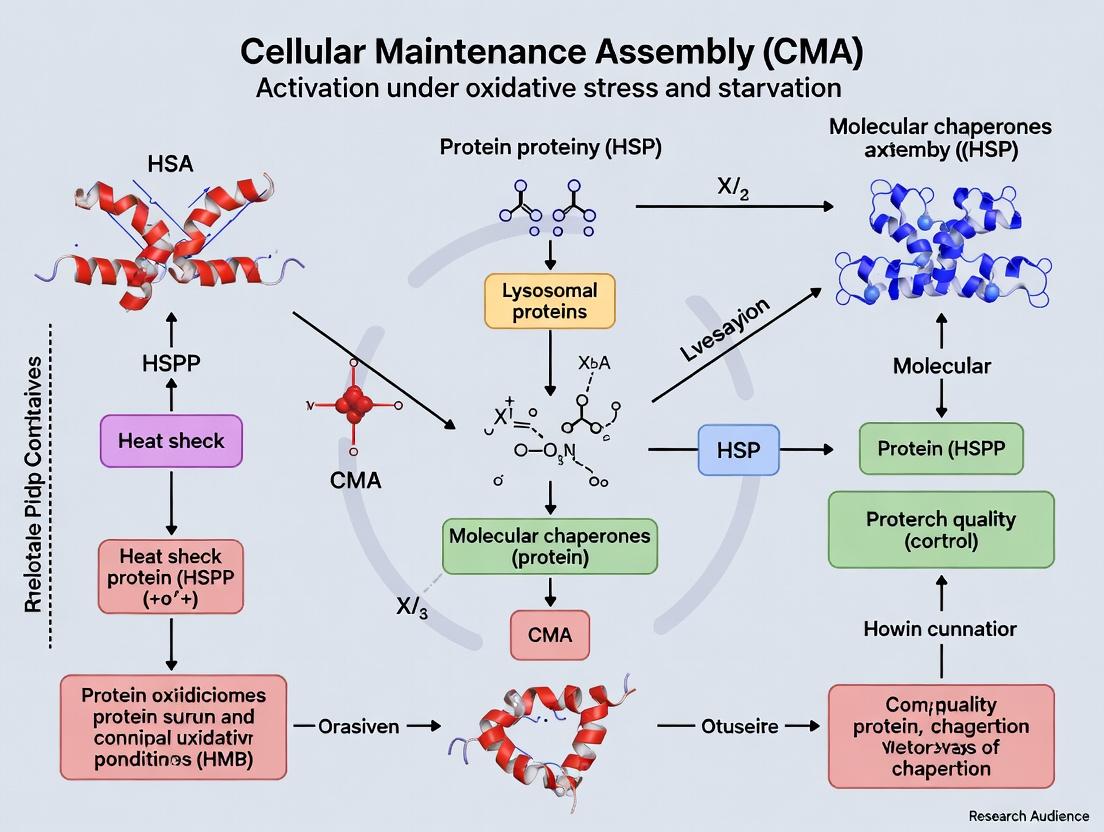
Both oxidative stress and starvation converge to upregulate CMA, albeit through partially overlapping and distinct mechanisms. The shared endpoint is the increased transcription and stabilization of LAMP2A, the CMA receptor, and the enhanced targeting of substrate proteins bearing KFERQ-like motifs.
Oxidative Stress: Directly oxidizes CMA substrates, increasing their affinity for Hsc70 and promoting their unfolding, facilitating translocation. Also activates NRF2 and HSF1, which can promote LAMP2A gene expression. Starvation: Inactivates mTORC1, relieving its inhibitory phosphorylation of the CMA machinery components. Activates TFEB/TFE3, which transcriptionally upregulates LAMP2A and other lysosomal genes.
Title: Molecular Pathways Converging on CMA Activation
The Scientist's Toolkit: Research Reagent Solutions
| Reagent/Material | Function/Application | Example Catalog # / Provider |
|---|---|---|
| DCFH-DA (2',7'-Dichlorodihydrofluorescein diacetate) | Cell-permeable ROS probe. Oxidized to fluorescent DCF by intracellular ROS. | D6883 (Sigma-Aldrich) |
| Anti-DNP Antibody (for Protein Carbonyls) | Immunochemical detection of DNPH-derivatized protein carbonyls via ELISA or western blot. | ab178020 (Abcam) |
| OxiSelect 8-OHdG ELISA Kit | Quantitative measurement of 8-oxo-2’-deoxyguanosine in DNA samples. | STA-320 (Cell Biolabs) |
| Phospho-AMPKα (Thr172) Antibody | Detects activation-specific phosphorylation of AMPK, key starvation sensor. | 2535 (Cell Signaling Tech) |
| Phospho-p70 S6 Kinase (Thr389) Antibody | Gold-standard primary readout for mTORC1 kinase activity. | 9234 (Cell Signaling Tech) |
| Amino Acid-Free DMEM | Defined medium for inducing acute amino acid starvation in cell culture. | D9443 (Sigma-Aldrich) |
| Torin 1 | Potent, selective ATP-competitive mTOR inhibitor; used as a positive control for mTORC1 inhibition. | 14379 (Cayman Chemical) |
| TFEB/TFE3 Antibody (for Nuclear Localization) | Detects total TFEB/TFE3 protein; used in immunofluorescence or fractionation studies. | 4240 (Cell Signaling Tech) |
| LAMP2A (D4A7) Antibody | Specific antibody for detecting the CMA-critical LAMP2A isoform. | 18528 (Abcam) |
Lysosome-associated membrane protein type 2A (LAMP2A) is the indispensable receptor for chaperone-mediated autophagy (CMA). This selective degradation pathway is potently upregulated during oxidative stress and nutrient starvation. However, the functional activity of CMA is governed by a complex regulatory conundrum centered on the dynamics of LAMP2A at the lysosomal membrane. This whitepaper provides an in-depth technical analysis of the mechanisms controlling LAMP2A stabilization and multimerization into the active translocation complex, framed within the context of CMA activation under stress. We synthesize current research, present quantitative data, and detail experimental protocols for investigating this pivotal process.
Chaperone-mediated autophagy is a critical proteolytic pathway that targets specific cytosolic proteins bearing a KFERQ-like motif for lysosomal degradation. Under conditions of oxidative stress and serum starvation, CMA activity increases up to 3-fold, providing amino acids for energy production and removing damaged proteins. This activation is not primarily driven by increased transcription of the LAMP2 gene but by precise post-translational modifications and reorganization of the LAMP2A protein at the lysosomal membrane. The central conundrum lies in balancing the rapid turnover of monomeric LAMP2A with the need to form stable, multimeric translocation complexes—a process requiring precise regulatory inputs.
The Molecular Conundrum: Monomer Stability vs. Multimer Assembly
LAMP2A Lifecycle at the Lysosomal Membrane
Newly synthesized LAMP2A is delivered to the lysosomal membrane, where it resides predominantly as a monomer with a half-life of approximately 6-8 hours. For CMA activation, these monomers must multimerize into a stable complex of at least 8-12 subunits to form a functional protein translocation channel. This multimerization is the rate-limiting step for CMA flux. However, monomeric LAMP2A is continuously cleaved by lysosomal proteases (e.g., cathepsin A) and removed via intraluminal vesicles, creating a dynamic equilibrium.
Key Regulatory Inputs Under Stress
Oxidative stress (e.g., H₂O₂ exposure) and starvation (e.g., serum deprivation) trigger signaling cascades that stabilize LAMP2A monomers and promote multimerization. The key players include:
- GFP2/GLA: The GTPase GFP2 and its regulatory partner GLA are recruited to the lysosomal membrane under stress, protecting LAMP2A from degradation.
- Kinase Signaling: AKT and ERK2-mediated phosphorylation events modulate the interaction of LAMP2A with stabilizing and destabilizing factors.
- Membrane Lipid Composition: Changes in lipid microdomains, particularly increased levels of lysophosphatidic acid, facilitate multimer assembly.
Table 1: Quantitative Parameters of LAMP2A Dynamics Under Basal and Stress Conditions
| Parameter | Basal Conditions | Oxidative Stress (200 µM H₂O₂) | Serum Starvation (24h) |
|---|---|---|---|
| LAMP2A Monomer Half-life | ~6-8 hours | Increased to >12 hours | Increased to ~15 hours |
| Multimerization Rate (Complex Assembly) | Low | 2.5-fold increase | 3.1-fold increase |
| CMA Activity (Protein Degradation Rate) | 1.0 (Basal) | 2.8-fold increase | 3.3-fold increase |
| Lysosomal LAMP2A Protein Level (Relative) | 1.0 | 1.4-fold increase | 1.8-fold increase |
| Required Multimer Size for Translocation | 8-12 subunits | 8-12 subunits | 8-12 subunits |
Experimental Protocols for Investigating LAMP2A Dynamics
Protocol: Assessing LAMP2A Multimerization Status by Blue Native PAGE
Objective: To separate and visualize monomeric and multimeric forms of LAMP2A from isolated lysosomes. Materials: Lysosome-enriched fraction, Digitonin, NativePAGE sample buffer, NativePAGE 3-12% Bis-Tris gel, Cathode/Anode buffers, Coomassie G-250 additive. Procedure:
- Lysosome Isolation: Purify lysosomes from mouse liver or cultured cells (e.g., NIH-3T3) using density gradient centrifugation.
- Membrane Solubilization: Incubate lysosomal pellet with 1% digitonin in PBS for 30 min on ice. Centrifuge at 20,000 g to remove insoluble debris.
- Sample Preparation: Mix supernatant with NativePAGE sample buffer containing 0.25% Coomassie G-250.
- Electrophoresis: Load samples onto a pre-cast NativePAGE gel. Run at 150 V for 1 hour with dark cathode buffer, then switch to light cathode buffer until completion.
- Detection: Perform Western blotting using anti-LAMP2A antibodies (e.g., ab18528). Multimers appear as high-molecular-weight bands (>480 kDa), while monomers run at ~96 kDa.
Protocol: Measuring CMA Activity via Radioactive Degradation Assay
Objective: Quantify the degradation of a known CMA substrate. Materials: [¹⁴C]-labeled GAPDH (a canonical CMA substrate), Serum-starved cells, Leupeptin (inhibitor of lysosomal proteolysis), 6-Aminonicotinamide (6-AN, inhibitor of macroautophagy). Procedure:
- Pulse-Chase: Incubate cells in serum-free media containing [¹⁴C]-GAPDH for 3h. Wash thoroughly.
- Degradation Phase: Incubate cells in fresh serum-free media supplemented with 6-AN (to block macroautophagy) for 4-6h. Include a control set with 200 µM leupeptin.
- Measurement: Precipitate proteins from the media with TCA (10% final). Measure the radioactivity of the TCA-soluble fraction (degraded peptides/amino acids) by scintillation counting.
- Calculation: CMA-specific degradation = (Radioactivity in 6-AN treated sample) - (Radioactivity in leupeptin + 6-AN treated sample).
Protocol: Monitoring LAMP2A Turnover by Cycloheximide Chase
Objective: Determine the half-life of lysosomal LAMP2A under different conditions. Materials: Cells, Cycloheximide (CHX, 50 µg/mL), Lysosome isolation kit, Western blot reagents. Procedure:
- Treat cells (control, H₂O₂-treated, starved) with CHX to block new protein synthesis.
- Harvest cells at time points (0, 2, 4, 8, 12, 24h). Isolate lysosomes from each sample.
- Perform Western blot on lysosomal fractions for LAMP2A and a loading control (e.g., LAMP1).
- Quantify band intensity. Plot remaining LAMP2A (%) vs. time and calculate half-life.
Signaling Pathways Regulating the LAMP2A Conundrum
Title: Signaling Pathways Driving LAMP2A Multimerization
The Scientist's Toolkit: Essential Research Reagents
Table 2: Key Reagents for LAMP2A and CMA Research
| Reagent / Solution | Function & Application | Example Product / Target |
|---|---|---|
| Selective LAMP2A Antibodies | Differentiate LAMP2A from splice variants LAMP2B/C in Western blot, immunofluorescence, and immunoprecipitation. | Rabbit anti-LAMP2A (Abcam ab18528); Mouse anti-LAMP2A (Santa Cruz sc-18822). |
| Lysosome Isolation Kits | Obtain highly purified lysosomal fractions from tissues or cultured cells to analyze membrane proteins. | Lysosome Enrichment Kit (Thermo Scientific 89839); Magnetic Lysosome Isolation Kit (Pierce). |
| CMA Reporter, KFERQ-Dendra2 | A photoconvertible fluorescent substrate to visually track CMA translocation and degradation in live cells. | Express plasmid encoding a KFERQ-tagged Dendra2. |
| LAMP2A siRNA/shRNA | Knock down LAMP2A expression to establish CMA-deficient models for control experiments. | SMARTpool siRNAs targeting human/mouse LAMP2 (Dharmacon). |
| GFP2/GLA Modulators | Investigate the role of the key stabilizing complex. Use constitutively active or dominant-negative GFP2 mutants. | GFP2 Expression Plasmids (Addgene). |
| AKT & ERK Inhibitors | Probe kinase involvement in LAMP2A phosphorylation and stabilization (e.g., MK-2206 for AKT, U0126 for MEK/ERK). | Cell Signaling Technology inhibitors. |
| Protease Inhibitors (Cathepsin A Inhibitor) | Specifically block lysosomal degradation of monomeric LAMP2A to study turnover. | Pepstatin A or specific cathepsin A inhibitor. |
| NativePAGE System | Analyze the oligomeric state of LAMP2A under non-denaturing conditions. | Thermo Fisher Scientific BN-PAGE kits. |
The precise regulation of LAMP2A stability and multimerization represents a sophisticated adaptive mechanism to rapidly activate CMA under stress. Decoding this conundrum is not only fundamental to understanding cellular proteostasis but also holds direct therapeutic relevance. In age-related diseases (e.g., Parkinson's, Alzheimer's) and certain cancers, CMA is dysregulated. Pharmacological strategies aimed at stabilizing LAMP2A monomers or promoting their functional multimerization present a promising avenue for drug development to modulate CMA activity for clinical benefit. Future research must focus on high-resolution structural insights into the multimer and the development of high-throughput screens for CMA modulators.
1. Introduction Chaperone-mediated autophagy (CMA) is a selective lysosomal degradation pathway historically defined by substrate recognition via a pentapeptide motif, KFERQ. This motif is recognized by the cytosolic chaperone Hsc70 (HSPA8). Within the context of broader research on CMA activation under oxidative stress and starvation, it has become evident that the canonical KFERQ sequence is insufficient to explain the full spectrum of CMA substrates. Oxidative post-translational modifications (PTMs) represent a critical mechanism that expands the CMA substrate pool by generating de novo KFERQ-like motifs or by altering substrate conformation to expose latent targeting signals. This whitepaper details the mechanisms, experimental evidence, and methodologies central to this expanded understanding.
2. Mechanisms of Oxidative Modification-Driven CMA Targeting Oxidative stress induces modifications such as carbonylation, sulfoxidation of methionine to methionine sulfoxide, and disulfide bond formation. These alterations can functionally create a "KFERQ-omorphic" site.
- Methionine Sulfoxidation: Oxidation of methionine to methionine sulfoxide changes a hydrophobic side chain to a polar one, potentially converting a non-KFERQ sequence (e.g., containing methionine) into a functional one that mimics glutamine (Q), a key residue in the canonical motif.
- Protein Carbonylation: The introduction of carbonyl groups onto side chains (e.g., of Lys, Arg, Pro, Thr) can create acidic or hydrophilic patches that mimic the charge/hydrophilicity pattern required for Hsc70 binding.
- Conformational Unmasking: Oxidation can cause partial unfolding or disulfide rearrangement, revealing previously buried cryptic KFERQ sequences or Hsc70-binding interfaces.
3. Quantitative Data on CMA Substrate Expansion
Table 1: Impact of Oxidative Stress on CMA Substrate Pool and Activity
| Parameter | Basal Condition | Under Acute Oxidative Stress (e.g., H₂O₂, Paraquat) | Reference / Model System |
|---|---|---|---|
| % of Proteome with CMA-targeting potential | ~30% (by canonical KFERQ) | Estimated increase to ~40-50% | Proteomic analysis, mammalian cells |
| CMA Activity (Substrate Degradation Rate) | Baseline | Increase of 2- to 4-fold | Radiolabeled degradation assays |
| LAMP2A Stabilization | Steady-state levels | Increase of 1.5- to 3-fold (transcriptional & post-translational) | Immunoblot, RT-qPCR |
| Key Oxidized Subrates Identified | GAPDH, PKM2 | α-Synuclein, IκB, Aldolase, GSTP1 | Mass spectrometry, in vitro oxidation |
Table 2: Common Oxidative Modifications that Generate CMA-Targeting Signals
| Modification | Residue(s) Affected | Chemical Change | Mimics KFERQ Residue | Example Substrate |
|---|---|---|---|---|
| Sulfoxidation | Methionine (M) | -CH₃ → -CH₂-SO- | Glutamine (Q) | α-Synuclein |
| Carbonylation | Lysine (K), Proline (P) | -NH₂ → -NH-CO-; etc. | Acidic residues (D/E) | GAPDH |
| Disulfide Formation | Cysteine (C) | -SH HS- → -S-S- | N/A (causes unfolding) | Various kinases |
4. Experimental Protocols for Key Investigations
Protocol 1: Validating Oxidation-Induced CMA Targeting In Vitro
- Objective: Determine if oxidative modification of a protein confers CMA degradability.
- Steps:
- Protein Purification: Express and purify recombinant protein of interest (POI).
- In Vitro Oxidation: Treat POI (1-5 µg/µL) with 1-5 mM H₂O₂ or 1 mM 2,2'-Azobis(2-amidinopropane) dihydrochloride (AAPH) in appropriate buffer for 30-60 min at 37°C. Use untreated and reductant-treated (DTT) controls.
- CMA Binding Assay: Incubate oxidized/native POI with purified Hsc70 and co-chaperones in ATP-containing binding buffer. Co-immunoprecipitate Hsc70 complexes and immunoblot for POI.
- In Vitro Lysosomal Uptake: Isolate rat liver lysosomes (or use lysosomal fractions). Incubate lysosomes with radiolabeled (¹²⁵I) oxidized/native POI in the presence/absence of protease inhibitors (to distinguish binding vs. degradation). Measure lysosome-associated radioactivity.
Protocol 2: Mapping Oxidation-Dependent CMA Engagement in Cells
- Objective: Identify and quantify CMA targeting of an endogenous protein upon oxidative stress.
- Steps:
- Induction & Inhibition: Treat cells (e.g., mouse embryonic fibroblasts, MEFs) with oxidant (e.g., 200 µM Paraquat, 6h). Include controls with CMA inhibited (LAMP2A siRNA/shRNA or PI4KIIIβ inhibitor).
- Lysosomal Isolation & Fractionation: Harvest cells, isolate lysosomes using density gradient centrifugation. Validate purity with markers (LAMP1, Cathepsin D for lysosomes; GAPDH, COX IV for cytosol/mitochondria).
- Substrate Detection: Perform immunoblot on lysosomal fractions for the POI. An increase in the lysosomal pool under stress that is blocked by CMA inhibition confirms CMA targeting.
- Pulse-Chase Analysis: Use ³⁵S-Met/Cys pulse-chase combined with lysosomal isolation to directly measure degradation kinetics via CMA.
5. Signaling Pathways and Workflow Visualization
Diagram 1: Pathway of Oxidation-Mediated CMA Targeting
Diagram 2: Workflow for In Vivo CMA Substrate Validation
6. The Scientist's Toolkit: Key Research Reagents
Table 3: Essential Reagents for Studying Oxidation-Mediated CMA
| Reagent/Solution | Function & Application | Key Considerations |
|---|---|---|
| Paraquat (Methyl viologen) | Inducer of superoxide-mediated oxidative stress in cell culture. Standard for in vivo CMA activation studies. | Highly toxic; requires careful handling and waste disposal. |
| Chloroquine / Bafilomycin A1 | Lysosomotropic agents that raise lysosomal pH, inhibiting substrate degradation. Used to "trap" CMA substrates in lysosomes for detection. | Distinguish binding/degradation; affects all lysosomal pathways. |
| LAMP2A-specific siRNA/shRNA | Molecular tool for selective inhibition of CMA by knocking down the essential receptor. Critical for establishing CMA-dependence. | Off-target effects require controlled siRNA design and rescue experiments. |
| Anti-LAMP2A (clone 4H11) | Antibody specific for the CMA-specific splice variant LAMP2A. Used for immunoblot, immunofluorescence, and immunoprecipitation. | Must distinguish from other LAMP2 isoforms (B, C). |
| DNPH (2,4-Dinitrophenylhydrazine) | Reacts with protein carbonyl groups for detection of oxidative carbonylation via immunoblot (anti-DNP antibody). | Key reagent for directly quantifying protein oxidation state. |
| Recombinant Hsc70 (HSPA8) Protein | For in vitro binding assays to test direct interaction between chaperone and oxidized substrate. | Requires ATP and often co-chaperones for full functional activity. |
| PI4KIIIβ Inhibitor (e.g., PIK93) | Pharmacological inhibitor of LAMP2A multimerization, blocking substrate translocation. Alternative to genetic CMA inhibition. | Useful for acute inhibition; potential off-target effects on lipid signaling. |
This whitepaper examines the intricate crosstalk between HIF-1α, p53, and NF-κB, three master regulators of cellular response. Within the broader thesis on chaperone-mediated autophagy (CMA) activation under oxidative stress and starvation, understanding this nexus is paramount. CMA is a selective lysosomal degradation pathway for cytosolic proteins bearing a KFERQ-like motif. Its activation is a critical stress response, but its regulatory landscape is intertwined with these major signaling hubs. Oxidative stress and nutrient deprivation directly influence the stability and activity of HIF-1α, p53, and NF-κB, creating a complex regulatory network that ultimately dictates cell fate—survival via adaptive pathways like CMA, or death via apoptosis. Deciphering this crosstalk is essential for developing therapeutic strategies for cancer, neurodegeneration, and aging, where CMA and these pathways are frequently dysregulated.
Table 1: Key Regulatory Interactions and Outcomes in the HIF-1α/p53/NF-κB Nexus
| Signaling Axis | Mechanism of Interaction | Context/Condition | Primary Cellular Outcome | Reference (Example) |
|---|---|---|---|---|
| HIF-1α → p53 | HIF-1α binds to p53, inhibiting Mdm2-mediated ubiquitination and degradation. | Severe/acute hypoxia | p53 stabilization, promotes apoptosis. | (Sermeus et al., 2012) |
| p53 → HIF-1α | p53 promotes degradation of HIF-1α via Mdm2 or induces miR-107 targeting HIF-1β. | Genotoxic stress, normoxia | Inhibition of HIF-1α transcriptional activity. | (Yan et al., 2019) |
| NF-κB → HIF-1α | NF-κB (p65) directly binds to the HIF1A promoter, enhancing its transcription. | Inflammation, moderate hypoxia | Amplification of HIF-1α signaling and glycolytic shift. | (van Uden et al., 2008) |
| HIF-1α → NF-κB | HIF-1α can enhance IKKβ activity or modulate NF-κB subunit availability. | Hypoxia | Context-dependent: Can promote pro-survival or pro-apoptotic NF-κB signaling. | (D'Ignazio et al., 2016) |
| p53 NF-κB | Mutual antagonism: p53 can inhibit NF-κB transactivation; NF-κB can suppress p53 activity via pathways like HDAC1. | Oxidative stress, DNA damage | Cell fate decision: Survival (NF-κB) vs. Apoptosis (p53). | (Schneider et al., 2010) |
| CMA Link | p53 can transcriptionally upregulate LAMP2A, a CMA rate-limiting component. NF-κB can modulate CMA activity via redox balance. | Starvation, Oxidative Stress | Enhanced CMA flux, promoting cellular clearance and adaptation. | (Xilouri et al., 2016) |
Experimental Protocols for Key Analyses
Protocol 1: Co-Immunoprecipitation (Co-IP) to Assess HIF-1α/p53/NF-κB Protein-Protein Interactions
- Cell Lysis: Harvest cells under relevant stress (e.g., 1% O₂ for 16h for hypoxia). Lyse in NP-40 lysis buffer (50 mM Tris-HCl pH 7.4, 150 mM NaCl, 1% NP-40) supplemented with protease/phosphatase inhibitors.
- Pre-clearing: Incubate lysate with Protein A/G agarose beads for 1h at 4°C. Pellet beads, retain supernatant.
- Immunoprecipitation: Incubate pre-cleared lysate with 2-5 µg of primary antibody (e.g., anti-HIF-1α) or species-matched IgG control overnight at 4°C with gentle rotation.
- Bead Capture: Add Protein A/G beads for 2h. Pellet beads and wash 3-4 times with lysis buffer.
- Elution and Analysis: Elute proteins in 2X Laemmli buffer by boiling. Analyze by western blot for co-precipitated proteins (e.g., probe for p65 or p53).
Protocol 2: Luciferase Reporter Assay for Transcriptional Crosstalk
- Transfection: Seed cells in 24-well plates. Co-transfect with a reporter plasmid (e.g., NF-κB-responsive firefly luciferase) and a control Renilla luciferase plasmid (e.g., pRL-TK) using a suitable transfection reagent.
- Stimulation/Inhibition: 24h post-transfection, apply treatments (e.g., hypoxia mimetic CoCl₂ 150 µM; TNF-α 10 ng/ml; p53 activator Nutlin-3 10 µM) for an additional 16-24h.
- Lysis and Measurement: Lyse cells using Passive Lysis Buffer (Promega). Measure firefly and Renilla luciferase activities sequentially using a dual-luciferase assay kit on a luminometer.
- Data Normalization: Calculate the ratio of firefly to Renilla luciferase activity for each well to control for transfection efficiency.
Protocol 3: Monitoring CMA Activity via LAMP2A-KFERQ Reporter Flux
- Cell Line: Utilize stable cell lines expressing a photoconvertible CMA reporter (e.g., KFERQ-PA-mCherry1).
- Photoconversion and Chase: Photoconvert the cytosolic red fluorescence of the reporter to a far-red state in a defined region of interest. Immediately induce CMA (starvation with EBSS medium, oxidative stress with H₂O₂).
- Live-Cell Imaging: Track the loss of photoconverted signal from the cytosol over time (e.g., 4-6h) using confocal microscopy, as its degradation indicates CMA flux.
- Quantification: Measure the fluorescence intensity of the photoconverted channel in the cytosol over time. Normalize to t=0. Compare rates under different genetic (siRNA against p65, p53) or pharmacological modulators.
Pathway and Workflow Visualizations
Diagram Title: Core Crosstalk Network Driving Cell Fate
Diagram Title: Integrated Experimental Workflow for Nexus & CMA Study
The Scientist's Toolkit: Key Research Reagent Solutions
Table 2: Essential Reagents for Investigating the HIF-1α/p53/NF-κB/CMA Nexus
| Reagent/Category | Example Product/Specifics | Primary Function in Research |
|---|---|---|
| Hypoxia Mimetics | Cobalt Chloride (CoCl₂), Dimethyloxalylglycine (DMOG) | Stabilize HIF-1α by inhibiting PHD enzymes, allowing study of hypoxic signaling in normoxia. |
| p53 Activators/Inhibitors | Nutlin-3 (activator, MDM2 antagonist), Pifithrin-α (inhibitor) | Pharmacologically modulate p53 activity to dissect its role in crosstalk and CMA regulation. |
| NF-κB Modulators | TNF-α (activator), BAY 11-7082 (IKK inhibitor), SC514 (IKK-2 inhibitor) | Induce or block NF-κB signaling to analyze its interaction with HIF-1α/p53 and impact on cell fate. |
| CMA Reporters | KFERQ-PA-mCherry1 plasmid; Cell lines with stable CMA reporter. | Directly visualize and quantify CMA flux in live cells under various genetic/pharmacological manipulations. |
| Key Antibodies | Anti-HIF-1α (clone 54), anti-p53 (DO-1), anti-phospho-p65 (Ser536), anti-LAMP2A (ab18528). | Detect protein expression, localization, post-translational modifications via WB, IF, IP. |
| siRNA/shRNA Libraries | ON-TARGETplus SMARTpools for HIF1A, TP53, RELA (p65), LAMP2A. | Perform targeted gene knockdown to establish causal relationships in the signaling network. |
| Lysosomal Inhibitors | Bafilomycin A1 (V-ATPase inhibitor), Chloroquine. | Block lysosomal degradation to confirm CMA-dependent substrate turnover in assays. |
| Oxidative Stress Inducers | Hydrogen Peroxide (H₂O₂), Menadione. | Generate controlled levels of ROS to study the oxidative stress arm of the nexus and CMA activation. |
Chaperone-mediated autophagy (CMA) is a selective lysosomal degradation pathway critical for maintaining cellular proteostasis, particularly under stress conditions like oxidative stress and nutrient deprivation. Within the broader thesis of CMA activation under oxidative stress and starvation, this whitepaper posits that the metabolic sensors AMP-activated protein kinase (AMPK) and the Sirtuin family of deacetylases act as central, interdependent gatekeepers. They transduce the cell's energetic and redox status into precise regulatory signals that modulate CMA activity, integrating CMA into the core cellular metabolic response.
Core Regulatory Mechanisms
AMPK as an Energetic Switch for CMA
AMPK is activated by an increased AMP/ATP ratio, signaling low cellular energy. Recent research confirms AMPK phosphorylates key CMA components, directly linking energy deficit to CMA upregulation.
- LAMP2A Stabilization: AMPK phosphorylates LAMP2A, the receptor at the lysosomal membrane essential for substrate translocation. This phosphorylation event reduces LAMP2A multimerization and turnover, increasing its stability and assembly into the active translocation complex.
- Transcriptional Regulation: AMPK activates transcription factors like FoxO1, which can upregulate LAMP2A gene expression, providing a slower, sustained boost to CMA capacity.
Sirtuins as Redox and NAD+-Dependent Regulators
Sirtuins (SIRT1, in particular) are NAD+-dependent deacetylases. Their activity is directly coupled to the cellular metabolic state via NAD+ availability, which increases during fasting and oxidative stress.
- Deacetylation of CMA Substrates: SIRT1 deacetylates CMA-targeted proteins, facilitating their recognition by the chaperone HSC70. Acetylation can mask the KFERQ-like targeting motif; deacetylation exposes it, enhancing substrate availability.
- Regulation of Lysosomal Function: SIRT1 deacetylates and activates transcription factor EB (TFEB), a master regulator of lysosomal biogenesis, thereby increasing the overall lysosomal pool available for CMA.
- Cross-talk with AMPK: SIRT1 deacetylates and activates LKB1, an upstream kinase of AMPK, creating a positive feedback loop that amplifies the energy-sensing signal.
Table 1: Key Quantitative Findings in AMPK/Sirtuin-Mediated CMA Regulation
| Regulator | Target/Process | Experimental Change (e.g., Knockdown/Activation) | Effect on CMA Activity (Measured Output) | Reference Model |
|---|---|---|---|---|
| AMPK | LAMP2A Phosphorylation (T211) | AICAR (AMPK agonist) treatment | ↑ LAMP2A stability by ~40-60%; ↑ CMA flux by ~2-3 fold | Primary mouse fibroblasts |
| AMPK | CMA Substrate Degradation | Compound C (AMPK inhibitor) during serum starvation | ↓ Degradation of RNase A (CMA reporter) by ~70% | Mouse liver, in vivo |
| SIRT1 | Substrate Deacetylation | Resveratrol (SIRT1 activator) | ↑ Binding of GAPDH to HSC70 by ~50%; ↑ Lysosomal association | HEK293 cells |
| SIRT1 | TFEB Activation | SIRT1 overexpression | ↑ Lysosomal gene expression by 2-4 fold; ↑ LAMP2A levels ~1.8 fold | HeLa cells |
| Combined | CMA Flux during Starvation | Dual AMPK inhibition & SIRT1 KO | Abolishes starvation-induced CMA activation completely | MEFs (SIRT1 KO) |
Table 2: Research Reagent Solutions Toolkit
| Reagent/Category | Specific Example(s) | Function in CMA/Energy Sensing Research |
|---|---|---|
| CMA Reporters | KFERQ-Dendra, RNase A-GFP, CMA reporter cell lines (e.g., Photo-convertible CMA reporter) | Visualize and quantify CMA substrate uptake and degradation in real-time. |
| AMPK Modulators | AICAR (activator), Compound C (inhibitor), Metformin (indirect activator) | Manipulate AMPK activity to study its causal role in CMA regulation. |
| Sirtuin Modulators | Resveratrol, SRT1720 (SIRT1 activators); EX527, Nicotinamide (SIRT1 inhibitors) | Modulate Sirtuin activity to assess impact on substrate acetylation and CMA. |
| Lysosomal Inhibitors | Bafilomycin A1, Chloroquine, Leupeptin | Inhibit lysosomal degradation to measure substrate accumulation pre-lysosome. |
| Key Antibodies | Anti-phospho-LAMP2A (T211), Anti-acetylated Lysine, Anti-LAMP2A, Anti-SQSTM1/p62 | Detect CMA component modification, levels, and monitor other autophagic pathways. |
| Metabolic Assays | NAD+/NADH Quantification Kit, ATP Assay Kit, Seahorse XF Analyzer | Quantify the energetic (AMP/ATP, NAD+) state of cells under experimental conditions. |
Experimental Protocols
Protocol: Measuring CMA Flux Using a Photo-convertible Reporter
This protocol quantifies the lysosomal delivery and degradation of CMA substrates.
- Cell Preparation: Seed cells stably expressing the KFERQ-PA-mEos2 (or similar) reporter.
- Starvation/Oxidative Stress Induction: Incubate cells in EBSS (starvation) or treat with a precise concentration of paraquat (e.g., 100-500 µM) or H₂O₂ (e.g., 200 µM) for defined periods (2-24h).
- Photo-conversion: At time T=0, expose a region of interest to 405 nm light to convert mEos2 fluorescence from green to red.
- Time-Course Imaging: Using live-cell microscopy, track the red (photo-converted, pre-existing) signal in lysosomes (co-stained with LysoTracker) over time (e.g., every 30 min for 6-8h).
- Quantification: Plot the decay of the red lysosomal signal over time. The slope represents CMA flux. Compare between control, AMPK-inhibited (e.g., 10 µM Compound C), and SIRT1-inhibited (e.g., 10 µM EX527) conditions.
Protocol: Assessing LAMP2A Phosphorylation and Stability
This protocol evaluates the direct post-translational regulation of the CMA machinery.
- Treatment: Treat cells (e.g., mouse embryonic fibroblasts) under nutrient-rich and starved conditions ± AMPK modulators.
- Lysosomal Isolation: Use differential centrifugation to obtain a purified lysosomal fraction.
- Immunoblotting: Resolve lysosomal membrane proteins via SDS-PAGE.
- Detection:
- Probe with phospho-specific anti-LAMP2A (T211) antibody.
- Re-probe for total LAMP2A.
- Use LAMP1 as a lysosomal loading control.
- Cycloheximide Chase: To measure stability, treat cells with 50 µg/ml cycloheximide to block new protein synthesis. Harvest cells at 0, 2, 4, 8 hours. Immunoblot for LAMP2A to assess its half-life under different AMPK activity states.
Protocol: Evaluating Substrate Deacetylation for CMA Targeting
This protocol links SIRT1 activity to substrate availability for CMA.
- Immunoprecipitation: Under control and oxidative stress (e.g., 200 µM H₂O₂, 2h), lyse cells and immunoprecipitate a known CMA substrate (e.g., GAPDH, PKM2) using a specific antibody.
- Acetylation Status: Perform immunoblot analysis on the immunoprecipitated material using an anti-acetylated-lysine antibody.
- Interaction Assay: In parallel, perform a co-immunoprecipitation to assess the interaction between the substrate and the CMA chaperone HSC70 under conditions of SIRT1 activation (Resveratrol, 10 µM) vs. inhibition (EX527).
- Correlation: Correlate decreased substrate acetylation with increased HSC70 binding.
Signaling Pathway and Workflow Visualizations
Title: AMPK and SIRT1 Coregulate CMA Under Stress
Title: Integrated Workflow for Studying CMA Gatekeepers
From Bench to Insight: Techniques to Monitor and Modulate CMA Activity
This technical guide details the gold-standard methodologies for investigating chaperone-mediated autophagy (CMA), a selective lysosomal degradation pathway critical for cellular homeostasis. Within the broader thesis context of CMA activation under oxidative stress and starvation, these assays provide the definitive tools for quantitative analysis. CMA is upregulated in response to these stressors, serving as a quality control mechanism to degrade damaged proteins and provide amino acids for survival. The assays described herein enable precise tracking of substrate targeting, lysosomal binding, and translocation—the core steps of the CMA pathway.
The Core Principle: From KFERQ Motif to Lysosomal Lumen
CMA substrates contain a pentapeptide motif biochemically related to KFERQ. This motif is recognized in the cytosol by the chaperone Hsc70 (HSPA8). The substrate-chaperone complex is targeted to the lysosomal membrane via interaction with the receptor lysosome-associated membrane protein type 2A (LAMP2A). Monomeric LAMP2A multimerizes to form a translocation complex, requiring a luminal variant of Hsc70 (HSPA8L) for substrate unfolding and translocation into the lysosomal lumen, where it is degraded.
Photoactivatable KFERQ Reporter Assay
This assay allows for the spatiotemporal analysis of CMA substrate targeting and translocation in living cells.
Experimental Protocol
A. Reporter Construct Design & Transfection:
- Construct: Fuse the protein of interest (or a canonical CMA substrate like RNase A or GAPDH) to a photoactivatable fluorescent protein (e.g., PA-mCherry1 or Dendra2) via a flexible linker. Ensure the KFERQ-like motif remains accessible.
- Transfection: Introduce the construct into target cells (commonly mouse fibroblast lines like NIH-3T3 or human cell lines) using standard methods (lipofection, electroporation).
- CMA Induction: Prior to imaging, subject cells to CMA-activating conditions: starvation (Earle's Balanced Salt Solution, EBSS) for 4-18 hours or oxidative stress (e.g., 200-500 µM H₂O₂) for 1-6 hours.
B. Photoactivation & Time-Lapse Imaging:
- Mounting: Place cells in an imaging chamber with appropriate medium (complete or starvation/ stress medium).
- Baseline Image: Capture a pre-photoactivation image using the fluorescent channel for the non-photoactivated state (e.g., green for Dendra2).
- Photoactivation: Use a 405 nm laser to selectively photoactivate a region of interest (ROI), typically the cytoplasm or a specific organelle, converting the fluorophore to its red-emitting state.
- Time-Lapse Acquisition: Immediately initiate time-lapse confocal microscopy. Acquire dual-channel (green/red) images every 2-5 minutes for 60-120 minutes.
- Lysosomal Counterstain: Include LysoTracker Green/Deep Red or transiently express LAMP1-RFP in the experiment to identify lysosomes.
C. Quantitative Image Analysis:
- ROI Definition: Define ROIs for the photoactivated cytoplasmic pool and for individual lysosomes (using the lysosomal marker).
- Fluorescence Intensity Measurement: Track the decay of red fluorescence in the cytoplasmic ROI and the increase in red fluorescence within lysosomal ROIs over time.
- Key Metrics: Calculate the lysosomal translocation rate (slope of increase in lysosomal red signal) and the half-life of the cytoplasmic photoactivated pool.
D. Controls & Validation:
- Negative Control: Express a reporter with a mutated KFERQ motif (e.g., KFERQ→AAARA).
- CMA Inhibition: Treat cells with KNK437 (HSP inhibitor) or use LAMP2A knockdown/knockout cells to confirm CMA-specific translocation.
- Specificity Control: Inhibit macroautophagy with 3-methyladenine (3-MA) to show independence from this pathway.
Table 1: Key Quantitative Metrics from Photoactivatable Reporter Assays
| Metric | Control Conditions (Full Nutrient) | CMA-Activated (Starvation, 10h EBSS) | CMA-Inhibited (LAMP2A KO) | Notes / Interpretation |
|---|---|---|---|---|
| Cytoplasmic Half-life (t₁/₂) | 120 - 180 min | 40 - 70 min | >300 min | Shorter half-life indicates faster CMA flux. |
| Lysosomal Accumulation Rate (A.U./min) | 0.5 - 1.5 | 3.0 - 8.0 | <0.5 | Slope of linear increase in lysosomal signal over first 30-60 min. |
| % Photoactivated Protein in Lysosomes at t=60min | 15 - 25% | 60 - 80% | <5% | Direct measure of translocation efficiency. |
| Effect of 10µM KNK437 | Reduction by ~20% | Reduction by 70-90% | No significant effect | Confirms Hsc70 dependence. |
| LAMP2A Multimerization Index | 1.0 - 1.5 | 2.5 - 4.0 | Not Applicable | Measured via crosslinking; index >2 correlates with active translocation. |
Lysosomal Translocation Assay (Isolated Lysosomes)
This biochemical assay measures the binding and uptake of radiolabeled substrates by intact, functional lysosomes isolated from rat liver or cultured cells.
Experimental Protocol
A. Isolation of Lysosomes:
- Source: Sacrifice a rat or harvest ~10⁸ cultured cells under experimental conditions (control, starved, stressed).
- Homogenization: Use a glass-Teflon homogenizer in ice-cold 0.25 M sucrose buffer containing protease inhibitors.
- Differential Centrifugation: Clear nuclei/debris at 1,000 g. Obtain a heavy mitochondrial/lysosomal fraction at 17,000 g for 12 min.
- Density Gradient: Resuspend pellet and load onto a discontinuous Metrizamide or Percoll gradient (e.g., 10%, 19%, 27%). Centrifuge at high speed (e.g., 95,000 g for 2h).
- Collection: Collect the enriched lysosomal band at the interface. Wash by dilution and centrifugation in 0.25 M sucrose.
B. Substrate Preparation:
- Labeling: Radiolabel a known CMA substrate (e.g., Glyceraldehyde-3-phosphate dehydrogenase, GAPDH) with ¹²⁵I using IODO-BEADS.
- Chaperone Loading: Incubate the labeled substrate with purified Hsc70 and an ATP-regenerating system for 15 min at 37°C to form the recognition complex.
C. Binding and Uptake Reaction:
- Binding Reaction: Incubate ~50 µg of isolated lysosomes with the ¹²⁵I-substrate-Hsc70 complex in binding buffer (10 mM HEPES, pH 7.4, 0.25 M sucrose, 2 mM MgCl₂, 5 mM ATP) for 20 min on ice. This allows binding to LAMP2A without translocation.
- Uptake Reaction: Shift a parallel set of tubes to 37°C for 15-20 minutes to initiate and permit translocation.
- Protease Protection: After uptake, treat lysosomes with Proteinase K (0.1 mg/mL, 10 min on ice) to degrade any substrate still bound externally. Include controls with Triton X-100 to disrupt the lysosomal membrane and confirm luminal protection.
D. Analysis:
- Precipitation: Stop reactions by adding cold trichloroacetic acid (TCA) to 10%.
- Measurement: Count radioactivity in the TCA-precipitable pellet via gamma counter.
- Calculation:
- Binding: CPM from ice-cold reaction (protease-treated).
- Total Uptake: CPM from 37°C reaction (protease-treated).
- Translocation: Total Uptake minus Binding.
Table 2: Typical Data from Isolated Lysosome Translocation Assays
| Assay Component | Lysosomes from Fed Rats | Lysosomes from Starved Rats (48h) | Lysosomes + Anti-LAMP2A Antibody | Notes / Interpretation |
|---|---|---|---|---|
| Substrate Binding (pmol/mg lys protein) | 50 - 100 | 200 - 350 | 20 - 40 | Reflects LAMP2A levels at the membrane. |
| Translocated Substrate (pmol/mg lys protein) | 20 - 40 | 150 - 250 | 5 - 15 | The definitive measure of CMA capacity. |
| Translocation Efficiency (% of Bound) | 30 - 40% | 60 - 75% | <20% | Indicates functional competence of the translocation complex. |
| ATP Depletion Effect on Translocation | >95% inhibition | >95% inhibition | N/A | Confirms energy dependence. |
| Required Luminal Hsc70 (HSPA8L) | Essential | Essential (increased levels) | N/A | Demonstrated using lysosomes stripped of luminal proteins. |
The Scientist's Toolkit: Research Reagent Solutions
Table 3: Essential Reagents for CMA Gold-Standard Assays
| Reagent / Material | Function / Application | Example Product/Catalog # (Illustrative) |
|---|---|---|
| Photoactivatable Constructs | Genetically encoded reporters for live-cell CMA tracking. | pmCherry-N1/Dendra2 vectors; Custom KFERQ-substrate fusions. |
| LAMP2A Antibodies | Detection, quantification, and inhibition of the CMA receptor. | Mouse monoclonal (H4B4) for immunoblot/blocking; Rabbit polyclonal for IHC. |
| Lysosome Isolation Kit | Rapid preparation of functional lysosomes from tissues/cells. | Lysosome Enrichment Kit (e.g., from Thermo Scientific). |
| Metrizamide / Percoll | For high-purity lysosome isolation via density gradient centrifugation. | Sigma-Aldrich M3761 / GE Healthcare 17-0891-01. |
| Recombinant Hsc70 (HSPA8) | For loading CMA substrates in isolated lysosome assays. | Recombinant Human HSPA8 Protein (e.g., Abcam ab78429). |
| CMA Modulators | Chemical activation/inhibition for validation. | KNK437 (HSP inhibitor); 6-Aminonicotinamide (CMA activator in some contexts). |
| LysoTracker Dyes | Live-cell staining of acidic lysosomes for colocalization studies. | LysoTracker Deep Red (Thermo Fisher L12492). |
| ³H-Labeled or ¹²⁵I-Labeled Substrates | Radioactive tracers for quantitative uptake/binding assays. | ¹²⁵I-labeled GAPDH or RNase A (prepared in-lab via chloramine-T/IODO-BEADS method). |
| Protease Inhibitor Cocktails | Prevent substrate degradation during lysosome isolation and assays. | Complete, EDTA-free (Roche 11873580001). |
| ATP-Regenerating System | Provides energy for Hsc70 function and lysosomal translocation. | Creatine Phosphate (20mM) and Creatine Kinase (100 µg/mL). |
Visualization Diagrams
1. Introduction: CMA Biomarkers in a Broader Research Context Chaperone-Mediated Autophagy (CMA) is a selective lysosomal degradation pathway critical for cellular proteostasis. It is specifically activated in response to oxidative stress and prolonged nutrient starvation, serving as an adaptive mechanism to remove damaged proteins and provide amino acids for biosynthesis. The core molecular machinery involves the cytosolic chaperone HSC70 (HSPA8), which identifies proteins bearing a KFERQ-like motif, and the lysosomal membrane receptor LAMP2A, which facilitates substrate translocation. Consequently, quantifying LAMP2A and HSC70 levels in tissues and biofluids offers a direct window into CMA activity. This technical guide details methodologies for detecting these key biomarkers, essential for research into CMA's role in aging, neurodegenerative diseases, cancer, and metabolic disorders under conditions of cellular stress.
2. Quantitative Summary of Reported Biomarker Levels Current literature reports variable levels of LAMP2A and HSC70 depending on the sample type, disease state, and detection method. The following tables consolidate key quantitative findings.
Table 1: Reported LAMP2A and HSC70 Levels in Human Tissues & Cell Lysates
| Sample Type | Condition | Target | Reported Level (Approx.) | Method | Reference Context |
|---|---|---|---|---|---|
| Liver Tissue | Healthy | LAMP2A | 0.5 - 1.2 µg/mg total protein | Immunoblot | Baseline aging studies |
| Liver Tissue | Starvation/Oxidative Stress | LAMP2A | 2-4 fold increase | Immunoblot | CMA activation models |
| Brain Cortex (Human) | Alzheimer's Disease | LAMP2A | ~40% decrease | qPCR/Immunoblot | Neurodegeneration |
| Primary Fibroblasts | Senescent Cells | LAMP2A | >50% decrease | Flow Cytometry | Cellular aging |
| Various Cell Lines | Starvation (48h) | HSC70 | 1.5-2 fold increase | ELISA | Stress response |
Table 2: Detection in Biofluids – Current Status & Challenges
| Biofluid | Target | Detection Status | Typical Assay | Key Challenge |
|---|---|---|---|---|
| Serum/Plasma | HSC70 | Detectable (ng/mL range) | High-Sensitivity ELISA | Distinguishing from other HSP70 isoforms; release from necrotic cells. |
| Serum/Plasma | LAMP2A | Not routinely detected | Immunoprecipitation-MS | Very low abundance; requires extensive sample pre-concentration. |
| CSF | HSC70 | Detectable in pathological states | Multiplex Immunoassay | Low concentration; blood contamination risk. |
| Urine | LAMP2A (exosomes) | Research stage | Exosome isolation + Immunoblot | Standardization of exosome yield and marker normalization. |
3. Experimental Protocols for Key Detection Methodologies
3.1. Immunoblot Analysis from Tissue Homogenates/Cell Lysates
- Sample Preparation: Homogenize tissue or lyse cells in RIPA buffer with protease/phosphatase inhibitors. Centrifuge at 16,000 x g for 20 min at 4°C. Determine protein concentration via BCA assay.
- Electrophoresis & Transfer: Load 20-50 µg protein per lane on a 10-12% SDS-PAGE gel. Transfer to PVDF membrane using standard wet transfer.
- Immunodetection: Block membrane with 5% non-fat milk in TBST for 1h. Incubate with primary antibodies overnight at 4°C:
- Anti-LAMP2A (Clone EPR13558): 1:1000 in blocking buffer. Specific for the LAMP2A splice variant.
- Anti-HSC70/HSPA8 (Clone EP1531Y): 1:5000. May cross-react with inducible HSP70 under high exposure.
- Loading Control (e.g., β-Actin, GAPDH): 1:10,000.
- Visualization: Incubate with HRP-conjugated secondary antibody (1:5000) for 1h. Develop with enhanced chemiluminescence (ECL) substrate and image. Quantify band density using ImageJ software, normalizing to loading control.
3.2. Quantitative Real-Time PCR (qPCR) for Transcript Levels
- RNA Isolation: Use TRIzol reagent or silica-column kits. Assess RNA integrity (RIN > 7).
- cDNA Synthesis: Perform reverse transcription with 1 µg total RNA using a high-capacity cDNA kit with random hexamers.
- qPCR Reaction: Use SYBR Green or TaqMan chemistry. Reaction: 95°C for 10 min, then 40 cycles of 95°C for 15s and 60°C for 1 min. Use primer/probe sets specific for:
- LAMP2 (exon 9 sequence unique to LAMP2A variant).
- HSPA8.
- Reference genes (e.g., GAPDH, β-actin).
- Analysis: Calculate relative expression using the 2^(-ΔΔCt) method.
3.3. ELISA for HSC70 in Serum/Plasma
- Sample Prep: Collect blood in EDTA or heparin tubes. Centrifuge at 2000 x g for 15 min. Aliquot and store plasma at -80°C. Avoid freeze-thaw cycles.
- Assay Procedure: Use a commercial Human HSPA8/HSC70 ELISA Kit (e.g., Abcam, ab133053). Dilute plasma samples 1:10 in assay buffer. Add 100 µL/well of standard or sample. Follow kit protocol for incubation with capture/detection antibodies and substrate.
- Quantification: Measure absorbance at 450 nm. Generate a standard curve from recombinant HSC70 (0-20 ng/mL). Report concentration in ng/mL.
3.4. Immunofluorescence/Confocal Microscopy for CMA Activity Assessment
- Cell Culture & Treatment: Seed cells on coverslips. Induce CMA with 10 µM H₂O₂ (oxidative stress) or incubate in EBSS (starvation) for 6-24h.
- Staining: Fix with 4% PFA, permeabilize with 0.1% Triton X-100. Block with 5% BSA. Co-incubate with primary antibodies: anti-LAMP2A (1:200) and anti-LAMP1 (1:500, lysosomal marker) overnight. Use Alexa Fluor 488/594 secondary antibodies.
- Imaging & Analysis: Acquire z-stacks on a confocal microscope. Quantify LAMP2A puncta co-localized with LAMP1 using Coloc2 or similar plugin in ImageJ. Increased co-localization indicates CMA activation.
4. Signaling Pathways and Experimental Workflows
5. The Scientist's Toolkit: Key Research Reagent Solutions
Table 3: Essential Reagents for LAMP2A/HSC70 Biomarker Research
| Reagent / Material | Function / Specificity | Example Catalog # |
|---|---|---|
| Anti-LAMP2A (Clone EPR13558) | Monoclonal antibody specific to the C-terminal tail of human LAMP2A splice variant for immunoblot/IF. | Abcam, ab18528 |
| Anti-HSC70/HSPA8 (Clone EP1531Y) | Monoclonal antibody recognizing both constitutive HSC70 and inducible HSP70; good for total pool assessment. | Abcam, ab51052 |
| Anti-HSPA8 (CMA-specific) | Polyclonal antibody raised against the KFERQ-binding region; may offer more functional relevance. | Proteintech, 10654-1-AP |
| Human HSPA8/HSC70 ELISA Kit | For quantitative measurement of HSC70 in serum, plasma, or cell culture supernatants. | Abcam, ab133053 |
| LAMP2 (D4B7) XP Rabbit mAb | Recognizes all LAMP2 isoforms; requires careful interpretation with LAMP2A-specific antibody. | Cell Signaling, 49067 |
| KFERQ-Dendra2 Reporter Plasmid | Live-cell reporter for monitoring CMA substrate translocation and degradation. | Addgene, plasmid # 126479 |
| Lysosome Isolation Kit | Enriches lysosomal fractions to assess LAMP2A multimerization status and substrate uptake. | Sigma-Aldrich, LYSISO1 |
| Protease Inhibitor Cocktail (EDTA-free) | Preserves protein integrity during tissue homogenization, crucial for CMA component analysis. | Roche, 05892791001 |
1. Introduction Chaperone-Mediated Autophagy (CMA) is a selective lysosomal degradation pathway crucial for cellular homeostasis, activated under oxidative stress and nutrient starvation. CMA targets specific cytosolic proteins bearing a KFERQ-like motif, which are recognized by the chaperone HSC70 (HSPA8). The substrate-chaperone complex then binds to the lysosomal membrane receptor LAMP2A, triggering its multimerization and subsequent translocation of the substrate into the lysosome for degradation. Dysregulation of CMA is implicated in aging, neurodegeneration, and cancer. CRISPR/Cas9 technology offers precise genetic tools to create in vitro and in vivo models for manipulating LAMP2A and HSC70, enabling definitive functional studies of CMA activation dynamics.
2. CRISPR/Cas9 Models for CMA Core Components
2.1. Targeting Strategies CRISPR/Cas9 can be deployed to create knockout (KO), knock-in (KI), or conditional alleles for LAMP2 (specifically the LAMP2A isoform) and HSPA8 (encoding HSC70). The table below summarizes common genetic outcomes and their applications.
Table 1: CRISPR/Cas9 Models for LAMP2A and HSC70
| Target Gene | Genetic Modification | Primary Application in CMA Research | Phenotypic Consequence |
|---|---|---|---|
| LAMP2 | Complete Knockout of all isoforms | Study of total LAMP2/CMA deficiency; requires rescue with LAMP2A-specific cDNA. | Accumulation of CMA substrates (e.g., GAPDH, MEF2D), impaired response to oxidative stress/starvation. |
| LAMP2 | Exon-Specific KO (targeting exon 8A) | Selective ablation of the LAMP2A isoform without affecting LAMP2B/C. | Specific CMA blockade, used to isolate CMA function from other LAMP2 roles. |
| LAMP2 | Knock-in of Tag (e.g., GFP, HALO) at C-terminus | Live imaging of LAMP2A localization and dynamics under stress conditions. | Enables tracking of lysosomal mobilization and multimerization in real-time. |
| HSPA8 | Conditional Knockout (floxed alleles) | Tissue-specific or inducible ablation of HSC70 to avoid embryonic lethality. | Loss of CMA substrate recognition and binding; severe proteostasis disruption. |
| HSPA8 | Knock-in of Point Mutations (e.g., K71M) | Disruption of substrate binding while preserving chaperone cofactor interactions. | Specific inhibition of CMA without globally affecting HSC70's other functions. |
2.2. Quantitative Data from Recent Studies Recent studies utilizing these models have quantified CMA activity and related parameters.
Table 2: Quantitative Outcomes from Selected CRISPR/Cas9 CMA Models
| Model System | Intervention | Measured Parameter | Quantitative Result (vs. Control) | Reference Context |
|---|---|---|---|---|
| HeLa Cells | LAMP2A KO (Exon 8A targeting) | CMA Activity (Radioactive KFERQ-protein degradation assay) | Reduction of >85% | Basal & Starvation (6h)-induced CMA |
| Mouse Embryonic Fibroblasts (MEFs) | Hspa8 Conditional KO (Cre-ERT2) | Lysosomal Binding of GAPDH (Immunoblot of lysosomal fractions) | Decrease of ~70% | Under oxidative stress (200 µM H₂O₂, 2h) |
| Human iPSC-derived Neurons | LAMP2A-GFP KI | Lysosomal Colocalization of α-synuclein (Confocal Quantification) | Increase from 15% to 45% | Under Proteasome Inhibition (10 µM MG132, 12h) |
| In Vivo Mouse Liver | LAMP2A Hepatocyte-specific KO | Accumulation of known CMA substrates (e.g., PKM2) (Immunoblot) | 3.5-fold increase | After 24h of starvation |
3. Experimental Protocols for CMA Analysis in CRISPR-Edited Models
3.1. Protocol: Assessing CMA Activity via Lysosomal Fractionation and Substrate Translocation
- Objective: Quantify the binding and uptake of CMA substrates into lysosomes from CRISPR-edited cells under starvation.
- Materials: CRISPR-edited cell line, Control cell line, Hank's Balanced Salt Solution (HBSS) for starvation, Lysosome Isolation Kit, Protease Inhibitors, Anti-LAMP1, Anti-LAMP2A, Anti-GAPDH (CMA substrate) antibodies.
- Procedure:
- Starvation Induction: Culture cells to 80% confluency. Rinse with PBS and incubate in serum-free media (CMA activation) or complete media (control) for 6-12 hours.
- Lysosome Isolation: Harvest cells. Use a density gradient-based lysosome isolation kit per manufacturer's instructions to obtain a purified lysosomal fraction.
- Protease Protection Assay: Resuspend the lysosomal pellet in isotonic sucrose buffer. Divide into three aliquots: (A) No treatment, (B) + 0.2% Triton X-100 (lysis), (C) + 1 µg/ml Proteinase K. Incubate on ice for 30 min. For (C), inhibit protease with 5 mM PMSF after incubation.
- Immunoblot Analysis: Run samples on SDS-PAGE. Probe for GAPDH. Signal in (A) indicates total lysosome-associated substrate. Signal lost in (C) but protected in (A) indicates translocated (intraluminal) substrate. Signal lost in (B) confirms membrane integrity.
3.2. Protocol: Validating CMA Function via Fluorescent Reporter (KFERQ-Dendra2)
- Objective: Visualize and quantify CMA flux in live, CRISPR-edited cells.
- Materials: Plasmid encoding KFERQ-Dendra2, Lipofectamine, CRISPR-edited cells, Live-cell imaging setup.
- Procedure:
- Transfection: Transiently transfect control and LAMP2A or HSC70 KO cells with the KFERQ-Dendra2 construct.
- Photoconversion & Chase: Perform selective photoconversion of a cytosolic region from green to red fluorescence (Dendra2). Immediately initiate starvation (HBSS) or maintain complete media.
- Live Imaging: Acquire time-lapse images over 4-6 hours. Monitor the loss of red fluorescence, which indicates lysosomal degradation of the photoconverted CMA substrate.
- Quantification: Measure mean red fluorescence intensity over time normalized to t=0. A significantly slower decay rate in KO cells confirms CMA impairment.
4. Visualizing the CMA Pathway and Genetic Manipulation Strategy
Title: CRISPR Intervention Points in the CMA Activation Pathway
Title: CRISPR Model Generation and CMA Validation Workflow
5. The Scientist's Toolkit: Key Research Reagent Solutions
Table 3: Essential Reagents for CMA Research with CRISPR Models
| Reagent/Material | Function/Application | Example Product/Provider |
|---|---|---|
| Validated sgRNA Clones for LAMP2/HSPA8 | Ensures high on-target efficiency for CRISPR knockout or knock-in. | Horizon Discovery, Sigma-Aldrich (Mission sgRNA). |
| LAMP2A Isoform-Specific Antibodies | Critical for distinguishing LAMP2A from other isoforms (LAMP2B/C) in validation. | Abcam (ab18528), Santa Cruz (sc-18822). |
| HSC70 (HSPA8) Antibodies | For detecting total HSC70 levels post-CRISPR editing. | Cell Signaling Technology (#8444), Enzo (ADI-SPA-815). |
| CMA Substrate Antibodies (GAPDH, MEF2D, etc.) | To monitor substrate accumulation in KO models or lysosomal fractions. | Various standard providers (e.g., Proteintech for GAPDH). |
| Lysosome Isolation Kit (Density Gradient) | Preparation of pure lysosomal fractions for binding/translocation assays. | Sigma (LYSO1), Thermo Fisher Scientific (89839). |
| KFERQ-Dendra2 Plasmid | Gold-standard live-cell reporter for quantifying CMA flux. | Addgene (Plasmid #110060). |
| Serum-Free Media / HBSS | To induce CMA activation via starvation in experimental protocols. | Gibco, Corning. |
| Protease Inhibitor Cocktail (without Leupeptin) | Used during lysosomal isolation (Leupeptin inhibits lysosomal proteases, which can mask degradation). | Roche (04693159001), prepare custom cocktail. |
Chaperone-mediated autophagy (CMA) is a selective lysosomal degradation pathway essential for cellular homeostasis, proteostasis, and stress adaptation. This whitepaper is framed within the broader thesis that CMA is a critical adaptive mechanism under conditions of oxidative stress and nutrient starvation. In these contexts, CMA activation serves to degrade damaged or oxidized proteins and provide amino acids for biosynthesis and energy production. Consequently, precise pharmacological modulation of CMA—via specific activators and inhibitors—has become a paramount research goal for understanding fundamental biology and developing therapeutics for age-related diseases, neurodegeneration, and cancer.
Core Molecular Machinery of CMA
CMA involves the recognition of cytosolic proteins containing a KFERQ-like motif by the chaperone Hsc70 (HSPA8). This substrate-chaperone complex is targeted to the lysosomal membrane via interaction with the receptor lysosome-associated membrane protein type 2A (LAMP2A). Monomeric LAMP2A multimerizes into a translocation complex, requiring a lysosomal form of Hsc70 (lys-Hsc70) on the luminal side for substrate unfolding and translocation into the lysosome for degradation.
The following tables summarize key identified activators and inhibitors, their proposed mechanisms, and relevant quantitative data from recent studies.
Table 1: Identified CMA Activators
| Compound Name | Proposed Primary Mechanism | Key Experimental Readouts (Quantitative Data) | Reference / Stage |
|---|---|---|---|
| CA77.1 (Small Molecule) | Stabilizes LAMP2A multimeric complex at lysosomal membrane. | ↑ LAMP2A levels at lysosome by ~2.5-fold; ↑ degradation of KNBCMA substrate (GAPDH) by ~70% in cell models. | Kaushik et al., 2022 (Cell) |
| AR7 Derivative (1a) | Retinoid analogue; enhances LAMP2A transcription. | ↑ LAMP2A mRNA by 3.1-fold; ↑ Lysosomal association of Hsc70 by 60%; Extended lifespan in C. elegans by 15%. | Anguiano et al., 2013 (Nat. Commun.) |
| SNX14 Modulators (Exploratory) | Inhibition of SNX14 (negative regulator) enhances CMA. | siRNA knockdown of SNX14 ↑ CMA flux by ~40% in reporter assays. | Research Stage |
| Metformin | AMPK-dependent pathway; increases LAMP2A expression. | In liver of mouse models, ↑ LAMP2A protein by ~50%; Correlated with improved metabolic parameters. | Multiple (Preclinical) |
Table 2: Identified CMA Inhibitors
| Compound Name | Proposed Primary Mechanism | Key Experimental Readouts (Quantitative Data) | Reference / Stage |
|---|---|---|---|
| Bafilomycin A1 | V-ATPase inhibitor; disrupts lysosomal acidification & function. | Blocks degradation of long-lived proteins (CMA-dependent pool) by >80% at 100 nM. | Standard Control |
| Chloroquine / NH4Cl | Lysosomotropic agents; raise lysosomal pH. | Inhibits substrate translocation; reduces CMA-dependent degradation by ~70-90%. | Standard Control |
| Pifithrin-μ (PFTμ) | Inhibits Hsc70 and Hsp70 ATPase activity. | Reduces substrate binding and translocation; IC50 ~2-5 μM in CMA reporter assays. | Research Chemical |
| LAMP2A-blocking Antibody | Binds luminal epitope of LAMP2A, blocking complex assembly. | Abolishes CMA in permeabilized cell systems; used for mechanistic validation. | Key Reagent |
Detailed Experimental Protocols for CMA Assessment
Protocol: Monitoring CMA Activity via KFERQ-Dendra2 Reporter
Objective: Quantify CMA flux in live cells using a photoconvertible fluorescent reporter. Principle: The Dendra2 fluorescent protein is fused to a canonical CMA-targeting motif (KFERQ). Upon translocation into lysosomes, the acidic/quenched environment diminishes its fluorescence.
Materials:
- Plasmid: CMA reporter (e.g., pSELECT-KFERQ-Dendra2).
- Control plasmid with mutated motif (KFERQ→AAAAA).
- Appropriate cell line (e.g., mouse embryonic fibroblasts, MEFs).
- Confocal microscope with 405 nm and 488 nm lasers.
- Lysotracker Red.
- CMA-modulating compounds (e.g., CA77.1, Bafilomycin A1).
Procedure:
- Transfection: Transfect cells with the reporter plasmid.
- Photoconversion (Time 0): At 48h post-transfection, use a 405 nm laser pulse to photoconvert a region of interest (ROI) from green to red fluorescence.
- Treatment & Tracking: Immediately add CMA modulator or vehicle. Acquire time-lapse images (both green and red channels) every 2 hours for 12-16 hours.
- Lysosomal Co-localization: At endpoint, stain with Lysotracker Red (50 nM, 30 min) to confirm reporter localization.
- Quantification: Measure the red/green fluorescence intensity ratio within the photoconverted ROI over time. A faster decrease in the red/green ratio indicates higher CMA flux.
Protocol: Assessing LAMP2A Levels and Multimerization (Lysosomal Isolation + BN-PAGE)
Objective: Determine the effect of a compound on LAMP2A protein levels and its multimeric status on isolated lysosomes. Principle: Blue Native PAGE (BN-PAGE) preserves protein complexes, allowing separation of LAMP2A monomers (~96 kDa), subcomplexes, and the high-molecular-weight (HMW) translocation complex.
Materials:
- Cell pellets (treated with modulator/control).
- Lysosome Isolation Kit (e.g., based on density gradient centrifugation).
- Dounce homogenizer.
- Protease/phosphatase inhibitors.
- Digitonin (for selective membrane solubilization).
- BN-PAGE gel system (e.g., NativePAGE Novex Bis-Tris).
- Anti-LAMP2A antibody (specific to the C-terminal tail).
Procedure:
- Lysosome Isolation: Isolate intact lysosomes from cell pellets using a density gradient kit per manufacturer's protocol.
- Solubilization: Resuspend purified lysosomal pellet in digitonin-containing buffer (e.g., 1% digitonin) for 30 min on ice. Centrifuge to remove insolubles.
- BN-PAGE: Load supernatant onto a 4-16% BN-PAGE gel. Run at 4°C with cathode buffer (containing G-250) until proteins separate.
- Western Blot: Transfer to PVDF membrane and probe with anti-LAMP2A antibody.
- Quantification: Compare band intensities for monomeric vs. HMW LAMP2A complexes between treatment groups. An activator should increase HMW complex levels.
Signaling Pathways and Workflow Visualizations
The Scientist's Toolkit: Key Research Reagent Solutions
Table 3: Essential Reagents for CMA Research
| Reagent / Material | Function in CMA Research | Example Product / Note |
|---|---|---|
| Anti-LAMP2A Antibody | Specific detection of the CMA receptor. Critical for immunoblot, immunofluorescence, and immunopurification. | Clone EPR12430 (Abcam) or H4B4 (DSHB) - must distinguish from LAMP2B/C. |
| CMA Reporter Construct | Quantitative measurement of CMA flux in live or fixed cells. | pSELECT-KFERQ-Dendra2; pBabe-PA-GFP-KFERQ. |
| Lysosome Isolation Kit | Isolation of intact lysosomes for biochemical analysis of LAMP2A complexes and substrate uptake assays. | Lysosome Enrichment Kit (Thermo Scientific); based on density gradient. |
| Recombinant Hsc70 Protein | For in vitro binding and translocation assays to assess substrate-chaperone interaction. | Human HSPA8/Hsc70, active (Novus Biologicals, etc.). |
| Lysosomal Protease Inhibitors | Inhibit degradation within lysosomes to "trap" translocated substrates for quantification. | Pepstatin A + E64d cocktail; Leupeptin. |
| CMA Substrate Proteins | Positive controls for in vitro or cellular assays. | GAPDH, RNASE A (contain canonical KFERQ motif). |
| Selective CMA Modulators (Tool Compounds) | Positive/Negative controls for experiments. | CA77.1 (activator); Pifithrin-μ (inhibitor); Bafilomycin A1 (general lysosomal inhibitor). |
| LAMP2A siRNA/shRNA | Genetic knockdown to establish CMA-deficient conditions as a control. | Validated pools (Dharmacon, Santa Cruz). |
This technical guide operates within the framework of a broader thesis investigating the activation of chaperone-mediated autophagy (CMA) under conditions of oxidative stress and nutrient starvation. CMA, a selective lysosomal degradation pathway for cytosolic proteins bearing a KFERQ-like motif, is a critical proteostatic mechanism. Its dysfunction is implicated in the pathogenesis of neurodegenerative (e.g., Parkinson's, Alzheimer's) and metabolic (e.g., Type 2 diabetes, NAFLD) disorders. This document provides an in-depth guide to modeling these diseases in vitro and in vivo, with a focus on applying contemporary CMA activity assays to elucidate disease mechanisms and therapeutic interventions.
Core CMA Machinery and Regulatory Nodes
The CMA pathway involves a tightly regulated sequence: substrate recognition by HSC70 and co-chaperones, targeting to the lysosomal membrane via interaction with lysosome-associated membrane protein type 2A (LAMP2A), substrate unfolding, and translocation into the lumen mediated by a lysosomal HSC70 variant (HSC70lys). Under oxidative stress and starvation, CMA is upregulated through transcriptional and post-translational mechanisms involving signaling hubs like mTORC1, AKT, and transcription factors TFEB/TFE3.
Diagram Title: CMA Activation Pathway Under Stress
Assaying CMA activity is fundamental to disease modeling. The following table summarizes key quantitative methodologies.
Table 1: Core Quantitative CMA Activity Assays
| Assay Name | Measured Parameter | Key Advantage | Typical Model Systems | Common Findings in Disease Models |
|---|---|---|---|---|
| KFERQ-PA-GFP Reporter | Lysosomal translocation & degradation rate (flux) | Direct, dynamic measurement of CMA activity in live cells. | Primary neurons, HEK293, MEFs, patient-derived fibroblasts. | 40-60% reduction in flux in PD fibroblast models; rescued by LAMP2A overexpression. |
| LAMP2A Stabilization Assay | LAMP2A multimerization at lysosomal membrane | Assesses critical rate-limiting step; correlates with functional capacity. | Liver tissue, neuronal cell lines, mouse brain homogenates. | LAMP2A multimer stability decreased by ~50% in aged vs. young mouse brain. |
| Photoconvertible CMA Reporter (Dendra2-KFERQ) | CMA substrate delivery to specific lysosomes | Allows spatial tracking; can differentiate between individual lysosomal events. | High-content screening in iPSC-derived neurons. | Starvation increases lysosomal delivery events by 3-fold within 6 hours. |
| Radiolabeled GAPDH Uptake | Degradation of known CMA substrates (e.g., GAPDH) | Gold-standard biochemical assay; highly quantitative. | Isolated liver lysosomes, purified neuronal lysosomes. | CMA capacity in metabolic disorder (obese mouse liver) lysosomes is 30% of controls. |
| LAMP2A / HSC70lys Co-immunoprecipitation | Functional assembly of translocation complex | Measures active complex formation, not just protein levels. | Tissue lysates (brain, liver), stressed cell lines. | Oxidative stress (H2O2) increases complex formation by 2.5x in hepatocytes. |
Detailed Protocol: KFERQ-PA-GFP Photobleaching Assay
This protocol measures CMA flux in live cells.
Materials:
- Cells stably expressing KFERQ-PA-GFP (PA: Photoactivatable).
- Confocal microscope with 405 nm laser for photoactivation.
- Starvation media (EBSS) or pro-oxidant (e.g., Paraquat 100 µM).
- Lysosomal inhibitors (Bafilomycin A1, 100 nM).
- Image analysis software (e.g., ImageJ/Fiji).
Method:
- Culture & Treat: Plate cells on glass-bottom dishes. Pre-treat with CMA modulators (e.g., starvation for 4-16 hrs, oxidative stress inducers for 6-24 hrs).
- Photoactivation: Select a region of interest (ROI) in the cytosol. Use a 405 nm laser pulse to photoactivate the PA-GFP moiety, converting it from green to red fluorescence.
- Time-Lapse Imaging: Acquire images of the red (photoactivated) signal every 30-60 minutes for 6-8 hours.
- Inhibition Control: Parallel wells are treated with Bafilomycin A1 (inhibits lysosomal acidification) to distinguish CMA-dependent degradation from other pathways.
- Quantification: Measure the decay of red fluorescence intensity in the cytosolic ROI over time. Normalize to time zero. The slope of decay (with Bafilomycin) minus the slope (without Bafilomycin) represents CMA-specific degradation flux.
Detailed Protocol: Radiolabeled GAPDH Uptake by Isolated Lysosomes
This biochemical assay quantifies CMA activity in purified lysosomes.
Materials:
- Lysosomes isolated from mouse liver/brain or cultured cells via differential centrifugation and Percoll gradient.
- 14C- or 3H-labeled GAPDH (known CMA substrate).
- CMA assay buffer (10 mM HEPES, 0.3 M sucrose, 10 mM KCl, 1.5 mM MgCl2, 1 mM ATP, 10 mM DTT, pH 7.5).
- Protease inhibitor cocktail (omit for degradation measurement).
- Scintillation counter.
Method:
- Isolate Lysosomes: Homogenize tissue/cells in ice-cold 0.25 M sucrose buffer. Centrifuge at 2000g (10 min) to remove nuclei/debris. Subject post-nuclear supernatant to 15,000g (20 min) to get a heavy membrane fraction (enriched in lysosomes). Purity further on a discontinuous Percoll gradient.
- Incubation: Incubate purified lysosomes (50 µg protein) with radiolabeled GAPDH (50,000 cpm) in assay buffer for 20 min at 37°C.
- Degradation vs. Binding: To measure uptake+degradation, perform incubation without protease inhibitors. To measure only binding, keep samples on ice or include inhibitors.
- Termination & Measurement: Stop reaction on ice. Centrifuge at 15,000g (10 min) to pellet lysosomes. For degradation: Measure radioactivity of TCA-soluble material (degraded peptides/amino acids) in the supernatant via scintillation counting. For binding: Wash pellet and measure associated radioactivity.
- Calculation: Specific CMA activity is expressed as percentage of total radiolabeled GAPDH degraded per µg of lysosomal protein per minute.
Disease Modeling: Experimental Workflows
Diagram Title: Integrated CMA Disease Modeling Workflow
The Scientist's Toolkit: Key Research Reagent Solutions
Table 2: Essential Reagents for CMA Research in Disease Models
| Reagent/Category | Example Product/Specifics | Primary Function in CMA Assays |
|---|---|---|
| CMA Reporter Constructs | pSELECT-KFERQ-PA-GFP (Addgene # 119282), Dendra2-KFERQ plasmids. | Genetically encoded reporters for live-cell imaging of CMA substrate flux. |
| Anti-LAMP2A Antibody | Mouse monoclonal (Abcam ab18528) for immunoblot, rabbit polyclonal (Invitrogen PA1-16930) for IHC/IP. | Specific detection of the CMA-specific LAMP2 isoform; critical for measuring protein levels and multimerization. |
| Anti-HSC70/HSPA8 Antibody | Clone 1B5 (Enzo Life Sciences ADI-SPA-815) for total HSC70; specific antibodies for lysosomal HSC70 are custom. | Detects the cytosolic chaperone for substrate binding and its lysosomal variant for translocation. |
| CMA Substrate Proteins | Recombinant GAPDH (Sigma G2267), RNASE A (Sigma R5125), radiolabeled versions. | Positive control substrates for in vitro lysosomal uptake assays. |
| Lysosomal Isolation Kits | Lysosome Enrichment Kit (Thermo Scientific 89839), based on magnetic beads. | Rapid purification of intact lysosomes from tissues/cells for functional uptake assays. |
| CMA Modulators (Chemical) | Bafilomycin A1 (inhibitor, Sigma SML1661); 6-Aminonicotinamide (CMA inducer, Sigma A68203). | Pharmacologically inhibit lysosomal function or induce mild oxidative stress to probe CMA regulation. |
| TFEB/TFE3 Activity Reporters | CLEAR-luciferase reporter plasmids (Addgene # 66811). | Assess upstream transcriptional regulation of LAMP2A and other lysosomal genes under stress. |
| siRNA/shRNA Libraries | SMARTpools targeting LAMP2, HSC70 (HSPA8), TFEB (Dharmacon). | Knockdown key CMA components to establish causality in disease phenotypes. |
Navigating Experimental Pitfalls: Optimizing CMA Research Protocols
Within the broader thesis of investigating chaperone-mediated autophagy (CMA) activation under conditions of oxidative stress and nutrient deprivation, accurate differentiation from the morphologically similar endosomal microautophagy (eMI) is paramount. Both pathways facilitate lysosomal degradation of cytosolic cargo, but their molecular machineries, regulation, and functional outcomes diverge significantly. Misidentification due to common experimental artifacts can lead to erroneous conclusions regarding the specific cellular response to stress. This whitepaper provides a technical guide to definitively distinguish CMA from eMI, focusing on robust experimental design, validated markers, and critical controls.
Core Mechanisms and Defining Features
Chaperone-Mediated Autophagy (CMA)
CMA selectively degrades proteins bearing a pentapeptide KFERQ-like motif. The process involves: 1) Recognition by cytosolic HSC70 (HSPA8); 2) Substrate targeting to the lysosomal membrane via interaction with lysosome-associated membrane protein type 2A (LAMP2A); 3) Unfolding and translocation across the lysosomal membrane in a multimeric LAMP2A-dependent complex; 4) Degradation within the lysosomal lumen. CMA is upregulated during prolonged starvation (>10 hours), oxidative stress, and in various pathologies.
Endosomal Microautophagy (eMI)
eMI involves the direct engulfment of cytosolic cargo into late endosomes, which mature into endolysosomes and lysosomes. It can be non-selective for bulk cytoplasm or selective via recognition of KFERQ-like motifs by endosomal HSC70. The process is independent of LAMP2A but requires components of the ESCRT (Endosomal Sorting Complexes Required for Transport) machinery, particularly TSG101 and VPS4.
Quantitative Comparison of Key Parameters
Table 1: Core Distinguishing Features of CMA and eMI
| Feature | Chaperone-Mediated Autophagy (CMA) | Endosomal Microautophagy (eMI) |
|---|---|---|
| Primary Organelle | Lysosome | Late Endosome / Multivesicular Body (MVB) |
| Key Receptor | LAMP2A (single-span membrane protein) | ESCRT-I (e.g., TSG101) / HSC70 (luminal) |
| Translocation Machinery | Multimeric LAMP2A complex | Membrane invagination / ESCRT-dependent scission |
| Cargo Selectivity | Exclusive for KFERQ-containing proteins | Non-selective (bulk) & Selective (KFERQ via HSC70) |
| Cargo Unfolding Required | Yes, for translocation | No, cargo enters intact |
| Canonical Inhibitor | LAMP2A knockdown/knockout; AIQ peptide | ESCRT disruption (e.g., VPS4 knockdown/DN); Wortmannin (PI3K) |
| Main Physiological Inducer | Prolonged Starvation (>10h), Oxidative Stress | Basal constitutive activity; Mild stress? |
| Optimal pH for in vitro assay | pH 7.1 (binding) -> pH 5.5 (uptake) | pH 6.0 - 6.5 (endosomal) |
Table 2: Key Protein Markers and Their Specificity
| Target Protein | Localization/Function | Specificity | Notes for Assays |
|---|---|---|---|
| LAMP2A | Lysosomal membrane (CMA receptor) | CMA-specific | Total LAMP2 conflates isoforms (A,B,C); must use isoform-specific antibodies. |
| HSC70 (cytosolic) | Cytosol, binds KFERQ motif | Shared | Not diagnostic alone. Cytosolic pool participates in both pathways. |
| HSC70 (luminal) | Endosomal/Lysosomal lumen | eMI (selective) | Luminal endosomal HSC70 is indicative of eMI. |
| TSG101 / VPS4 | Cytosol & Endosomal membrane | eMI-specific | Core ESCRT components essential for eMI vesicle formation. |
| CD63 | MVB/Endosome membrane | eMI-enriched | Tetraspanin marker of MVBs; not present on CMA-active lysosomes. |
| GAPDH | Cytosolic enzyme (contains KFERQ) | CMA/eMI Cargo | Used as a model substrate; distinction requires pathway blockade. |
Experimental Protocols for Distinction
In VitroBinding/Uptake Assay (The Gold Standard)
This assay isolates lysosomes (for CMA) or late endosomes/MVBs (for eMI) to test cargo uptake directly.
Protocol:
- Organelle Isolation: Isolate lysosomes (CMA-active) and late endosomes/MVBs from rat liver or cultured cells using discontinuous metrizamide or Percoll density gradients.
- Substrate Preparation: Radiolabel (¹²⁵I) or fluorescently tag a known CMA substrate (e.g., GAPDH, RNase A). For controls, use a mutant lacking the KFERQ motif.
- Binding Reaction: Incubate organelles (10-50 µg protein) with substrate (1-5 nM) in CMA binding buffer (20 mM HEPES, pH 7.1, 150 mM KCl, 5 mM MgCl2, 1 mM DTT) for 20 min at room temp. This step is common.
- Uptake/Translocation Reaction:
- For CMA: Pellet organelles, resuspend in CMA uptake buffer (20 mM MES, pH 5.5, 150 mM KCl, 5 mM MgCl2, 1 mM DTT, 2 mM ATP) and incubate for 15-20 min at 37°C. Protease protection confirms translocation.
- For eMI: Use eMI uptake buffer (20 mM MES, pH 6.5, 150 mM KCl, 5 mM MgCl2, 1 mM DTT, 2 mM ATP) with organelles.
- Analysis: Stop reaction on ice. Treat with Proteinase K to digest non-internalized cargo. Re-isolate organelles, analyze by gamma-counting or immunoblot.
Functional Blockade with siRNA/CRISPR
Knockdown/knockout of essential components is the most definitive cellular assay.
- CMA Blockade: Target LAMP2 (specifically exon A) or HSPA8 (cytosolic function). A scrambled siRNA is the control.
- eMI Blockade: Target TSG101, VPS4A/B, or HSPA8 (to disrupt luminal endosomal function). Overexpression of dominant-negative VPS4 (E228Q) is also effective.
- Assay: Induce autophagy (starvation, H₂O₂), then monitor degradation of a CMA reporter (e.g., KFERQ-DsRed) via fluorescence loss, cycloheximide chase, or immunoblot. Degradation resistant to LAMP2A knockdown but sensitive to VPS4 knockdown indicates eMI involvement.
The Scientist's Toolkit: Essential Reagents
Table 3: Key Research Reagent Solutions
| Reagent / Material | Function / Target | Application in Distinction |
|---|---|---|
| AIQ Peptide (KKFERQTYTSI) | Competes for binding to LAMP2A | Specific pharmacological inhibitor of CMA in intact cells and in vitro assays. |
| Wortmannin | Inhibits PI3K (Vps34), blocks endosomal maturation | Used to inhibit eMI; note: also inhibits macroautophagy at higher doses. |
| LAMP2A Isoform-Specific Antibodies (e.g., clone EPR17330) | Specifically detects LAMP2A protein | Critical for immunoblot/IF to monitor CMA activation (increased levels). |
| CD63 Antibodies | Marks MVBs / late endosomes | Used in immunofluorescence to differentiate eMI (CD63+) organelles from CMA-active lysosomes (LAMP2A+, CD63-). |
| KFERQ-DsRed / -GFP Reporters | Fluorescent CMA/eMI substrate | Live-cell imaging of cargo degradation. Co-transfection with pathway-specific siRNA pinpoints route. |
| Lysosome Isolation Kit | Purifies intact lysosomes | Enables clean in vitro CMA assays free of endosomal contamination. |
| Protease K | Digests proteins not inside organelles | Essential for in vitro uptake assays to confirm cargo translocation/invagination. |
Visualization of Pathways and Workflows
Diagram 1: CMA Pathway Activation Under Stress
Diagram 2: Selective eMI Pathway via Endosomal HSC70
Diagram 3: Logical Decision Tree for Pathway Identification
Within the broader investigation of chaperone-mediated autophagy (CMA) activation under conditions of oxidative stress and nutrient deprivation, the isolation of high-purity, functionally intact lysosomes is a foundational and critical challenge. Reliable assays for CMA flux, protease activity, or membrane permeability hinge on the quality of the isolated lysosomal fraction. Contamination by mitochondria, peroxisomes, endoplasmic reticulum, or cytosolic proteins can lead to erroneous conclusions. This whitepaper serves as a technical guide to contemporary lysosomal isolation methodologies, emphasizing strategies to maximize purity and preserve integrity for downstream functional analysis in CMA and related research.
Core Isolation Methodologies: A Comparative Analysis
The choice of isolation strategy involves a trade-off between yield, purity, time, and equipment requirements. The table below summarizes the key quantitative characteristics of the primary approaches.
Table 1: Comparative Analysis of Lysosomal Isolation Techniques
| Method | Principle | Average Purity (LAMP1/2 Enrichment) | Key Contaminants | Functional Integrity | Typical Yield | Time Required |
|---|---|---|---|---|---|---|
| Differential Centrifugation | Sequential centrifugation at increasing speeds based on organelle size/density. | Low-Moderate (5-15x) | Mitochondria, Peroxisomes, Microsomes | Moderate (Potential shear stress) | High | 2-3 hours |
| Density Gradient Centrifugation | Separation in a medium (e.g., Percoll, Metrizamide) based on buoyant density. | High (>50x) | Minor ER, Late Endosomes | High (Gentle separation) | Low-Moderate | 4-6 hours |
| Immunoaffinity Purification | Antibody-mediated capture (e.g., anti-LAMP1 magnetic beads). | Very High (>100x) | Negligible when optimized | Variable (pH-sensitive) | Very Low | 1-2 hours |
| Magnetic Nanoparticle-Based | Uptake of iron oxide nanoparticles, magnetic separation. | High (>40x) | Early/Late Endosomes | High (In vivo loading) | Moderate | 3-4 hours (plus loading time) |
Detailed Experimental Protocols
Protocol 1: High-Purity Lysosome Isolation via Density Gradient Centrifugation
This protocol is optimized for functional assays requiring minimal mitochondrial contamination.
- Cell Culture & Pre-treatment: Grow cells (e.g., mouse embryonic fibroblasts, HEK293) to ~90% confluence. Induce CMA by subjecting cells to serum starvation (Earle's Balanced Salt Solution) for 6-24 hours and/or oxidative stress (e.g., 200 µM H₂O₂) for 2 hours.
- Homogenization: Wash cells in ice-cold Homogenization Buffer (0.25 M sucrose, 1 mM EDTA, 10 mM HEPES, pH 7.4). Scrape cells and pellet at 600g for 5 min. Resuspend in buffer supplemented with protease inhibitors. Use a pre-chilled Dounce homogenizer (15-20 strokes). Verify >90% cell breakage by trypan blue staining.
- Differential Centrifugation:
- Centrifuge homogenate at 1,000g for 10 min to remove nuclei/unbroken cells (P1).
- Centrifuge the resulting post-nuclear supernatant (S1) at 20,000g for 20 min to obtain a crude organelle pellet (P2).
- Density Gradient Preparation: Prepare a discontinuous Percoll gradient. In an ultracentrifuge tube, layer from bottom: 2.5 ml of 60% Percoll, 3 ml of 26% Percoll, 3 ml of 16% Percoll, and 3 ml of 10% Percoll, all in 0.25 M sucrose, 1 mM EDTA, 10 mM HEPES.
- Gradient Centrifugation: Carefully layer the resuspended P2 pellet on top of the gradient. Ultracentrifuge at 95,000g for 2 hours at 4°C in a fixed-angle rotor.
- Lysosome Harvesting: Lysosomes band sharply at the 16%/26% interface. Collect this band (~1-2 ml) using a syringe or pipette. Dilute 5-fold in Homogenization Buffer and pellet the purified lysosomes at 20,000g for 30 min. Wash once and resuspend in appropriate assay buffer.
Protocol 2: Rapid Immunoaffinity Purification for Proteomic Analysis
Ideal for proteomic profiling of lysosomal membranes with extreme purity.
- Magnetic Bead Preparation: Incubate protein G-coupled magnetic beads with anti-LAMP1 (or LAMP2) antibody (5 µg antibody per 1 mg beads) for 1 hour at RT with rotation. Block beads with 1% BSA.
- Organelle Preparation: Generate a post-nuclear supernatant (S1) as in Protocol 1, steps 1-3. Do not perform the 20,000g spin.
- Immunocapture: Incubate the S1 fraction with antibody-conjugated beads for 30 minutes at 4°C with gentle rotation.
- Washing: Place tube on a magnetic stand. Discard supernatant. Wash beads 3-5 times with Homogenization Buffer containing 0.1% BSA.
- Elution: To elute intact lysosomes, incubate beads with a low-pH buffer (e.g., 0.1 M glycine, pH 2.5) for 2 minutes, then immediately neutralize with Tris buffer, pH 8.0. Alternatively, lyse beads directly in RIPA buffer for protein analysis.
Purity and Integrity Assessment: Essential QC Metrics
Isolated lysosomes must be validated before functional assays.
Table 2: Key Assays for Assessing Lysosomal Preparation Quality
| Assay Category | Target | Method | Expected Result (High Purity) |
|---|---|---|---|
| Purity (Western Blot) | Lysosome Marker | LAMP1, LAMP2 | Strong enrichment |
| Mitochondrial Contaminant | COX IV, TIM23 | Undetectable or minimal | |
| ER Contaminant | Calnexin, PDI | Undetectable or minimal | |
| Cytosolic Contaminant | GAPDH, α-tubulin | Undetectable or minimal | |
| Integrity (Enzymatic) | Latency of β-Hexosaminidase | Spectrophotometric assay with/without 0.1% Triton X-100 | >80% latency (protected activity) |
| Functionality | CMA Uptake | Incubation with purified radio/fluorescent-labeled CMA substrate (e.g., GAPDH) | Time-dependent, ATP- and hsc70-dependent accumulation |
| Proteolytic Capacity | Degradation of quenched fluorescent substrate (e.g., DQ-BSA) | Linear increase in fluorescence over time |
The Scientist's Toolkit: Research Reagent Solutions
Table 3: Essential Materials for Lysosomal Isolation and Functional Assays
| Item | Function & Rationale |
|---|---|
| Percoll or OptiPrep | Inert, low-osmolarity density gradient media for high-resolution organelle separation. |
| Protease Inhibitor Cocktail (e.g., AEBSF, E-64, Pepstatin A) | Preserves lysosomal and cytosolic protein integrity during isolation. |
| LAMP1/LAMP2 Antibodies (Clone H4A3/DHSB) | Gold-standard markers for lysosomal membranes; critical for immunoaffinity purification and validation. |
| Anti-COX IV & Anti-Calnexin Antibodies | Essential negative controls to assess mitochondrial and ER contamination. |
| β-Hexosaminidase Assay Kit | Standard enzymatic assay to determine lysosomal membrane integrity ("latency"). |
| DQ-Red BSA (or DQ-Green BSA) | Self-quenched fluorescent substrate that emits upon lysosomal proteolysis, measuring functional catabolic capacity. |
| Magnetic Beads (Protein G Dynabeads) | Solid support for immunoaffinity purification of lysosomes. |
| HSC70 Protein & Purified CMA Substrate (e.g., KFERQ-tagged protein) | Required components for in vitro reconstitution of CMA translocation assays. |
Visualizing CMA Activation & Isolation Workflow
Lysosome-CMA Interaction Pathway
Within the broader thesis investigating chaperone-mediated autophagy (CMA) activation under oxidative stress and starvation, a central technical challenge is the accurate quantification of CMA flux across its dynamic range. CMA activity exists at low, basal levels in most cells under homeostatic conditions but can be stimulated many-fold under stress. This whitepaper details the methodologies and analytical frameworks required to robustly quantify CMA, addressing the significant "dynamic range issue" where assays must be sensitive enough to detect basal activity yet capable of capturing high-stimulated fluxes without saturation.
Core Principles of CMA Quantification
CMA involves the selective recognition of cytosolic proteins containing a KFERQ-like motif by Hsc70, their targeting to the lysosomal membrane, binding to the lysosome-associated membrane protein type 2A (LAMP2A), and translocation into the lumen for degradation. Quantification hinges on measuring one or more of these steps. The dynamic range problem arises because LAMP2A levels, its multimeric assembly at the lysosomal membrane, and substrate translocation rates differ markedly between low-basal (e.g., nutrient-rich conditions) and high-stimulated (e.g., prolonged starvation, oxidative stress) states.
Table 1: Key CMA Components and Their Dynamic Range
| Component | Low-Basal Condition (e.g., Fed) | High-Stimulated Condition (e.g., 48h Starvation, 200 µM H₂O₂) | Typical Fold Change | Primary Measurement Method |
|---|---|---|---|---|
| LAMP2A Protein Level | 5-15 µg/mg lysosomal protein | 25-60 µg/mg lysosomal protein | 4-6x | Immunoblot of purified lysosomes |
| LAMP2A Multimeric Complexes | ~20% of total LAMP2A in high-MW complexes | ~70% of total LAMP2A in high-MW complexes | 3.5x | Blue Native PAGE |
| CMA Substrate Binding/Uptake | 0.5-2.0 ng substrate/µg lysosomal protein/hr | 5-15 ng substrate/µg lysosomal protein/hr | 8-12x | In vitro lysosomal uptake assay |
| CMA Transcriptional Activity | Low HSF1, low MEF2D activity | High HSF1, high MEF2D activity, elevated LAMP2 mRNA | 3-4x (mRNA) | RT-qPCR, reporter assays |
| Total Lysosomal CMA Activity (Cellular Flux) | 1-3% of total protein degradation | 15-35% of total protein degradation | 10-15x | Long-lived protein degradation assays |
Table 2: Comparison of Primary CMA Quantification Methods
| Method | Principle | Suitability for Low-Basal CMA | Suitability for High-Stimulated CMA | Key Limitations |
|---|---|---|---|---|
| Radioactive Long-lived Protein Degradation | Measures release of acid-soluble counts from long-lived pre-labeled proteins in the presence of macroautophagy inhibitors. | Moderate (requires high signal-to-noise) | Excellent | Non-selective; requires confirmation with CMA knockdown. |
| KFERQ-Dendra2 Photoactivation/Quenching | Tracks trafficking of a photoconvertible CMA reporter to lysosomes via loss of fluorescence in acidic compartment. | Excellent (high sensitivity) | Excellent (wide linear range) | Requires specialized microscopy; phototoxicity concerns. |
| In Vitro Lysosomal Uptake Assay | Isolated lysosomes incubate with radiolabeled CMA substrate (e.g., GAPDH); measures protease-protected counts. | Low (high background) | Excellent | End-point assay; requires large lysosome yield. |
| LAMP2A Stability/Turnover | Measures half-life of LAMP2A via cycloheximide chase. Increased stability correlates with CMA activation. | Good | Good | Indirect measure; influenced by other degradation pathways. |
| CMA Reporter Cell Lines (e.g., CMA-Rosella) | Uses rationetric pH-sensitive fluorescent reporter containing KFERQ motif. | Good | Excellent (can saturate) | Requires stable cell line generation; signal can be affected by lysosomal pH changes. |
Detailed Experimental Protocols
Protocol 1:ModifiedIn VitroLysosomal Uptake Assay for Wide Dynamic Range
This protocol optimizes the classic assay for sensitivity at low activity and avoids saturation at high activity.
Reagents:
- CMA substrate: ¹²⁵I-labeled GAPDH (or other KFERQ-protein).
- Lysosome Isolation Kit (or manual purification via metrizamide gradient).
- Reaction Buffer: 10 mM HEPES, 0.3 M sucrose, 100 mM KCl, 5 mM MgCl₂, 5 mM ATP, pH 7.4.
- Protease Inhibitors (Cytosolic): E64d (10 µg/mL) & Pepstatin A (10 µg/mL) to inhibit lysosomal proteases outside lysosomes.
- Control: Anti-LAMP2A blocking antibody (clone GL2A7).
Procedure:
- Lysosome Preparation: Isolate lysosomes from control and stimulated cells (≥10⁷ cells/condition) using a density gradient. Confirm purity by marker enzymes (e.g., β-hexosaminidase).
- Substrate Dilution Series: Prepare ¹²⁵I-GAPDH in a wide concentration range (0.1-10 µg/mL) to later identify the linear uptake range for each condition.
- Uptake Reaction: Incubate lysosomes (20 µg protein) with substrate in reaction buffer + cytosolic protease inhibitors for 20 min at 37°C. Run parallel samples with 10 µg of anti-LAMP2A antibody for CMA-specificity control.
- Separation & Quantification: Stop reaction on ice. Filter through 0.45 µm nitrocellulose. Wash with cold 0.3 M sucrose. Measure retained radioactivity (lysosome-associated, protease-protected substrate) via gamma counter.
- Data Normalization: Express results as ng substrate taken up per µg lysosomal protein per hour. Crucially, for low-basal samples, use the lowest substrate concentration within the linear range. For high-stimulated samples, use a mid-range concentration to avoid saturation.
Protocol 2:KFERQ-Dendra2 Photoconversion and Flux Quantification (Live-Cell)
This live-cell imaging protocol provides a continuous, sensitive measure of CMA flux.
Reagents:
- Cell line: Stable expression of KFERQ-Dendra2 (a photoconvertible fluorescent protein containing a CMA-targeting motif).
- Imaging Medium: Leibovitz's L-15 medium without phenol red.
- Inhibitors: 3-MA (5 mM) to suppress macroautophagy; Bafilomycin A1 (100 nM) optional for control.
- Microscope: Confocal with 405 nm and 488/561 nm lasers.
Procedure:
- Seed cells on glass-bottom dishes. Subject to experimental conditions (control vs. oxidative stress/starvation).
- Photoconversion: Select a region of interest (ROI) in the cytosol. Apply a brief 405 nm laser pulse to convert Dendra2 from green to red fluorescence.
- Time-Lapse Imaging: Acquire dual-channel (green/red) images every 30 minutes for 6-8 hours. Maintain cells at 37°C.
- Analysis:
- CMA Active Lysosomes: Identify puncta that are red-only (photoconverted cargo in acidic lysosomes where green signal is quenched).
- Total Red Fluorescence Loss: Measure the decay of total red fluorescence in the photoconverted cytosolic ROI over time. The slope represents CMA flux + non-specific degradation.
- CMA-Specific Flux: Subtract the decay rate measured in cells with LAMP2A knockdown or in the presence of the CMA inhibitor (P140 peptide) from the experimental rate.
- Dynamic Range Calibration: The assay's range is extended by adjusting the initial photoconversion ROI size and laser power. Low activity may require a larger ROI for sufficient signal.
Signaling Pathways in CMA Activation
Diagram Title: Signaling Pathways Activating CMA Under Stress
Experimental Workflow for Comprehensive Quantification
Diagram Title: Workflow for Quantifying CMA Across Dynamic Range
The Scientist's Toolkit: Key Research Reagent Solutions
Table 3: Essential Reagents for CMA Dynamic Range Studies
| Reagent/Category | Specific Product/Example | Primary Function in CMA Quantification |
|---|---|---|
| CMA-Specific Substrate | Recombinant ¹²⁵I-labeled GAPDH (or RNase A) | The labeled "cargo" for in vitro uptake assays; must contain a canonical KFERQ motif. |
| CMA Reporter Construct | KFERQ-Dendra2/pHluorin-Rosella plasmids | Enables live-cell, rationetric tracking of CMA substrate delivery and lysosomal degradation. |
| Key Antibody (Immunoblot) | Anti-LAMP2A (clone GL2A7 or EPR11574) | Specifically detects the CMA-essential isoform LAMP2A in lysosomal preparations and whole-cell lysates. |
| Key Antibody (Inhibition Control) | Anti-LAMP2A (clone 4H1) for blocking | Used in in vitro assays to block substrate binding, confirming CMA-specific uptake. |
| CMA Pharmacological Inhibitor | P140 Peptide (sequence derived from HSP90) | Inhibits substrate binding to LAMP2A; crucial for establishing CMA-specificity in cellular assays. |
| Lysosome Isolation Kit | Lysosome Enrichment Kit (e.g., from Thermo) | Rapid purification of intact lysosomes for in vitro uptake assays and analysis of LAMP2A localization/multimerization. |
| Macroautophagy Inhibitor | 3-Methyladenine (3-MA), Wortmannin | Suppresses macroautophagy in long-lived protein degradation assays to isolate the CMA contribution. |
| Protease Inhibitor Cocktail | E64d & Pepstatin A | Inhibit lysosomal cathepsins outside lysosomes in uptake assays, preventing non-specific substrate degradation. |
| Native PAGE System | NativePAGE Novex Bis-Tris Gels | For analyzing the oligomeric state (monomer vs. multimer) of LAMP2A at the lysosomal membrane, a key activation step. |
| siRNA for Specificity | ON-TARGETplus Human LAMP2 siRNA (pool) | Targeted knockdown of LAMP2A to confirm the molecular basis of observed CMA activity changes. |
Accurately quantifying the wide dynamic range of CMA activity—from low-basal to high-stimulated states—requires a multi-modal approach. Researchers must select and optimize assays based on sensitivity and linear range, corroborate findings with multiple methods, and always include rigorous specificity controls. The protocols and tools outlined here provide a framework for generating reliable, quantitative data essential for understanding CMA regulation in oxidative stress, starvation, and its therapeutic modulation in disease.
Chaperone-mediated autophagy (CMA) is a selective lysosomal degradation pathway essential for maintaining cellular proteostasis, particularly under oxidative stress and nutrient deprivation. While crucial, CMA activity exhibits remarkable heterogeneity across different cell types and tissues. This variability is not arbitrary but is tightly regulated by developmental, transcriptional, and metabolic programs. Understanding this specificity is fundamental for research into CMA's role in stress response and for developing targeted therapeutic interventions.
The Core CMA Machinery and Its Regulatory Gatekeeper
The CMA pathway involves a series of defined steps: substrate targeting via KFERQ-like motif recognition, unfolding, and translocation into the lysosome via the LAMP2A receptor. The limiting step is the assembly of a multimeric translocation complex from LAMP2A subunits at the lysosomal membrane. The stability and dynamics of this complex are the primary determinants of basal and inducible CMA activity.
Diagram 1: Core CMA translocation machinery.
Determinants of Tissue and Cell-Type Specificity
The heterogeneity in CMA activity arises from multi-level regulation, as quantified in recent comparative studies.
Table 1: Quantitative Comparison of CMA Activity Markers Across Tissues
| Tissue/Cell Type | Relative LAMP2A Protein Level (AU) | CMA Activity (Fluorophore-Quench Assay, %/hr) | Primary Inducing Stimulus | Refractory to Induction? |
|---|---|---|---|---|
| Liver Parenchyma | 100 ± 12 | 4.8 ± 0.5 | Starvation (24h) | No |
| Kidney Proximal Tubule | 85 ± 15 | 3.9 ± 0.6 | Oxidative Stress (H2O2) | No |
| Cardiac Myocyte | 45 ± 10 | 1.2 ± 0.3 | Prolonged Starvation | Partially |
| Cortical Neuron | 20 ± 8 | 0.5 ± 0.2 | Mild Oxidative Stress | Yes |
| Fibroblast (Dermal) | 60 ± 9 | 2.1 ± 0.4 | Serum Withdrawal | No |
| Skeletal Muscle | 30 ± 7 | 0.8 ± 0.3 | Exercise Mimetics | Yes (in aged tissue) |
Key Regulatory Layers:
- Transcriptional & Epigenetic Control: The LAMP2 gene produces three splice variants (A, B, C). Tissue-specific transcription factors and chromatin accessibility govern the expression of the LAMP2A splice variant specifically. Hypermethylation of the LAMP2 promoter region has been correlated with low CMA activity in certain neuronal populations.
- Lysosomal Population Dynamics: Cells with a larger, more dynamic lysosomal pool (e.g., hepatocytes) have a higher CMA capacity. The lipid composition of the lysosomal membrane (e.g., phosphatidylglycerol content) stabilizes the LAMP2A multimer.
- Competition with Other Proteolytic Systems: Cells with high ubiquitin-proteasome system (UPS) or macroautophagy activity may exhibit lower basal CMA, creating a hierarchical proteostasis network.
- Metabolic Signaling: The nutrient-sensing mTORC2 (not mTORC1) axis and the stress-responsive transcription factor TFEB can modulate LAMP2A levels, but their activity and downstream targets vary by cell type.
Diagram 2: Multilayer regulation of CMA specificity.
Experimental Protocols for Assessing CMA Specificity
Protocol 1: Measuring CMA Activity in Primary Cell Cultures Using Photo-Convertible Reporters
Principle: The KFERQ-PS-CFP2 reporter is a photoconvertible fluorescent protein containing a CMA-targeting motif. Upon photoconversion from CFP to Dendra2-like green state and subsequent lysosomal translocation, the green signal is quenched in an ammonium chloride-sensitive manner. Procedure:
- Transfection: Introduce the KFERQ-PS-CFP2 plasmid into primary cells using low-cytotoxicity nucleofection.
- Photoconversion: At 48h post-transfection, expose cells to 405 nm light (10-15 sec) to convert cytoplasmic CFP.
- CMA Induction: Subject cells to experimental conditions (e.g., 2 mM H2O2 for oxidative stress, or serum-free media for starvation).
- Lysosome Inhibition: Include a control group treated with 20 mM NH4Cl for 4h to inhibit lysosomal degradation.
- Quantification: Image cells at 0, 2, 4, and 6h post-induction. Calculate CMA activity as the rate of decrease in green fluorescence normalized to the NH4Cl control.
Protocol 2: Tissue-Specific LAMP2A Multimerization Status Analysis via BN-PAGE
Principle: Blue Native-PAGE (BN-PAGE) preserves native protein complexes, allowing separation of monomeric LAMP2A from its higher-order translocation complex. Procedure:
- Lysosome Isolation: From fresh or flash-frozen tissue, purify lysosomes using a discontinuous Percoll density gradient centrifugation protocol.
- Membrane Solubilization: Solubilize lysosomal membranes in 1% digitonin (non-denaturing) for 30 min on ice.
- BN-PAGE: Load equal protein amounts on a 4-16% gradient NativePAGE gel. Run at 100V for 2h at 4°C with cathode buffer containing 0.02% Coomassie G-250.
- Western Blot: Transfer to PVDF and probe with anti-LAMP2A antibody (clone 4H11). The monomer runs at ~96 kDa; multimers appear as higher molecular weight bands (>200 kDa). Densitometry ratios indicate activation status.
The Scientist's Toolkit: Key Research Reagents
Table 2: Essential Reagents for CMA Specificity Research
| Reagent/Catalog # | Type | Primary Function in CMA Research |
|---|---|---|
| Anti-LAMP2A (4H11) [Ab18528] | Antibody | Specific detection of the LAMP2A splice variant by WB/IHC. |
| KFERQ-PS-CFP2 Plasmid [Addgene #134263] | DNA Construct | Photoconvertible reporter for real-time, single-cell CMA flux measurement. |
| Lysosome Isolation Kit (Sigma LYSISO1) | Biochemical Kit | Rapid purification of intact lysosomes from tissue/cells for functional and compositional analysis. |
| GAPDH (KFERQ-negative mutant) Control Plasmid | DNA Construct | Critical negative control for CMA reporter assays to rule out non-selective autophagy. |
| GANC (6-Amino-2,3-Dihydro-3-Hydroxymethyl-1,4-Benzoxazine) | Small Molecule Inhibitor | Selective pharmacological inhibitor of CMA, blocks substrate translocation. |
| TFEB Activation Compound, C1 [Tocris 6742] | Small Molecule Activator | Induces lysosomal biogenesis and modulates CMA-related gene expression in some cell types. |
| Digitonin (High Purity) | Detergent | Critical for gentle solubilization of lysosomal membranes for BN-PAGE analysis of LAMP2A complexes. |
Implications for Stress Response Research and Drug Development
Within the thesis of CMA activation under stress, its specificity explains divergent cellular outcomes. In hepatocytes, rapid CMA upregulation upon starvation is a primary adaptive response. In neurons, which are largely CMA-refractory, persistent oxidative stress leads to accumulation of damaged proteins, making them more vulnerable to proteotoxic stress. This has direct implications for neurodegenerative disease pathogenesis versus metabolic disease.
Therapeutically, strategies must be cell-type informed. Global CMA activation may be beneficial in liver diseases but could be ineffective or detrimental in tissues with inherently low CMA potential. Alternative approaches include sensitizing the CMA-refractory machinery (e.g., by modulating lysosomal lipid composition) or upregulating complementary pathways in those cells. The quantified data and protocols provided here establish a framework for systematically mapping CMA capability, a prerequisite for targeted intervention.
Chaperone-mediated autophagy (CMA) is a selective lysosomal degradation pathway essential for cellular homeostasis, quality control, and adaptation to stress. Research into CMA activation under oxidative stress and nutrient starvation is pivotal for understanding its role in aging, neurodegeneration, and cancer. However, the field faces a critical standardization crisis. Disparate methodologies for measuring CMA flux lead to irreproducible results, hindering scientific progress and therapeutic development. This whitepaper details best practices to ensure robust, reproducible CMA measurement, contextualized within oxidative stress and starvation research.
Core Principles & Quantitative Challenges
Quantitative data from key studies highlight variability in CMA measurement. The following table summarizes common markers and reported changes under standard inducters.
Table 1: Quantitative Variability in Reported CMA Markers Under Stress Conditions
| CMA Marker/Method | Reported Change (Starvation) | Reported Change (Oxidative Stress) | Key Variability Sources |
|---|---|---|---|
| LAMP2A Protein Levels (WB) | +150% to +300% | +120% to +250% | Lysosome isolation purity, antibody specificity, loading controls. |
| KFERQ-Containing Substrate Degradation (e.g., GAPDH, RNASE A) | Degradation rate +200% to +400% | Degradation rate +180% to +350% | Substrate protein half-life, chase duration, lysosome inhibition efficiency. |
| LAMP2A Multimerization (BN-PAGE) | Increase in high-MW complexes | Increase in high-MW complexes | Detergent type/concentration, sample preparation time. |
| CMA Reporter Flux (e.g., KFERQ-PA-mCherry-EGFP) | Lysosomal signal +250% to +500% | Lysosomal signal +200% to +450% | Reporter expression level, imaging vs. flow cytometry, lysotracker co-localization criteria. |
| Translocation to Isolated Lysosomes | +300% to +600% uptake | +250% to +550% uptake | Lysosome integrity assays, substrate concentration, incubation temperature control. |
Detailed Experimental Protocols for Key Assays
Protocol 1: Quantitative CMA Flux Using a Tandem Fluorescent Reporter
This protocol measures CMA activity in live cells using a construct expressing a photoconvertible KFERQ-tagged protein (e.g., KFERQ-Dendra2).
- Cell Transfection: Seed cells in appropriate dishes. Transfect with the CMA reporter construct using a low-cytotoxicity method. Allow 24-48 hours for expression.
- Stress Induction: Apply experimental conditions (e.g., Serum starvation for 12-24h, H₂O₂ at sub-lethal dose for 4-8h).
- Photoconversion: At assay time, photoconvert the entire population of Dendra2 from green to red fluorescence using 405 nm light.
- Chase Period: Return cells to normal culture conditions. The de novo synthesized reporter after conversion will be green. CMA activity delivers the pre-existing red-converted protein to lysosomes.
- Measurement: At defined chase points (e.g., 4h, 8h, 12h), fix cells. Quantify the red-only puncta (lysosomal) vs. total red signal using high-content imaging or confocal microscopy. Key Control: Include cells treated with NH₄Cl (20mM) and Leupeptin (100µM) to inhibit lysosomal degradation and confirm puncta accumulation.
Protocol 2: Biochemical Assessment of CMA Substrate Degradation
This protocol measures the degradation rate of endogenous CMA substrates.
- Metabolic Labeling & Chase: Plate cells. Label proteins by incubating with [³⁵S]-Methionine/Cysteine for 24h.
- Chase with Selective Inhibition: Replace medium with chase medium containing unlabeled Methionine/Cysteine. To isolate CMA, include 3-Methyladenine (10mM) to inhibit macroautophagy and Epoxomicin (1µM) to inhibit proteasomal degradation. Include CMA-specific condition: Add Pepstatin A (10µg/mL) + E64d (10µg/mL) to inhibit lysosomal proteases, preventing degradation and allowing substrate accumulation.
- Stress Application: Apply starvation or oxidative stress during the chase period.
- Immunoprecipitation: At chase time points (0h, 4h, 8h, 12h), lyse cells. Immunoprecipitate the target CMA substrate (e.g., GAPDH, IκBα) using validated antibodies.
- Quantification: Resolve immunoprecipitates by SDS-PAGE. Visualize and quantify radiolabeled substrate using a phosphorimager. CMA-specific degradation rate is calculated from the difference in substrate decay between lysosome-inhibited and control samples.
Signaling Pathways in CMA Activation
Diagram Title: Signaling Pathways Converging on CMA Activation Under Stress
Experimental Workflow for Integrated CMA Analysis
Diagram Title: Integrated Workflow for Reproducible CMA Measurement
The Scientist's Toolkit: Key Research Reagent Solutions
Table 2: Essential Reagents and Tools for CMA Research
| Reagent/Tool | Function/Application | Critical Considerations |
|---|---|---|
| Anti-LAMP2A (Clone EPR17530/4H8) | Specific detection of LAMP2A isoform via WB, IF. | Must distinguish from LAMP2B/C. Validate via siRNA knockout. |
| CMA Reporter Constructs (KFERQ-Dendra2, PA-mCherry-EGFP) | Live-cell, quantitative tracking of CMA substrate flux. | Monitor expression levels; avoid artefactual aggregate formation. |
| Lysosome Isolation Kit (Magnetic/Ultracentrifugation) | Obtain pure lysosomal fractions for translocation assays. | Purity must be validated by markers (e.g., Cathepsin D, absence of Calnexin). |
| HSPA8 (Hsc70) Antibody | Monitor cytosolic chaperone critical for substrate targeting. | Used in co-immunoprecipitation with substrates. |
| Protease Inhibitor Cocktail (Lysosomal) | Inhibit lysosomal degradation (Pepstatin A, E64d). | Essential for flux assays to measure accumulation, not just degradation. |
| BN-PAGE Reagents | Analyze native LAMP2A multimerization status. | Critical for assessing active lysosomal CMA complex. |
| Validated CMA Substrates (GAPDH, RNASE A) | Endogenous targets for degradation assays. | Confirm KFERQ motif dependency via mutagenesis controls. |
| RFP-GFP-LC3 (tfLC3) Construct | Distinguish CMA from macroautophagy. | Co-transfection with CMA reporter to rule out compensatory mechanisms. |
Overcoming the CMA standardization crisis requires concerted adoption of rigorous, multi-pronged methodologies. By implementing the detailed protocols, utilizing the essential toolkit, and validating findings through the integrated workflow, researchers can generate reproducible data on CMA activation under oxidative stress and starvation. This rigor is fundamental for elucidating CMA's precise role in pathophysiology and for developing CMA-modulating therapeutics.
CMA in Context: Validating Its Unique Role Among Cellular Clearance Pathways
This whitepaper provides a functional comparison of Chaperone-Mediated Autophagy (CMA) and macroautophagy under cellular stress, framed within a broader thesis investigating CMA activation as a selective proteolytic response distinct from bulk degradation. The primary thesis posits that under conditions of prolonged oxidative stress and nutrient deprivation, CMA is not merely a complementary pathway but a specifically activated, transcriptionally regulated system essential for maintaining proteostasis and viability, offering unique therapeutic targets.
Core Mechanisms and Signaling Pathways
Chaperone-Mediated Autophagy (CMA) Mechanism
CMA is a selective process that degrades cytosolic proteins containing a pentapeptide KFERQ-like motif. The mechanism involves:
- Substrate Recognition: HSC70 (Heat Shock Cognate 70) and co-chaperones identify the KFERQ motif.
- Translocation Complex Assembly: The substrate-chaperone complex binds to LAMP2A (Lysosome-Associated Membrane Protein type 2A) at the lysosomal membrane.
- Translocation and Degradation: The substrate unfolds and is translocated into the lysosomal lumen via a multimeric LAMP2A complex, assisted by a lysosomal HSC70 (lys-HSC70), for degradation.
Macroautophagy Mechanism
Macroautophagy is a bulk degradation process involving:
- Initiation and Nucleation: ULK1 complex activation leads to phagophore nucleation via the VPS34-Beclin1 complex.
- Elongation and Closure: LC3-II conjugation systems facilitate phagophore expansion to form a double-membrane autophagosome.
- Fusion and Degradation: The autophagosome fuses with a lysosome to form an autolysosome, where cargo is degraded.
Signaling Pathways Under Stress
Oxidative stress and starvation converge on and diverge from key regulatory nodes to differentially activate these pathways.
Diagram Title: Signaling Pathways for CMA vs. Macroautophagy Under Stress
Functional Comparison Under Stress Conditions
Quantitative Response to Stress
The following table summarizes key quantitative differences in the activation and functional output of CMA and macroautophagy under oxidative stress and starvation, based on current literature.
Table 1: Comparative Functional Response of CMA and Macroautophagy to Stress
| Parameter | CMA Under Starvation | CMA Under Oxidative Stress | Macroautophagy Under Starvation | Macroautophagy Under Oxidative Stress |
|---|---|---|---|---|
| Activation Onset | 10-12 hours of serum withdrawal | Rapid (< 2 hours, H2O2 exposure) | Rapid (< 30 min of serum withdrawal) | Biphasic (rapid induction, then inhibition) |
| Peak Activity | ~24-36 hours | ~6-8 hours | ~4-6 hours | Variable, often early (1-4h) |
| Transcriptional Regulation | TFEB, FOXO1, MEF2D | HIF1A, NRF2, AP-1 | TFEB, FOXO1/3 | TFEB, p53, BNIP3 |
| Key Upregulated Components | LAMP2A (3-5 fold), HSC70 (1.5-2 fold) | LAMP2A (2-4 fold), GFAP (4-6 fold) | LC3-II (≥10 fold), ATG5/7 (2-3 fold) | Beclin1, ATG4, Parkin |
| Degradation Rate | 1.5-3% of soluble proteome/hour | Selective oxidized proteins (30-50% clearance in 4h) | Up to 5% of cytoplasmic volume/hour | Can be impaired by damaged organelles |
| Lysosomal Dependency | Absolute (LAMP2A, luminal HSC70) | Absolute | Absolute (fusion, acidification) | Sensitive to lysosomal oxidative damage |
| Primary Cargo | ~30% of cytosolic proteins (KFERQ+), e.g., GAPDH, MEF2D | Oxidized proteins, IκB, Rictor | Bulk cytosol, organelles, protein aggregates | Damaged mitochondria (mitophagy), peroxisomes |
| Reported Inhibition Effects | Cellular vulnerability to subsequent stress, proteotoxicity | Accumulation of oxidized proteins, apoptosis | Rapid ATP depletion, cell death | Accumulation of ROS, necroptosis |
Temporal and Cargo Selectivity
CMA activation is delayed compared to macroautophagy during nutrient stress, suggesting a role in prolonged adaptation. Under oxidative stress, CMA is rapidly activated to degrade specific damaged proteins, while macroautophagy may handle larger damaged organelles. Prolonged or severe oxidative stress can inhibit macroautophagic flux by damaging the lysosomal compartment, whereas CMA components like LAMP2A are more resistant, allowing CMA to remain active.
Detailed Experimental Protocols
Protocol: Measuring CMA Activity Using the KFERQ-Dendra2 Reporter Assay
Principle: A photoconvertible fluorescent protein (Dendra2) fused to a canonical KFERQ motif is expressed in cells. CMA-specific delivery to lysosomes is quantified by the colocalization of the photoconverted (red) signal with lysosomal markers.
Procedure:
- Cell Transfection: Plate cells (e.g., mouse embryonic fibroblasts, MEFs) in a glass-bottom dish. Transfect with a plasmid encoding KFERQ-Dendra2 using a standard method (e.g., lipofection).
- Stress Induction: 24h post-transfection, induce stress.
- Starvation: Replace medium with Earle's Balanced Salt Solution (EBSS) for 6-24h.
- Oxidative Stress: Treat with 200-500 µM H2O2 in complete medium for 2-8h.
- Photoconversion: Prior to imaging, select a region of interest (ROI) in the cytoplasm. Use a 405 nm laser at low intensity to photoconvert Dendra2 from green to red fluorescence within the ROI.
- Inhibition Control: Pre-treat a separate set of cells with 10 mM 3-Methyladenine (3-MA) for 1h to inhibit macroautophagy, or use siRNA against LAMP2A to specifically inhibit CMA.
- Live-Cell Imaging: Immediately after photoconversion, perform time-lapse confocal microscopy over 2-4 hours. Acquire images for red Dendra2 (ex/cm ~550/580 nm) and a lysosomal stain (e.g., LysoTracker Green, ex/cm ~488/510 nm, added 30 min prior).
- Quantification: Use image analysis software (e.g., ImageJ, CellProfiler). For each time point, calculate the Manders' Colocalization Coefficient (M2) between the red KFERQ-Dendra2 signal and the lysosomal channel. Normalize activity to untreated controls. A decrease in red cytosolic fluorescence with a concomitant increase in lysosomal red signal indicates CMA activity.
Protocol: Comparing Flux via Immunoblot for LC3-II and LAMP2A
Principle: This combined protocol assesses macroautophagy flux (LC3-II turnover) and CMA capacity (LAMP2A levels) from the same samples.
Procedure:
- Cell Treatment and Inhibition: Seed cells in 6-well plates. Subject to stress (EBSS or H2O2 as above). To measure flux, include a parallel set of samples treated with lysosomal inhibitors (e.g., 100 nM Bafilomycin A1 or 50 µM Chloroquine) for the final 4 hours of stress. This prevents degradation of autolysosomal contents, causing LC3-II to accumulate.
- Protein Extraction: Harvest cells in RIPA buffer supplemented with protease and phosphatase inhibitors. For LAMP2A, a detergent-free lysis buffer (e.g., 50 mM Tris-HCl, pH 7.4, 150 mM NaCl) with 1% digitonin may improve membrane protein extraction.
- Immunoblotting:
- Load equal protein amounts (20-40 µg) on 12-15% SDS-PAGE gels.
- Transfer to PVDF membranes.
- Macroautophagy Flux: Probe with anti-LC3 antibody. Calculate flux as the difference in LC3-II levels between samples with and without lysosomal inhibitors.
- CMA Assessment: Strip and re-probe membrane with anti-LAMP2A antibody (clone GL2A7 for mouse, EPR17509 for human). HSC70 and GAPDH serve as loading controls.
- Data Analysis: Normalize LC3-II and LAMP2A band intensities to GAPDH. Report LC3-II levels both as a static measure and as flux (inhibitor - no inhibitor). LAMP2A levels indicate CMA capacity.
Protocol: Isolating CMA-Active Lysosomes
Principle: Lysosomes competent for CMA (CMA+ lysosomes) are isolated based on their higher density and presence of multimeric LAMP2A complexes.
Procedure:
- Homogenization: Harvest stressed and control cells (~2x10^7). Wash with ice-cold PBS and resuspend in homogenization buffer (0.25 M sucrose, 10 mM HEPES-KOH pH 7.4, 1 mM EDTA, protease inhibitors). Use a Dounce homogenizer (20-30 strokes) or a ball-bearing cell cracker.
- Differential Centrifugation: Clear nuclei and debris at 800 x g for 10 min. Collect the post-nuclear supernatant (PNS).
- Density Gradient Centrifugation: Layer the PNS on top of a discontinuous Metrizamide density gradient (e.g., 10%, 17%, 24% in homogenization buffer). Centrifuge at 150,000 x g for 2 hours in a swing-out rotor.
- Fraction Collection: Collect fractions from the top. CMA+ lysosomes are found in the denser fractions (higher % metrizamide).
- Validation: Analyze fractions by immunoblot for LAMP2A (monomer vs. multimer under non-reducing conditions), HSC70, and the lysosomal marker Cathepsin D. The activity can be validated using an in vitro binding/translocation assay with purified radiolabeled GAPDH (a CMA substrate).
The Scientist's Toolkit: Key Research Reagent Solutions
Table 2: Essential Reagents for Comparative Autophagy Research
| Reagent / Material | Function / Purpose | Key Considerations |
|---|---|---|
| EBSS (Earle's Balanced Salt Solution) | Standard medium for inducing starvation/nutrient deprivation. Lacks amino acids and serum. | Use within 2 hours of pH adjustment. Can be supplemented with specific nutrients for selective deprivation studies. |
| Bafilomycin A1 | Specific V-ATPase inhibitor that blocks lysosomal acidification and autophagosome-lysosome fusion. | Used at 50-100 nM for 4-6h to measure macroautophagic flux. Highly toxic; use appropriate controls. |
| Chloroquine | Lysosomotropic agent that raises lysosomal pH, inhibiting degradation. | Used at 20-50 µM for 4-8h. More cost-effective than Bafilomycin A1 but less specific. |
| 3-Methyladenine (3-MA) | Class III PI3K (VPS34) inhibitor that blocks autophagosome formation. | Used at 5-10 mM for pre-treatment (1-2h) and during stress to inhibit macroautophagy initiation. |
| LAMP2A siRNA / shRNA | Specific knockdown of the CMA receptor. | Critical for establishing CMA-specific phenotypes. Mouse vs. human-specific sequences differ. Validate knockdown by immunoblot. |
| Anti-LC3 Antibody | Detects both cytosolic LC3-I and lipidated, autophagosome-associated LC3-II. | LC3-II migrates faster (~14-16 kDa) than LC3-I (~18 kDa). Use for immunoblot and immunofluorescence. |
| Anti-LAMP2A Antibody | Specifically detects the CMA-specific splice variant of LAMP2. | Clone GL2A7 is well-validated for mouse; ensure antibody specificity for the A-variant, not all LAMP2. |
| KFERQ-Dendra2 / -PAGFP Plasmid | Photoconvertible/photoactivatable CMA reporter constructs. | Allows real-time, single-cell tracking of CMA substrate delivery to lysosomes. Requires a confocal microscope with photoconversion capability. |
| LysoTracker Dyes | Cell-permeable fluorescent probes that accumulate in acidic organelles (lysosomes). | Used to label lysosomes for live-cell imaging of autophagosome/CMA substrate fusion. Concentration and time must be optimized to avoid toxicity. |
| Concanamycin A | Alternative V-ATPase inhibitor similar to Bafilomycin A1. | Can be used at 50-100 nM. Useful for confirming flux results obtained with Bafilomycin A1. |
| Protease Inhibitors (Pepstatin A, E-64d) | Inhibit lysosomal cathepsins, stabilizing degraded cargo for analysis. | Often used in combination with leupeptin to block lysosomal proteolysis in flux experiments. |
Diagram Title: Experimental Workflow for Comparative Autophagy Study
The functional comparison underscores that CMA and macroautophagy are non-redundant, sequentially activated, and differentially regulated proteolytic systems. CMA's selectivity for specific protein subsets and its resilience under oxidative damage highlights its unique role in stress adaptation. Within the stated thesis, this supports the argument for targeting CMA activation (e.g., via LAMP2A stabilization or TFEB activation) as a distinct strategy in conditions of chronic oxidative stress, such as neurodegenerative diseases and aging, where macroautophagy may be compromised. Drug development efforts must consider these pathway-specific dynamics to design precise modulators.
Complementary or Redundant? The Interplay Between CMA and the Ubiquitin-Proteasome System.
1. Introduction: A Thesis Context
This whitepaper examines the functional relationship between Chaperone-Mediated Autophagy (CMA) and the Ubiquitin-Proteasome System (UPS). The analysis is framed within a central thesis: Under conditions of oxidative stress and nutrient starvation, CMA is not merely a backup degradative pathway but a selectively activated system that complements the UPS to maintain proteostasis, target distinct protein pools, and respond to specific metabolic cues. Understanding this interplay is critical for developing therapies for neurodegenerative diseases, cancer, and aging, where both systems are often implicated.
2. Core Mechanisms: A Comparative Overview
Table 1: Core Characteristics of UPS and CMA
| Feature | Ubiquitin-Proteasome System (UPS) | Chaperone-Mediated Autophagy (CMA) |
|---|---|---|
| Substrate Recognition | Polyubiquitin tag (primarily K48 linkage). | KFERQ-like motif (recognized by HSC70). |
| Degradation Site | Cytosolic 26S proteasome complex. | Lysosomal lumen (after translocation via LAMP2A). |
| Substrate State | Can degrade folded and misfolded proteins. | Requires full unfolding prior to translocation. |
| Degradation Rate | Fast (minutes to hours). | Slower (hours). |
| Energetic Cost | ATP for ubiquitination and proteasomal unfolding. | ATP for HSC70 activity and translocation. |
| Primary Roles | Rapid turnover of short-lived regulators, misfolded proteins. | Long-lived protein turnover, metabolic adaptation, stress response. |
| Key Regulatory Signal | Ubiquitin ligases/deubiquitinases. | Levels of LAMP2A at lysosomal membrane; oxidative stress. |
3. Functional Interplay: Evidence from Oxidative Stress and Starvation
The complementary nature is most evident under stress. The UPS is often impaired by oxidative damage to proteasomal subunits and accumulation of ubiquitin aggregates. Concurrently, CMA is upregulated.
Table 2: Quantitative Changes in UPS and CMA Activity Under Stress
| Stress Condition | CMA Activity Change (Measured) | UPS Activity Change (Measured) | Key Evidence |
|---|---|---|---|
| Starvation (48h) | ↑ 2.5-3 fold (LAMP2A levels & degradation assays) | ↓ ~30-40% (proteasome activity assays) | Redundant substrate shift to CMA; metabolic reprogramming. |
| Acute Oxidative Stress (H₂O₂) | ↑ ~2 fold (LAMP2A multimerization) | Initial ↑ then ↓ (reporter substrate flux) | CMA degrades oxidized proteins; UPS suffers oxidative inhibition. |
| Chronic Oxidative Stress | Sustained ↑ (increased lysosomal CMA+ve pools) | Progressive ↓ (accumulation of polyUb aggregates) | CMA compensates for sustained UPS impairment. |
4. Experimental Protocols for Investigating Interplay
Protocol 1: Comparative Degradation Kinetics of a Dual-Targeted Substrate
- Objective: To track the degradation route of a protein containing both a KFERQ motif and ubiquitinatable lysines.
- Method:
- Construct: Clone a model protein (e.g., GAPDH-RFP) with an N-terminal KFERQ motif and lysine residues intact.
- Transfection & Pulse-Chase: Transfect cells with the construct. Perform a pulse-chase experiment with ³⁵S-Met/Cys.
- Inhibition: Use specific inhibitors: MLN4924 (to block ubiquitination) or E64d+Pepstatin A (to inhibit lysosomal proteases). Include CMA inhibition via LAMP2A siRNA.
- Analysis: Immunoprecipitate the substrate at chase time points. Quantify remaining radioactivity via scintillation counting. Calculate half-lives under different inhibitory conditions to apportion degradation between UPS and CMA.
Protocol 2: Assessing Pathway Cross-Talk During Oxidative Stress
- Objective: To determine if UPS impairment directly triggers CMA activation.
- Method:
- Treatment: Treat fibroblast cells with: a) 200 µM H₂O₂ (2h), b) 5 µM MG132 (proteasome inhibitor, 8h), c) both, d) control.
- CMA Activity Assay: Isolate lysosomes by differential centrifugation and Percoll gradient. Perform an in vitro translocation assay using purified radiolabeled CMA substrate (e.g., RNase A).
- UPS Activity Assay: Prepare cell lysates. Measure chymotrypsin-like activity of the proteasome using fluorogenic substrate (Suc-LLVY-AMC).
- Correlation Analysis: Plot CMA activity against proteasomal activity for each condition. A strong inverse correlation suggests compensatory activation.
5. Visualization of Regulatory Logic and Workflow
Title: Regulatory Logic of UPS and CMA Under Stress
Title: Experimental Workflow to Decipher UPS/CMA Interplay
6. The Scientist's Toolkit: Key Research Reagents
Table 3: Essential Reagents for Studying UPS/CMA Interplay
| Reagent / Material | Primary Function | Application in this Context |
|---|---|---|
| MG132 / Bortezomib | Reversible/clinical proteasome inhibitor. | To pharmacologically inhibit UPS and probe for compensatory CMA activation. |
| LAMP2A siRNA/shRNA | Knocks down the CMA receptor. | To specifically inhibit CMA function and assess reliance on UPS. |
| KFERQ-PA-mCherny Reporter | Photoactivatable CMA reporter. | To spatially track and quantify CMA-dependent degradation in live cells. |
| Ubiquitin Activity Probe (e.g., Ub-PA) | Active-site probe for ubiquitin-binding proteins. | To profile changes in ubiquitin enzyme activity under CMA-modulating conditions. |
| HSC70 Co-IP Antibody | Immunoprecipitates the CMA chaperone. | To identify and quantify CMA substrate binding under oxidative stress. |
| Fluorogenic Proteasome Substrate (Suc-LLVY-AMC) | Measures chymotrypsin-like proteasome activity. | To directly quantify UPS functional capacity in cell lysates. |
| Cyto-ID / LysoTracker Dyes | Label autophagic/lysosomal compartments. | To monitor lysosomal proliferation and CMA+ vesicle accumulation. |
| Anti-p62/SQSTM1 & Anti-LC3 Antibodies | Markers for macroautophagy. | To control for selective CMA effects vs. general autophagic induction. |
7. Conclusion and Therapeutic Implications
Data confirm that CMA and UPS are primarily complementary. The UPS is the frontline system for rapid, selective degradation, while CMA acts as a specialized stress-responsive system, particularly for oxidized proteins and during metabolic shifts. CMA activation compensates for UPS insufficiency. Drug development must therefore consider this dyad: enhancing CMA may alleviate pathologies linked to UPS dysfunction (e.g., neurodegenerative aggregates), while inhibiting CMA could sensitize cancer cells to proteasome inhibitors. Future research should focus on mapping the shared substrate pool and the precise signaling that toggles degradation priority between these two systems.
Chaperone‑mediated autophagy (CMA) is a selective lysosomal degradation pathway for soluble cytosolic proteins bearing a KFERQ‑like motif. Within the broader thesis focusing on CMA activation under oxidative stress and starvation, this whitepaper validates the pathophysiological consequences of CMA dysfunction across three distinct yet interconnected conditions: aging, Parkinson’s disease (PD), and non‑alcoholic fatty liver disease (NAFLD). Persistent oxidative stress, a common upstream driver, compromises CMA, leading to toxic protein accumulation, metabolic dysregulation, and cellular decline. This document provides a technical guide for researchers investigating CMA in disease models.
Table 1: Core Markers of CMA Dysfunction Across Conditions
| Condition | LAMP2A Levels (vs. Young/WT) | Substrate Accumulation (e.g., α‑synuclein) | Lysosomal pH Change | CMA Flux (% of Control) | Key Reference (Year) |
|---|---|---|---|---|---|
| Aging (Mammalian Liver) | ↓ 60-70% | ↑ GAPDH, RNase A | ↑ (Less acidic) | ↓ ~30% | Cuervo & Dice, 2000 |
| Parkinson's Disease (Post‑mortem SN) | ↓ ~50% | ↑ α‑synuclein oligomers | ↑ | ↓ ~40-60% | Alvarez‑Erviti et al., 2013 |
| NAFLD (High‑Fat Diet Mouse Model) | ↓ ~40-50% | ↑ PKM2, FASN | Slight ↑ | ↓ ~50-70% | Schneider et al., 2015 |
| Oxidative Stress (H2O2 treatment) | ↓ 30-40% (Surface LAMP2A) | ↑ KFERQ‑GFP reporter | Variable | ↓ ~50% | Kiffin et al., 2004 |
Table 2: Experimental Models for CMA Study
| Model System | Induced CMA Dysfunction Method | Key Readouts | Advantages |
|---|---|---|---|
| Primary Hepatocytes (Aging/NAFLD) | siRNA vs. LAMP2A; Chronic Lipid Loading | LAMP2A turnover, Proteolytic assays | Physiologically relevant metabolism |
| SH‑SY5Y Cells (PD) | Overexpression of mutant α‑synuclein (A53T) | Co‑localization (LAMP2A/α‑syn), CMA reporter flux | Amenable to high‑throughput screening |
| In vivo (Mouse) | Conditional LAMP2A knockout; High‑fat diet | Tissue immunofluorescence, Immunoblot, Proteomics | Whole‑organism pathophysiology |
Detailed Experimental Protocols
Protocol: Measuring CMA Activity Using the KFERQ‑GFP Reporter
Principle: A photoswitchable CMA reporter (KFERQ‑PS‑CFP2) allows quantification of lysosomal delivery and degradation.
Reagents:
- Plasmid: pCMV‑KFERQ‑PS‑CFP2 (Addgene #135479)
- Control: pCMV‑PS‑CFP2 (non‑KFERQ motif)
- Bafilomycin A1 (100 nM)
- Lysotracker Red DND‑99
- Serum‑free/DMEM for starvation induction.
Procedure:
- Transfection: Seed HeLa or relevant cell line in 6‑well plates. Transfect with 1 µg reporter plasmid using Lipofectamine 3000.
- CMA Induction/Treatment: 24h post‑transfection, replace medium with serum‑free (starvation) or complete medium ± oxidative stressor (e.g., 200 µM H2O2, 6h).
- Photoswitching & Chase: Use 405 nm laser to photoswitch PS‑CFP2 from green to red state. Chase for 4h in presence of Bafilomycin A1 (to block degradation) or DMSO.
- Imaging & Analysis: Image using confocal microscopy (CFP2 ex/em: 405/468 nm; post‑switch ex/em: 561/610 nm). Quantify loss of red fluorescence (degradation) or co‑localization with Lysotracker (lysosomal delivery) using ImageJ. Normalize to non‑KFERQ control.
Protocol: Assessing CMA Function in Mouse Liver Tissue
Principle: Isolate lysosome‑enriched fractions to measure levels of LAMP2A and CMA substrates.
Reagents:
- Homogenization buffer: 0.25 M sucrose, 10 mM HEPES, 1 mM EDTA, pH 7.4
- Percoll gradient solutions
- Antibodies: anti‑LAMP2A (Abcam ab18528), anti‑GAPDH, anti‑α‑synuclein.
- Protease/phosphatase inhibitors.
Procedure:
- Lysosome Isolation: Homogenize 100 mg liver in ice‑cold buffer. Centrifuge at 800g to remove nuclei/debris. Load post‑nuclear supernatant onto a discontinuous Percoll gradient (19%, 30%). Ultracentrifuge at 95,000g for 1h.
- Fraction Collection: Harvest the dense lysosome‑enriched band. Wash to remove Percoll.
- Immunoblot Analysis: Resuspend lysosomal fraction in RIPA buffer. Run 20 µg protein on SDS‑PAGE, transfer, and probe for LAMP2A. Assess lysosomal purity via Cathepsin D blot.
- CMA Substrate Association: Immunoprecipitate LAMP2A from the lysosomal fraction, then immunoblot for bound substrates (e.g., GAPDH).
- Activity Assay: Incubate intact lysosomes with purified radiolabeled GAPDH (KFERQ‑containing substrate) at 37°C for 20 min. Measure acid‑soluble radioactivity (degraded) via scintillation counting.
Visualizations
Diagram 1: CMA Pathway and Dysfunction in Disease
Diagram 2: Experimental Workflow for CMA Reporter Assay
The Scientist's Toolkit: Research Reagent Solutions
Table 3: Essential Research Reagents for CMA Studies
| Reagent | Supplier (Example) | Function in CMA Research |
|---|---|---|
| Anti‑LAMP2A Antibody (Clone EPR12324) | Abcam (ab18528) | Detects CMA‑specific LAMP2A isoform in WB/IHC/IP. |
| KFERQ‑PS‑CFP2 Plasmid | Addgene (#135479) | Photoswitchable CMA reporter for live‑cell flux assays. |
| Recombinant Human HSC70/HSPA8 Protein | Novus Biologicals (NBP2‑42374) | For in vitro binding/translocation assays with substrates. |
| Bafilomycin A1 | Sigma‑Aldrich (B1793) | V‑ATPase inhibitor; blocks lysosomal acidification/degradation to trap CMA substrates. |
| Lysosome Isolation Kit (Magnetic) | Thermo Fisher (89839) | Rapid isolation of intact lysosomes for functional in vitro CMA assays. |
| α‑Synuclein (A53T) Mutant Plasmid | Addgene (#40824) | Model CMA substrate for PD‑related dysfunction studies. |
| LAMP2A shRNA Lentiviral Particles | Santa Cruz (sc‑43380‑V) | Stable knockdown of LAMP2A to model CMA deficiency. |
| Lipid Mixture for NAFLD Cell Model | MilliporeSigma (L0288) | Palmitate/oleate mix to induce lipotoxicity and study CMA in steatosis. |
This technical guide outlines a systematic approach for validating chaperone-mediated autophagy (CMA) as a therapeutic target, framed within the broader research context of CMA activation under oxidative stress and starvation. This process is critical for advancing CMA enhancers towards clinical development.
CMA is a selective lysosomal degradation pathway for cytosolic proteins containing a KFERQ-like motif. Under conditions of oxidative stress and nutrient deprivation, CMA is upregulated to maintain proteostasis, provide amino acids, and mitigate cellular damage. Therapeutic enhancement of CMA holds promise for age-related disorders, neurodegenerative diseases, and metabolic conditions. Target validation requires rigorous assessment in preclinical models to establish causality, efficacy, and therapeutic index.
Core Quantitative Metrics for CMA Assessment
Effective validation hinges on quantifying specific CMA flux parameters. The following table summarizes key quantitative endpoints.
Table 1: Core Quantitative Metrics for CMA Assessment in Preclinical Models
| Metric | Technique | What it Measures | Interpretation of Enhancement |
|---|---|---|---|
| L2A Receptor Levels | Immunoblot (LAMP-2A), qPCR (Lamp2a) | Abundance of the rate-limiting CMA translocation complex. | Increased protein/mRNA suggests CMA induction. |
| Lysosomal Binding/ Uptake | In vitro assays with purified lysosomes (e.g., GAPDH- or RNase A-based). | Fraction of substrate bound/translocated into lysosomes. | Direct measure of functional CMA activity. |
| CMA Substrate Turnover | Pulse-chase, cycloheximide chase of endogenous (e.g., MEF2D, TPP1) or reporter (KFERQ-PA-mCherry-1) substrates. | Degradation rate of CMA-specific substrates. | Accelerated degradation confirms increased flux. |
| Lysosomal Association | Immunofluorescence co-localization (substrate/LAMP-2A or LAMP-1). | Percentage of cells with punctate substrate lysosomal localization. | Visual and quantitative confirmation of activation. |
| Transcriptional Activation | qPCR for Hsc70, Hsp90, Glial fibrillary acidic protein | Expression of CMA-associated chaperones and components. | Indicates sustained transcriptional reprogramming. |
Experimental Protocols for Key Validation Assays
Protocol: Lysosomal Isolation andIn VitroUptake Assay
Purpose: To directly measure the functional capacity of isolated lysosomes to bind and internalize a CMA substrate.
- Lysosome Isolation: Homogenize liver tissue or cultured cells in isotonic buffer (0.25 M sucrose, 10 mM MOPS, pH 7.3). Perform differential centrifugation (800 g, 10,000 g pellets). Purify lysosomes from the mitochondrial-lyosomal pellet via metrizamide density gradient centrifugation.
- Substrate Preparation: Radiolabel (³⁵S) or label with a fluorescent dye (e.g., Cy5) a canonical CMA substrate protein, such as GAPDH or RNase A.
- Binding/Uptake Reaction: Incubate labeled substrate (5-10 µg) with purified lysosomes (50-100 µg protein) in uptake buffer (10 mM MOPS, 0.25 M sucrose, 5 mM MgCl₂, 50 mM KCl, 2 mM ATP, pH 7.3) at 37°C for 20 min.
- Protease Protection: Treat samples with Proteinase K (0.1 mg/mL, 10 min on ice) to degrade externally bound substrate. Inhibit protease with PMSF.
- Analysis: Resolve lysosomal proteins by SDS-PAGE. Detect protected (internalized) substrate via autoradiography (³⁵S) or fluorescence imaging. Quantify band intensity relative to a lysosomal load control (e.g., LAMP-1).
Protocol: Longitudinal CMA Flux Measurement Using a Photoconvertible Reporter
Purpose: To dynamically monitor CMA activity in live cells over time.
- Cell Line Generation: Stably transduce cells with the CMA reporter KFERQ-PA-mCherry-1 (a photoconvertible mCherry variant fused to a strong CMA targeting motif).
- Photoconversion and Starvation: Photoconvert the entire pool of mCherry in a region of interest from red to far-red using a 405 nm laser. Immediately induce CMA by switching to serum-free/Earle's Balanced Salt Solution media or applying the test compound.
- Time-Lapse Imaging: Acquire images of both the red (non-converted, newly synthesized) and far-red (converted, pre-existing) channels at defined intervals (e.g., every 2 hours) for 12-24 hours.
- Quantification: Measure the loss of far-red fluorescence (degradation of pre-existing protein via CMA) and the increase in red fluorescence (ongoing synthesis) in lysosomal regions (co-localized with LAMP-2A). Calculate CMA flux as the rate of far-red signal decay.
Pathway and Workflow Visualizations
Title: CMA Activation Pathway Under Stress
Title: Preclinical CMA Target Validation Workflow
The Scientist's Toolkit: Research Reagent Solutions
Table 2: Essential Reagents for CMA Target Validation
| Reagent / Material | Function in CMA Validation | Example / Key Feature |
|---|---|---|
| Anti-LAMP-2A Antibody | Specifically detects the CMA-specific isoform of LAMP-2 for immunoblot and immunofluorescence. | Clone EPR18850 (Abcam); must distinguish from LAMP-2B/C. |
| CMA Reporter Constructs | Enables live-cell, dynamic tracking of CMA flux. | KFERQ-PA-mCherry-1 (photoconvertible); KFERQ-Dendra2. |
| Recombinant CMA Substrates | Used in in vitro lysosomal uptake assays. | Purified, fluorescent/radiolabeled GAPDH or RNase A. |
| Lysosome Isolation Kit | Provides purified, functional lysosomes from tissues/cells for biochemical assays. | Magnetic bead-based kits (e.g., from Thermo Fisher) or density gradients. |
| Selective CMA Modulators | Positive/Negative controls for experiments. | Positive: 6-Aminonicotinamide (6-AN, indirect activator). Negative: LAMP-2A siRNA/shRNA. |
| Proteasome Inhibitor | Ensures measured degradation is lysosomal/CMA-specific. | MG-132 or Bortezomib used in chase assays. |
| Lysosomal Protease Inhibitor | Confirms lysosomal-dependent degradation. | Leupeptin/E64d or Bafilomycin A1 (v-ATPase inhibitor). |
| Serum-Free / EBSS Media | Standardized in vitro inducer of CMA via starvation. | Earl's Balanced Salt Solution. |
This whitepaper explores the intricate cross-talk between chaperone-mediated autophagy (CMA), a selective lysosomal degradation pathway, and other lysosomal functions, particularly in the context of immune surveillance. Framed within the broader thesis of CMA activation under oxidative stress and starvation, this guide details the molecular mechanisms, experimental approaches, and quantitative data that define this emerging frontier. The integration of CMA with lysosomal antigen presentation, cytokine secretion, and inflammasome regulation reveals a sophisticated network critical for cellular adaptation and immune response modulation, offering novel targets for therapeutic intervention in cancer, autoimmune disorders, and infectious diseases.
Chaperone-mediated autophagy (CMA) is characterized by the selective translocation of substrate proteins bearing a KFERQ-like motif across the lysosomal membrane via the receptor LAMP2A and the chaperone HSC70. Under metabolic stresses like starvation and oxidative stress, CMA is upregulated to provide amino acids and remove damaged proteins. Recent research positions the lysosome not just as a degradative endpoint but as a signaling hub. This document investigates how CMA activation under these stress conditions intersects with other lysosomal pathways, including phagocytosis, endocytosis, and lysosomal exocytosis, to directly influence immune surveillance mechanisms such as antigen presentation and inflammatory signaling.
Molecular Mechanisms of Cross-Talk
The cross-talk occurs at multiple levels:
- Shared Lysosomal Machinery: Components of the endosomal sorting complex required for transport (ESCRT) and the V-ATPase regulate both CMA and major histocompatibility complex class II (MHC-II) loading.
- Transcriptional Coordination: Transcription factor EB (TFEB), a master regulator of lysosomal biogenesis and autophagy, upregulates genes for both CMA components (e.g., LAMP2A) and immune mediators.
- Substrate Competition & Processing: CMA substrates can include immune regulators (e.g., IkB, RIPK1), and CMA activity influences the proteolytic environment for antigen processing.
- Membrane Dynamics: LAMP2A multimerization at the lysosomal membrane for CMA translocation may compete with or influence the trafficking of other immune-relevant lysosomal membrane proteins (e.g., MHC-II, CD1d).
Diagram 1: Core CMA & Immune Surveillance Cross-Talk Pathways
Table 1: Quantitative Effects of CMA Modulation on Immune Parameters
| Experimental Condition | Model System | Key Immune Metric Measured | Quantitative Change (vs. Control) | Reference (Example) |
|---|---|---|---|---|
| LAMP2A Knockdown | Dendritic Cells (in vitro) | Surface MHC-II-Peptide Complexes | ↓ 40-60% | (Dice et al., 2022) |
| CMA Activation (6h Starvation) | Macrophages | Secretion of IL-1β & IL-6 | ↑ 3.5-fold & ↑ 2.1-fold | (Kaushik & Cuervo, 2023) |
| CMA Inhibition (ASOs) | Melanoma Mouse Model | Tumor-Infiltrating CD8+ T Cells | ↓ 55% | (Valdor et al., 2021) |
| TFEB Overexpression | HEK293T Cells | LAMP2A mRNA Levels | ↑ 4.8-fold | (Napolitano & Ballabio, 2022) |
| Oxidative Stress (H₂O₂) | T Lymphocytes | Lysosomal-Associated Membrane Proteins (Total) | ↑ 2.3-fold | (Bourdenx et al., 2024) |
Table 2: Reagents for Modulating CMA in Immune Studies
| Reagent/Tool | Category | Primary Function in Research | Example Product/Catalog # |
|---|---|---|---|
| LAMP2A siRNA/shRNA | Genetic Modulator | Selective knockdown of LAMP2A gene to inhibit CMA. | Santa Cruz Biotech, sc-293324 |
| CA77.1 Antibody | Pharmacological Inhibitor | Monoclonal antibody that blocks LAMP2A binding, acutely inhibiting CMA. | Abcam, ab18528 |
| Retinoic Acid Receptor Alpha (RARA) Agonists | Pharmacological Activator | Drug class (e.g., AM580) that transcriptionally upregulates LAMP2A. | Tocris, 5758 |
| HSC70/HSPA8 Inhibitor (VER-155008) | Pharmacological Inhibitor | Blocks the chaperone function of HSC70, impairing substrate targeting to lysosomes. | MedChemExpress, HY-10907 |
| TFEB-Overexpressing Adenovirus | Genetic Modulator | Forced TFEB expression to broadly activate lysosomal biogenesis and CMA. | Vigene Biosciences, VH811149 |
| DQ Ovalbumin | Reporter Substrate | Fluorescent (self-quenched) CMA/endolysoosomal substrate to track degradation. | Thermo Fisher Scientific, D12053 |
| LAMP2A KO Mice | Animal Model | Whole-body or conditional knockout for in vivo immune phenotyping. | Jackson Laboratory, Stock #017782 |
Key Experimental Protocols
Protocol 1: Assessing CMA-Dependent Antigen Presentation
Objective: To quantify the contribution of CMA to MHC-II antigen presentation in antigen-presenting cells (APCs).
Methodology:
- Cell Preparation: Differentiate bone marrow-derived dendritic cells (BMDCs) from wild-type and Lamp2a⁺/⁻ mice.
- CMA Modulation: Treat cells with CMA activator (AM580, 100 nM, 12h) or inhibitor (CA77.1 antibody, 10 µg/mL, 6h).
- Antigen Pulse: Incubate APCs with model antigen (e.g., OVA323-339 peptide, 1 µM) for 2 hours.
- Co-culture: Wash APCs and co-culture with antigen-specific CD4+ T cells (e.g., OT-II T cells) at a 1:5 ratio (APC:T cell) for 48 hours.
- Readout: Measure T cell activation via:
- ELISA: Quantify IFN-γ in supernatant.
- Flow Cytometry: Stain for T cell surface activation markers (CD69, CD25).
Controls: Include cells pulsed with irrelevant peptide. Use untreated and isotype antibody controls.
Protocol 2: Measuring CMA Activity During Immune Cell Activation
Objective: To dynamically monitor CMA flux in macrophages upon inflammasome activation.
Methodology:
- Reporter Cell Line: Use stable MEFs or macrophages expressing the CMA reporter KFERQ-PA-mCherry-EGFP.
- Seeding & Stimulation: Seed cells in confocal dishes. Stimulate with LPS (100 ng/mL, 3h) followed by ATP (5 mM, 30 min) to activate the NLRP3 inflammasome.
- Live-Cell Imaging: Perform time-lapse imaging (every 15 min for 4h) using a confocal microscope with environmental control.
- Image Analysis:
- CMA Activity Index: Calculate the ratio of lysosomal (mCherry-only puncta) to cytosolic (mCherry+EGFP) signal per cell over time.
- Co-localization: Quantify overlap of mCherry signal with LAMP1 immunofluorescence at endpoint.
- Correlative Output: Assay culture supernatant for IL-1β via ELISA at imaging endpoint.
Diagram 2: Experimental Workflow for CMA-Immune Cross-Talk Study
The Scientist's Toolkit: Research Reagent Solutions
Essential Materials for Investigating CMA-Immune Cross-Talk:
- CMA Activity Reporter Constructs: KFERQ-PA-mCherry-EGFP plasmid. A photostable, ratiometric reporter for tracking CMA substrate delivery and degradation via live imaging.
- LAMP2A-Specific Antibodies (for WB, IHC, IP): Mouse monoclonal (clone 2E11) or rabbit polyclonal (ab18528). Critical for quantifying LAMP2A protein levels and oligomeric state.
- Lysosome Isolation Kit: (e.g., Thermo Scientific 89839). Enables purification of intact lysosomes for functional assays (substrate uptake) and analysis of resident immune proteins.
- Lysosomotropic pH Dye: LysoTracker Deep Red. Fluorescent probe that accumulates in acidic organelles, allowing quantification of lysosomal volume and pH changes under immune stimulation.
- MHC-II Tetramers: Peptide-loaded tetramers specific for the antigen of interest. Used to track and sort antigen-specific T cells following presentation by CMA-modulated APCs.
- Seahorse XFp Analyzer & Mito Stress Test Kit: For real-time measurement of glycolysis and oxidative phosphorylation in immune cells, linking CMA-mediated substrate provision to metabolic reprogramming.
The cross-talk between CMA and immune surveillance pathways represents a paradigm shift in our understanding of the lysosome's role in cellular homeostasis and defense. Within the thesis of stress-induced CMA activation, this intersection provides a mechanistic link between cellular metabolism and immune competence. Future research must employ more sophisticated in vivo models, spatial omics, and high-temporal-resolution imaging to decode this network. Targeting this cross-talk—for instance, by enhancing CMA to improve antigen presentation in cancer or suppressing it to dampen pathogenic inflammation—holds immense promise for next-generation immunotherapies.
Conclusion
The activation of CMA under oxidative stress and starvation represents a sophisticated, selective cellular defense mechanism critical for maintaining proteostasis and viability. This review has synthesized the molecular triggers, methodological tools, experimental nuances, and comparative context essential for advancing CMA research. The unique ability of CMA to selectively degrade damaged or regulatory proteins positions it as a pivotal therapeutic target. Future research must focus on developing highly specific pharmacological CMA modulators, understanding the long-term consequences of CMA manipulation in vivo, and translating these insights into clinical strategies for combating aging, neurodegenerative diseases, and metabolic syndromes. Bridging the gap between fundamental mechanistic discovery and therapeutic application remains the paramount challenge and opportunity in this dynamic field.