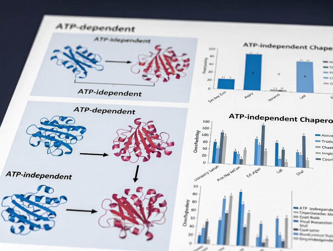
ATP vs. ATP-Independent Chaperones: Energy-Dependent and Energy-Neutral Protein Folding Mechanisms in Health and Disease
This article provides a comprehensive analysis of ATP-dependent and ATP-independent molecular chaperone systems, tailored for researchers and drug development professionals.
ATP vs. ATP-Independent Chaperones: Energy-Dependent and Energy-Neutral Protein Folding Mechanisms in Health and Disease
Abstract
This article provides a comprehensive analysis of ATP-dependent and ATP-independent molecular chaperone systems, tailored for researchers and drug development professionals. It explores the foundational biology of these protein homeostasis guardians, details cutting-edge methodologies for their study, discusses troubleshooting common experimental challenges, and offers a comparative validation of their mechanisms. The synthesis of this information aims to inform targeted therapeutic strategies against protein misfolding diseases, including neurodegenerative disorders and cancer, by highlighting the distinct vulnerabilities and opportunities presented by each chaperone class.
Chaperone Fundamentals: Defining ATP-Driven and Conformation-Driven Protein Folding Pathways
The Protein Folding Crisis and the Essential Role of Molecular Chaperones
The "protein folding crisis" refers to the fundamental cellular challenge that a significant portion of newly synthesized polypeptides and stress-denatured proteins fail to reach their native, functional conformations. This intrinsic inefficiency and susceptibility to misfolding is a primary source of toxic aggregates implicated in neurodegenerative diseases (e.g., Alzheimer's, Parkinson's) and other proteinopathies. Molecular chaperones are the essential cellular machinery that mitigates this crisis, facilitating de novo folding, preventing aggregation, disaggregating clumps, and directing irreversibly damaged proteins for degradation.
Current research is framed by a critical thesis: delineating the mechanisms, specific roles, and therapeutic potential of ATP-dependent chaperone systems versus ATP-independent chaperones (holdases). This distinction is central to developing targeted interventions—where ATP-dependent machines (e.g., Hsp70, Hsp90, AAA+ disaggregases) offer points for cycle modulation, ATP-independent holdases (e.g., small Hsps, Spy) provide immediate aggregation suppression.
Core Chaperone Systems: ATP-Dependent vs. ATP-Independent Mechanisms
ATP-Dependent Chaperones: Function as molecular machines where ATP binding/hydrolysis drives conformational changes essential for client protein binding, folding, or translocation.
- Hsp70 System (DnaK in E. coli): Central hub for de novo folding and refolding. Cycle regulated by co-chaperones (J-domain proteins stimulate ATPase; Nucleotide Exchange Factors promote ADP release).
- Hsp90 System: Matures late-folding clients (kinases, steroid hormone receptors). Involves a complex ATP-driven conformational clamp.
- AAA+ Disaggregases (e.g., Hsp104 in yeast, ClpB in bacteria): Use ATP hydrolysis to thread aggregated proteins through a central pore, disentangling them.
ATP-Independent Chaperones (Holdases): Stabilize unfolding clients by binding exposed hydrophobic patches, preventing aggregation. Activity is often regulated by stress-sensitive conditions (pH, temperature).
- Small Heat Shock Proteins (sHsps): Form dynamic oligomers that act as reservoirs for unfolded proteins, presenting them to ATP-dependent systems.
- Trigger Factor (Prokaryotes): Ribosome-associated chaperone that interacts with nascent chains without ATP.
Table 1: Comparative Analysis of Key Chaperone Systems
| Chaperone Class | Prototype | Energy Source | Primary Function | Key Co-chaperones/Regulators | Therapeutic Target Potential |
|---|---|---|---|---|---|
| Hsp70 System | DnaK (Hsp70) | ATP | De novo folding, refolding, translocation, prevention of aggregation | J-domain proteins (Hsp40), NEFs (GrpE, BAG) | High (Cancer, Neurodegeneration) |
| Hsp90 System | Hsp90 | ATP | Late-stage maturation & stabilization of client proteins ("clients") | Cochaperones (p23, Aha1, immunophilins) | High (Cancer, numerous inhibitors) |
| AAA+ Disaggregase | Hsp104/ClpB | ATP | Disaggregation of amyloids & stress granules, thermotolerance | Hsp70 system for full in vivo activity | High (Neurodegeneration) |
| sHSP Holdase | Hsp27 (Human), IbpA (E. coli) | None (ATP-independent) | Immediate binding of unfolded proteins, aggregation suppression | Oligomeric dynamics (pH/temp sensitive) | Medium (Prevent initial aggregation) |
| Ribosome-Associated | Trigger Factor | None | Nascent chain stabilization, co-translational folding | Ribosome (SecB in some pathways) | Low |
Detailed Experimental Protocols
Protocol 1: Measuring ATPase Activity of Hsp70 via NADH-Coupled Assay
- Objective: Quantify the ATP hydrolysis rate of an Hsp70 chaperone, modulated by client protein and J-domain protein.
- Reagents: Purified Hsp70, Hsp40 (J-domain protein), client protein (e.g., reduced, carboxymethylated α-lactalbumin), ATP, Phosphoenolpyruvate (PEP), Pyruvate Kinase, Lactate Dehydrogenase (LDH), NADH.
- Procedure:
- Prepare assay buffer (50 mM HEPES-KOH pH 7.5, 50 mM KCl, 10 mM MgCl₂).
- Add coupling system: 2 mM PEP, 0.2 mM NADH, 20 U/ml Pyruvate Kinase, 28 U/ml LDH.
- Initiate reaction with 2 mM ATP.
- Add experimental components: Hsp70 (1 µM) ± Hsp40 (0.5 µM) ± client protein (5 µM).
- Monitor absorbance at 340 nm (A₃₄₀) for 30-60 min at 25°C. NADH oxidation (decrease in A₃₄₀) is stoichiometric with ADP production.
- Calculate ATPase rate using NADH extinction coefficient (ε₃₄₀ = 6220 M⁻¹cm⁻¹).
Protocol 2: Assessing Holdase Activity via Aggregation Suppression Assay
- Objective: Visualize and quantify the ability of an ATP-independent chaperone (e.g., sHsp) to prevent client protein aggregation under stress.
- Reagents: Purified holdase (sHsp), client protein (e.g., citrate synthase, insulin), DTT (for insulin reduction), light scattering buffer.
- Procedure:
- Set up reaction mixtures containing client protein (e.g., 0.15 µM citrate synthase) in appropriate buffer.
- Add varying concentrations of the holdase chaperone (0-10 µM) to separate reactions.
- Induce aggregation: For citrate synthase, heat to 43°C; for insulin, add 20 mM DTT at 25°C.
- Immediately monitor aggregation by measuring light scattering (turbidity) at 320 or 360 nm for 60+ minutes.
- Control: Client protein alone (maximum aggregation). Reduced initial slope and final turbidity indicate holdase efficacy.
Protocol 3: Disaggregation/Refolding Assay for AAA+ Systems
- Objective: Measure the recovery of active enzyme from an aggregated state by an AAA+ disaggregase (e.g., Hsp104/ClpB) with the Hsp70 system.
- Reagents: Aggregated luciferase (heat-denatured), purified Hsp104, Hsp70 (DnaK), Hsp40 (DnaJ), NEF (GrpE), ATP-regenerating system.
- Procedure:
- Prepare aggregated firefly luciferase by heating at 42°C for 10 min.
- In refolding buffer, combine aggregated luciferase with the full chaperone system: Hsp70 (1 µM), Hsp40 (0.5 µM), NEF (0.2 µM), Hsp104 (0.5 µM).
- Initiate refolding with 2 mM ATP and an ATP-regenerating system (10 mM creatine phosphate, 20 µg/ml creatine kinase).
- Incubate at 25°C. At timed intervals, aliquot the reaction and measure recovered luciferase activity by adding its substrate (D-luciferin) and quantifying luminescence.
- Controls: Omit individual chaperone components (e.g., -Hsp104, -Hsp70 system) to delineate their contributions.
The Scientist's Toolkit: Key Research Reagent Solutions
Table 2: Essential Research Reagents for Chaperone Studies
| Reagent / Material | Function & Application | Example / Key Supplier |
|---|---|---|
| Recombinant Chaperone Proteins | Purified, active components for in vitro assays. | Human/yeast/E. coli Hsp70, Hsp90, Hsp104, sHsps (Sigma, Enzo, homemade). |
| ATP-Regenerating System | Maintains constant [ATP] in long assays, crucial for ATP-dependents. | Creatine Phosphate + Creatine Kinase; or Pyruvate Kinase + PEP. |
| ATPase Activity Assay Kits | Colorimetric/Malachite Green or coupled enzymatic assays for high-throughput screening. | Sigma-Aldrich ATPase Assay Kit (Colorimetric). |
| Thermal Shift Dyes | Monitor protein thermal stability (melting curve) with/without chaperones or drugs. | SYPRO Orange, Thermofluor dyes. |
| Client Proteins for Aggregation | Model substrates for folding/aggregation assays. | Citrate Synthase, Luciferase, Insulin, α-Lactalbumin. |
| Chaperone Inhibitors | Tool compounds for mechanistic and therapeutic studies. | VER-155008 (Hsp70), Geldanamycin/17-AAG (Hsp90), KUS (Hsp105). |
| Size-Exclusion Chromatography (SEC) | Analyze chaperone-client complex formation and oligomeric state. | Superose 6, Superdex 200 columns (Cytiva). |
| Surface Plasmon Resonance (SPR) Chips | Measure real-time kinetics of chaperone-client and chaperone-cochaperone interactions. | CMS Sensor Chip (Cytiva). |
Visualizations of Chaperone Pathways and Workflows
Diagram 1: ATP-Driven Hsp70 Chaperone Cycle
Diagram 2: Chaperone Network Response to Prot Folding Crisis
Diagram 3: Protocol for Aggregation Suppression Assay
Cellular protein homeostasis is maintained by chaperones, which are broadly categorized by their energy requirements. ATP-independent chaperones (e.g., small Hsps, trigger factor) primarily prevent aggregation through passive shielding. In contrast, ATP-dependent chaperones utilize nucleotide hydrolysis to drive conformational changes, enabling active folding, remodeling, disaggregation, and translocation of client proteins. This whitepaper focuses on three central ATP-dependent systems—Hsp70, Hsp90, and AAA+ ATPase machines—detailing their mechanisms, quantitative dynamics, and experimental interrogation. Research contrasting these with ATP-independent mechanisms is crucial for understanding proteostasis partitioning and for developing targeted therapeutics.
Core Machinery: Mechanisms and Quantitative Dynamics
Hsp70 (DnaK) System
Hsp70 chaperones bind hydrophobic peptide segments in an open, ATP-bound state with low affinity and fast exchange. ATP hydrolysis, stimulated by J-domain co-chaperones (e.g., Hsp40/DnaJ), induces a conformational shift to a closed, ADP-bound state with high client affinity. Nucleotide exchange factors (NEFs) catalyze ADP release, resetting the cycle. This cycle drives iterative "holdase" and "foldase" functions.
Table 1: Quantitative Parameters of Human Hsp70 (HSPA1A) Function
| Parameter | Value | Experimental Context |
|---|---|---|
| ATPase Rate (kcat) | 0.01 - 0.1 min-1 (basal) | Isothermal Titration Calorimetry (ITC) |
| ATPase Rate (stimulated) | 1 - 5 min-1 | With Hsp40 (DNAJB1) & client peptide |
| Kd for Client (ATP-state) | ~1 - 10 µM | Peptide library screening, SPR |
| Kd for Client (ADP-state) | ~0.1 - 0.5 µM | Fluorescence anisotropy |
| KM for ATP | 5 - 20 µM | Enzyme kinetics assay |
Hsp90 System
Hsp90 functions as a flexible dimer, orchestrating the late-stage maturation of "client" proteins (e.g., kinases, steroid receptors). Its ATP-driven conformational cycle is tightly regulated by co-chaperones. The cycle progresses from an open, apo-state through a series of intermediates to a closed, ATP-bound state that transiently dimerizes the N-terminal domains, facilitating client remodeling.
Table 2: Quantitative Parameters of Human Hsp90 (HSP90AA1) Function
| Parameter | Value | Experimental Context |
|---|---|---|
| ATPase Rate (kcat) | 0.5 - 1.0 min-1 (per dimer) | Malachite Green Phosphate Assay |
| KM for ATP | 50 - 150 µM | Enzyme kinetics assay |
| Client Activation Rate | Varies widely by client | Luciferase refolding/kinase activation assay |
| Effect of Inhibitor (Geldanamycin) IC50 | 10 - 50 nM | Cell viability/proteomics |
AAA+ ATPase Machines
AAA+ (ATPases Associated with diverse cellular Activities) proteins form ring-shaped hexamers that mechanically unfold and translocate substrate proteins through a central pore. Key families include Hsp104/ClpB (disaggregases), NSF (membrane fusion), and the proteasome regulatory particle. Power strokes from sequential ATP hydrolysis around the ring drive substrate threading.
Table 3: Quantitative Parameters of Key AAA+ Chaperones
| Parameter | Hsp104 (Yeast) | VCP/p97 (Human) | Experimental Context |
|---|---|---|---|
| ATPase Rate (kcat) | ~80 min-1 (per hexamer) | ~100 min-1 (per hexamer) | NADH-coupled ATPase assay |
| Unfolding/Translocation Rate | ~50 aa/s | N/A (extracts/remodels) | FRET-based unfolded assay |
| Step Size | 2 aa/ATP (per protomer) | N/A | Single-molecule optical tweezers |
| Cooperative ATP Binding | Positive (hexameric) | Positive (hexameric) | ITC & kinetic modeling |
Detailed Experimental Protocols
Protocol: Measuring Hsp70 ATPase Kinetics (Coupled Enzyme Assay)
Objective: Determine kcat and KM for ATP hydrolysis.
- Reagent Setup: Prepare Assay Buffer (40 mM HEPES-KOH pH 7.6, 50 mM KCl, 5 mM MgCl2). Prepare 10x ATP solution (0-10 mM in buffer). Prepare Coupling System: 2 mM phospho(enol)pyruvate (PEP), 0.2 mM NADH, 50 µg/mL pyruvate kinase (PK), 50 µg/mL lactate dehydrogenase (LDH).
- Reaction Assembly: In a 96-well plate, mix 80 µL of Assay Buffer containing 1 µM Hsp70, 1 µM Hsp40, and 10 µM model peptide substrate (e.g., NRLLLTG). Add 10 µL of ATP solution (varying concentration). Initiate reaction with 10 µL of Coupling System.
- Data Acquisition: Monitor absorbance at 340 nm (A340) for NADH consumption every 15 sec for 30 min at 30°C using a plate reader.
- Analysis: Calculate ATP hydrolysis rate from the linear slope of A340 decrease (ε340 NADH = 6220 M-1cm-1). Fit rates vs. [ATP] to the Michaelis-Menten equation using GraphPad Prism.
Protocol: Client Refolding Assay for Hsp90
Objective: Quantify Hsp90-dependent refolding of denatured client protein (e.g., firefly luciferase).
- Client Denaturation: Dilute purified luciferase to 1 µM in 25 mM HEPES pH 7.5, 5 mM DTT, 6 M guanidine-HCl. Incubate 30 min at 25°C.
- Refolding Reaction: Rapidly dilute denatured luciferase 100-fold into Refolding Buffer (40 mM HEPES-KOH pH 7.4, 50 mM KCl, 5 mM MgCl2, 2 mM ATP) containing 2 µM Hsp90, 2 µM Hsp70, 1 µM Hsp40, 1 µM Hop, and 3 µM p23.
- Kinetic Measurement: At time points (0, 5, 15, 30, 60, 90 min), remove 10 µL aliquot and mix with 50 µL luciferase assay reagent (Promega). Measure luminescence immediately with a luminometer.
- Controls: Include reactions lacking ATP, Hsp90, or with Hsp90 inhibitor (20 µM radicicol). Normalize activity to native luciferase control.
- Analysis: Plot % luciferase activity recovered vs. time. Fit data to a first-order exponential to determine the refolding rate constant.
Protocol: Single-Molecule Substrate Translocation by AAA+ Protease (ClpXP)
Objective: Visualize real-time, mechanical unfolding and translocation.
- Substrate Engineering: Engineer a substrate protein with an N-terminal ssrA degradation tag and a C-terminal tandem dye pair (e.g., Cy3-Cy5) for FRET.
- Surface Immobilization: Passivate a quartz microfluidic chamber with PEG-biotin. Incubate with 0.2 mg/mL NeutrAvidin. Anchor biotinylated substrate via a flexible linker.
- Data Acquisition: Use a TIRF microscope with alternating laser excitation (532 nm & 640 nm). Perfuse reaction buffer (50 mM Tris-HCl pH 7.5, 100 mM KCl, 10 mM MgCl2, 1 mM ATP, oxygen scavenger system) containing 100 nM ClpX hexamer.
- Analysis: Record FRET signal (Cy3 donor, Cy5 acceptor) over time. A high-to-low FRET transition indicates substrate unfolding/engagement. Subsequent processive translocation manifests as equidistant, stepwise changes in FRET. Analyze step dwell times and sizes using change-point algorithms (e.g., vbFRET).
Diagrams of Chaperone Pathways and Workflows
Diagram 1: Hsp70 ATPase Cycle and Client Interaction
Diagram 2: Hsp90 Chaperone Cascade for Client Maturation
Diagram 3: AAA+ Machine Unfolding and Translocation Mechanism
The Scientist's Toolkit: Research Reagent Solutions
Table 4: Essential Reagents for ATP-Dependent Chaperone Research
| Reagent | Function/Application | Example Product (Supplier) |
|---|---|---|
| Non-hydrolyzable ATP analogs (ATPγS, AMP-PNP) | Trap chaperones in specific nucleotide states for structural studies. | ATPγS, Sodium Salt (Sigma-Aldrich, A1388) |
| Hsp90 Inhibitors | Mechanistic probes and cancer therapeutic leads. | Geldanamycin (Cayman Chemical, 11415) |
| Fluorescent ATP analogs (e.g., N6-etheno-ATP) | Real-time monitoring of ATP binding and release. | 1,N6-Ethenoadenosine 5'-triphosphate (Sigma-Aldrich, 03907) |
| J-domain Peptide (Hsp40 mimetic) | Standardized stimulator for Hsp70 ATPase assays. | Recombinant Human DNAJB1 J-domain (Abcam, ab212896) |
| BAG Domain Protein (NEF) | Study nucleotide exchange in Hsp70 cycle. | Recombinant Human BAG1 (ProSpec, CHR-453) |
| Casein, FITC-labeled | Universal, unfolded model substrate for AAA+ proteases. | FITC-Casein (Thermo Fisher, C2990) |
| ATP Regeneration System (PK/LDH or CP/CK) | Maintain constant [ATP] in extended kinetic assays. | Pyruvate Kinase/Lactate Dehydrogenase from rabbit muscle (Sigma-Aldrich, P0294) |
| Malachite Green Phosphate Assay Kit | Colorimetric detection of inorganic phosphate release. | Malachite Green Phosphate Assay Kit (Sigma-Aldrich, MAK307) |
| Tetra-Cysteine Tag System (FlAsH/ReAsH) | Site-specific protein labeling for single-molecule FRET studies of conformational changes. | Lumio Green In-Cell Labeling Kit (Thermo Fisher, P30153) |
This whitepaper provides an in-depth technical examination of ATP-independent molecular chaperones, a critical class of proteins that maintain proteostasis under both normal and stress conditions. The discussion is framed within the broader research thesis comparing the mechanistic and functional paradigms of ATP-dependent versus ATP-independent chaperone systems. While ATP-dependent chaperones (e.g., Hsp70/DnaK, Hsp60/GroEL, Hsp90) utilize cycles of ATP binding and hydrolysis to actively fold substrates, ATP-independent chaperones function as "holdases" or "stabilizers." Their primary role is to prevent aggregation by binding to exposed hydrophobic regions of non-native client proteins, maintaining them in a folding-competent state until conditions permit refolding, often in cooperation with ATP-dependent foldases. This guide focuses on three archetypal groups: General Holdases (e.g., Hsp33, which is redox-regulated), Small Heat Shock Proteins (sHSPs), and the ribosome-associated Trigger Factor.
Core Mechanisms & Structural Biology
Small Heat Shock Proteins (sHSPs): These are a ubiquitous family of ATP-independent chaperones (~12-42 kDa) characterized by a conserved α-crystallin domain flanked by variable N- and C-terminal regions. They form dynamic, large oligomers (9 to >32 subunits) that act as reservoirs for substrate binding. Under stress, sHSPs undergo controlled dissociation, exposing hydrophobic surfaces to bind a wide array of unfolding clients. They do not refold substrates but hold them in a soluble, amyloid-like complex, preventing irreversible aggregation.
Holdases (e.g., Hsp33): Hsp33 is a well-studied redox-regulated chaperone activated by oxidative stress. It remains inactive in reducing conditions. Upon oxidation, zinc is released, and disulfide bonds form, triggering a conformational change that exposes a high-affinity hydrophobic substrate-binding site. This allows it to bind unfolded proteins promptly under conditions where ATP-dependent systems may be compromised.
Trigger Factor (TF): In bacteria, TF is a ribosome-associated chaperone that interacts with nascent polypeptide chains as they emerge from the ribosomal exit tunnel. It operates without ATP, providing a first line of defense against cytosolic aggregation by shielding hydrophobic segments. Its activity is coordinated with the translational machinery.
Quantitative Data Comparison
Table 1: Key Characteristics of Major ATP-Independent Chaperone Families
| Feature | Small HSPs (e.g., Hsp27, αB-crystallin) | Holdases (e.g., Hsp33) | Trigger Factor (TF) |
|---|---|---|---|
| Primary Function | Prevent aggregation; maintain solubility | Redox-regulated client binding & holdase activity | Nascent chain stabilization; prolyl isomerization |
| Regulatory Mechanism | Oligomeric dynamics (phosphorylation, pH, temp) | Redox-switch (disulfide bond formation) | Ribosome binding cycle |
| Typical Oligomeric State | Large, polydisperse oligomers (9-40 subunits) | Dimer (inactive) → monomer/dimer (active) | Monomer |
| Key Structural Domain | α-Crystallin domain | Zinc-binding domain & linker region | Peptidyl-prolyl cis/trans isomerase (PPIase) domain |
| Substrate Specificity | Broad, hydrophobic surfaces | Broad, hydrophobic surfaces (oxidation-unfolded) | Nascent chains (≥100 aa), hydrophobic regions |
| Cooperation with ATP-Systems | Transfers clients to Hsp70/DnaK and Hsp60/GroEL | Transfers clients to DnaK/J (Hsp70/40) system | Cooperates with DnaK/J-GrpE and GroEL/ES |
| Reported Kd for Client Binding | Low µM range (e.g., ~1-5 µM for αB-crystallin with βL-crystallin) | nM to µM range upon activation | Not applicable; operates co-translationally |
Table 2: Comparison of Key Experimental Parameters in Functional Assays
| Assay Type | Typical Substrate (Model Client) | Key Readout | Parameters for sHSPs/Holdases | Parameters for Trigger Factor |
|---|---|---|---|---|
| Aggregation Suppression | Citrate Synthase (CS), Insulin, MDH | Light Scattering (OD 360 nm) | 0.1-1 µM chaperone, 0.1-0.5 µM client, 43°C (CS) | 0.5-2 µM TF, 0.25 µM Luciferase, 42°C |
| Holdase Activity (Filter Trap) | Chemically denatured Luciferase | Retained aggregates on cellulose acetate filter | 2 µM chaperone, 50 nM denatured luc, 25°C incubation | Not commonly used for TF |
| Chaperone-Client Complex Analysis | Fluorescently labeled α-lactalbumin | Size-Exclusion Chromatography (SEC) or Native PAGE | 10 µM chaperone, 5 µM client, 37°C, 30 min | 5 µM TF, 5 µM RNCs (Ribosome-Nascent Chains), 4°C |
| Isothermal Titration Calorimetry (ITC) | Peptide (e.g., WFI/P) | Binding enthalpy (∆H), Kd | 50 µM peptide in syringe, 5 µM chaperone in cell, 25°C | 100 µM peptide in syringe, 10 µM TF in cell, 25°C |
Detailed Experimental Protocols
Protocol 4.1: Light Scattering Assay for Aggregation Suppression
- Objective: Quantify the ability of an ATP-independent chaperone to prevent the heat- or chemical-induced aggregation of a model substrate.
- Materials: Purified chaperone (e.g., Hsp27, Hsp33), model client (e.g., Citrate Synthase), aggregation buffer (e.g., 40 mM HEPES-KOH, pH 7.5), spectrophotometer with thermostatted cuvette holder.
- Procedure:
- Prepare chaperone samples in aggregation buffer at 2x the final desired concentration (e.g., 2 µM).
- Prepare the client protein (Citrate Synthase) in the same buffer at 2x final concentration (e.g., 0.6 µM).
- Pre-incubate the chaperone sample (or buffer control) in a quartz cuvette at the assay temperature (e.g., 43°C) for 5 minutes in the spectrophotometer.
- Initiate aggregation by rapidly adding an equal volume of pre-warmed client protein to the cuvette, mixing quickly.
- Immediately start monitoring light scattering by recording the optical density at 360 nm (OD₃₆₀) every 10-15 seconds for 30-60 minutes.
- The initial slope and final plateau of the scattering curve are indicators of aggregation kinetics and total aggregated mass, respectively. Compare curves with/without chaperone.
Protocol 4.2: Redox Activation of Hsp33 and Client Binding Assay
- Objective: Activate the holdase function of Hsp33 via oxidation and assess client binding.
- Materials: Reduced, zinc-bound Hsp33, reducing agent (DTT), oxidant (H₂O₂ or diamide), model client (e.g., chemically denatured Luciferase), non-reducing SDS-PAGE gel.
- Procedure:
- Activation: Incubate 10 µM reduced Hsp33 with 2 mM H₂O₂ (or 5 mM diamide) in buffer (e.g., 50 mM HEPES, pH 7.5, 50 mM KCl) at 30°C for 30-60 minutes.
- Quenching: Remove excess oxidant by buffer exchange using a desalting column or repeated centrifugal concentration.
- Client Denaturation: Denature 5 µM Luciferase in 6 M Guanidine-HCl for 1 hour at 25°C.
- Binding Reaction: Rapidly dilute the denatured Luciferase 100-fold into a solution containing 2 µM oxidized (or reduced control) Hsp33. This initiates refolding/aggregation. Incubate at 25°C for 10 minutes.
- Analysis: Analyze the mixture via non-reducing SDS-PAGE. Client binding is often indicated by co-migration of the client with Hsp33 in the stacking gel or high molecular weight region, as stable complexes survive sample preparation. A filter trap assay can also be used as a complementary readout.
Protocol 4.3: Co-Translational Binding Assay for Trigger Factor
- Objective: Demonstrate TF binding to nascent polypeptide chains on ribosomes.
- Materials: E. coli PURExpress in vitro transcription-translation system, DNA template encoding a protein of interest with a C-terminal affinity tag (e.g., His₆), ³⁵S-Methionine, purified Trigger Factor, anti-TF antibody, Ni-NTA beads.
- Procedure:
- Set up a translation stall by using a DNA template lacking a stop codon to produce Ribosome-Nascent Chain complexes (RNCs). Perform the reaction in PURExpress mix supplemented with ³⁵S-Met according to manufacturer's instructions. Incubate at 37°C for 20 min.
- Isolate RNCs by centrifugation through a high-salt sucrose cushion.
- Incubate purified RNCs with or without excess purified TF (e.g., 5 µM) in binding buffer (50 mM HEPES-KOH, pH 7.5, 150 mM KOAc, 10 mM Mg(OAc)₂) for 15 min at 4°C.
- Capture RNCs via the nascent chain's affinity tag using Ni-NTA beads.
- Wash beads thoroughly and elute bound material. Analyze the eluate by SDS-PAGE and autoradiography. Co-precipitation of TF (detectable by Western blot) with the radiolabeled RNCs indicates specific binding.
Visualizations
Diagram 1: ATP-Independent vs Dependent Chaperone Functional Axis
Diagram 2: Redox Activation Mechanism of Hsp33 Holdase
Diagram 3: sHSP Oligomeric Dynamics & Substrate Sequestration
The Scientist's Toolkit: Research Reagent Solutions
Table 3: Essential Reagents for Studying ATP-Independent Chaperones
| Reagent/Material | Function & Application | Example Product/Source |
|---|---|---|
| Recombinant Chaperone Proteins | Purified sHSPs, Hsp33, Trigger Factor for in vitro assays. Critical for functional studies. | Express in E. coli or purchase from recombinant protein vendors (e.g., Abcam, StressMarg). |
| Model Client/Substrate Proteins | Well-characterized proteins prone to aggregation (Citrate Synthase, Malate Dehydrogenase, Insulin, Luciferase). Used in aggregation suppression assays. | Sigma-Aldrich (Citrate Synthase, Insulin), Promega (Luciferase). |
| Chemical Chaperones / Denaturants | Guanidine HCl and Urea for client denaturation; DTT and H₂O₂/Diamide for redox regulation studies of Hsp33. | Thermo Fisher Scientific, Sigma-Aldrich. |
| Size-Exclusion Chromatography (SEC) Columns | Analyze oligomeric state of sHSPs and chaperone-client complexes under native conditions. | Superdex 200 Increase, Superose 6 (Cytiva). |
| Crosslinking Reagents | Capture transient interactions, stabilize sHSP oligomers or chaperone-client complexes for analysis. | BS³, DSS (homobifunctional NHS-esters), Thermo Fisher. |
| In Vitro Translation System | Study co-translational chaperone function (e.g., Trigger Factor). Generate radiolabeled nascent chains. | PURExpress (NEB), E. coli S30 Extract. |
| Anti-Chaperone Antibodies | Detect endogenous chaperones, perform co-immunoprecipitation (Co-IP), Western blotting. | Commercial antibodies for Hsp27 (Enzo), αB-crystallin (Cell Signaling), Trigger Factor (in-house common). |
| Specialized Buffers & Cofactors | Zinc chloride (for Hsp33 activity), specific ATP-removal systems (apyrase) for strict ATP-independent validation. | Sigma-Aldrich. |
| Fluorescent Dyes (ANS, Bis-ANS) | Probe hydrophobic surface exposure, a key indicator of chaperone activation and client binding. | Sigma-Aldrich, Thermo Fisher. |
This whitepaper elucidates the core mechanistic divergence in protein homeostasis, contrasting ATP-dependent (energy-coupled) and ATP-independent (passive) chaperone systems. The central thesis posits that these represent two fundamental paradigms for managing protein folding, aggregation, and disaggregation, with profound implications for cellular stress response, disease pathology (e.g., neurodegenerative diseases, cancer), and therapeutic intervention. Energy-coupled cycles, exemplified by Hsp70, Hsp90, and AAA+ disaggregases like Hsp104, utilize ATP hydrolysis to drive conformational changes and perform mechanical work on client proteins. Passive systems, including small heat shock proteins (sHsps) and holdases, rely on selective binding and surface effects to shield hydrophobic regions, preventing aggregation without active remodeling.
Core Mechanistic Principles
ATP-Dependent, Energy-Coupled Cycles
These systems function as molecular machines. ATP binding and hydrolysis are coupled to precise, cyclic conformational changes in the chaperone. This energy input allows for:
- Active unfolding/Refolding: Application of mechanical force to disentangle misfolded aggregates or unfold misfolded domains.
- Directed Allostery: Controlled binding and release of client proteins, often regulated by cochaperones and nucleotide exchange factors.
- High Specificity & Regulation: The cycle can be tuned via nucleotide state (ATP, ADP, apo) and cochaperone interaction, allowing for temporal control.
ATP-Independent, Passive Binding & Surface Effects
These systems operate via equilibrium thermodynamics, providing a rapid, first-line defense:
- Kinetic Stabilization: High-capacity, multivalent binding to exposed hydrophobic surfaces on non-native clients, effectively raising the energy barrier for aggregation.
- Formation of Storage Complexes: sHsps form dynamic, polydisperse oligomers that encapsulate clients, keeping them in a folding-competent state for later ATP-dependent processing.
- Surface Activity: Acts as "surfactants" for proteins, coating aggregation-prone interfaces without consuming cellular energy.
Quantitative Comparison of Key Systems
Table 1: Core Characteristics of Representative Chaperone Mechanisms
| Feature | ATP-Dependent (Hsp70 System) | ATP-Dependent (AAA+ Disaggregase, Hsp104) | ATP-Independent (Small HSP, αB-Crystallin) |
|---|---|---|---|
| Energy Source | ATP hydrolysis (~50 kJ/mol per cycle) | ATP hydrolysis (hexamer, ~300 kJ/mol) | None (passive) |
| Core Action | Peptide binding/release cycle; partial unfolding | Threading client through central pore; mechanical pulling | Surface coating; kinetic trapping |
| Typical Stoichiometry | 1:1 (Chaperone:Client peptide) but processive | Hexameric ring (6:1 or client engagement) | Large oligomer (24-32 subunits : many clients) |
| Key Rate Constants | (k{hyd}) (ATP→ADP): ~0.02 min⁻¹; (k{ex}) (ADP release): ~1.0 min⁻¹ | ATPase rate: ~100 min⁻¹ per protomer; Translocation: ~50 aa/s | Association ((k{on})): diffusion-limited; Dissociation ((k{off})): slow (min-hr) |
| Functional Role | Foldase, translocation, prevention | Disaggregase, reactivation | Holdase, storage |
| Aggregate Disassembly | Yes (with cochaperones like DnaJB1 & Hsp110) | Yes (direct, powerful) | No (requires transfer to ATP-system) |
Table 2: Experimental Readouts Differentiating the Mechanisms
| Experimental Assay | Energy-Coupled Cycle Signature | Passive Binding Signature |
|---|---|---|
| ATPase Activity Assay | Stimulated by client/cochaperone. Michaelis-Menten kinetics observed. | No ATPase activity detected. |
| Single-Molecule FRET (smFRET) | Discrete, stepwise client conformational changes synchronized with ATP cycles. | Static or slow, stochastic FRET fluctuations. |
| Aggregation Light Scattering | Reduction in scattering over time (active disaggregation). | Immediate suppression of scattering increase (prevention). |
| Isothermal Titration Calorimetry (ITC) | Exothermic/endothermic peaks coupled to nucleotide state. | Simple binding isotherm; no nucleotide effect. |
Detailed Experimental Protocols
Protocol: Measuring ATPase Activity Stimulation (Hsp70 System)
Objective: Quantify the coupling efficiency between client binding and ATP hydrolysis. Reagents: Purified Hsp70 (DnaK), DnaJ cochaperone (Hsp40), model client (e.g., reduced, carboxymethylated α-lactalbumin, RCMLA), ATP, NADH, phosphoenolpyruvate (PEP), pyruvate kinase/lactate dehydrogenase (PK/LDH) enzyme mix. Procedure:
- Prepare reaction buffer (40 mM HEPES-KOH pH 7.6, 50 mM KCl, 5 mM MgCl₂).
- Set up a coupled enzymatic assay: ATP hydrolysis is linked to NADH oxidation, monitored at A₃₄₀.
- In a 96-well plate, mix: 2 µM Hsp70, 0-10 µM RCMLA, 0.4 µM DnaJ, 2 mM ATP, 0.2 mM NADH, 1 mM PEP, 10 U/ml PK/LDH.
- Initiate reaction with ATP. Monitor A₃₄₀ decrease at 30°C for 30 minutes.
- Calculate ATP hydrolysis rate from the linear slope (ε₃₄₀(NADH) = 6220 M⁻¹cm⁻¹). Interpretation: Increased negative slope with client/J-protein confirms energy coupling.
Protocol: Aggregation Suppression Assay (Passive Holdase Activity)
Objective: Distinguish passive prevention from active disaggregation. Reagents: Target aggregation-prone protein (e.g., citrate synthase, CS), holdase (e.g., αB-crystallin), ATP (as control), thermostatted spectrophotometer. Procedure:
- Prepare CS at 0.15 µM in 40 mM HEPES-KOH pH 7.5.
- Pre-incubate CS with or without 0.3 µM (oligomer) αB-crystallin for 10 min at 25°C.
- Split each mixture into two cuvettes. To one, add ATP (2 mM final). The other receives buffer.
- Induce thermal aggregation by rapidly shifting temperature to 43°C.
- Monitor light scattering at 360 nm for 60 minutes. Interpretation: Immediate suppression of scattering by αB-crystallin, unaffected by ATP, confirms passive holdase activity. An ATP-dependent decrease would indicate contamination/activation of an energy-coupled system.
Protocol: Single-Molecule Disaggregase Pulling Assay
Objective: Visualize direct mechanical work by an AAA+ chaperone. Reagents: Surface-immobilized, polyprotein client with fluorescent handles (e.g., tandem repeats of a protein domain like I27), purified, fluorescently labeled AAA+ hexamer (e.g., ClpB/Hsp104), oxygen scavenging system, TIRF microscope. Procedure:
- Construct a DNA handle-linked polyprotein client and tether it to a functionalized coverslip.
- Image fluorescent spots using TIRF microscopy in imaging buffer with 2 mM ATP.
- Inject fluorescently labeled AAA+ chaperone and record movies.
- Analyze time traces of fluorescence intensity and FRET between client handles. Interpretation: Sudden, stepwise changes in fluorescence/FRET coinciding with chaperone binding demonstrate processive, mechanical unfolding driven by ATP hydrolysis.
The Scientist's Toolkit: Research Reagent Solutions
Table 3: Essential Reagents for Mechanistic Chaperone Studies
| Reagent | Function/Description | Example Use Case |
|---|---|---|
| Non-hydrolyzable ATP analogs (AMP-PNP, ATPγS) | Traps chaperone in ATP-bound conformation; dissects cycle steps. | Distinguishing ATP binding effects from hydrolysis in structural studies. |
| Fluorescent ATP analogs (e.g., Mant-ATP) | Reports on nucleotide binding and dissociation kinetics via FRET/fluorescence change. | Measuring nucleotide exchange rates in Hsp70 systems. |
| Photo-crosslinkable Amino Acids (Bpa) | Site-specific, UV-induced crosslinking to capture transient chaperone-client interactions. | Mapping binding interfaces during different stages of the ATPase cycle. |
| Aggregation-Sensitive Dyes (e.g., Thioflavin T, SYPRO Orange) | Report on formation of amyloid fibrils or exposed hydrophobic surfaces. | High-throughput screening for chaperone inhibitors or enhancers. |
| FRET-Optimized Client Proteins | Engineered with donor/acceptor pairs to report on conformational state. | Single-molecule or bulk analysis of unfolding/refolding kinetics. |
| Nucleotide Exchange Factor (NEF) / Cochaperone Proteins (e.g., Bag1, Hsp110, Hsp40s) | Essential modulators of ATPase cycle timing and client specificity. | Reconstituting complete functional cycles in vitro. |
Visualizations of Mechanisms & Workflows
Cellular Niches and Primary Functions of Each Chaperone Class
This technical guide details the cellular localization and functional roles of molecular chaperone classes. The analysis is framed within a critical thesis in proteostasis research: the comparative study of ATP-dependent versus ATP-independent chaperone mechanisms. Understanding the specific niches of each class is fundamental to deciphering how these two mechanistic paradigms cooperate, compete, or specialize to maintain proteome integrity across cellular compartments. This distinction has profound implications for developing targeted therapeutics for protein misfolding diseases, cancer, and neurodegeneration.
Molecular chaperones are classified based on structure, mechanism, and cellular localization. Their primary function is to prevent aggregation and facilitate the proper folding, assembly, transport, and degradation of client proteins.
Table 1: Chaperone Classes, Their Cellular Niches, and Primary Functions
| Chaperone Class / Complex | Key Members (Examples) | Primary Cellular Niche(s) | Core Function(s) | ATP Dependence |
|---|---|---|---|---|
| Hsp70 System | Hsp70 (DnaK), Hsp40 (J-proteins), NEFs (GrpE, BAG) | Cytosol, Nucleus, Mitochondrial matrix, ER Lumen (BiP) | De novo folding, translocation across membranes, prevention of aggregation, disaggregation (with Hsp100), regulation of folding intermediates. | ATP-dependent (Hsp70 ATPase cycle regulated by co-chaperones) |
| Hsp60 Chaperonins | GroEL/GroES (prokaryotes), TRiC/CCT (eukaryotic cytosol), Hsp60/Hsp10 (mitochondria) | Bacterial cytosol, Eukaryotic cytosol (TRiC), Mitochondrial matrix | Encapsulation-assisted folding of small, obligate clients (~30-60 kDa) within an isolated cage; folds proteins that cannot fold via Hsp70. | ATP-dependent (ATP hydrolysis drives conformational changes and cage cycling) |
| Hsp90 System | Hsp90, Co-chaperones (p23, Aha1, Hop, Cdc37) | Cytosol, Nucleus, ER-associated | Maturation and activation of metastable "client" proteins (e.g., kinases, steroid receptors, transcription factors); involved in signal transduction. | ATP-dependent (ATPase-driven conformational clamping cycle) |
| Small Heat Shock Proteins (sHsps) | αA- and αB-Crystallin, Hsp27, HspB5 | Cytosol, Nucleus, Mitochondria, | ATP-independent aggregation suppression. Form dynamic, large oligomers that bind unfolding clients, holding them in a refolding-competent state for ATP-dependent chaperones. | ATP-independent (Holdases) |
| ATP-independent Holders/Folders | Spy, Skp, Trigger Factor (TF) | Bacterial cytosol (periplasm for Skp), Ribosome-associated (TF) | Co-translational folding (TF), periplasmic chaperoning (Skp), prevention of aggregation for specific clients (Spy). Often specialized for local environments. | ATP-independent (Intrinsic folding energy or passive binding) |
| Disaggregases | Hsp100 (ClpB in bacteria, Hsp104 in yeast), Hsp70-Hsp40-NEF system | Cytosol, Nucleus | Disassembly of protein aggregates and fibrils. Hsp100 machines thread clients through a pore, feeding disentangled polypeptides to Hsp70 for refolding. | ATP-dependent (Requires ATP hydrolysis for mechanical unfolding/threading) |
| Nucleoplasmins | NPM1, Nucleophosmin | Nucleolus, Nucleus | Chaperone for histone assembly, ribosomal biogenesis, genome stability. Prevents aggregation in the dense nucleolar environment. | Largely ATP-independent |
| ER-Resident Chaperones | BiP (Hsp70), Grp94 (Hsp90), Calnexin/Calreticulin, Protein Disulfide Isomerase (PDI) | Endoplasmic Reticulum Lumen | Glycoprotein folding & quality control (Calnexin/Calreticulin cycle), disulfide bond formation (PDI), general folding & ERAD targeting (BiP). | Mixed: Calnexin cycle is ATP-independent; BiP/Grp94 are ATP-dependent. |
Key Thesis Insight: The cellular niche often dictates the mechanistic requirement. ATP-dependent systems (Hsp70, Hsp60, Hsp90) dominate in compartments requiring active, regulated, and iterative conformational remodeling. ATP-independent chaperones (sHsps, some holders) are crucial in stressful or constrained environments (cytosol during heat shock, periplasm, nucleolus) or for rapid, co-translational interactions, providing a first line of defense by sequestering clients until ATP-dependent resources are available.
Experimental Protocols for Key Studies
Protocol: Differentiating ATP-Dependent vs. Independent Holdase Activity (sHsps vs. Hsp70)
Objective: To quantify the ability of a chaperone to suppress client protein aggregation in the presence or absence of ATP.
Methodology (Based on Light Scattering Assay):
- Reagents: Purified chaperone (e.g., αB-Crystallin or Hsp70/Hsp40), client protein (e.g., Citrate Synthase or Luciferase), ATP regeneration system (ATP, Creatine Phosphate, Creatine Kinase), reaction buffer.
- Aggregation Induction: Client protein is chemically denatured (e.g., with Guanidine HCl) or thermally denatured (heated to 43-45°C).
- Experimental Setup: In a cuvette, mix:
- Control: Client protein + buffer.
- Test 1 (ATP-independent): Client protein + sHsp chaperone + buffer.
- Test 2 (ATP-dependent): Client protein + Hsp70 system (Hsp70, Hsp40, NEF) +/- ATP regeneration system.
- Measurement: Aggregation is monitored in real-time by measuring light scattering (turbidity) at 320-360 nm using a spectrophotometer with a temperature-controlled cuvette holder.
- Data Analysis: The initial slope or plateau of the scattering curve indicates aggregation kinetics/capacity. sHsps suppress aggregation independently of ATP. The Hsp70 system shows suppression only in the presence of ATP and co-chaperones.
Protocol: Assessing Chaperonin Cage-Mediated Folding (GroEL/ES)
Objective: To demonstrate the de novo folding of an obligate chaperonin client inside the GroEL/ES cage.
Methodology (Based on Refolding of Denatured MDH):
- Reagents: GroEL, GroES, denatured client (e.g., Mitochondrial Malate Dehydrogenase, MDH), ATP, ATP-regeneration system, assay buffer.
- Client Denaturation: MDH is fully denatured in 6M Guanidine HCl.
- Refolding Reaction:
- Control: Dilute denatured MDH into refolding buffer (leads to aggregation/no activity).
- + GroEL: Dilute denatured MDH into buffer containing GroEL. MDH binds to GroEL's apical domains but does not fold.
- + GroEL/ES/ATP: Dilute denatured MDH into buffer containing GroEL, then add GroES and ATP to initiate the folding cycle.
- Folding Assessment: After incubation (15-30 mins, 25°C), measure recovered enzymatic activity of MDH via its specific spectrophotometric assay (NADH oxidation at 340 nm).
- Interpretation: Significant activity recovery only in the GroEL/ES/ATP condition demonstrates the requirement for the encapsulated, ATP-dependent folding cycle.
Visualizations (Graphviz DOT)
Diagram 1: ATP-Dependent vs. Independent Chaperone Pathways
Diagram 2: Key Experimental Workflow for Chaperone Mechanism Study
The Scientist's Toolkit: Key Research Reagent Solutions
Table 2: Essential Reagents for Chaperone Mechanism Research
| Reagent / Material | Supplier Examples | Function in Experimentation |
|---|---|---|
| Recombinant Chaperone Proteins (Hsp70, Hsp40, GroEL/ES, Hsp90, sHsps) | Sigma-Aldrich, Enzo Life Sciences, Assay Designs, in-house purification | Purified, active components for in vitro reconstitution of chaperone functions and mechanistic studies. |
| Thermolabile Client Proteins (Citrate Synthase, Luciferase, MDH) | Sigma-Aldrich, Promega | Standardized, well-characterized substrates for aggregation suppression and refolding assays. |
| ATP Regeneration System (ATP, Creatine Phosphate, Creatine Kinase) | Roche, Sigma-Aldrich, Thermo Fisher | Maintains constant [ATP] during long kinetic experiments, crucial for studying ATP-dependent cycles. |
| ANS (1-Anilinonaphthalene-8-sulfonate) | Thermo Fisher, Sigma-Aldrich | Hydrophobic fluorescent dye used to monitor client protein unfolding or exposure of hydrophobic patches on chaperones. |
| Native Gel Electrophoresis Kits (e.g., NativePAGE) | Thermo Fisher | Analyze intact, non-denatured chaperone-client complexes and their oligomeric states. |
| Size Exclusion Chromatography (SEC) Columns (e.g., Superose, Superdex) | Cytiva | Separate and analyze the size distribution of chaperone oligomers and chaperone-client complexes. |
| Real-Time PCR Thermocyclers with FRET capabilities | Bio-Rad, Thermo Fisher | Monitor protein aggregation (light scattering) or conformational changes (FRET-based biosensors) in real-time. |
| Proteostat or Thioflavin T (ThT) | Enzo Life Sciences, Sigma-Aldrich | Fluorescent dyes for specific detection of aggregated/amyloid structures in cellular or biochemical assays. |
| ATPase/GTPase Activity Assay Kits (Colorimetric) | Sigma-Aldrich, Abcam | Quantify the ATP hydrolysis rates of chaperones like Hsp70 or Hsp90, a key functional metric. |
| Chaperone-Specific Inhibitors (VER-155008 (Hsp70), Radicicol/Geldanamycin (Hsp90), JG-98 (Hsp70)) | Tocris, Sigma-Aldrich | Pharmacological tools to disrupt specific chaperone functions in cells, linking mechanism to phenotype. |
Investigating Chaperone Action: From In Vitro Assays to In Vivo Drug Discovery
This whitepaper details three core biochemical assays fundamental to dissecting ATP-dependent versus ATP-independent chaperone mechanisms. Understanding these mechanisms is critical for elucidating protein homeostasis in health and disease, informing therapeutic strategies for conditions like neurodegeneration and cancer.
ATPase Activity Assay
Purpose: Quantifies the ATP hydrolysis rate of a chaperone, a direct measure of its ATP-dependent enzymatic function. This assay distinguishes ATP-consuming chaperones (e.g., Hsp70, Hsp90) from ATP-independent ones (e.g., small Hsps, trigger factor).
Detailed Protocol: Colorimetric Phosphate Release Assay
Principle: Measures inorganic phosphate (Pi) released from hydrolyzed ATP using a malachite green reagent.
- Reaction Setup: Prepare a 50-100 µL reaction containing:
- Assay Buffer: 20-50 mM HEPES, pH 7.4, 50-100 mM KCl, 5-10 mM MgCl₂.
- ATP: 1-5 mM final concentration.
- Chaperone: 0.1-2 µM purified protein.
- Optional: Client protein or co-chaperone (e.g., 1-5 µM J-domain protein for Hsp70).
- Incubation: Incubate at 30-37°C for 0, 5, 10, 20, and 30 minutes.
- Reaction Stop & Detection: At each time point, transfer 10-25 µL to a well containing 100-150 µL of malachite green reagent (0.034% malachite green, 1.05% ammonium molybdate, 1 M HCl). Incubate for 10-30 minutes at room temperature.
- Measurement: Read absorbance at 620-650 nm.
- Calculation: Generate a standard curve using known KH₂PO₄ concentrations. Calculate the rate of Pi release (nmol/min/µg chaperone).
Table 1: Comparative ATPase Activity of Chaperone Systems
| Chaperone System | Basal ATPase Rate (min⁻¹) | Stimulated ATPase Rate (min⁻¹) | Stimulus | Primary Mechanism |
|---|---|---|---|---|
| DnaK (Hsp70) | 0.02 - 0.05 | 0.5 - 2.0 | J-domain protein + peptide | ATP-Dependent |
| Hsp90 | 0.01 - 0.03 | 0.1 - 0.3 | Client + co-chaperone (Aha1) | ATP-Dependent |
| GroEL (Hsp60) | 0.05 - 0.15 | 10 - 20 | GroES encapsulation | ATP-Dependent |
| Hsp33 | Not Detectable | Not Detectable | N/A | ATP-Independent (Oxidation) |
| αB-Crystallin | Not Detectable | Not Detectable | N/A | ATP-Independent (Holdase) |
Visualization: ATPase Cycle of a Generic ATP-Dependent Chaperone
Title: ATPase Cycle of a Generic ATP-Dependent Chaperone
Luciferase Refolding Assay
Purpose: A functional assay measuring the ability of chaperones to renature a chemically denatured substrate (firefly luciferase), directly assessing foldase activity.
Detailed Protocol: Chaperone-Assisted Luciferase Reactivation
- Luciferase Denaturation: Dilute purified firefly luciferase (5 µM) into denaturation buffer (6 M guanidine-HCl, 30 mM HEPES-KOH pH 7.4, 50 mM KCl) to 2 µM. Incubate at 25°C for 60 minutes.
- Refolding Reaction: Rapidly dilute denatured luciferase 100-fold into refolding buffer (30 mM HEPES-KOH pH 7.4, 50 mM KCl, 5 mM MgCl₂, 2 mM DTT, 1-5 mM ATP if required) containing the test chaperone system (e.g., 1-5 µM Hsp70, 1 µM Hsp40, 1-2 µM NEF). Include controls: no chaperone (negative), native luciferase (100% control).
- Incubation: Incubate at 25-30°C. Withdraw aliquots at various time points (e.g., 0, 15, 30, 60, 120 minutes).
- Activity Measurement: Mix aliquot with luciferase assay reagent (containing luciferin, ATP, Mg²⁺). Measure luminescence immediately.
- Data Analysis: Express recovered luminescence as a percentage of native luciferase activity. Plot % activity vs. time.
Table 2: Luciferase Refolding by Different Chaperone Systems
| Chaperone System | ATP Required? | Max % Recovery (at 60 min) | Half-time of Recovery (t₁/₂, min) | Functional Class |
|---|---|---|---|---|
| Hsp70 + Hsp40 + NEF | Yes | 60 - 80% | 15 - 25 | ATP-Dependent Foldase |
| GroEL + GroES + ATP | Yes | 70 - 90% | 10 - 20 | ATP-Dependent Foldase |
| Hsp90 + Co-chaperones | Yes | 20 - 40% | 40 - 60 | ATP-Dependent Maturase |
| Hsp33 | No | < 5% | N/A | ATP-Independent Holdase |
| αB-Crystallin | No | < 5% | N/A | ATP-Independent Holdase |
Visualization: Luciferase Refolding Experimental Workflow
Title: Experimental Workflow for Luciferase Refolding Assay
Client Co-Immunoprecipitation (Co-IP)
Purpose: Captures direct physical interactions between a chaperone and its client protein, often under different nucleotide or stress conditions, to assess binding dependency.
Detailed Protocol: Magnetic Bead-Based Co-IP
- Lysis & Pre-clearing: Lyse cells expressing tagged chaperone (e.g., FLAG-Hsp70) and client in mild lysis buffer (40 mM HEPES pH 7.4, 100 mM KCl, 5 mM MgCl₂, 0.5% NP-40, 1 mM DTT, protease inhibitors). Pre-clear lysate with control IgG beads for 30 minutes at 4°C.
- Binding Conditions: Aliquot lysate. Treat with ATP (5 mM), ADP (5 mM), or non-hydrolyzable ATP analogue (ATPγS, 5 mM) for 15 minutes on ice.
- Immunoprecipitation: Incubate each aliquot with anti-FLAG magnetic beads for 1-2 hours at 4°C with gentle rotation.
- Washing: Wash beads 3-4 times with ice-cold wash buffer (identical to lysis buffer but with 0.1% NP-40 and respective nucleotide if desired).
- Elution & Analysis: Elute proteins with 2X Laemmli buffer containing 5% β-mercaptoethanol. Boil samples. Analyze by SDS-PAGE and immunoblotting for the chaperone tag and putative client protein.
Table 3: Effect of Nucleotides on Chaperone-Client Co-IP
| Chaperone-Client Pair | Binding in ATP | Binding in ADP | Binding in ATPγS | Interpretation |
|---|---|---|---|---|
| Hsp70 - Tau protein | Weak / None | Strong | Strong | ATP hydrolysis releases client; ADP state has high affinity. |
| Hsp90 - Kinase Client | Intermediate | Strong | Strong | ATP-bound state dynamic; stable in ADP/ATPγS locked state. |
| GroEL - Unfolded Rubisco | Strong | Strong | Strong | ATP binding not strictly required for initial hydrophobic binding. |
| αB-Crystallin - β-Amyloid | Strong | Strong | Strong | ATP-independent binding (constitutively bound holdase). |
Visualization: Co-IP Strategy for Chaperone-Client Interaction Analysis
Title: Co-IP Strategy for Chaperone-Client Interaction Analysis
The Scientist's Toolkit: Essential Research Reagents
Table 4: Key Reagent Solutions for Chaperone Mechanism Studies
| Reagent / Material | Function / Purpose | Example in Assays |
|---|---|---|
| Malachite Green Reagent | Colorimetric detection of inorganic phosphate (Pi). | Quantifying Pi release in ATPase assays. |
| Firefly Luciferase | Model substrate for refolding assays. Sensitive reporter of native conformation. | Denatured client in functional refolding assays. |
| Non-hydrolyzable ATP Analogues (e.g., ATPγS, AMP-PNP) | Lock chaperones in specific nucleotide-bound states for mechanistic studies. | Co-IP binding condition; probing ATPase cycle steps. |
| Tag-Specific Affinity Beads (e.g., Anti-FLAG, Strep-Tactin) | High-specificity capture of tagged chaperones for interaction studies. | Co-Immunoprecipitation of chaperone-client complexes. |
| Recombinant J-domain Proteins (Hsp40s) | Stimulate ATPase activity and target clients to Hsp70 systems. | Essential component in Hsp70 ATPase and refolding assays. |
| Nucleotide Exchange Factors (NEFs) (e.g., Bag1, GrpE) | Catalyze ADP/ATP exchange on chaperones, regulating cycle progression. | Critical for efficient Hsp70-mediated refolding in assays. |
| Chemical Chaperones / Denaturants (e.g., GdnHCl, Betaine) | Induce unfolding or stabilize proteins to probe folding pathways. | Denaturing luciferase; testing holdase activity under stress. |
| Protease/Phosphatase Inhibitor Cocktails | Maintain integrity of chaperones, clients, and post-translational modifications during lysis. | Essential for all cell-based assays and Co-IP experiments. |
Understanding the mechanistic divergence between ATP-dependent and ATP-independent chaperone systems is a central theme in proteostasis research. Structural biology provides the definitive framework for elucidating these mechanisms. This whitepaper details the application of Cryo-Electron Microscopy (Cryo-EM) and X-ray Crystallography in determining high-resolution structures of chaperone complexes, offering a technical guide for researchers probing these critical cellular machines.
Core Techniques: Principles and Comparative Analysis
Table 1: Comparative Analysis of Cryo-EM and X-ray Crystallography for Chaperone Studies
| Feature | X-ray Crystallography | Cryo-Electron Microscography (Single Particle Analysis) |
|---|---|---|
| Optimal Resolution | Typically 1.5 – 3.0 Å | Typically 2.5 – 4.0 Å for complexes >200 kDa |
| Sample State | Crystalline lattice | Frozen-hydrated, solution-like (vitreous ice) |
| Sample Requirement | High-purity, homogeneous, crystallizable protein. Often requires truncations/constructs. | High-purity, homogeneous protein. Tolerates some heterogeneity and flexibility. |
| Size Suitability | Small to large complexes, but crystallization becomes challenging for large, flexible systems. | Ideal for large (>100 kDa), flexible, or transient complexes (e.g., chaperone-substrate complexes). |
| ATP-State Capture | Requires trapping specific state via inhibitors (e.g., AMPPNP), mutations, or time-resolved methods. | Can often resolve multiple conformational states from a single sample (3D classification). |
| Key Limitation | Crystal packing may distort flexible regions; difficult for membrane proteins or complexes with inherent asymmetry. | Lower signal-to-noise; requires high particle counts; small proteins (<50 kDa) remain challenging. |
| Typical Data Collection Time | Hours to days (synchrotron). | Days to weeks for high-resolution maps (modern K3 detectors). |
| Primary Output | Atomic model based on electron density map. | Atomic model based on Coulomb potential map. |
Experimental Protocols for Chaperone Complex Structural Analysis
Protocol for X-ray Crystallography of an ATP-Dependent Chaperone (e.g., Hsp70/Hsp40/Substrate Complex)
- Sample Preparation: Express and purify chaperone components (Hsp70, Hsp40, nucleotide) and a model substrate peptide. Form the complex by incubating Hsp70 with ATP/ADP, Hsp40, and substrate in a stabilizing buffer.
- Crystallization: Use robotic vapor-diffusion screening (sitting drop). Common screens: PEG/Ion, JCSG+, MembFac (if applicable). Co-crystallize with non-hydrolyzable ATP analog (AMPPNP, ATPγS) to trap specific state.
- Cryo-protection & Flash-Cooling: Transfer crystal to mother liquor supplemented with 20-25% glycerol or other cryoprotectant. Mount in a loop and flash-cool in liquid nitrogen.
- Data Collection: Collect 360° of data at a synchrotron microfocus beamline (e.g., 1.0 Å wavelength) with high detector distance for resolution.
- Data Processing: Index, integrate, and scale data with XDS or HKL-3000. Solve phase problem by Molecular Replacement (MR) using an existing Hsp70 structure (e.g., PDB 2KHO) as a search model in Phaser.
- Model Building & Refinement: Build in Coot, refine with Phenix.refine or BUSTER. Validate with MolProbity.
Protocol for Cryo-EM of a Large ATP-Independent Chaperone Complex (e.g., Small Heat Shock Protein)
- Sample Preparation & Vitrification: Apply 3-4 µL of purified sHSP oligomer (at ~0.5-1 mg/mL in low-salt buffer) to a glow-discharged Quantifoil R1.2/1.3 300-mesh Au grid. Blot for 3-5 seconds at 100% humidity, 4°C, and plunge-freeze in liquid ethane using a Vitrobot.
- Screening & Data Collection: Screen for ice quality and particle distribution on a 200 keV Talos Arctica. For high-resolution, collect ~5,000 movies on a 300 keV Titan Krios with a K3/GIF BioQuantum detector at 105,000x magnification (~0.82 Å/pixel). Use a defocus range of -0.8 to -2.2 µm. Total exposure dose: ~50 e⁻/Ų.
- Image Processing (RELION Workflow):
- Motion Correction & CTF Estimation: Use MotionCor2 and Gctf/Gautomatch.
- Particle Picking: Template-based or neural-net picking (cryoSPARC Live or Topaz).
- 2D Classification: Select classes showing clear secondary structure features.
- Ab initio Reconstruction & 3D Classification: Generate initial model and separate conformational or compositional heterogeneity (e.g., empty vs. substrate-bound oligomers).
- High-Resolution Refinement & Post-processing: Perform Bayesian polishing, CTF refinement, and map sharpening to yield the final map.
- Atomic Model Building: Fit available crystal structures of domains into the map as rigid bodies in UCSF ChimeraX. Build de novo loops and flexible regions. Iteratively refine using real-space refine in Phenix and manual adjustment in Coot.
Visualization of Workflows and Chaperone Mechanisms
Cryo-EM Single Particle Analysis Workflow
Title: Cryo-EM Single Particle Analysis Pipeline
ATP-Dependent vs. ATP-Independent Chaperone Functional Paradigm
Title: ATP-Dependent vs. Independent Chaperone Mechanisms
The Scientist's Toolkit: Research Reagent Solutions
Table 2: Essential Reagents for Structural Studies of Chaperone Complexes
| Reagent/Material | Function & Application | Example Product/Catalog |
|---|---|---|
| Non-hydrolyzable ATP Analogs | Trap ATP-bound state of chaperones for crystallization or Cryo-EM. Critical for capturing "on" conformation. | AMPPNP (A2647, Sigma), ATPγS ( Roche), ADP·AlFx (mimics transition state). |
| J-Domain Protein (Hsp40) Constructs | Essential co-chaperone for Hsp70 systems. Truncated functional constructs (J-domain alone) aid crystallization. | Recombinant human DNAJA1 (residues 1-70) for complex formation. |
| Grids for Cryo-EM | Support film for sample vitrification. Holey carbon gold grids reduce motion and improve stability. | Quantifoil R1.2/1.3 Au 300 mesh, UltrauFoil. |
| SEC Columns | Achieve monodispersity, critical for both techniques. Size-exclusion chromatography separates functional oligomers. | Superose 6 Increase 10/300 GL (Cytiva) for large complexes. |
| Crosslinkers (GraFix) | Stabilize weak or transient chaperone-substrate complexes for Cryo-EM via gradient fixation. | BS3 (suberimidate) or GraFix kits (Thermo). Use sparingly. |
| Detergents/Amphiphiles | Solubilize and stabilize membrane-interacting chaperones (e.g., Hsp70 in ER). | GDN (Glyco-diosgenin), DDM, LMNG for Cryo-EM; CHAPS for crystallization. |
| Crystallization Screens | First-line screening for identifying crystallization conditions of chaperone domains/complexes. | MemGold2 (for membrane-associated), PEG/Ion (Hampton), JC SG+. |
| Fluorescent Dyes (nDSF) | Assess protein stability and ligand binding (e.g., nucleotide) to guide construct design and buffer optimization. | Prometheus NT.48 (NanoTemper) using intrinsic tryptophan fluorescence. |
This technical guide details the integration of single-molecule Förster Resonance Energy Transfer (smFRET) and Hydrogen-Deuterium Exchange Mass Spectrometry (HDX-MS) to dissect the conformational dynamics and energy landscapes of molecular chaperones. Framed within a thesis investigating ATP-dependent versus ATP-independent chaperone mechanisms, we provide a comparative analysis of how these orthogonal techniques elucidate nucleotide-driven conformational changes, client protein interactions, and allosteric regulation.
Molecular chaperones are essential for proteostasis, assisting in protein folding, assembly, and disaggregation. Their mechanisms are broadly classified as ATP-dependent (e.g., Hsp70, Hsp90, GroEL) or ATP-independent (e.g., small heat shock proteins, trigger factor). Understanding their real-time functional dynamics is critical for elucidating disease mechanisms and developing therapeutics. This guide focuses on the synergistic application of smFRET, which provides nanometer-scale distance dynamics on millisecond timescales, and HDX-MS, which offers residue-level insights into solvent accessibility and conformational flexibility.
Core Techniques: Principles and Integration
Single-Molecule FRET (smFRET)
smFRET measures the non-radiative energy transfer between a donor and an acceptor fluorophore. The efficiency (E) is inversely proportional to the sixth power of the distance (r) between the dyes, providing a sensitive molecular ruler (~3-8 nm range).
Key Quantitative Relationship: ( E = 1 / [1 + (r/R0)^6] ) where ( R0 ) is the Förster radius (distance at 50% transfer efficiency).
Hydrogen-Deuterium Exchange Mass Spectrometry (HDX-MS)
HDX-MS measures the rate at which backbone amide hydrogens exchange with deuterium in a solvent. Exchange rates are dependent on hydrogen bonding and solvent accessibility, reporting on protein dynamics, folding, and interactions.
Table 1: Comparative Dynamics of ATP-Dependent vs. ATP-Independent Chaperones
| Parameter | ATP-Dependent (e.g., Hsp70) | ATP-Independent (e.g., sHSP) | Technique |
|---|---|---|---|
| Conformational Timescale | 1 ms - 1 s (nucleotide-dependent) | >1 s (often static ensembles) | smFRET |
| Deuterium Uptake Increase upon Client Binding | 15-25% (specific domains) | 5-10% (broad, distributed) | HDX-MS |
| Allosteric Coupling Strength | High (ΔE ~ 0.4-0.6) | Low/None (ΔE < 0.2) | smFRET |
| Nucleotide-Induced Protection Factor (logPF) | ΔlogPF: 2.0 - 3.5 (ATP vs. ADP) | Not Applicable | HDX-MS |
| Client-Induced Stabilization (ΔΔG) | -3 to -8 kcal/mol | -1 to -3 kcal/mol | HDX-MS Kinetics |
Table 2: smFRET Dye Pairs and Properties for Chaperone Studies
| Dye Pair (Donor-Acceptor) | R₀ (Å) | Dynamic Range (Å) | Suited For |
|---|---|---|---|
| Cy3B - Alexa Fluor 647 | ~60 Å | 40-80 Å | Subdomain movements |
| Cy5 - Cy7 | ~70 Å | 50-100 Å | Large-scale rearrangements |
| ATTO 550 - ATTO 647N | ~62 Å | 42-82 Å | High-stability measurements |
Detailed Experimental Protocols
smFRET for Monitoring Chaperone Cycling
Objective: To observe real-time conformational changes in an ATP-dependent chaperone (e.g., Hsp70) during its ATPase cycle. Key Reagents: Site-specifically labeled chaperone (Cys-lights with maleimide-dye conjugates), ATP/ADP, client peptide, oxygen scavenger system (PCA/PCD), triplet-state quencher (Trolox).
Protocol:
- Labeling: Introduce cysteine mutations at strategic helical/domain interfaces. Label with donor (Cy3B) and acceptor (Alexa647) dyes via maleimide chemistry. Purify using size-exclusion chromatography.
- Imaging Setup: Use a total-internal-reflection fluorescence (TIRF) microscope. Immobilize labeled chaperone (~50 pM) on a PEG-passivated quartz slide via a biotin-streptavidin linkage (e.g., biotinylated on a non-essential residue).
- Data Acquisition: Record movies at 10-100 ms time resolution. Alternating laser excitation (ALEX) is used to identify stoichiometry and correct for static heterogeneity.
- Initiation of Cycle: Introduce imaging buffer containing:
- 2 mM ATP (or ADP for control)
- 100 nM client peptide (e.g., NRLLLTG)
- Oxygen scavenger system (1 mg/mL glucose oxidase, 0.04 mg/mL catalase, 3 mg/mL glucose)
- 2 mM Trolox
- Analysis: Generate FRET efficiency (E) histograms and build transition density plots (TDPs) using hidden Markov modeling (e.g., vbFRET) to identify discrete states and transition rates.
HDX-MS for Mapping Chaperone-Client Interactions
Objective: To identify regions of stabilization/destabilization in an ATP-independent chaperone (e.g., Hsp27) upon client binding. Key Reagents: Chaperone and client proteins, deuterium oxide (D₂O) buffer (pD 7.0, 25 mM phosphate, 50 mM NaCl), quench buffer (2M guanidine HCl, 0.8% formic acid, 3 °C).
Protocol:
- Labeling Reaction: Dilute chaperone (10 µM) +/- client protein (15 µM) 1:10 into D₂O buffer. Incubate at 25°C for 10 sec, 1 min, 10 min, 1 hr, and 4 hr.
- Quenching: At each time point, mix 50 µL labeling reaction with 50 µL ice-cold quench buffer.
- Digestion & Analysis: Inject quenched sample into a cooled (0°C) online pepsin column. Digest peptides are captured on a C8 trap and separated by a C18 UPLC column (8-minute gradient, 0.1% formic acid in water/acetonitrile). Analyze with a high-resolution mass spectrometer (e.g., Q-TOF).
- Data Processing: Use software (e.g., HDExaminer, DynamX) to identify peptides, correct for back-exchange, and calculate deuterium uptake for each peptide at each time point.
- Interpretation: Calculate relative fractional uptake differences. Peptides showing decreased uptake upon client binding indicate interaction interfaces or stabilization. Increased uptake indicates allosteric destabilization or structural loosening.
Visualizing Pathways and Workflows
Diagram Title: Integrated smFRET-HDX-MS Workflow for Chaperone Analysis
Diagram Title: ATP-Dependent vs. ATP-Independent Chaperone Dynamics
The Scientist's Toolkit: Research Reagent Solutions
Table 3: Essential Reagents and Materials for Integrated smFRET/HDX-MS Studies
| Item/Category | Specific Example/Product | Function & Rationale |
|---|---|---|
| Site-Specific Labeling | Maleimide-derivatized Cy3B, Alexa Fluor 647 | Covalent, specific attachment of FRET pair to engineered cysteines. High photostability and brightness. |
| Microscopy Surface Passivation | PEG-Biotin / PEG-Silane (e.g., from Microsurfaces) | Creates a non-adhesive surface, minimizing non-specific protein binding for single-molecule imaging. |
| Oxygen Scavenging System | Protocatechuate 3,4-Dioxygenase (PCD) / Protocatechuic Acid (PCA) | Reduces photobleaching and dye blinking by removing dissolved oxygen. Preferred over glucose oxidase for stable pH. |
| Deuterium Oxide Buffer | 99.9% D₂O (Cambridge Isotope Labs) | Source of deuterium for HDX labeling. Purity is critical for accurate background subtraction. |
| HDX Quench Buffer | 2M Guanidine HCl, 0.8% Formic Acid (pH ~2.3), 3°C | Rapidly lowers pH and temperature, denatures protein, and minimizes back-exchange (<10%). |
| Proteolytic Enzyme | Immobilized Porcine Pepsin (e.g., from Pierce) | Provides rapid, low-pH digestion for HDX-MS. Immobilized format prevents autolysis. |
| Chaperone Client Model | NRLLLTG peptide (Hsp70) or Citrate Synthase (sHSP) | Well-characterized, standardized client substrates for comparative mechanistic studies. |
| Data Analysis Software | SPARTAN (smFRET), HDExaminer (HDX-MS) | Specialized platforms for rigorous, reproducible analysis of complex dynamic datasets. |
Cellular & Animal Models for Chaperone Function and Dysfunction
This technical guide explores the utility of cellular and animal models in elucidating the mechanisms of molecular chaperones. The research is framed within a pivotal thesis distinction: ATP-dependent chaperone systems (e.g., Hsp70, Hsp90, chaperonins) versus ATP-independent chaperones and holdases (e.g., small HSPs, Spy). Understanding the functional output and pathological dysfunctions of these classes in disease-relevant models is critical for developing targeted therapeutic interventions.
Key Cellular Models and Their Applications
Immortalized Cell Lines
- HEK293 (Human Embryonic Kidney): Workhorse for protein overexpression, studying client protein folding, and interrogating chaperone-co-chaperone interactions via co-immunoprecipitation.
- SH-SY5Y (Human Neuroblastoma): Predominant model for neurodegenerative diseases (Alzheimer's, Parkinson's) to study chaperone role in mitigating amyloid-β, α-synuclein, and tau aggregation.
- C2C12 (Mouse Myoblast): Model for muscular dystrophies and sarcopenia to investigate chaperone function in muscle cell differentiation and response to proteotoxic stress.
Primary Cell Cultures
- Primary Neurons: Essential for studying cell-type-specific chaperone responses in neuronal proteostasis.
- Primary Cardiomyocytes: Model for cardiac proteotoxicity in diseases like desmin-related myopathy.
Patient-Derived Induced Pluripotent Stem Cells (iPSCs)
- Application: Enable disease modeling with patient-specific genetic backgrounds. Differentiated into neurons, cardiomyocytes, or hepatocytes to study chaperone dysfunction in context.
Key Animal Models and Their Applications
Invertebrate Models
- Caenorhabditis elegans: Transgenic worms expressing human disease proteins (e.g., Aβ, polyQ) are used for genetic screens to identify chaperone modifiers of aggregation and toxicity.
- Drosophila melanogaster: Models for neurodegenerative diseases and aging; allow tissue-specific manipulation of chaperone genes to assess organismal phenotypes.
Vertebrate Models
- Zebrafish (Danio rerio): Used for real-time, in vivo imaging of chaperone-GFP reporters and developmental phenotypes.
- Mouse (Mus musculus): The cornerstone for in vivo pathophysiology.
- Transgenic Overexpression: e.g., HSP70-overexpressing mice tested for neuroprotection.
- Knockout/Knockin Models: e.g., Hspb1 (HSP27) or Hspb5 (αB-crystallin) knockouts to study protein aggregation diseases.
- Disease Models: e.g., R6/2 mouse (Huntington's disease) to assess chaperone induction efficacy.
Experimental Protocols for Core Investigations
Protocol: Assessing ATP-Dependence in Client RefoldingIn Vitro
Aim: To distinguish ATP-dependent from ATP-independent chaperone activity. Method:
- Denaturation: Purified client protein (e.g., Luciferase) is chemically denatured in 6M Guanidine-HCl.
- Dilution & Refolding: Denatured client is rapidly diluted 100-fold into refolding buffer containing the purified chaperone of interest.
- ATP Manipulation:
- Condition A: Refolding buffer contains 5mM ATP and an ATP-regenerating system.
- Condition B: Refolding buffer contains ATPase-deficient mutant chaperone or is supplemented with Apyrase (ATP hydrolyzing enzyme).
- Condition C: Buffer contains a non-hydrolyzable ATP analog (e.g., ATPγS).
- Kinetic Assay: Aliquots are taken over time (0-120 min) and client enzyme activity is measured. The recovery rate quantifies chaperone-assisted refolding efficiency.
- Control: Refolding in buffer alone (spontaneous refolding).
Protocol:In VivoAggregation Suppression Assay in C. elegans
Aim: To test if a chaperone modulates aggregation of a disease-linked protein. Method:
- Strains: Use transgenic C. elegans strain expressing polyQ::YFP in body wall muscle (e.g., AM141 rmIs133 [Punc-54::Q40::YFP]).
- Chaperone Modulation:
- Overexpression: Generate a cross with a strain overexpressing the chaperone of interest in muscle.
- Knockdown: Feed worms HT115 E. coli expressing dsRNA targeting the chaperone gene (RNAi).
- Quantification: At Day 1 adult stage, immobilize worms and image YFP fluorescence using a confocal microscope.
- Analysis: Count the number of visible fluorescent aggregates per worm (n>20). Compare mean aggregates between experimental and control groups using Student's t-test.
Protocol: Co-Immunoprecipitation (Co-IP) of Chaperone-Client Complexes
Aim: To identify and validate physical interactions between chaperones and client proteins in cells. Method:
- Cell Lysis: Lyse HEK293 cells expressing tagged client and chaperone in mild, non-denaturing lysis buffer (e.g., 1% Triton X-100, 150mM NaCl, protease/phosphatase inhibitors). Centrifuge to clear debris.
- Pre-Clearing: Incubate lysate with Protein A/G beads for 30 min to remove non-specific binders.
- Immunoprecipitation: Incubate pre-cleared lysate with antibody against the tag (or endogenous protein) overnight at 4°C. Add Protein A/G beads for 2 hours.
- Washing: Pellet beads and wash 3x with lysis buffer.
- Elution: Boil beads in 2X Laemmli sample buffer.
- Analysis: Analyze eluate and input lysates by SDS-PAGE and Western blot, probing for both the chaperone and the client.
Data Presentation
Table 1: Comparison of Key Animal Models for Chaperone Research
| Model Organism | Genetic Tractability | Throughput | In Vivo Imaging Ease | Key Disease Modeling Applications | Cost & Lifespan |
|---|---|---|---|---|---|
| C. elegans | Very High (RNAi, CRISPR) | Very High | High (transparent) | Neurodegeneration (polyQ, Aβ), Aging | Low / 2-3 weeks |
| Drosophila | High (Gal4/UAS) | High | Moderate | Neurodegeneration, Muscular Dystrophy, Cardiac Aging | Low / ~70 days |
| Zebrafish | High (CRISPR, Morpholinos) | Moderate | Very High (embryonic transparency) | Developmental Disorders, Cardiomyopathy | Moderate / ~2 years |
| Mouse | Moderate (Complex transgenics) | Low | Low (requires instrumentation) | Neurodegeneration, Cardiomyopathy, Complex Systemic Diseases | High / ~2 years |
Table 2: Example Quantitative Outcomes from Refolding & Aggregation Assays
| Assay Type | Chaperone Class (Example) | Experimental Condition | Quantitative Readout | Typical Result (Relative to Control) | Implied Mechanism |
|---|---|---|---|---|---|
| In Vitro Luciferase Refolding | ATP-dependent (Hsp70/DnaK) | + ATP | % Activity Recovery at 60 min | 70-90% | ATP-hydrolysis drives iterative folding |
| + ATPγS (non-hydrolyzable) | % Activity Recovery at 60 min | 10-20% | |||
| ATP-independent (sHSP/HSP27) | No ATP | % Activity Recovery at 60 min | 40-60% (after subsequent Hsp70 addition) | ATP-independent holdase activity | |
| C. elegans PolyQ Aggregation | Genetic Modifier (Hsp70 overexpression) | Mean Aggregates/Worm | 5 ± 2 | Significant suppression | |
| Control (Q40::YFP only) | Mean Aggregates/Worm | 15 ± 3 | Baseline aggregation |
Visualizations
Diagram 1 Title: ATP-Dependent vs Independent Chaperone Mechanisms
Diagram 2 Title: C. elegans Chaperone Screening Workflow
The Scientist's Toolkit: Research Reagent Solutions
| Item | Function / Application | Example Product / Type |
|---|---|---|
| Recombinant Chaperone Proteins | Purified proteins for in vitro refolding, ATPase, and binding assays. | Human Hsp70 (HSPA1A), Hsp90α, αB-Crystallin (HSPB5). |
| ATP-Regenerating System | Maintains constant [ATP] in ATP-dependent assays by regenerating ATP from ADP. | Creatine Kinase + Phosphocreatine or Pyruvate Kinase + Phosphoenolpyruvate. |
| Denaturants | Chemically denature client proteins for refolding assays. | Guanidine Hydrochloride (GdnHCl), Urea. |
| Proteasome Inhibitor | Blocks degradation, allowing accumulation of misfolded clients for Co-IP or aggregation studies. | MG132, Bortezomib. |
| Thermal Shift Dye | Monitors protein thermal stability; chaperone binding often shifts melting curve. | SYPRO Orange, NanoDSF. |
| Chaperone-Specific Antibodies | For Western blot, Co-IP, and Immunofluorescence to detect expression/localization. | Anti-HSP70 (clone C92F3A-5), Anti-HSP90, Anti-HSP27. |
| ATP-Analogs | Probe ATP-dependency. Non-hydrolyzable analogs (ATPγS) block cycle; hydrolysis-deficient mutants provide genetic control. | ATPγS, AMP-PNP. |
| Luciferase Renaturation Kit | Commercial kit for standardized chaperone refolding assays. | ThermoFisher Scientific "Luciferase Refolding Assay". |
| RNAi Libraries | Genome-wide or targeted knockdown screens in C. elegans or cells. | C. elegans ORFeome RNAi library, MISSION shRNA libraries. |
| Aggregation-Sensitive Dyes | Detect and quantify protein aggregates in cells or tissues. | Thioflavin T/S, ProteoStat Aggregation Assay. |
The cellular chaperone network is a fundamental proteostasis system, traditionally divided into ATP-dependent (e.g., Hsp70, Hsp90) and ATP-independent (e.g., small Hsps, trigger factor) mechanisms. Within the broader thesis of comparing these systems, therapeutic strategies have diverged. Targeting the ATP-dependent Hsp90 has yielded numerous clinical-grade inhibitors, while modulating ATP-independent systems presents a distinct, emerging challenge due to their lack of a conventional enzymatic pocket. This whitepaper provides a technical comparison of these two targeting paradigms, focusing on molecular mechanisms, experimental approaches, and translational data.
Core Biology and Therapeutic Targets
Hsp90: An ATP-Dependent Machinery
Hsp90 stabilizes and activates ~200 "client" proteins, many oncogenic (e.g., HER2, AKT, mutant p53). Its conformational cycle, driven by ATP hydrolysis, provides a well-defined druggable site in the N-terminal domain.
ATP-Independent Chaperone Systems
This class includes small heat shock proteins (sHsps, e.g., HSPB1, αB-crystallin) and other holdases. They prevent aggregation primarily through transient, dynamic interactions, acting as a first line of defense. Their "modulation" requires altering protein-protein interactions or oligomeric state.
Quantitative Comparison of Drug Classes
Table 1: Comparison of Hsp90 Inhibitors vs. ATP-Independent System Modulators
| Parameter | Hsp90 Inhibitors (e.g., Geldanamycin analogs, Radicicol) | ATP-Independent System Modulators (e.g., HSPB1 inhibitors, α-Crystallin peptides) |
|---|---|---|
| Target Binding Site | Defined N-terminal ATP-binding pocket. | Varied; often interfaces of oligomerization or client binding (e.g., β4-β8 groove of αB-crystallin). |
| Primary Mechanism | Competitive ATP inhibition, blocking chaperone cycle. | Allosteric modulation, disrupting oligomeric assemblies or protein-protein interactions. |
| Key Phenotypic Outcome | Client protein degradation via proteasome, induction of heat shock response. | Inhibition of anti-aggregation function or alteration of substrate selectivity. |
| Clinical Stage | Multiple candidates in Phase II/III (e.g., Ganetespib, Luminespib). | Predominantly preclinical; first candidates entering Phase I (e.g., compounds targeting HSPB1 in CMT2A). |
| Primary Indications | Oncology (breast, NSCLC, myeloma), neurodegenerative disease trials. | Oncology (metastasis), protein aggregation diseases (neurodegeneration, cataract). |
| Major Challenge | On-target toxicity, induction of pro-survival heat shock response, compensatory mechanisms. | Identifying specific, druggable pockets; achieving selectivity within sHsp family. |
Table 2: Selected Quantitative Data from Recent Preclinical Studies (2023-2024)
| Compound / Agent | Target | IC50 / KD | Key Experimental Model | Outcome Metric |
|---|---|---|---|---|
| Ganetespib (STA-9090) | Hsp90 N-terminal | ~5 nM (cell-free) | Triple-negative breast cancer PDX | 78% tumor growth inhibition vs. control |
| RDC-11 (Peptide) | αB-Crystallin β4-β8 groove | 1.2 µM (SPR) | In vitro lens aggregation assay | 85% reduction in light scattering |
| BR-1018 (Small Molecule) | HSPB1 dimer interface | ~15 µM (ITC) | CMT2A Schwann cell model | 60% reduction in mutant MFN2 aggregation |
| 17-AAG (Tanespimycin) | Hsp90 N-terminal | ~8 nM (cell-free) | Glioblastoma stem-like cells | 90% depletion of client protein (EGFRvIII) |
Experimental Protocols
Protocol: Assessing Hsp90 Inhibitor Efficacy and Compensatory Response
Objective: To evaluate direct inhibition and subsequent cellular stress response. Detailed Methodology:
- Treatment: Seed target cancer cells (e.g., SKBR3 for HER2). At 60% confluence, treat with gradient concentrations of inhibitor (e.g., 0, 10, 50, 100 nM Ganetespib) for 6, 12, and 24 hours.
- Client Protein Depletion (Western Blot): Lyse cells in RIPA buffer with protease/phosphatase inhibitors. Resolve 30 µg protein on SDS-PAGE, transfer to PVDF. Probe for Hsp90 clients (e.g., HER2, AKT, CDK4) and loading control (β-actin). Use Hsp90 as a control for gel loading.
- Heat Shock Response (HSR) Induction (qPCR): Extract total RNA (TRIzol), synthesize cDNA. Perform qPCR for HSPA1A (Hsp70) and HSPB1 (Hsp27) using SYBR Green. Normalize to GAPDH. Fold induction calculated via ΔΔCt method.
- Proliferation Assay (Real-time): Use impedance-based systems (e.g., xCELLigence). Monitor cell index every 15 minutes for 72 hours post-treatment. Calculate normalized cell index curves.
Protocol: Evaluating Modulators of ATP-Independent sHsps
Objective: To test the effect of a putative modulator on sHsp oligomeric state and chaperone holdase activity. Detailed Methodology:
- Oligomer Disruption (Size-Exclusion Chromatography - SEC): Incubate recombinant human αB-crystallin (50 µM in PBS) with compound (e.g., RDC-11 peptide at 200 µM) for 1h at 37°C. Inject onto Superdex 200 Increase column pre-equilibrated with PBS + 1 mM DTT. Monitor at 280 nm. Compare elution profiles (oligomer size shift).
- Chaperone Activity Assay (Light Scattering): Use insulin reduction aggregation model. Prepare insulin (0.2 mg/mL) in sodium phosphate buffer (pH 7.4) with 20 mM DTT. Pre-incubate αB-crystallin (10 µM) with/without modulator for 30 min. Mix in cuvette, immediately start recording scattered light at 360 nm (25°C, 60 min). Plot intensity over time.
- Cellular Thermal Shift Assay (CETSA): Treat HEK293T cells with 50 µM test compound for 4h. Harvest, aliquot cell lysate, heat aliquots at a temperature gradient (37-65°C, 3 min). Centrifuge, analyze soluble fraction by Western blot for target sHsp. Calculate melt curve and ∆Tm.
Visualization of Pathways and Workflows
Title: Hsp90 ATP-Dependent Inhibition Pathway
Title: ATP-Independent sHsp Modulation
Title: Experimental Workflow: sHsp Modulator Characterization
The Scientist's Toolkit: Research Reagent Solutions
Table 3: Essential Research Materials for Chaperone-Targeted Studies
| Reagent / Material | Supplier Examples | Function in Experiments |
|---|---|---|
| Recombinant Human Hsp90α/β Protein | StressMarq, Enzo Life Sciences | Biochemical ATPase assays, ligand binding studies (SPR, ITC). |
| Hsp90 Inhibitor (Geldanamycin, 17-AAG) | Cayman Chemical, MedChemExpress | Positive control for client degradation and HSR induction experiments. |
| Recombinant Human sHsps (HSPB1, αB-Crystallin) | ProSpec, Abcam | In vitro oligomerization and chaperone activity assays. |
| Anti-Hsp90 Client Antibody (e.g., anti-HER2, anti-AKT) | Cell Signaling Technology | Detection of client depletion via Western blot. |
| Anti-Phospho-HSF1 (Ser326) Antibody | Abcam | Marker for HSR activation in cell-based assays. |
| Proteasome Inhibitor (MG-132) | Sigma-Aldrich, Selleckchem | Confirms client degradation is proteasome-dependent. |
| Size-Exclusion Chromatography Column (Superdex 200 Increase) | Cytiva | Analysis of sHsp oligomeric state shifts. |
| Real-time Cell Analysis (RTCA) System (e.g., xCELLigence) | Agilent | Label-free, dynamic monitoring of cell proliferation/toxicity. |
| CETSA-Compatible Cell Lysis Buffer | Thermo Fisher Scientific | Optimized for protein stability measurements post-thermal challenge. |
| Thioflavin T (ThT) | Sigma-Aldrich | Fluorescent dye for detection of protein aggregates in cellular models. |
Overcoming Experimental Hurdles in Chaperone Research and Assay Development
Thesis Context: This guide examines methodological challenges within the broader thesis of elucidating ATP-dependent versus ATP-independent chaperone mechanisms. A precise distinction between direct client binding and indirect, often ATP-modulated, effects is critical for defining a chaperone's fundamental mechanism and its potential as a therapeutic target.
In chaperone research, an observed phenotypic change (e.g., suppression of aggregation, altered folding yield) following chaperone introduction is often interpreted as direct client binding. This assumption is a significant pitfall. Effects can be indirect, arising from chaperone interactions with other components (e.g., other chaperones, co-factors, the solvent) or from ATP hydrolysis-driven environmental changes. Misattribution confounds mechanistic models—particularly in classifying ATP-dependence—and can derail drug discovery aimed at modulating client binding sites.
Core Concepts and Pitfalls
- Direct Client Binding: Physical, stoichiometric interaction between the chaperone and its client protein, often protecting metastable regions. Confirmation requires demonstration of a binding interface and complex formation.
- Indirect Effects: Alteration of client behavior without persistent physical complexation. Examples include:
- Holdase Activity: Transient, often non-specific collision complexes that prevent aggregation but may not involve a defined binding pocket.
- Solvent/Crowding Modulation: Chaperone surfaces altering local water structure or effective cellular crowding.
- Upstream Pathway Activation: Chaperone signaling (e.g., HSF1 activation) leading to increased expression of other folding factors.
- ATP Hydrolysis Byproducts: Local changes in ADP/ATP ratios or inorganic phosphate concentration affecting client stability.
Primary Pitfall: Using aggregation suppression or co-elution in non-dissociating conditions as sole proof of direct binding. These readouts are susceptible to indirect mechanisms.
Table 1: Key Assays for Distinguishing Direct vs. Indirect Effects
| Assay Category | Measures Direct Binding? | Primary Output | Potential for Indirect Effect Artifact |
|---|---|---|---|
| Aggregation Suppression | No | Turbidity (A340), Light Scattering | High. Holdases and crowders can suppress aggregation without specific binding. |
| Size-Exclusion Chromatography (SEC) | Conditional | Elution volume/complex size | Medium-High. Co-elution may indicate complex stabilization during separation, not native binding. |
| Surface Plasmon Resonance (SPR) | Yes | Binding kinetics (kon, koff), Affinity (KD) | Low. Real-time measurement of complex formation/dissociation. |
| Isothermal Titration Calorimetry (ITC) | Yes | Binding stoichiometry (N), Enthalpy (ΔH), KD | Low. Measures heat from direct molecular interaction. |
| NMR Chemical Shift Perturbation | Yes | Residue-specific binding interface mapping | Very Low. Pinpoints atomic-level interactions. |
| Crosslinking + MS | Yes | Proximity-labeled interaction partners | Medium. Requires careful controls to distinguish proximal from directly bound. |
| ATPase Activity Modulation | Indirect Evidence | ATP hydrolysis rate (kcat) | N/A. Client-induced change in ATPase rate suggests interaction but not its nature. |
Table 2: Comparative Signatures in Model Chaperone Systems
| Chaperone | Canonical Mechanism | Direct Binding Signature (KD, Method) | Indirect Effect Signature | ATP-Dependence |
|---|---|---|---|---|
| Hsp70 (DnaK) | ATP-dependent binder | Low µM (ITC, NMR) with peptide substrates | ATP-hydrolysis required for client release, not initial binding. | Yes (Cycle) |
| Hsp90 | ATP-dependent conformer | Client-specific, often weak (SPR) | Large conformational shifts driven by ATP and co-chaperones dictate client engagement. | Yes (Cycle) |
| SpHsp16.5 (sHSP) | ATP-independent holdase | No saturable binding (SEC, ITC) | Forms non-specific, polydisperse complexes; suppresses aggregation via collision. | No |
| Trigger Factor | ATP-independent binder | nM-µM (NMR) for unfolded chains | Binds ribosome-proximal; activity is ATP-independent but location-specific. | No |
| GroEL/ES | ATP-dependent cage | Encapsulation within cage (EM) | Aggregated clients not bound; requires unfolding/encapsulation. | Yes (Cage) |
Experimental Protocols for Distinction
Orthogonal Binding Validation Protocol
Aim: To confirm direct binding using two biophysical methods. Method A: Surface Plasmon Resonance (SPR)
- Immobilize purified chaperone on a CMS sensor chip via amine coupling.
- Use a series of client protein concentrations (e.g., 0.1-10 x expected KD) in running buffer (often HEPES buffer with 150mM NaCl, 5mM MgCl2, +/- 1mM ATP).
- Inject client for 120s association, monitor dissociation for 300s.
- Regenerate surface with mild acid (10mM Glycine, pH 2.0) or high salt.
- Fit sensograms to a 1:1 Langmuir binding model to derive kon, koff, and KD.
Method B: Isothermal Titration Calorimetry (ITC)
- Dialyze chaperone and client into identical buffer.
- Load cell with 20µM chaperone. Fill syringe with 200µM client.
- Perform 19 injections of 2µL each at 180s intervals at 25°C.
- Integrate heat peaks, subtract dilution heat, and fit to a single-site binding model to obtain N, KD, ΔH, and ΔS.
Interpretation: Concordant KD values from SPR (kinetic) and ITC (thermodynamic) provide strong evidence for direct binding.
ATP-Dependence Decoupling Protocol
Aim: To separate ATP's role in binding versus conformational cycling. Method:
- Use ATP hydrolysis-deficient mutants (e.g., Hsp70 D10N, Hsp90 E47A) or non-hydrolyzable analogs (ATPγS, AMP-PNP).
- Perform binding assays (SPR, ITC) in three conditions: a) No nucleotide, b) +ATPγS, c) +ATP.
- For holdase activity, measure aggregation suppression of a model client (e.g., citrate synthase) under the same nucleotide conditions.
Interpretation: If binding occurs in the absence of ATP or with ATPγS but client release requires ATP hydrolysis, the mechanism is ATP-fueled cycling. If binding affinity is unchanged by nucleotide, the mechanism is ATP-independent.
Visualizing Pathways and Workflows
The Scientist's Toolkit: Research Reagent Solutions
Table 3: Essential Reagents for Distinguishing Chaperone Mechanisms
| Reagent / Material | Function & Rationale |
|---|---|
| Non-hydrolyzable ATP Analogs (ATPγS, AMP-PNP, ADP-AlFx) | To trap chaperone in specific nucleotide states, decoupling binding/conformational changes from hydrolysis. |
| Chaperone Point Mutants (ATPase-deficient, Client-binding deficient) | Definitive genetic tools to isolate specific functional steps (e.g., Hsp70 T199A for substrate binding, D10N for ATPase). |
| Model Client Proteins (Citrate Synthase, Luciferase, Rhodanese) | Well-characterized, aggregation-prone proteins for standardized functional (holdase/aggregation) assays. |
| Nucleotide-Free Apo-State Preparation Kits (e.g., columns with immobilized phosphatase) | Critical for preparing chaperones free of endogenous nucleotides for clean baseline binding studies. |
| Site-Specific Crosslinkers (e.g., DSS, BMH) with MS-grade reagents | To capture transient chaperone-client complexes for mass spectrometry identification of binding interfaces. |
| Biosensor Chips for SPR (CM5, NTA for His-tag capture) | For immobilizing chaperones in a native orientation to measure real-time client binding kinetics. |
| ITC-Compatible High-Purity Buffers | Essential to eliminate heats of dilution/mixing artifacts in thermodynamic binding measurements. |
| Labelling Kits for NMR (¹⁵N, ¹³C isotopes) | For producing isotopically labeled chaperones or clients for residue-level binding mapping via NMR. |
Optimizing Conditions for Studying ATP-Independent Mechanisms (e.g., preventing ATP contamination)
Research into protein folding and maintenance of proteostasis is often dichotomized into ATP-dependent and ATP-independent chaperone mechanisms. A central thesis in modern chaperone biology posits that ATP-dependent systems (e.g., Hsp70, Hsp90, chaperonins) provide the primary, regulated folding machinery, while ATP-independent chaperones (e.g., small Hsps, trigger factor, some holdases) act as rapid, first-line responders to stress, preventing aggregation. To rigorously test this thesis and delineate the precise contributions of ATP-independent mechanisms, it is imperative to establish experimental conditions that completely eliminate confounding ATP contamination. This guide details the protocols and controls required to achieve this.
ATP is ubiquitous in biological systems and common laboratory reagents. Key contamination sources include:
- Cellular Lysates: Endogenous ATP from incomplete depletion.
- Commercial Enzyme Preparations: Contaminating ATPase/kinase activities (e.g., in creatine kinase, pyruvate kinase used in coupling systems).
- Biochemical Reagents: ADP/ATP in nucleotide stocks, impurities in buffer components like Mg²⁺ salts.
- Handling: Introduction via pipettes, tubes, or skin contact.
Title: ATP Contamination Pathways in Experiments
Core Experimental Protocols for ATP Depletion and Control
Comprehensive ATP Depletion from Biological Samples
Objective: Remove all endogenous and exogenous ATP from a protein/chaperone preparation.
Detailed Protocol:
- Sample Preparation: Lysate cells in a ATP-depletion buffer (20 mM HEPES-KOH pH 7.4, 50 mM KCl, 5 mM MgCl₂, 1 mM DTT) excluding any ATP-regenerating systems.
- Enzymatic Scavenging: Treat the lysate with a highly purified hexokinase/glucose trap.
- Add hexokinase (Roche, 2 U/µl final) and 20 mM D-glucose.
- Incubate at 30°C for 45 minutes.
- Size-Exclusion Chromatography (Desalting):
- Pass the scavenged lysate through a Sephadex G-25 column (e.g., PD-10, Cytiva) pre-equilibrated with ATP-depletion buffer + 2 mM glucose.
- This removes hydrolyzed phosphate and any residual small molecules.
- Validation via Luciferase Assay: Immediately test flow-through fractions using a sensitive luminescent ATP assay kit (e.g., Promega BacTiter-Glo). Acceptable threshold: < 0.1 nM ATP.
ATP-Independent Holdase Activity Assay (Aggregation Suppression)
Objective: Measure chaperone ability to prevent client protein aggregation without ATP.
Detailed Protocol:
- Client Protein Preparation: Purify a thermolabile client (e.g., citrate synthase, MDH). Pre-clear by centrifugation (100,000 x g, 10 min).
- Chaperone Preparation: Purify the ATP-independent chaperone (e.g., human αB-crystallin) and subject it to Protocol 3.1.
- Aggregation Reaction:
- Set up in a quartz cuvette with constant stirring at 43°C.
- Final Mix: 150 nM client protein, 5 µM chaperone, in ATP-depletion buffer.
- Critical Control: Include a reaction with chaperone + 5 U/mL apyrase (ATP-diphosphohydrolase) as an additional scavenger.
- Monitoring: Measure light scattering at 360 nm for 60 minutes. Compare initial slopes between client alone and client + chaperone.
The Scientist's Toolkit: Research Reagent Solutions
| Reagent/Material | Function & Rationale | Example Product/Catalog # |
|---|---|---|
| Apyrase (Grade VII) | Hydrolyzes ATP and ADP to AMP + 2Pi. Essential for final, rigorous scavenging in assay buffers. | Sigma A6535 |
| Hexokinase (High-Purity) | Catalyzes ATP + Glucose → ADP + Glucose-6-P. Core of enzymatic depletion traps. | Roche 11426362001 |
| Recombinant Luciferase | Ultra-sensitive ATP detection for validation. Requires ATP levels far below endogenous Km of chaperones. | Promega FF2021 |
| Sephadex G-25 Resin | Rapid desalting to remove hydrolyzed phosphates and small molecules post-scavenging. | Cytiva 17004201 |
| ATP Depletion Buffer | Base buffer lacking any nucleotide regeneration components. Contains Mg²⁺ to support scavenger enzymes. | N/A (In-house) |
| Glucose-6-Phosphate Dehydrogenase (G6PDH) | Can be used in a coupled scavenging system (Hexokinase/G6PDH) for continuous, amplified ATP consumption. | Roche 10127671001 |
| Charcoal-Stripped BSA | Provides protein stability in assays without introducing nucleotide contaminants. | Thermo Fisher AM9420 |
Validation and Data Presentation
Quantitative validation is non-negotiable. Below is a summary of expected outcomes from a well-controlled experiment.
Table 1: Key Metrics for Validating ATP-Independent Conditions
| Parameter | Target Value | Measurement Method | Significance |
|---|---|---|---|
| Residual [ATP] | < 0.1 nM | Luciferase-based bioluminescence | Below detection limit; ensures no activation of ATPases. |
| Holdase Activity | > 70% suppression | Light scattering at 360 nm | Confirms ATP-independent chaperone function. |
| ATP-Spike Recovery | 0% Activity Restoration | Add 1 mM ATP to positive control. | Verifies that observed activity is truly ATP-independent. |
| Apyrase Control | Identical to Depleted | Compare assay +/- apyrase. | Confirms no latent ATP is present in the assay mix. |
Table 2: Comparison of ATP-Depletion Methods
| Method | Principle | Efficiency (Residual ATP) | Pros | Cons |
|---|---|---|---|---|
| Hexokinase/Glucose | Phosphorylation of glucose. | ~1-10 nM | Highly specific, gentle. | Can be reversible at very low [ATP]. |
| Apyrase Treatment | Hydrolysis to AMP. | < 0.1 nM | Irreversible, broad (ATP/ADP). | May contain trace phosphatase activity. |
| Coupled Scavenger System | Hexo/G6PDH; consumes NADP+. | < 0.01 nM | Extremely efficient, driven by NADP+ consumption. | More complex, adds more protein to system. |
| Size-Exclusion | Physical separation by size. | Varies (complementary) | Removes hydrolyzed products. | Incomplete alone; must follow enzymatic step. |
Integrated Experimental Workflow
A robust study requires a sequential, validated workflow.
Title: ATP Depletion and Assay Workflow
Unambiguous investigation of ATP-independent chaperone mechanisms demands meticulous attention to ATP contamination. By implementing the enzymatic scavenging protocols, validation metrics, and layered controls outlined here, researchers can decisively separate ATP-independent holdase or foldase activity from ATP-dependent processes. This rigorous approach is fundamental to testing the overarching thesis on the division of labor within the cellular chaperone network and for identifying novel, ATP-independent targets for therapeutic intervention in protein aggregation diseases.
Challenges in Identifying Native, Physiological Client Proteins
Within the broader thesis on ATP-dependent versus ATP-independent chaperone mechanisms, a fundamental and persistent challenge is the unequivocal identification of a chaperone's true, native client proteins under physiological conditions. Chaperones interact transiently with a vast array of partially folded or misfolded polypeptides. Distinguishing these generic, stress-induced interactions from specific, functional interactions with physiological clients that are essential for folding, assembly, or regulation in a healthy cell is non-trivial. This challenge directly impacts our understanding of chaperone mechanism specificity and the development of targeted therapeutics.
Core Conceptual and Technical Hurdles
1. Transient and Low-Affinity Interactions: Physiological interactions are often brief and have low micromolar to millimolar binding constants, making them difficult to capture and stabilize for analysis.
2. Cellular Context Dependence: A client in one cell type or under one condition may not be a client in another. The native interactome is dynamic, changing with the cell cycle, metabolic state, and differentiation.
3. Overexpression Artifacts: Common pulldown or co-immunoprecipitation experiments following chaperone or candidate client overexpression can lead to non-physiological saturation and promiscuous binding.
4. Differentiation from Substrates: ATP-dependent chaperones like Hsp70 and Hsp90 have distinct conformational cycles. Capturing clients at a specific step (e.g., ADP-bound, high-affinity state) is required to differentiate stable clients from substrates in transit.
5. Validation of Functional Outcome: Identifying an interacting protein is insufficient; proving the interaction is necessary for the client's proper native-state folding, stability, or activity is required.
Quantitative Comparison of Key Methodologies
Table 1: Comparison of Primary Experimental Approaches for Client Identification
| Method | Key Principle | Advantages | Limitations | Best Suited For |
|---|---|---|---|---|
| Co-Immunoprecipitation (Co-IP) / Affinity Purification | Capture chaperone complex using antibodies or tags. | Well-established; can use endogenous levels; identifies direct/indirect partners. | Bias towards stable interactions; antibody specificity; requires washing. | Initial interactome mapping under controlled conditions. |
| Crosslinking + MS (e.g., XL-MS) | Covalently stabilize transient interactions in situ with chemical crosslinkers. | "Freezes" transient interactions; provides proximity/contact information. | Complex analysis; low crosslinking efficiency; can miss dynamic clients. | Mapping direct binding interfaces and transient complexes. |
| Stable Isotope Labeling by Amino Acids in Cell Culture (SILAC) + Pulse-Chase | Metabolic labeling to track de novo protein synthesis and folding. | Quantifies chaperone effect on synthesis & degradation rates of potential clients. | Technically demanding; does not prove direct interaction. | Identifying clients whose stability is chaperone-dependent. |
| ATPase/Trap Mutants | Use chaperone mutants locked in high-affinity conformational state (e.g., Hsp70 D10N, Hsp90 E47A). | Enriches for bona fide clients by stabilizing the interaction. | May alter normal cycling; potential for artifactual binding. | Enriching native client pools for proteomic analysis. |
| Cellular Thermal Shift Assay (CETSA) | Monitor protein thermal stability changes upon chaperone inhibition. | In-cellulo; detects functional, stability-based client relationships. | Does not prove direct binding; can be indirect effects. | Validating client identity post-discovery in a physiological context. |
Detailed Experimental Protocols
Protocol 1: Identification of Native Clients using ATPase-Deficient Trap Mutants
This protocol leverages ATPase-deficient mutants of Hsp70 or Hsp90 to arrest the chaperone cycle and enrich physiological clients.
1. Cell Lysis under Near-Physiological Conditions:
- Grow cells in appropriate media to 70-80% confluence.
- Wash with PBS and lyse in Lysis Buffer: 40 mM HEPES-KOH (pH 7.4), 50 mM KCl, 5 mM MgCl₂, 0.5% NP-40, 1 mM DTT, 10% glycerol, supplemented with EDTA-free protease inhibitors and 1 mM ADP (to stabilize high-affinity state). Critical: Avoid strong detergents (e.g., SDS) and high salt (>150 mM) to preserve weak interactions.
2. Affinity Purification:
- Clarify lysate by centrifugation (16,000 x g, 15 min, 4°C).
- Incubate supernatant with pre-washed beads conjugated to an antibody against the endogenous chaperone or with streptavidin beads for biotin-tagged chaperone mutants for 2 hours at 4°C with gentle rotation.
3. Stringent but Gentle Washing:
- Wash beads 4 times with 10 bead volumes of Wash Buffer: 40 mM HEPES-KOH (pH 7.4), 100 mM KCl, 5 mM MgCl₂, 0.1% NP-40, 1 mM DTT, 10% glycerol, 1 mM ADP. This removes nonspecific binders while preserving client interactions.
4. Elution and Analysis:
- Elute bound complexes using Laemmli buffer (for immunoblot) or via on-bead trypsin digestion for Mass Spectrometry (MS).
- Analyze eluates by Western blot for known clients or by liquid chromatography-tandem MS (LC-MS/MS) for unbiased identification. Compare results from trap mutant vs. wild-type chaperone purifications.
Protocol 2: In-cellulo Crosslinking with Formaldehyde for Transient Interaction Capture
This protocol stabilizes transient chaperone-client complexes in living cells prior to lysis.
1. In-cellulo Crosslinking:
- Treat cells with 1% formaldehyde in serum-free media for 10 minutes at room temperature. Optimization Note: Time and concentration are critical; excessive crosslinking creates aggregates.
- Quench the reaction by adding 125 mM glycine (final concentration) for 5 minutes.
2. Lysis and Sonication:
- Wash cells with cold PBS and lyse in RIPA buffer.
- Sonicate lysates to shear DNA and break apart large aggregates not linked to the target interaction.
3. Immunoprecipitation and Reverse Crosslinking:
- Perform standard IP with chaperone-specific antibody.
- Wash beads stringently (e.g., high-salt, RIPA-based washes).
- Elute proteins in Laemmli buffer and boil at 95°C for 20-30 minutes to reverse formaldehyde crosslinks.
4. Proteomic Sample Preparation:
- Run eluates a short distance into an SDS-PAGE gel. Excise the entire lane, digest with trypsin, and prepare peptides for LC-MS/MS analysis.
Visualization of Conceptual and Experimental Frameworks
Diagram 1: Conceptual roadmap for identifying native client proteins.
Diagram 2: Experimental workflow for client ID using chaperone trap mutants.
The Scientist's Toolkit: Key Research Reagents
Table 2: Essential Reagents for Native Client Identification Studies
| Reagent / Material | Function / Role in Experiment | Example & Notes |
|---|---|---|
| ATPase-Deficient Chaperone Mutants | "Traps" the chaperone in a high-affinity conformational state to stabilize client interactions for capture. | Hsp70 (D10N): Locked in ADP-state. Hsp90 (E47A): ATPase-deficient, traps clients. Essential for Protocol 1. |
| Crosslinking Reagents | Covalently stabilizes transient protein-protein interactions within the native cellular environment. | Formaldehyde: For in-cellulo crosslinking (Protocol 2). DSS/BS3: For amine-reactive crosslinking in lysates. |
| Mass Spectrometry-Grade Proteases | Digests captured protein complexes into peptides for LC-MS/MS analysis and identification. | Trypsin/Lys-C: Most common enzymes for proteomic sample prep. Must be sequencing-grade purity. |
| Chaperone-Specific Inhibitors | Pharmacologically perturbs chaperone function to assess client dependency via stability or interaction loss. | Hsp90: Geldanamycin, Radicicol. Hsp70: VER-155008. Used in CETSA and validation experiments. |
| Stable Isotope Amino Acids (SILAC) | Allows quantitative comparison of protein synthesis, degradation, and abundance between experimental conditions. | Lysine-8 (⁸Lys), Arginine-10 (¹⁰Arg): Metabolic labeling to track client protein turnover in response to chaperone inhibition. |
| Native Lysis & Wash Buffers | Maintains weak, native interactions during cell lysis and purification by mimicking intracellular conditions. | HEPES/KCl/MgCl₂/Glycerol/ADP buffers: Low detergent, physiological pH and ionic strength. Critical for all purifications. |
Managing Redundancy and Overlap in Chaperone Networks
The cellular proteostasis network is a robust system of molecular chaperones and folding catalysts, characterized by significant functional redundancy and overlap. This architectural principle ensures resilience against genetic or environmental perturbation. Understanding its regulation, however, is paramount for therapeutic intervention in protein aggregation diseases. This analysis is framed within a critical thesis in chaperone biology: the dichotomy between ATP-dependent and ATP-independent mechanisms.
- ATP-dependent chaperones (e.g., Hsp70, Hsp90, chaperonins) undergo conformational cycles driven by ATP hydrolysis, enabling active folding, remodeling, or disaggregation of client proteins.
- ATP-independent chaperones (e.g., small heat shock proteins (sHsps), nucleoplasmin) often function as "holdases," preventing aggregation through passive, stoichiometric binding, often regulated by post-translational modifications.
The redundancy and overlap within and between these two classes pose a fundamental question: Is it a mere safety net, or a finely tuned, regulatable system allowing for nuanced cellular responses? This guide dissects the experimental approaches to deconvolute this network, directly linking methodologies to the core ATP-dependency thesis.
Quantitative Landscape of Chaperone Network Redundancy
Recent systems-level studies have quantified chaperone interactions and dependencies. The data below summarizes key findings from genetic interaction maps and proteomic studies.
Table 1: Quantitative Profiling of Chaperone Network Properties
| Property | Experimental Method | Key Finding (Representative Data) | Implication for Redundancy |
|---|---|---|---|
| Genetic Interactions | Synthetic Genetic Array (SGA) in yeast | ~20% of chaperone gene deletions show synthetic sickness/lethality with another chaperone deletion. | Reveals functional backup; most redundancy is partial, not complete. |
| Client Overlap | Affinity Purification-MS (AP-MS) / Cross-linking MS | Hsp70 (SSA1) interacts with >1,000 clients; ~30% overlap with Hsp90 (HSC82) interactome. | Defines nodes of convergence for diverse folding pathways. |
| Expression Coordination | RNA-seq / scRNA-seq under stress | Co-regulation clusters: ATP-dependent (HSPA1A, HSPH1) vs. ATP-independent (HSPB1, DNAJB1) show distinct kinetic profiles. | Suggests layered, temporally regulated redundancy. |
| Abundance | Quantitative Mass Spectrometry | sHsps (HSPB1) can constitute up to 2% of total cellular protein under severe stress. | Highlights capacity for massive, passive protection. |
Experimental Protocols for Deconvoluting Redundancy
Protocol 3.1: Mapping Functional Overlap Using Dual-Knockdown Profiling
Aim: To assess compensatory relationships between an ATP-dependent (HSPA1A/Hsp70) and an ATP-independent (HSPB1/Hsp27) chaperone.
- Cell Line: Generate a stable HEK293 cell line with a doxycycline-inducible shRNA targeting HSPA1A.
- Primary Knockdown: Induce HSPA1A knockdown with 1 µg/mL doxycycline for 72h.
- Secondary Perturbation: Transfert cells with siRNA targeting HSPB1 or non-targeting control (siNT) 24h after doxycycline addition.
- Stress Challenge: At 96h, apply proteotoxic stress (42°C heat shock for 1h or 100 µM arsenite for 30 min).
- Viability Assay: Measure cell viability 24h post-stress using CellTiter-Glo luminescent assay.
- Aggregation Readout: Lyse cells in detergent-free buffer, separate insoluble fraction by centrifugation (16,000 x g, 20 min), and analyze by SDS-PAGE. Interpretation: Synergistic reduction in viability and increased aggregation in dual-knockdown vs. single indicates non-redundant, essential overlap in stress defense.
Protocol 3.2: Characterizing Client Handoff Mechanisms
Aim: To trace the transfer of a model aggregation-prone client (e.g., mutant huntingtin exon1-polyQ) between chaperone systems.
- Fluorescent Tagging: Label client (mHTT-Q74) with monomeric GFP and each chaperone (HSPA1A, DNAJB1, HSPB1) with distinct tags (e.g., HaloTag, SNAP-tag).
- Live-Cell Imaging & FRAP: Use fluorescence recovery after photobleaching (FRAP) on client aggregates.
- Bleach a region of interest (ROI) within an aggregate.
- Monitor recovery kinetics, which indicates dynamic exchange of chaperones.
- Pharmacological Inhibition: Treat cells with 5 µM VER-155008 (Hsp70 inhibitor) or 5 µ μM JG-98 (Hsp70 co-chaperone DNAJB1 inhibitor) prior to FRAP.
- Quantification: Fit recovery curves to calculate mobile fraction and half-time. Compare kinetics with/without inhibitors and across chaperones. Interpretation: ATP-dependent chaperones (Hsp70/DNAJ) will show ATP-inhibition-sensitive recovery, indicating active cycling. ATP-independent (Hsp27) recovery may be insensitive, indicating passive exchange.
Visualization of Pathways and Workflows
Chaperone Network Redundancy & Client Fate
Dual-Knockdown Functional Redundancy Assay
The Scientist's Toolkit: Key Research Reagents
Table 2: Essential Reagents for Studying Chaperone Redundancy
| Reagent / Material | Category | Primary Function in Research | Example Product / Target |
|---|---|---|---|
| ATP-competitive Inhibitors | Small Molecule Inhibitors | To acutely inhibit ATP-dependent chaperone cycles and probe for functional compensation. | VER-155008 (Hsp70), PU-H71 (Hsp90). |
| Chaperone-specific siRNAs/shRNAs | Genetic Tools | For selective, long-term knockdown of specific chaperones to map genetic interactions. | ON-TARGETplus siRNA pools (Dharmacon). |
| CRISPR/Cas9 Knockout Cell Pools | Genetic Tools | To generate complete loss-of-function backgrounds for studying essential redundancy. | Commercially available KO cell lines (e.g., Horizon Discovery). |
| SNAP-tag / HaloTag Chaperone Constructs | Protein Tagging | For covalent, specific fluorescent labeling of chaperones in live-cell imaging/FRAP experiments. | New England Biolabs SNAP-tag, Promega HaloTag. |
| Aggregation-Sensitive Dyes | Fluorescent Probes | To quantify protein aggregation in situ as a functional readout of proteostasis failure. | ProteoStat Aggregation Assay, Thioflavin T. |
| ATP Depletion Cocktails | Metabolic Modulators | To differentiate ATP-dependent from ATP-independent chaperone functions acutely. | Sodium Azide/2-Deoxy-D-glucose mix. |
| Phosphomimetic Mutants (sHsps) | Protein Variants | To study regulation of ATP-independent holdase activity (sHsps are often phosphorylation-regulated). | HSPB1 phosphomutant plasmids (S15D, S78D, S82D). |
| Client Protein Constructs | Disease Models | Defined aggregation-prone clients to trace chaperone engagement and handoff. | GFP-tagged Huntingtin exon1 (polyQ length variants). |
Best Practices for Validating Chaperone-Targeting Compounds In Vitro and In Cellulo
This guide outlines a rigorous framework for validating compounds that target molecular chaperones, a critical class of proteins involved in protein homeostasis. The validation strategy must be contextualized within the mechanistic dichotomy of ATP-dependent (e.g., Hsp70, Hsp90) and ATP-independent (e.g., small Hsps, chaperonins) chaperone systems. Effective validation bridges biochemical potency with functional efficacy in cellular models, ensuring target engagement and downstream biological consequence.
Core Validation Principles and Quantitative Benchmarks
Validation requires a multi-tiered approach, from isolated protein systems to complex cellular environments. Key quantitative benchmarks for promising compounds are summarized below.
Table 1: Key Validation Benchmarks for Chaperone-Targeting Compounds
| Validation Tier | Assay Type | Key Metrics | Benchmark for Progression | Mechanistic Insight |
|---|---|---|---|---|
| In Vitro (Biochemical) | ATPase Activity (Hsp70/Hsp90) | IC50, Ki, Km(ATP) modulation | IC50 < 10 µM; >10-fold selectivity over related ATPases | Confirms ATP-competitive or allosteric mechanism for ATP-dependent chaperones. |
| Client Protein Stabilization/Release | % client released (gel quantification), TR-FRET signal shift | >50% effect at 10x IC50 (ATPase) | Demonstrates functional consequence of inhibition/stimulation. | |
| Thermal Shift Assay (TSA) | ΔTm (shift in melting temp.) | Indicates direct compound binding (stabilizes ΔTm >2°C). | ||
| In Cellulo (Target Engagement) | Cellular Thermal Shift Assay (CETSA) | ΔTm in cell lysate or intact cells | ΔTm > 2°C at 10 µM compound | Confirms target engagement in a relevant milieu. |
| Luciferase Refolding Assay | % Recovery of luciferase activity | Significant recovery (p<0.01) vs. stressed control | Measures functional chaperone inhibition in cells. | |
| Phosphoprotein Biomarkers (for Hsp90) | p-AKT, p-ERK levels (Western Blot) | >70% reduction at 24h, IC50 < 1 µM | Surrogate for client protein degradation (e.g., kinase clients). | |
| In Cellulo (Phenotypic & Viability) | HSF-1 Translocation Assay | % cells with nuclear HSF-1 | >50% at 6h, EC50 < 5 µM | Induces proteotoxic stress & heat shock response. |
| Cytotoxicity (Proliferation) | IC50 (72h), Selectivity Index (SI) | IC50 < 5 µM in cancer cells; SI > 5 vs. normal line | Context-dependent therapeutic window. | |
| Synergy with Proteasome Inhibitors | Combination Index (CI) | CI < 0.7 suggests synergy | Validates mechanism via protein homeostasis disruption. |
Detailed Experimental Protocols
Protocol 1: In Vitro Hsp90 ATPase Activity Assay
Objective: Determine the inhibitory potency of a compound on the ATP-hydrolyzing function of Hsp90. Reagents: Recombinant human Hsp90α protein, ATP, NADH, phosphoenolpyruvate (PEP), pyruvate kinase/lactate dehydrogenase (PK/LDH) enzyme mix, assay buffer (50 mM HEPES-KOH pH 7.4, 150 mM KCl, 10 mM MgCl2). Procedure:
- Prepare a reaction mix containing 1 µM Hsp90, 2 mM ATP, 0.2 mM NADH, 1 mM PEP, and 10 U/mL each of PK and LDH in assay buffer.
- Incubate with a dilution series of the test compound (e.g., 0.1 nM to 100 µM) or DMSO control for 15 min at 37°C.
- Initiate the reaction by adding ATP. Monitor the oxidation of NADH by measuring absorbance at 340 nm every minute for 60 minutes using a plate reader.
- Calculate ATPase activity from the linear decrease in A340 (ε340 for NADH = 6220 M⁻¹cm⁻¹). Fit dose-response data to determine IC50.
Protocol 2: Cellular Thermal Shift Assay (CETSA)
Objective: Confirm direct engagement of the chaperone target by the compound in a cellular context. Reagents: Cultured cells, compound, PBS, lysis buffer (with protease inhibitors), PCR tubes, equipment for Western Blot or AlphaLISA. Procedure (Lysate CETSA):
- Harvest and lyse cells. Centrifuge to obtain clear lysate.
- Aliquot lysate into PCR tubes. Treat with compound or vehicle for 30 minutes at room temperature.
- Heat aliquots at a range of temperatures (e.g., 37°C to 65°C in 2°C increments) for 3 minutes in a thermal cycler.
- Cool tubes to 25°C, then centrifuge at 20,000 x g for 20 min to pellet aggregated protein.
- Analyze the soluble fraction (supernatant) for the target chaperone by immunoblotting. Quantify band intensity.
- Plot fraction remaining soluble vs. temperature. A rightward shift (ΔTm) in the compound-treated sample indicates stabilization via binding.
Protocol 3: Luciferase Refolding Assay
Objective: Assess the functional impact of chaperone inhibition on protein folding capacity in living cells. Reagents: HEK293T cells, expression plasmid for firefly luciferase, compound, 42°C water bath, luciferase assay reagent, plate reader. Procedure:
- Transfect cells with the luciferase plasmid for 24h.
- Pre-treat cells with compound or vehicle for 2h.
- Induce thermal denaturation of luciferase by heating the culture plate at 42°C for 30 minutes.
- Return cells to 37°C to allow refolding. Incubate for varying recovery times (0-4h).
- Lyse cells and measure recovered luciferase activity using a luminometer.
- Express data as % activity recovered relative to a non-heated control. Inhibition of key chaperones (e.g., Hsp70/Hsp90) will significantly reduce refolding rates.
Visualization of Pathways and Workflows
Title: Mechanistic Impact of Chaperone-Targeting Compounds on Protein Homeostasis
Title: Multi-Tiered Validation Workflow for Chaperone-Targeting Compounds
The Scientist's Toolkit: Research Reagent Solutions
Table 2: Essential Reagents for Chaperone Compound Validation
| Reagent / Material | Function / Application | Example / Note |
|---|---|---|
| Recombinant Chaperone Proteins | In vitro ATPase, binding, and client interaction assays. | Human Hsp90α/β, Hsp70 (HSPA1A), Hsp27 (HSPB1). Ensure co-chaperone proteins for complex assays. |
| Coupled Enzyme ATPase Assay Kits | Sensitive, continuous monitoring of ATP hydrolysis kinetics. | PK/LDH-based system (measures ADP) or malachite green (phosphate detection). |
| TR-FRET Chaperone-Client Assay Kits | Quantify compound-induced client protein release in vitro. | Hsp90-p50Cdc37 or Hsp70-Bag3 TR-FRET kits. High throughput compatible. |
| CETSA / TSA Kits | Standardized reagents for cellular and in vitro thermal shift assays. | Includes optimized buffers, protease inhibitors, and detection antibodies. |
| Phospho-Specific Antibodies | Detect downstream biomarker changes from chaperone inhibition. | p-AKT (Ser473), p-ERK1/2 (Thr202/Tyr204), cleaved PARP (apoptosis). |
| HSF-1 Reporter Cell Lines | Monitor heat shock response activation dynamically. | Stable cell line with luciferase under HSE (Heat Shock Element) promoter. |
| Proteasome Inhibitors (Positive Controls) | For synergy experiments to validate proteostasis disruption. | Bortezomib, MG-132. |
| Validated Chaperone Inhibitors (Controls) | Positive controls for assay validation. | Hsp90: Geldanamycin, 17-AAG. Hsp70: VER-155008. |
Head-to-Head Analysis: Strengths, Limitations, and Synergies of Dual Chaperone Systems
Functional Overlap and Division of Labor in the Proteostasis Network
Within the broader context of research on ATP-dependent versus ATP-independent chaperone mechanisms, the proteostasis network (PN) represents a sophisticated cellular system for maintaining protein homeostasis. Its components—chaperones, co-chaperones, ubiquitin-proteasome system (UPS), autophagy, and stress response pathways—exhibit significant functional overlap, yet maintain a precise division of labor. This dynamic interplay is crucial for folding nascent polypeptides, refolding misfolded proteins, and degrading irreparable clients, with ATP utilization being a primary axis of differentiation between key mechanisms.
Core Components and Mechanisms of the Proteostasis Network
The PN integrates multiple subsystems, each with specialized roles that collectively buffer against proteotoxic stress.
ATP-Dependent Chaperone Systems
These molecular machines utilize ATP hydrolysis to drive conformational changes essential for substrate binding, folding, and release.
HSP70 System:
- Core Machinery: HSP70 (DnaK in bacteria), HSP40 (J-domain co-chaperones), Nucleotide Exchange Factors (NEFs like GrpE, BAG-1).
- ATP Cycle: HSP40 delivers substrate and stimulates ATP hydrolysis by HSP70, locking the substrate in. NEFs catalyze ADP/ATP exchange, promoting substrate release.
- Primary Functions: De novo folding, translocation across membranes, refolding of misfolded proteins, disaggregation (with HSP104/110), and triage for degradation.
HSP90 System:
- Core Machinery: HSP90, co-chaperones (Hop, Cdc37, p23, immunophilins).
- ATP Cycle: HSP90's ATPase activity drives a conformational cycle that remodels "late-folding" clients.
- Primary Functions: Maturation and activation of specific client proteins (e.g., kinases, steroid hormone receptors), role in stress adaptation.
Chaperonins (HSP60 Family):
- Core Machinery: GroEL/GroES in bacteria, TRiC/CCT in eukaryotes.
- ATP Cycle: Form encapsulated folding chambers. ATP binding and hydrolysis govern substrate encapsulation and release cycles.
- Primary Functions: Folding of complex, aggregation-prone proteins in an isolated environment.
ATP-Independent Chaperone Systems
These holdases stabilize non-native proteins to prevent aggregation but do not actively fold them.
Small Heat Shock Proteins (sHSPs):
- Form dynamic, polydisperse oligomers that act as "first responders" to stress.
- Mechanism: Bind exposed hydrophobic patches on misfolded proteins, sequestering them in reversible, non-aggregated complexes.
- Function: Prevent aggregation, creating a reservoir of substrates for later refolding by ATP-dependent systems (e.g., HSP70).
Intrinsically Disordered Chaperones:
- Example: Nucleophosmin (NPM1), ERD2.
- Mechanism: Use flexible regions to entropically shield hydrophobic surfaces of clients.
Degradation Pathways: The Final Triage
Irreparably damaged proteins are cleared via two major ATP-dependent pathways:
- Ubiquitin-Proteasome System (UPS): Polyubiquitinated proteins are recognized and unfolded/degraded by the 26S proteasome, a massive ATP-dependent protease complex.
- Autophagy-Lysosomal Pathway: Bulk cytoplasm or specific protein aggregates (e.g., via p62/SQSTM1) are sequestered in autophagosomes and delivered to lysosomes for degradation.
Quantitative Analysis of Functional Overlap
Recent studies have quantified the redundancy and specificity within the PN. The following table summarizes key interaction and genetic screening data.
Table 1: Quantitative Measures of Overlap and Specificity in Chaperone Systems
| System/Component | Estimated # of Client Proteins (Human) | Genetic Interaction Partners (Yeast) | Overlap Coefficient with HSP70* | Primary Stress Response |
|---|---|---|---|---|
| HSP70 (cytosolic) | 1,500 - 3,000 | > 200 | 1.00 (reference) | Heat Shock, Proteotoxic |
| HSP90 | ~400 (specific clients) | ~150 | 0.45 - 0.60 | Heat Shock, Chemical |
| TRiC/CCT | ~300 (obligate) | ~50 | 0.30 - 0.40 | Heat Shock, Cytoskeletal Disruption |
| Small HSPs (HSPB1) | Broad, non-specific | ~20 | 0.15 - 0.25 | Heat Shock, Oxidative Stress |
| Proteasome (26S) | Thousands (degradation) | > 300 | 0.65 - 0.75 | Proteasome Inhibition |
| Aggresome/Autophagy | Bulk/aggregates | ~100 | 0.50 - 0.65 | Misfolding, Aggregation |
*Coefficient calculated from co-dependency/co-localization studies; 1 = complete overlap, 0 = no overlap.
Table 2: Energetic Costs of Proteostasis Mechanisms
| Mechanism | Approx. ATP Cost per Client Molecule | Rate (Molecules/Cell/Minute) | Primary Trigger for Engagement |
|---|---|---|---|
| HSP70 Folding Cycle | 10 - 100 ATP | 10^3 - 10^4 | Nascent chain, mildly misfolded protein |
| HSP90 Maturation | >100 ATP | 10^2 - 10^3 | Partially folded kinase/receptor |
| TRiC-Mediated Folding | ~70 ATP per ring | 10^2 - 10^3 | Complex, WD40/beta-propeller proteins |
| sHSP Sequestration | 0 ATP | 10^4 - 10^5 (capacity) | Acute heat shock, oxidative stress |
| Proteasomal Degradation | Several 100 ATP | 10^4 - 10^5 | Irreversibly misfolded, ubiquitinated |
| Aggresome Formation | Variable (motor ATPase) | N/A | Saturation of UPS, chronic stress |
Experimental Protocols for Studying Overlap and Division of Labor
Protocol 1: Proximity-Dependent Biotinylation (BioID) for Mapping Chaperone-Client Interaction Landscapes
Objective: Identify proximal interactors of a target chaperone under normal and stress conditions to define its "clientele."
- Cell Line Generation: Stably express bait chaperone (e.g., HSP70) fused to a promiscuous biotin ligase (BirA*) in your model cell line.
- Biotinylation: Culture cells with 50 µM biotin for 24 hours. Include stress conditions (e.g., 42°C heat shock for 1 hour).
- Cell Lysis: Harvest and lyse cells in RIPA buffer with protease inhibitors.
- Streptavidin Pulldown: Incubate lysate with streptavidin-coated magnetic beads for 2 hours at 4°C.
- Washing: Wash beads stringently (e.g., 1% SDS, high salt buffers) to remove non-specific binders.
- Elution & Digestion: Elute biotinylated proteins with 2 mM biotin or boiling in SDS sample buffer. Perform on-bead trypsin digestion for MS.
- Mass Spectrometry Analysis: Identify captured proteins via LC-MS/MS. Compare spectral counts between bait and control (BirA* alone) and across conditions.
Protocol 2: ATP-Dependency Assay Using Non-Hydrolyzable ATP Analogs
Objective: Determine if a chaperone's function in refolding or preventing aggregation is ATP-dependent.
- Substrate Denaturation: Denature a model client (e.g., firefly luciferase) in 6 M guanidine-HCl for 30 minutes at 25°C.
- Refolding Reaction: Rapidly dilute denatured luciferase 1:100 into refolding buffer containing:
- Test chaperone system (e.g., HSP70/HSP40/NEF).
- Experimental Conditions: 2 mM ATP, 2 mM ATPγS (non-hydrolyzable analog), or no nucleotide.
- Regenerating system (for ATP condition): 20 mM creatine phosphate, 50 µg/mL creatine kinase.
- Kinetic Monitoring: Aliquot reactions at time points (0, 10, 30, 60, 120 min) into luciferase assay reagent.
- Activity Measurement: Measure recovered luminescence. Activity in ATPγS condition matching the no-nucleotide control indicates strict ATP-hydrolysis dependence.
Protocol 3: Sequential Chaperone Assay to Demonstrate Triage
Objective: Demonstrate handoff of a substrate from an ATP-independent holdase to an ATP-dependent foldase.
- Sequestration Phase: Incubate chemically denatured citrate synthase (CS) with a holdase (e.g., αB-crystallin/sHSP) for 15 min at 25°C to prevent aggregation.
- Handoff Phase: Add the ATP-dependent foldase system (HSP70, HSP40, NEF) ± ATP to the sHSP-substrate complex.
- Control: Parallel reactions lacking sHSP (direct aggregation) or lacking ATP/foldase system (continued holding).
- Monitoring: Measure light scattering at 360 nm (aggregation) for 60 minutes. Monitor enzymatic recovery of CS activity.
- Expected Outcome: In the complete system, sHSP suppresses initial aggregation, and upon addition of the ATP-dependent system, substrate is handed off, leading to refolding and recovery of activity.
Visualizing Proteostasis Networks and Pathways
Title: Proteostasis Network Triage Pathways
Title: ATP-Dependent vs. Independent Chaperone Cycles
The Scientist's Toolkit: Research Reagent Solutions
Table 3: Essential Reagents for Proteostasis Network Research
| Reagent Category | Specific Example | Function in Research | Key Supplier(s) |
|---|---|---|---|
| Recombinant Chaperones | Human HSP70 (HSPA1A), HSP90α, αB-Crystallin | Defined in vitro assays for folding, aggregation, and triage studies. | Enzo Life Sciences, StressMarq, Abcam |
| ATP Analogs/Inhibitors | ATPγS (non-hydrolyzable), VER-155008 (HSP70 inhibitor), 17-AAG (HSP90 inhibitor) | Dissect ATP-dependency and specific chaperone functions in cells and in vitro. | Tocris, Sigma-Aldrich, MedChemExpress |
| Proteasome Inhibitors | MG-132, Bortezomib, Epoxomicin | Induce proteotoxic stress, study UPS backlog, and aggresome formation. | Cayman Chemical, Selleckchem |
| Autophagy Modulators | Bafilomycin A1 (lysosome inhibitor), Rapamycin (inducer), Chloroquine | Probe autophagy-lysosomal pathway contribution to aggregate clearance. | MilliporeSigma, Cell Signaling Tech |
| Aggregation Sensors | ProteoStat Dye, Thioflavin T, GFP-tagged PolyQ constructs (e.g., Htt-Q74) | Detect and quantify protein aggregation in cells and in vitro. | Enzo Life Sciences, Sigma-Aldrich, Addgene |
| Ubiquitin System Reagents | Recombinant E1/E2/E3 enzymes, TUBE (Tandem Ubiquitin Binding Entity) resins, K48/K63-linkage antibodies | Study ubiquitination, chain topology, and substrate targeting to proteasome. | R&D Systems, Boston Biochem, Lifesensors |
| Live-Cell Reporters | ThermoLuc (HSP70 activity), SMAD-Luc (HSP90 activity), Ubiquitin-GFP (UPS sensor) | Real-time monitoring of specific PN arms in living cells. | Promega, Systems Biosciences |
| CRISPR/Cas9 Libraries | GeCKO, sgRNA libraries targeting chaperones/PN components | Genome-wide screens for genetic interactions and synthetic lethality in proteostasis. | Addgene, Horizon Discovery |
This analysis examines the fundamental trade-off between energetic efficiency and speed in biomolecular processes, framed within the critical paradigm of protein homeostasis and chaperone function. The cellular machinery for protein folding and disaggregation is divided into two principal classes: ATP-dependent chaperones (e.g., Hsp70, Hsp90, AAA+ disaggregases) and ATP-independent chaperones (e.g., small heat shock proteins, trigger factor). This whitepaper provides a comparative, data-driven dissection of their operational parameters, methodologies for their study, and implications for therapeutic targeting.
Quantitative Comparison of Chaperone Mechanisms
The core metrics defining the cost-benefit relationship are summarized below.
Table 1: Core Performance Metrics of Chaperone Systems
| Parameter | ATP-Dependent Systems (e.g., Hsp70/DnaK) | ATP-Independent Systems (e.g., sHsps, Spy) |
|---|---|---|
| Energetic Cost | High (Direct consumption of ATP, ~1-100s ATP/client) | Negligible (Relies on client binding energy, co-aggregation) |
| Max Speed/Throughput | Fast, catalytic (Turnover enabled by ATP hydrolysis) | Slow, stoichiometric (Function limited by chaperone:client ratio) |
| Primary Function | Active folding, unfolding, translocation, disaggregation | Passive holding, prevention of aggregation, kinetic trapping |
| Specificity & Control | High (Regulated by co-chaperones, nucleotide state) | Low (Broad substrate recognition, limited regulation) |
| Information Dependency | High (Uses iterative cycles to "test" client conformations) | Low (Provides a non-selective shielded environment) |
Table 2: Experimental Measurement Data (Representative Values)
| Measurement | Typical Experimental Value (ATP-Dependent) | Typical Experimental Value (ATP-Independent) | Key Assay |
|---|---|---|---|
| ATP Hydrolysis Rate | 0.1 - 5 min⁻¹ (per chaperone complex) | 0 | NADH-coupled assay, Phosphate detection |
| Client Refolding Yield | 70-95% (from denatured state) | <5% (alone); enables subsequent refolding | Light scattering, Fluorescence recovery |
| Aggregation Prevention EC₅₀ | 0.1 - 1 µM (catalytic) | 1 - 10 µM (stoichiometric, 1:1 client ratio) | Turbidity at 340/360 nm |
| Complex Lifespan | Seconds (transient, cyclic) | Minutes to Hours (stable, static) | SEC, SPR, FRET |
| In Vivo Abundance Change (Stress) | Increase 2-5x (transcriptionally regulated) | Increase 10-50x (rapid, massive upregulation) | Proteomics, Fluorescence tagging |
Experimental Protocols for Key Assays
1. ATPase Activity Assay (For ATP-Dependent Systems)
- Objective: Quantify the kinetic cost of chaperone function.
- Methodology (Coupled Enzymatic Assay):
- Prepare reaction buffer (50 mM HEPES-KOH pH 7.5, 50 mM KCl, 10 mM MgCl₂).
- In a 96-well plate, mix: 2 mM NADH, 2 mM Phospho(enol)pyruvate, 5 U/mL Pyruvate Kinase, 5 U/mL Lactate Dehydrogenase.
- Add chaperone complex (e.g., 1 µM DnaK, 0.5 µM DnaJ, 0.2 µM GrpE) ± client protein (e.g., 5 µM denatured luciferase).
- Initiate reaction with 2 mM ATP. Monitor NADH absorbance at 340 nm for 30-60 min at 30°C.
- Calculate ATP hydrolysis rate using NADH extinction coefficient (6220 M⁻¹cm⁻¹).
2. Aggregation Suppression Assay (Comparative)
- Objective: Measure the speed and capacity of aggregation prevention.
- Methodology (Light Scattering):
- Use a thermo-regulated fluorometer with stirring.
- In a cuvette, add chaperone (e.g., 0-10 µM Hsp26 or 0-1 µM DnaK/DnaJ/GrpE) in assay buffer.
- Initiate client protein aggregation by adding 5 µM chemically denatured citrate synthase or by rapidly heating to 43°C.
- Monitor light scattering (excitation/emission = 360 nm, slit widths 2-5 nm) for 60 minutes.
- Plot initial rate of aggregation increase and final plateau turbidity against chaperone concentration.
3. Client Refolding Kinetics Assay
- Objective: Distinguish between holding (speed-limited) vs. active folding (speed-enhanced).
- Methodology (Fluorescence Recovery):
- Denature client protein (e.g., GFP, luciferase) in 6 M Guanidine-HCl for 60 minutes.
- Rapidly dilute 100-fold into refolding buffer containing either:
- ATP-independent chaperone (e.g., 10 µM Spy) for defined time (0-60 min), then dilute further to measure spontaneous refolding.
- ATP-dependent chaperone (e.g., 2 µM DnaK/DnaJ/GrpE + 2 mM ATP).
- Continuously monitor client-specific fluorescence (GFP: Ex 488/Em 510; Luciferase: tryptophan intrinsic fluorescence) over time.
- Fit curves to exponential recovery models to obtain rate constants.
Visualizations
The Scientist's Toolkit: Research Reagent Solutions
Table 3: Essential Materials for Chaperone Mechanism Studies
| Reagent/Material | Function & Application | Example (Supplier) |
|---|---|---|
| Recombinant Chaperone Proteins | Purified components for in vitro reconstitution of systems. | Human Hsp70 (BPS Bioscience), E. coli DnaK/DnaJ/GrpE (Sigma-Aldrich), Spy (R&D Systems) |
| Model Client Proteins | Well-characterized, reporter-tagged proteins for folding assays. | Citrate Synthase (for aggregation), Firefly Luciferase (for refolding), GFP-variants (for folding speed) |
| ATP Regeneration System | Maintains constant [ATP] in long-turnover assays. | Phosphocreatine/Creatine Kinase or PEP/Pyruvate Kinase kits (Cytiva, Roche) |
| Coupled ATPase Assay Kit | Enables continuous, spectrophotometric monitoring of ATP hydrolysis. | ADP-Glo Kinase Assay (Promega) or Malachite Green Phosphate Assay Kit (Sigma-Aldrich) |
| Temperature-Controlled Fluorometer | For real-time monitoring of aggregation (light scattering) and folding (fluorescence). | Chirascan qPCR (Applied Photophysics) or Fluorolog (Horiba) |
| Surface Plasmon Resonance (SPR) Chip | For measuring binding kinetics and affinities in real-time without labels. | Biacore Series S sensor chips (Cytiva) |
| Site-Specific Fluorescent Dyes | For labeling chaperones/clients for FRET or anisotropy experiments. | Maleimide-derivatives of Alexa Fluor 488/555 (Thermo Fisher) |
| Proteostasis Modulator Compounds | Pharmacological tools to perturb chaperone function in cells. | VER-155008 (Hsp70 inhibitor), 17-AAG (Hsp90 inhibitor), KRIEG11 (sHsp modulator) |
The energetic efficiency versus speed dichotomy is intrinsic to the layered architecture of the cellular proteostasis network. ATP-independent chaperones act as a fast, energy-conserving first line of defense, sacrificially stabilizing clients at the cost of speed and final folding yield. ATP-dependent systems then provide the necessary energetic investment to resolve these stabilized intermediates into native, functional conformations. This cost-benefit analysis provides a framework for the targeted design of therapeutics, where inhibiting specific ATP-dependent chaperones is a validated anti-cancer strategy, while stabilizing ATP-independent holdases may address protein aggregation diseases. Future research must quantify these trade-offs with greater precision in living systems to fully exploit their therapeutic potential.
This whitepaper explores the distinct cellular defense mechanisms mobilized against acute proteotoxic shock versus chronic proteotoxicity, framed within the critical research dichotomy of ATP-dependent and ATP-independent chaperone networks. The fidelity of the proteome is maintained by a complex proteostasis network (PN), whose engagement is stressor-specific. Acute stressors, such as rapid heat shock, demand immediate, energy-intensive refolding or clearance of aggregates. Chronic stressors, like the persistent expression of aggregation-prone proteins in neurodegenerative diseases, necessitate sustained, often ATP-independent, management of misfolded species. Understanding this dichotomy is central to developing targeted therapeutic interventions that modulate specific nodes of the PN.
Core Mechanistic Divergence: ATP-Dependent vs. ATP-Independent Chaperone Systems
The cellular response is governed by two principal chaperone paradigms:
- ATP-Dependent Systems: Central to the acute heat shock response (HSR). These systems, like Hsp70 (with co-chaperones) and Hsp90, utilize ATP hydrolysis for iterative substrate binding-release cycles, enabling active refolding. This is a high-fidelity, energy-costly process.
- ATP-Independent Systems: Crucial for managing chronic proteotoxicity. These include small heat shock proteins (sHsps) and certain chaperones that function via regulated assembly-disassembly or passive holding. sHsps act as "holdases," preventing irreversible aggregation by forming dynamic, stable complexes with misfolded clients, sequestering them until ATP-dependent refolding capacity is available.
Acute Proteotoxic Shock: The ATP-Driven Emergency Response
Acute stress (e.g., temperature spike >42°C, oxidative burst) causes widespread protein unfolding, overwhelming basal proteostasis. The canonical HSR is rapidly triggered.
Key Signaling Pathway: The master regulator is Heat Shock Factor 1 (HSF1). Under non-stress conditions, HSF1 is monomeric and inhibited by a repressive complex including Hsp90. Upon stress, accumulating misfolded proteins sequester Hsp90, releasing HSF1. HSF1 trimerizes, translocates to the nucleus, undergoes post-translational modifications, and binds to Heat Shock Elements (HSEs) to drive transcription of molecular chaperones (e.g., HSPA1A, HSPB1) and proteasome subunits.
Diagram 1: HSF1 Activation in Acute Shock
Experimental Protocol: Monitoring the Acute HSR
- Objective: Quantify HSR activation after acute heat shock.
- Method:
- Cell Treatment: Culture HEK293 cells. Subject experimental group to 43°C for 30 minutes in a precision water bath. Maintain control at 37°C.
- Sample Harvest: Lyse cells at 0, 1, 2, 4, and 8 hours post-shock in RIPA buffer with protease/phosphatase inhibitors.
- Analysis:
- Western Blot: Probe for phospho-HSF1 (Ser326), HSF1, Hsp70, Hsp27.
- qRT-PCR: Extract RNA, reverse transcribe, perform SYBR Green assay for HSPA1A and HSPB1 mRNA levels.
- Immunofluorescence: Fix cells, stain for HSF1 localization (cytoplasmic vs. nuclear).
Quantitative Data Summary: Acute HSR Dynamics
| Parameter | Basal Level (37°C) | Peak Post-Shock (43°C, 1h) | Return to Baseline | Measurement Technique |
|---|---|---|---|---|
| HSF1 Nuclear Localization | <10% cells | >85% cells | ~24h | Immunofluorescence scoring |
| HSPA1A mRNA | 1.0 (relative) | 150-200x increase | 12-24h | qRT-PCR (ΔΔCt) |
| Hsp70 Protein | 1.0 (relative) | 8-10x increase | 24-48h | Western blot densitometry |
| Global Protein Aggregation | Low | High (>5x) at 2h | Partial clearance by 8h | Insoluble fraction assay |
Chronic Proteotoxicity: Sustained Management via ATP-Independent Mechanisms
Chronic stress (e.g., sustained expression of mutant Huntingtin or α-synuclein) leads to a persistent burden of misfolded proteins. The acute HSR is often attenuated. The response shifts toward constitutive and ATP-independent systems.
Key Signaling Pathways: The Unfolded Protein Response (UPR) in the ER and the Keap1-Nrf2 oxidative stress pathway are often co-opted. Critically, sHsps like Hsp27 (HSPB1) form large, dynamic oligomers that bind exposed hydrophobic patches on clients, preventing aberrant interactions.
Diagram 2: Chronic Proteostasis Management
Experimental Protocol: Modeling Chronic Proteotoxicity
- Objective: Assess chaperone engagement in a model of chronic polyglutamine (polyQ) toxicity.
- Method:
- Model System: Generate stable PC12 or SH-SY5Y cell lines expressing GFP-tagged Huntingtin exon 1 with 74 polyQ repeats (Q74) and a control with 25 repeats (Q25).
- Chronic Stress Induction: Maintain Q74 cells for 7-14 days to allow aggregate accumulation.
- Analysis:
- Proximity Ligation Assay (PLA): Fix cells, perform PLA using antibodies against Hsp27 and the polyQ protein to visualize direct interaction.
- Sequential Extraction: Lyse cells in buffers of increasing stringency (Triton X-100, Sarcosyl, Urea) to separate soluble, oligomeric, and aggregated fractions. Analyze by Western blot for polyQ protein, Hsp70, Hsp27, and HDAC6 (aggresome marker).
- Fluorescence Recovery After Photobleaching (FRAP): Bleach GFP in aggregates to measure protein mobility and compartmentalization.
Quantitative Data Summary: Chronic vs. Acute Stress Markers
| Chaperone / Pathway | Acute Shock (43°C) | Chronic PolyQ (Q74, 10d) | Functional Implication |
|---|---|---|---|
| Hsp70 (Inducible) | Strongly induced | Weak or attenuated induction | ATP-dependent refolding not prioritized. |
| Hsp27 Phosphorylation | Transient increase at Ser15/78 | Sustained high phosphorylation | Regulates sHSP oligomer size & holdase capacity. |
| HSF1 Activity | Strong, transient activation | Chronic, low-level or inactive | Transcriptional reprogramming differs. |
| Ubiquitin-Proteasome Activity | Initially increased | Often impaired | Contributes to toxic aggregate persistence. |
| Autophagic Flux | Stimulated | May be induced but often ineffective | Clearance pathway overwhelmed or defective. |
The Scientist's Toolkit: Research Reagent Solutions
| Reagent / Material | Function in Stress Response Research | Example Product/Catalog # (Illustrative) |
|---|---|---|
| HSF1 Inhibitor (KRIBB11) | Chemically inhibits HSF1 trimerization and DNA-binding. Used to dissect HSR-specific effects. | APExBIO, KRIBB11 |
| Hsp70 Inhibitor (VER-155008) | ATP-competitive inhibitor of Hsp70. Blocks ATP-dependent chaperone function in acute response. | Tocris, 3803 |
| Recombinant Human sHsps (Hsp27, αB-c) | For in vitro aggregation assays (e.g., with α-synuclein) to study holdase kinetics. | StressMarq, SPR-101/102 |
| Proteostat Aggresome Detection Kit | Fluorescent dye for flow cytometry or microscopy to detect protein aggregates in cells. | Enzo Life Sciences, ENZ-51035 |
| Bafilomycin A1 | V-ATPase inhibitor that blocks autophagosome-lysosome fusion. Used to measure autophagic flux. | Cell Signaling Tech, 54645 |
| Tunicamycin | Induces ER stress by inhibiting N-linked glycosylation. Triggers the UPR as a co-stress. | Sigma-Aldrich, T7765 |
| HSPB1 (Hsp27) Phospho-Specific Antibodies | Detect activating phosphorylation (Ser15, Ser78, Ser82) to monitor sHSP regulation. | Cell Signaling Tech, #2405 etc. |
| Tet-On Inducible Expression System | For controlled, long-term expression of aggregation-prone proteins to model chronic stress. | Takara Bio, 631317 |
The differential engagement of ATP-dependent and ATP-independent chaperone systems defines the cellular strategy for handling acute versus chronic proteotoxic stress. Acute shock mobilizes a rapid, powerful, and transient HSR fueled by ATP. Chronic proteotoxicity necessitates a sustained, holding pattern dominated by ATP-independent chaperones like sHsps, coupled with adaptive signaling. Drug development must be context-specific: boosting the HSR may protect against acute insults (e.g., ischemic injury), while modulating sHSP activity or enhancing clearance pathways (e.g., autophagy) is a more viable strategy for chronic neurodegenerative diseases. Future research must focus on the precise regulatory interfaces between these two chaperone paradigms to identify nodes amenable to pharmacological intervention.
Thesis Context: This whitepaper examines the dichotomous roles of molecular chaperones in protein homeostasis, framed within a broader research thesis on ATP-dependent versus ATP-independent chaperone mechanisms. The central hypothesis posits that the energetic requirements of chaperone systems dictate their functional specialization, with ATP-dependent machines driving malignant proliferation and ATP-independent holdases mitigating proteotoxic stress in neurodegenerative disorders. Understanding this dichotomy is critical for developing targeted therapeutic interventions.
Molecular chaperones are essential components of the proteostasis network, assisting in protein folding, preventing aggregation, and facilitating degradation. Their mechanisms fall into two primary categories: ATP-dependent chaperones (e.g., Hsp70, Hsp90, chaperonins) that utilize cycles of ATP binding and hydrolysis to perform mechanical work on client proteins, and ATP-independent chaperones (e.g., small heat shock proteins (sHsps), Hsp40/DnaJ in holdase function) that act as passive "holdases" or "shieldases" to prevent misfolding and aggregation without energy consumption.
Emerging research indicates a disease-specific relevance: ATP-dependent chaperones are frequently co-opted in cancers to stabilize oncoproteins, promote cell survival, and drive proliferation. In contrast, ATP-independent chaperones are frontline defenders in protein aggregation disorders like Alzheimer's, Parkinson's, and Huntington's diseases, where their failure or insufficiency is a key pathological feature.
ATP-Dependent Chaperones: Engines of Cancer Progression
Key Players and Oncogenic Functions
- Hsp90: Stabilizes a vast array of "client" proteins critical for cancer cell signaling, including AKT, HER2, BRAF, and mutant p53. It maintains these clients in a metastable, active state, allowing tumor cells to thrive under stress.
- Hsp70: Inhibits apoptosis by interacting with apoptosome components, promotes autophagy, and assists in the refolding of damaged proteins, conferring treatment resistance.
- Chaperonin TRiC/CCT: Essential for the folding of structurally complex proteins like actin, tubulin, and cell cycle regulators such as cyclin E and STAT3.
Quantitative Data on Chaperone Dysregulation in Cancer
Table 1: Expression and Clinical Correlation of ATP-Dependent Chaperones in Human Cancers
| Chaperone | Cancer Type | Expression Change (vs. Normal) | Correlation with Prognosis | Key Client/Function |
|---|---|---|---|---|
| Hsp90α | Breast, Lung, Glioma | Up to 5-10 fold increase | Poor overall survival | Stabilizes HER2, EGFR, HIF-1α |
| Hsp70 (HSPA1A) | Colorectal, Leukemia | 2-8 fold increase | Correlates with metastasis & drug resistance | Inhibits apoptosis, stabilizes mutant p53 |
| Hsp90β | Various | Consistently elevated | Marker of high-grade tumors | General proteostasis, cell proliferation |
| TRiC/CCT | Breast, Liver | Subunit-specific 2-6 fold increase | Associated with rapid proliferation | Folds actin, tubulin, oncogenic kinases |
Detailed Experimental Protocol: Co-Immunoprecipitation of Hsp90-Client Complexes in Cancer Cells
Objective: To validate the physical interaction between Hsp90 and an oncogenic client protein (e.g., mutant BRAF V600E) in melanoma cell lysates. Materials:
- Melanoma cell line (e.g., A375) expressing mutant BRAF V600E.
- Lysis Buffer: 50 mM Tris-HCl (pH 7.4), 150 mM NaCl, 1% NP-40, protease/phosphatase inhibitors.
- Anti-Hsp90 monoclonal antibody (precipitating antibody).
- Species-matched control IgG.
- Protein A/G magnetic beads.
- Wash Buffer: Lysis buffer with 0.1% NP-40.
- Elution Buffer: 1X SDS-PAGE sample buffer.
- Antibodies for Western blot: anti-BRAF, anti-Hsp90. Procedure:
- Lysate Preparation: Culture A375 cells to 80% confluency. Wash with PBS, lyse in 1 mL ice-cold lysis buffer for 30 min on ice. Clarify by centrifugation at 14,000g for 15 min at 4°C.
- Pre-clearing: Incubate 500 µg of lysate with 20 µL of protein A/G beads for 1 hr at 4°C. Pellet beads, keep supernatant.
- Immunoprecipitation: Aliquot pre-cleared lysate into two tubes. To Tube 1, add 2 µg of anti-Hsp90 antibody. To Tube 2 (control), add 2 µg of control IgG. Incubate overnight at 4°C with gentle rotation.
- Bead Capture: Add 30 µL of protein A/G beads to each tube. Incubate for 2 hrs at 4°C with rotation.
- Washing: Pellet beads magnetically. Wash 4 times with 500 µL wash buffer.
- Elution: Resuspend beads in 40 µL 1X SDS sample buffer. Heat at 95°C for 5 min.
- Analysis: Resolve eluates by SDS-PAGE. Perform Western blotting using anti-BRAF and anti-Hsp90 antibodies to detect co-precipitated client and chaperone.
ATP-Independent Chaperones: Guardians Against Aggregation in Neurodegeneration
Key Players and Neuroprotective Functions
- Small Heat Shock Proteins (sHsps; e.g., αB-crystallin, Hsp27): Form large, dynamic oligomers that act as first-line holdases. They bind exposed hydrophobic patches on misfolded proteins, preventing irreversible aggregation. Their activity is regulated by phosphorylation, not ATP.
- Hsp40/DnaJ (Holdase function): While some Hsp40s are ATP-dependent co-chaperones for Hsp70, certain isoforms can function independently to bind and sequester misfolded proteins.
- Clusterin: An extracellular ATP-independent chaperone that binds a wide range of amyloidogenic proteins, including Aβ and α-synuclein.
Quantitative Data on Chaperone Involvement in Aggregation Disorders
Table 2: Association of ATP-Independent Chaperones with Protein Aggregation Disorders
| Chaperone | Aggregation Disorder | Observed Change | Proposed Protective Mechanism | Pathological Aggregates Bound |
|---|---|---|---|---|
| αB-Crystallin | Alzheimer's Disease, Alexander's | Found co-localized in plaques, mutations cause disease | Sequesters Aβ oligomers, prevents fibril growth | Aβ, Tau, GFAP |
| Hsp27 (HSPB1) | Amyotrophic Lateral Sclerosis (ALS), CMT | Upregulated in surviving neurons | Binds and solubilizes mutant SOD1, TDP-43 aggregates | mSOD1, TDP-43 |
| Clusterin (ApoJ) | Alzheimer's Disease | Genetic variant is a strong AD risk factor | Binds soluble Aβ, promotes clearance across BBB | Aβ oligomers |
| DNAJB6 (Hsp40) | Huntington's Disease, Myofibrillar myopathy | Suppression enhances polyQ aggregation | Directly binds polyQ tracts, prevents nucleation | Huntingtin (polyQ) |
Detailed Experimental Protocol:In VitroAggregation Inhibition Assay using sHsps
Objective: To assess the ability of recombinant αB-crystallin to inhibit the aggregation of Aβ42 peptide. Materials:
- Recombinant human αB-crystallin protein.
- Synthetic Aβ42 peptide.
- 1,1,1,3,3,3-Hexafluoro-2-propanol (HFIP).
- Aggregation Buffer: 50 mM phosphate buffer, pH 7.4, 150 mM NaCl.
- Thioflavin T (ThT) dye stock solution (1 mM in water).
- 96-well black, clear-bottom plate with non-binding surface.
- Fluorescent plate reader with temperature control and orbital shaking. Procedure:
- Aβ42 Monomer Preparation: Dissolve Aβ42 peptide in cold HFIP to 1 mM. Aliquot, evaporate HFIP under a gentle stream of N₂, and store peptide films at -80°C. Before use, resuspend film in cold DMSO to 5 mM, then immediately dilute in aggregation buffer to 100 µM. Centrifuge at 16,000g for 10 min at 4°C to remove pre-formed aggregates; use supernatant.
- Sample Preparation: In low-binding tubes, mix Aβ42 monomer (final conc. 25 µM) with varying concentrations of αB-crystallin (e.g., 0, 1, 5, 25 µM) in aggregation buffer. Include a control with αB-crystallin alone. Final volume per reaction: 100 µL.
- ThT Assay Setup: Add 10 µL of 1 mM ThT stock to each 100 µL sample (final ThT conc. ~91 µM). Load 100 µL of each mixture into 3-4 replicate wells of the 96-well plate.
- Kinetic Measurement: Place plate in pre-heated (37°C) plate reader. Set measurement cycle: orbital shaking for 30 sec, then read fluorescence (Ex: 440 nm, Em: 485 nm). Repeat cycle every 5-10 min for 24-48 hrs.
- Data Analysis: Plot fluorescence vs. time. Calculate the elongation rate from the growth phase and the final ThT intensity. Compare between conditions to determine concentration-dependent inhibition of fibrillization by αB-crystallin.
Visualizing Key Pathways and Concepts
Diagram 1: ATP vs. ATP-Independent Chaperone Mechanisms in Disease
Diagram 2: Co-IP Workflow for Chaperone-Client Interaction
The Scientist's Toolkit: Key Research Reagent Solutions
Table 3: Essential Reagents for Chaperone Research
| Reagent/Material | Supplier Examples | Function in Experiment | Key Consideration |
|---|---|---|---|
| Recombinant Human Chaperone Proteins | Enzo, StressMarq, Abcam | Provide pure, active protein for in vitro assays (e.g., aggregation, ATPase). | Verify oligomeric state (for sHsps) and lack of endotoxin. |
| Selective Hsp90 Inhibitors (Geldanamycin, 17-AAG) | Tocris, Selleckchem | Tool compounds to disrupt Hsp90-client interactions in cancer cell studies. | High toxicity & off-target effects require controlled dosing. |
| Chaperone-Specific Antibodies (IP/IF/WB validated) | Cell Signaling Tech, Abcam, Santa Cruz | Detect endogenous chaperone expression, localization, and interactions. | Check validation for specific applications (IP vs. IF). |
| ATPase/GTPase Activity Assay Kit | Promega, Cytoskeleton, Inc. | Measure the enzymatic activity of ATP-dependent chaperones (Hsp70, Hsp90). | Sensitive to non-chaperone ATPases; include controls. |
| Thioflavin T (ThT) | Sigma-Aldrich, TCI | Fluorescent dye that binds amyloid fibrils; core reagent for aggregation kinetics. | Light-sensitive; prepare fresh stock solutions. |
| Proteostat Aggregation Detection Kit | Enzo Life Sciences | Fluorescent-based detection of aggregated proteins in cells and lysates. | Can detect both soluble oligomers and large aggregates. |
| Non-Binding Surface Microplates | Corning, Greiner Bio-One | Prevent loss of protein aggregates to plate walls in kinetic assays. | Essential for reliable in vitro aggregation data. |
| Protein A/G Magnetic Beads | Pierce, ChromoTek | Efficient capture of antibody-antigen complexes for Co-IP and pull-downs. | Superior washing efficiency vs. agarose beads. |
Protein homeostasis relies on molecular chaperones to ensure the correct folding, assembly, and localization of client proteins. A central debate in the field contrasts ATP-dependent chaperone systems (e.g., Hsp70, Hsp90, GroEL/ES) with ATP-independent mechanisms (e.g., Spy, Trigger Factor, small heat shock proteins). This whitepaper explores integrated models where sequential and collaborative actions between these systems enforce folding fidelity. Fidelity here refers to the accuracy of achieving a protein's native, functional conformation while avoiding aggregation or misfolding. The integration of ATP-fueled active folding with ATP-independent holdase or scaffold functions creates a robust, hierarchical quality control network critical for cellular viability and a target for therapeutic intervention.
Core Mechanisms: Sequential and Collaborative Action
2.1 The ATP-Dependent Folding Engine ATP-dependent chaperones utilize cycles of ATP binding, hydrolysis, and nucleotide exchange to exert mechanical work on clients. This allows for iterative, directed conformational changes.
- Hsp70 System: J-domain proteins (JDPs) target clients to Hsp70. ATP binding opens the substrate-binding domain (SBD); ATP hydrolysis closes it, trapping the client. Nucleotide Exchange Factors (NEFs) promote ADP release, resetting the cycle. This cycle transiently exposes hydrophobic patches, allowing incremental folding.
- GroEL/ES: Provides an Anfinsen cage. The GroEL ring binds an unfolded client in its hydrophobic apical cavity. ATP and the co-chaperone GroES bind, causing a dramatic conformational shift that encapsulates the client in a hydrophilic chamber for folding. The cycle is timed by ATP hydrolysis in the cis ring.
2.2 ATP-Independent Chaperones as Fidelity Safeguards These chaperones act as critical buffers, preventing off-pathway reactions.
- Holdases: e.g., sHSPs form dynamic, large oligomers that bind non-native proteins, preventing aggregation. They do not actively fold but present clients to ATP-dependent systems.
- Scaffolds/Coordinators: e.g., the ribosome-associated Trigger Factor provides a protected, hydrophobic platform for nascent chains, delaying folding until a sufficient length is synthesized.
2.3 Integrated Fidelity Models Fidelity emerges from handoffs and collaboration:
- Sequential Handoff: A nascent chain from Trigger Factor may be passed to DnaK (Hsp70) and then to GroEL for final folding in E. coli.
- Collaborative Buffering: Under stress, sHSPs bind misfolded proteins, preventing irreversible aggregation. The sHSP-client complex is then actively disaggregated and refolded by the ATP-dependent Hsp70/Hsp100 (ClpB in yeast) system.
- Kinetic Partitioning: A client protein can partition between different chaperone pathways based on its folding kinetics, topology, and cellular conditions, with the network ensuring the highest probability of native-state achievement.
Experimental Protocols & Data
3.1 Key Experimental Methodologies Protocol 1: Measuring ATPase Activity in Coupled Chaperone Systems.
- Objective: Quantify how an ATP-independent chaperone (e.g., sHSP) modulates the ATP hydrolysis rate of an ATP-dependent partner (e.g., Hsp70).
- Method (Enzymatic Coupled Assay):
- Prepare reaction buffer (50 mM HEPES-KOH pH 7.4, 50 mM KCl, 10 mM MgCl₂).
- Include an ATP-regeneration system: 2 mM phosphoenolpyruvate (PEP), 20 U/ml pyruvate kinase, 0.2 mM NADH, and 20 U/ml lactate dehydrogenase.
- Add purified Hsp70 (1 µM), Hsp40 (0.5 µM), and a model substrate (e.g., reduced, carboxymethylated α-lactalbumin, 5 µM).
- In test samples, add purified sHSP (e.g., Hsp25, at 0-10 µM).
- Initiate reaction with 1 mM ATP.
- Monitor NADH oxidation continuously at 340 nm for 30 minutes at 30°C. The rate of absorbance decrease is proportional to ATP consumption.
- Analysis: Compare ATPase rates (µM ATP/min) with and without sHSP.
Protocol 2: Single-Molecule FRET to Monitor Sequential Client Handoff.
- Objective: Visualize real-time transfer of a client protein from an ATP-independent to an ATP-dependent chaperone.
- Method:
- Label a client protein (e.g., actin) with a FRET pair: cysteine mutation for maleimide-conjugated donor (Cy3) and acceptor (Cy5) dyes.
- Immobilize the labeled client via a biotin tag on a PEG-passivated, streptavidin-coated quartz slide.
- In imaging buffer, first introduce the ATP-independent chaperone (e.g., Spy, 100 nM). Observe FRET state (low FRET indicates client binding/unfolding).
- Flush in solution containing ATP-dependent chaperone (e.g., DnaK/DnaJ/GrpE system, 100 nM) and 1 mM ATP.
- Use TIRF microscopy to track individual molecules. A sudden FRET shift indicates client conformation change or release/handoff.
- Analysis: Calculate dwell times for each chaperone-client complex and transition probabilities.
3.2 Quantitative Data Summary Table 1: Impact of ATP-Independent Chaperones on ATP-Dependent System Efficiency
| ATP-Dependent System | ATP-Independent Partner | Measured Parameter | Change (%) | Interpreted Role | Reference (Example) |
|---|---|---|---|---|---|
| Hsp70 (DnaK) | sHSP (IbpA) | Aggregation Prevention | +300% | Enhanced buffering capacity | 2023, Cell Reports |
| GroEL/ES | Trigger Factor | Yield of Native Protein | +40% | Improved client targeting | 2022, Nature Comm |
| Hsp104 (ClpB) | Hsp26 (sHSP) | Reactivation Yield | +250% | Holdase prevents aggregation prior to disaggregation | 2023, Mol. Cell |
| Hsp90 | FKBP38 (ATP-ind.) | Client Activation Rate | -60% | Kinetic delay for regulation | 2022, PNAS |
Table 2: Kinetic Parameters in Sequential Folding Pathways
| Chaperone Sequence | Client Protein | Step 1 Rate Constant (k₁, s⁻¹) | Step 2 Rate Constant (k₂, s⁻¹) | Overall Folding Yield |
|---|---|---|---|---|
| Trigger Factor → DnaK → GroEL | Rhodanese | 0.05 (binding) | 0.01 (translocation) | >85% |
| sHSP (Hsp25) → Hsp70/Hsp40 | Firefly Luciferase | 0.5 (complex formation) | 0.02 (release/refolding) | 70% |
| Spy → DnaKJ | SufI | 0.1 (hold) | 0.08 (handoff) | ~90% |
Visualization of Pathways and Workflows
The Scientist's Toolkit: Research Reagent Solutions
Table 3: Essential Reagents for Chaperone Fidelity Research
| Reagent / Material | Supplier Examples | Function in Experiment |
|---|---|---|
| Recombinant Chaperone Proteins | Abcam, Sigma-Aldrich, Enzo | Purified components (Hsp70, Hsp40, sHSPs, GroEL/ES) for in vitro reconstitution assays. |
| ATP Regeneration System | Sigma-Aldrich, Roche | Pyruvate kinase, lactate dehydrogenase, phosphoenolpyruvate (PEP) to maintain constant [ATP] in kinetic assays. |
| Maleimide-Activated Dyes (Cy3, Cy5, Alexa Fluor) | Cytiva, Thermo Fisher | Site-specific labeling of cysteine mutants in client proteins for single-molecule FRET studies. |
| PEG-Passivated Slides & Chambers | Microsurfaces, Grace Bio-Labs | Create inert surfaces for single-molecule microscopy to prevent non-specific binding. |
| Nucleotide Analogs (ATPγS, AMP-PNP) | Jena Bioscience, Sigma | Non-hydrolyzable ATP analogs to trap specific conformational states of ATP-dependent chaperones. |
| Aggregation-Prone Model Substrates | Sigma-Aldrich | Citrate synthase, insulin, α-lactalbumin. Used to measure chaperone holdase/refolding activity. |
| Chaperone Activity Assay Kits | Assay Biotech, BioVision | Commercial kits for fluorescent/luminescent measurement of ATPase or refolding activity. |
| Crosslinking Agents (BS³, DSS) | Thermo Fisher | Chemical crosslinkers to capture transient chaperone-client or chaperone-cochaperone complexes for structural analysis. |
Conclusion
ATP-dependent and ATP-independent chaperones represent two complementary, evolutionarily optimized strategies for maintaining proteome integrity. While ATP-driven machines offer powerful, directed remodeling, ATP-independent chaperones provide an essential, rapid first line of defense. The future of biomedical research lies in leveraging this comparative understanding. For therapeutic development, this means moving beyond broad Hsp90 inhibition towards precision strategies: designing stabilizers for ATP-independent chaperones to combat aggregation diseases, or developing context-specific modulators of ATPase cycles to disrupt oncogenic client proteins without global proteotoxic stress. Unlocking the full potential of chaperone biology requires a nuanced, systems-level approach that respects the unique and synergistic contributions of both energy paradigms.