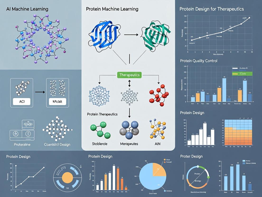
AI-Powered Protein Design: Revolutionizing Therapeutic Development with Machine Learning
This article provides a comprehensive overview of how artificial intelligence and machine learning are transforming the field of therapeutic protein design.
AI-Powered Protein Design: Revolutionizing Therapeutic Development with Machine Learning
Abstract
This article provides a comprehensive overview of how artificial intelligence and machine learning are transforming the field of therapeutic protein design. Tailored for researchers, scientists, and drug development professionals, we explore the foundational concepts, including deep learning architectures and the shift from structure-based to sequence-based design. We detail key methodologies like RFdiffusion and ProteinMPNN, their applications in creating novel enzymes, antibodies, and vaccines, and address common challenges in model training, data scarcity, and protein stability. Finally, we examine rigorous validation techniques and compare leading AI platforms, culminating in a synthesis of current achievements and future clinical implications, offering a roadmap for integrating AI into next-generation biotherapeutics pipelines.
From Sequences to Structures: Core AI Concepts Transforming Protein Design
Within therapeutic research, the central thesis is that machine learning (ML) and artificial intelligence (AI) are not merely incremental improvements but represent a foundational paradigm shift from reductionist, manual design to holistic, predictive generation of functional proteins. Traditional rational design operates on limited human-defined rules, while AI leverages high-dimensional pattern recognition across entire protein sequence space to discover novel solutions beyond human intuition.
Comparative Analysis: Core Methodologies & Data
Table 1: Paradigm Comparison: Traditional Rational vs. AI-Driven Design
| Aspect | Traditional Rational Design | AI-Driven Design |
|---|---|---|
| Philosophical Basis | Reductionist; structure-determines-function. | Holistic; statistical pattern recognition in sequence-structure-function landscape. |
| Starting Point | Known 3D structure of a natural template (e.g., wild-type protein). | Can start from scratch (de novo), a motif, or a disordered sequence. |
| Key Drivers | Site-directed mutagenesis based on evolutionary alignment, mechanistic hypotheses, & biophysical principles (e.g., ΔΔG calculations). | Generative models (e.g., ProteinMPNN, RFdiffusion), protein language models (e.g., ESM-2), and structure predictors (AlphaFold2, RosettaFold). |
| Throughput & Scale | Low-to-medium; iterative cycles of design, build, test for handfuls of variants. | High; can generate, in silico screen, and rank thousands to millions of designs in one cycle. |
| Primary Success Metric (Therapeutics) | Improved binding affinity (KD), stability (Tm), or activity (kcat/KM) of a known scaffold. | Discovery of novel folds, functional sites, and binders with no natural precedent. |
| Quantitative Success Rate | ~5-15% of designed variants show desired improvement. | ~20-50% of AI-generated proteins express and fold correctly, with ~1-10% showing high target function in first-round experimental validation. |
| Major Limitation | Heavily constrained by prior knowledge; poor at exploring novel conformations. | Training data dependency; potential for "hallucinations" that are physically unrealistic. |
Table 2: Quantitative Benchmark: Design of Novel Protein Binders
| Design Target | Traditional Method (e.g., Rosetta) | AI Method (e.g., RFdiffusion/ProteinMPNN) | Reported Outcome |
|---|---|---|---|
| SARS-CoV-2 Spike RBD | Months of design cycles; low yield of high-affinity miniproteins. | Weeks of in silico generation; high yield. | AI: Multiple designs with KD < 100 nM, some < 10 nM. (Nature, 2023) |
| Cancer Antigen (e.g., HER2) | Focus on humanization and affinity maturation of existing antibodies (mAbs). | De novo design of small binding proteins to epitopes inaccessible to mAbs. | AI: Novel binders with sub-nanomolar affinity and enhanced tissue penetration. |
| G Protein-Coupled Receptor (GPCR) | Extremely challenging due to dynamic structure; limited success. | Diffusion models conditioned on inactive/active states. | AI: First de novo designed agonists and positive allosteric modulators for specific GPCRs. (Science, 2024) |
Detailed Application Notes & Protocols
Protocol 1: Traditional Rational Design for Thermostabilization
Aim: Increase the melting temperature (Tm) of an enzyme by 10°C. Workflow:
- Template Analysis: Obtain crystal structure of wild-type (WT) enzyme. Perform molecular dynamics (MD) simulation to identify flexible regions.
- Evolutionary Analysis: Run multiple sequence alignment (MSA) of homologous proteins. Identify conserved residues and positions with correlated mutations.
- Hypothesis Generation: Select 5-10 target positions for mutagenesis based on: (a) Replacing flexible, non-conserved residues with Proline (rigidifies), (b) Introducing salt bridges or disulfide bonds in flexible loops.
- Energy Calculation: Use computational tools like Rosetta or FoldX to calculate predicted ΔΔG of folding for each single-point mutant.
- Library Construction: Use site-directed mutagenesis (e.g., KLD method) to create the 5-10 single mutants.
- Expression & Purification: Express each variant in E. coli and purify via His-tag affinity chromatography.
- Validation: Measure Tm via differential scanning fluorimetry (DSF). Test activity via spectrophotometric assay.
Protocol 2: AI-DrivenDe NovoBinder Design
Aim: Generate a novel small protein that binds to a therapeutically relevant target with high affinity and specificity. Workflow:
- Target Specification: Define the target protein's structure (experimental or AlphaFold2 prediction) and specify the binding epitope (a set of residues).
- Conditional Generation: Use a diffusion model (e.g., RFdiffusion) conditioned on the target epitope coordinates to generate 1,000-10,000 backbone scaffolds that geometrically complement the surface.
- Sequence Design: For each generated backbone, use a protein language model (e.g., ProteinMPNN) to design the optimal amino acid sequence that stabilizes the fold and the interface. Filter designs by in silico confidence scores (pLDDT, pAE).
- In Silico Screening: Dock top 500 designs back to the target using fast docking tools (e.g., AlphaFold-Multimer) and rank by predicted interface score (pTM or iScore). Select top 50-100 designs for experimental testing.
- DNA Synthesis & High-Throughput Build: Encode selected designs into genes and synthesize via pooled oligo library synthesis.
- HT Expression & Screening: Use a yeast surface display or mammalian cell display system to screen the library for target binding. Isulate hits via fluorescence-activated cell sorting (FACS).
- Characterization: Express and purify hits. Characterize via Surface Plasmon Resonance (SPR) for kinetics (KD, kon, koff) and DSF for stability.
Visualizations
Title: Workflow Comparison: Traditional vs. AI Protein Design
Title: AI De Novo Binder Design Protocol Flow
The Scientist's Toolkit: Key Research Reagent Solutions
| Reagent / Material | Function in Protein Design | Example/Supplier |
|---|---|---|
| Rosetta Software Suite | Computational modeling for energy calculation, docking, and traditional design. | University of Washington RosettaCommons. |
| AlphaFold2 / ColabFold | Accurate protein structure prediction from sequence; essential for target and design analysis. | DeepMind; Public Colab notebooks. |
| RFdiffusion & ProteinMPNN | AI models for de novo backbone generation and sequence design, respectively. | Publicly available on GitHub (Baker Lab). |
| NEB Gibson Assembly Master Mix | Seamless cloning of designed gene variants into expression vectors. | New England Biolabs. |
| Cytiva HisTrap HP Columns | Standardized affinity purification of His-tagged recombinant protein variants. | Cytiva. |
| Promega Nano-Glo HiBiT Blotting System | Rapid, high-sensitivity quantitation of protein stability and solubility in lysates. | Promega. |
| Cytiva Biacore 8K Series | Gold-standard SPR system for label-free kinetics (KD) analysis of protein-protein interactions. | Cytiva. |
| Unchained Labs Uncle | High-throughput thermal stability (Tm) and aggregation measurement. | Unchained Labs. |
| Twist Bioscience Gene Synthesis | Reliable synthesis of designed gene sequences, including large variant libraries. | Twist Bioscience. |
| ClonePlus Yeast Display Kit | Display and screening platform for isolating high-affinity binders from designed libraries. | ProteoGen. |
Application Notes for Protein Design
1. Convolutional Neural Networks (CNNs) Primary Therapeutic Application: Local structural motif and binding pocket prediction from protein 2D contact maps or 3D voxelized grids. CNNs excel at identifying spatial hierarchies and local patterns critical for understanding secondary structure elements (alpha-helices, beta-sheets) and catalytic sites.
2. Transformers (Attention-Based Models) Primary Therapeutic Application: Sequence-to-property prediction and de novo protein sequence generation. By processing entire amino acid sequences with self-attention, Transformers model long-range dependencies crucial for understanding non-local interactions that determine protein folding and function.
3. Diffusion Models Primary Therapeutic Application: Generative design of novel protein backbones and 3D structures. These probabilistic models iteratively refine noise into valid structures, enabling the sampling of diverse, thermodynamically stable protein folds conditioned on desired functional specifications.
Table 1: Performance Benchmarks of Architectures on Key Protein Design Tasks
| Architecture | Task (Dataset) | Key Metric | Reported Performance | Year |
|---|---|---|---|---|
| CNN (3D ResNet) | Protein-Ligand Affinity Prediction (PDBBind) | Pearson's R | 0.82 | 2023 |
| Transformer (ProteinBERT) | Protein Function Prediction (Gene Ontology) | F1 Max | 0.65 | 2022 |
| Diffusion Model (RFdiffusion) | De Novo Protein Scaffold Design | Design Success Rate | ~20% (high accuracy) | 2023 |
| Geometric CNN | Protein-Protein Interface Prediction (DockGround) | AUC-ROC | 0.91 | 2024 |
| Transformer (ESM-2) | Variant Effect Prediction | Spearman's ρ | 0.73 | 2023 |
| Diffusion (Chroma) | Protein Complex Generation | Tm Score (>0.5) | 41% | 2023 |
Table 2: Computational Resource Requirements for Training
| Architecture | Typical Model Size (Params) | Minimum GPU VRAM | Approx. Training Time (Dataset Size) |
|---|---|---|---|
| 2D/3D CNN | 10M - 100M | 8 GB | 2-5 days (~100k samples) |
| Standard Transformer | 100M - 10B | 40 GB+ | 1-4 weeks (~1M sequences) |
| Diffusion Model (Protein) | 50M - 500M | 24 GB+ | 1-3 weeks (~100k structures) |
Experimental Protocols
Protocol 1: Training a CNN for Binding Pocket Detection Objective: Train a 3D CNN to identify and segment ligand-binding pockets from protein structure voxel grids.
- Data Preparation: Obtain protein structures from the PDB. Pre-process into 1Å-resolution 3D grids (e.g., 64x64x64 voxels). Channels represent atom type densities (C, N, O, S, etc.) and electrostatic potential.
- Label Generation: Use a tool like
fpocketto generate ground-truth binary masks for binding pockets. - Model Architecture: Implement a 3D U-Net with residual blocks. Use 3D convolutions, batch normalization, and ReLU activations.
- Training: Loss: Dice Loss. Optimizer: Adam (lr=1e-4). Batch size: 8 (subject to VRAM). Train for 200 epochs with early stopping.
- Validation: Evaluate on held-out set using DICE coefficient and precision-recall on voxel-wise segmentation.
Protocol 2: Fine-Tuning a Transformer for Stability Prediction Objective: Adapt a pre-trained protein language model (e.g., ESM-2) to predict the thermostability (ΔΔG) of protein variants.
- Base Model: Load the
esm2_t30_150M_UR50Dmodel and its tokenizer. - Dataset: Use the S669 or a proprietary variant stability dataset. Format:
[wildtype_sequence], [mutation], [experimental_ddG]. - Input Representation: For a variant "M1A", construct input as
<seq>: M1Aor use a specialized token. The wildtype sequence is tokenized. - Head Addition: Replace the LM head with a regression head (pooled output -> linear layer -> single output).
- Training: Freeze most transformer layers, only fine-tune the last 2 layers and the head. Loss: Mean Squared Error. Optimizer: AdamW (lr=5e-5). Train for 10-20 epochs.
Protocol 3: Generating Novel Protein Folds with a Diffusion Model Objective: Use a conditional diffusion model (e.g., RFdiffusion) to generate a backbone structure for a specified function.
- Environment Setup: Install the RFdiffusion software (e.g., from GitHub repository) and its dependencies, including PyRosetta or AlphaFold2 for structure refinement.
- Conditioning: Define the functional motif. This could be a specified protein-protein interface (via a motif pdb), a catalytic triad geometry, or a set of secondary structure constraints.
- Inference: Run the diffusion model with the appropriate conditioning flags (e.g.,
--contigs="A1-100",--binders="B1-50"for a binder). The model will perform iterative denoising from a random cloud of Ca atoms. - Post-processing & Filtering: Refine the generated backbone with AlphaFold2 to predict sidechains and an all-atom structure. Filter outputs using predicted local-distance difference test (pLDDT) and/or predicted template modeling (pTM) scores (>0.7 acceptable).
- Validation: Run in silico docking if applicable, and molecular dynamics (MD) simulations (≥100 ns) to assess stability.
Visualizations
CNN Protein Analysis Workflow
Transformer Self-Attention for Proteins
Diffusion Model Denoising Process
The Scientist's Toolkit: Research Reagent Solutions
Table 3: Essential Computational Tools for ML-Based Protein Design
| Tool/Solution | Primary Function | Relevance to Architecture |
|---|---|---|
| AlphaFold2 (ColabFold) | Protein structure prediction from sequence. | Provides ground truth & validation for CNN/Diffusion models; fine-tuning base. |
| PyTorch / JAX | Deep learning frameworks. | Essential for implementing and training all CNN, Transformer, and Diffusion models. |
| ESM (Evolutionary Scale Modeling) | Pre-trained protein language models. | Transformer-based foundational models for transfer learning on therapeutic tasks. |
| RFdiffusion / Chroma | Diffusion models for protein generation. | Specialized software for de novo protein backbone and complex design. |
| Rosetta / PyRosetta | Molecular modeling suite. | Used for physics-based refinement, scoring, and design validation post-ML generation. |
| MD Simulation (GROMACS/AMBER) | Molecular Dynamics. | Critical for in silico validation of generated proteins' stability and dynamics. |
| PDB & UniProt | Public protein structure/sequence databases. | Primary sources of training data for all architectures. |
| Docker/Singularity | Containerization. | Ensures reproducibility of complex ML and molecular modeling pipelines. |
Within the paradigm of AI-driven therapeutic protein design, the selection and representation of structural data are foundational. This document provides application notes and protocols for utilizing the Protein Data Bank (PDB) and AlphaFold Database (AFDB) as primary data sources, focusing on their integration into machine learning pipelines for structure-based design and functional prediction.
Table 1: Core Characteristics of PDB and AlphaFold DB (as of 2024)
| Feature | Protein Data Bank (PDB) | AlphaFold DB (EMBL-EBI) |
|---|---|---|
| Primary Content | Experimentally determined 3D structures (X-ray, Cryo-EM, NMR). | Computationally predicted protein structures (AI/DeepMind). |
| Size (Entries) | ~220,000 (with redundancy). | >200 million (proteome-scale predictions). |
| Resolution (Typical) | Atomic (e.g., 1.0Å - 3.5Å for X-ray). | Predicted Local Distance Difference Test (pLDDT) score (0-100). |
| Metadata | Rich experimental details, ligands, crystallization conditions. | Prediction metadata (pLDDT, per-residue confidence, predicted aligned error). |
| Key File Format | PDB, mmCIF. | PDB, mmCIF (with custom fields for confidence metrics). |
| Therapeutic Relevance | Gold standard for binding sites, drug-protein complexes, mechanistic studies. | Enables work on proteins with no experimental structure (e.g., novel targets, orphan receptors). |
| Update Frequency | Weekly. | Major releases quarterly, with periodic updates. |
| Access | REST API, FTP, RCSB PDB website. | REST API, Google Cloud Public Dataset, AFDB website. |
Table 2: Key Confidence Metrics in AlphaFold DB Outputs
| Metric | Range | Interpretation for Therapeutic Design |
|---|---|---|
| pLDDT | 0 - 100 | Per-residue confidence. >90: High (backbone reliable). 70-90: Confident (side chains may vary). <50: Low confidence (use with caution). |
| PAE (Predicted Aligned Error) | 0 - 30+ Å | Expected positional error between residues. Low inter-domain PAE suggests reliable relative orientation. |
| Model Confidence (Global) | High/Medium/Low | Overall model quality based on pLDDT distribution. |
Experimental Protocols
Protocol 3.1: Curating a High-Quality Structural Dataset for Training a Binding Site Predictor
Objective: Assemble a non-redundant set of protein-ligand complexes from the PDB for training a graph neural network.
Materials:
- High-performance computing cluster or cloud instance.
biopython,pandas,mdanalysisPython libraries.- RCSB PDB REST API access.
- PDB FTP archive.
Procedure:
- Query Generation: Use the RCSB API to query all structures with:
- Resolution ≤ 2.5 Å.
- Contains a non-polymeric ligand (HETATM records).
- No NMR structures.
- Release date after 2010.
- Example query:
https://search.rcsb.org/rcsbsearch/v2/query?json={"query":{"type":"group","logical_operator":"and","nodes":[{"type":"terminal","service":"text","parameters":{"attribute":"rcsb_entry_info.resolution_combined","operator":"less_or_equal","value":2.5}},{"type":"terminal","service":"text","parameters":{"attribute":"rcsb_entry_info.deposition_date","operator":"greater_or_equal","value":"2010-01-01"}},{"type":"terminal","service":"text","parameters":{"attribute":"rcsb_struct_symmetry.symbol","operator":"equals","value":"C1"}},{"type":"terminal","service":"text","parameters":{"attribute":"entity_poly.rcsb_entity_polymer_type","operator":"equals","value":"Protein"}}]},"return_type":"entry"}
Redundancy Reduction: Download the list of PDB IDs. Use MMseqs2 or CD-HIT at 40% sequence identity to cluster proteins. Select one representative structure per cluster, prioritizing higher resolution and newer deposition date.
Data Download & Processing: For each selected PDB ID:
- Download the structure file (mmCIF format recommended).
- Use
BiopythonorMDTrajto isolate the protein chain(s) and all non-water, non-ion ligands within 5Å of the protein. - Extract atomic coordinates, element types, and residue types.
- Parse the
site_recordin the PDB file to annotate known functional/binding sites.
Feature Encoding: For each residue/atom, compute and store:
- Geometric: Solvent accessible surface area (SASA), dihedral angles, secondary structure (DSSP).
- Chemical: One-hot encoding of residue type, atomic partial charges (from force field), hydrophobicity index.
- Neighborhood: Radial basis function (RBF) distances to k-nearest neighbors.
Graph Construction: Represent each complex as a graph where nodes are residues (or atoms) and edges connect residues within a 10Å cutoff. Node features are the computed descriptors; edge features include distance and direction vectors.
Protocol 3.2: Integrating AlphaFold DB Predictions for a Target of Unknown Structure
Objective: Obtain, validate, and prepare an AlphaFold-predicted structure for in silico docking.
Materials:
- AlphaFold DB REST API or Google Cloud Public Dataset access.
- Molecular visualization software (PyMOL, UCSF ChimeraX).
- Structure preparation software (OpenBabel, Schrödinger's Protein Preparation Wizard, or
pdbfixer).
Procedure:
- Retrieval: Query the AlphaFold DB (
https://alphafold.ebi.ac.uk/api/prediction/{UNIPROT_ID}) using the canonical UniProt identifier of your target. Download the ranked PDB files and the associated JSON file containing pLDDT and PAE data.
Confidence Assessment:
- Load the ranked model 1 in visualization software. Color the structure by the pLDDT b-factor field.
- Identify low-confidence regions (pLDDT < 70). These loops or termini may require modeling refinement or deletion for docking.
- Analyze the PAE matrix plot (from the JSON file) to check for domain-level errors. Low confidence in inter-domain orientation may necessitate using only a single, high-confidence domain.
Structure Preparation:
- Use
pdbfixerto add missing hydrogens at physiological pH (7.4). - If low-confidence regions are not near the putative active site (based on literature or homology), delete them to simplify the model.
- Run a brief energy minimization (e.g., using OpenMM or GROMACS with a simple force field like AMBERff14SB) to relieve steric clashes, keeping the majority of the backbone restrained to preserve the AF2 prediction.
- Use
Active Site Definition: If no experimental site is known, use computational methods (e.g., fpocket, DeepSite) on the prepared structure to predict potential binding pockets. Prioritize pockets with high conservation (from ConSurf analysis) and high average pLDDT scores.
Diagrams
Title: Data Flow from PDB and AlphaFold DB to ML Models
Title: Protocol for Using an AlphaFold DB Prediction
The Scientist's Toolkit
Table 3: Essential Research Reagents & Computational Tools
| Item | Category | Function in Protocol |
|---|---|---|
| RCSB PDB REST API | Web Service | Programmatic querying and metadata retrieval from the PDB. |
| MMseqs2 / CD-HIT | Software Tool | Rapid clustering of protein sequences to reduce dataset redundancy. |
| Biopython / MDTraj | Python Library | Parsing structural files (PDB, mmCIF), geometric calculations, and data extraction. |
| AlphaFold DB API | Web Service | Programmatic retrieval of predicted structures and confidence metrics. |
| PyMOL / UCSF ChimeraX | Visualization Software | Visual inspection of structures, coloring by confidence (pLDDT), and active site analysis. |
| PDBFixer / OpenBabel | Software Tool | Adding missing atoms, hydrogens, and performing basic structure cleanup. |
| OpenMM / GROMACS | Molecular Dynamics Engine | Energy minimization to relieve steric clashes in predicted models. |
| fpocket / DeepSite | Software Tool | Predicting potential ligand-binding pockets on protein surfaces. |
| PyTorch Geometric / DGL | Python Library | Building and training graph neural network models on structural data. |
The inverse folding problem—determining an amino acid sequence that will fold into a predetermined three-dimensional protein structure—represents a core challenge in computational biology. Within the broader thesis of employing AI and machine learning (ML) for protein design in therapeutics, solving this problem is pivotal. It enables the de novo design of novel protein therapeutics, enzymes, and vaccines with tailored functions and stabilities, moving beyond natural evolutionary constraints. Recent breakthroughs in deep learning architectures have transformed this field from a theoretical pursuit into a practical pipeline for drug development.
Current State of AI/ML Models for Inverse Folding
The following table summarizes key quantitative performance metrics for leading deep learning models in protein inverse folding, based on recent benchmarks.
Table 1: Performance Comparison of Recent Inverse Folding Models
| Model Name (Year) | Architecture Core | Key Training Data | Design Success Rate (Top-1 Recovery) | Sequence Recovery on Native Pairs | Computational Speed (Per Design) | Key Therapeutic Application Focus |
|---|---|---|---|---|---|---|
| ProteinMPNN (2022) | Message Passing Neural Network | CATH, PDB | ~52% (CATH 4.2) | ~33.5% | ~0.2 seconds | High-accuracy de novo scaffolds, symmetric assemblies |
| RFdiffusion (2023) | Diffusion Model + RosettaFold | PDB, synthetic | High (varies by task) | N/A | Minutes (GPU) | De novo binder design, motif scaffolding |
| ESM-IF1 (2022) | Inverse Folding Transformer | PDB | ~51% (CATH 4.2) | ~32.8% | Seconds | Fixed-backbone design, variant generation |
| Chroma (2023) | Diffusion Model (Latent) | PDB, AlphaFold DB | State-of-the-art on complex tasks | N/A | Minutes (GPU) | Large protein complexes, functional site design |
Core Experimental Protocol: Validating AI-Designed Sequences
This protocol details the experimental validation pipeline for sequences generated by inverse folding models, a critical step for therapeutic development.
Protocol: In Silico and In Vitro Validation of Designed Protein Sequences
Objective: To express, purify, and biophysically characterize a protein from an AI-designed sequence to confirm it adopts the target structure.
Materials & Reagents:
- Synthetic Gene Fragment: Codon-optimized for expression system (e.g., E. coli).
- Cloning Vector: (e.g., pET series with His-tag for purification).
- Competent Cells: For cloning (DH5α) and expression (BL21(DE3)).
- LB Media & Antibiotics: For bacterial culture.
- Induction Agent: Isopropyl β-d-1-thiogalactopyranoside (IPTG).
- Lysis & Purification Buffers: Including imidazole for immobilized metal affinity chromatography (IMAC).
- Size Exclusion Chromatography (SEC) Column: For final polishing.
- Circular Dichroism (CD) Spectrometer.
- Differential Scanning Calorimetry (DSC) or Fluorimeter.
- SEC-Multi-Angle Light Scattering (SEC-MALS) system.
Procedure:
Part A: In Silico Folding Confidence Check
- Input: Generate candidate sequences using your chosen inverse folding model (e.g., ProteinMPNN) and your target backbone structure (PDB file).
- Folding Prediction: Submit the designed sequences to a structure prediction network (e.g., AlphaFold2, ESMFold). Use the model's confidence metrics (pLDDT, pTM).
- Analysis: Select sequences where the predicted structure has a high root-mean-square deviation (RMSD) < 2.0 Å to the target backbone and high per-residue confidence (pLDDT > 80).
Part B: Gene Synthesis, Cloning, and Expression
- Gene Synthesis: Order the top 3-5 selected sequences as codon-optimized gene fragments.
- Cloning: Subclone each gene into an expression vector using restriction enzyme/ligation or Gibson assembly. Transform into cloning cells, screen colonies by colony PCR, and verify plasmid by Sanger sequencing.
- Expression: Transform verified plasmid into expression cells. Grow a 50 mL overnight culture, inoculate 1 L of main culture. Grow at 37°C to OD600 ~0.6-0.8, induce with 0.5-1.0 mM IPTG, and express at appropriate temperature (often 18-20°C) for 16-20 hours.
Part C: Purification and Biophysical Characterization
- Lysis and IMAC: Harvest cells by centrifugation, lyse by sonication, and clarify by centrifugation. Pass the supernatant over a Ni-NTA column. Wash with buffer containing 20-40 mM imidazole, elute with 250-500 mM imidazole.
- Polishing: Further purify the eluate by size exclusion chromatography (SEC). Analyze the SEC elution profile for a single, monodisperse peak.
- Secondary Structure (CD): Dilute purified protein to ~0.2 mg/mL in appropriate buffer. Acquire CD spectrum from 260-190 nm. Compare the spectrum's shape (double minima at ~208 nm & ~222 nm for α-helix) to that expected from the target structure.
- Thermal Stability (DSC/DSF):
- DSC: Load protein at >0.5 mg/mL. Perform a temperature ramp (e.g., 20-100°C). Record the melting temperature (Tm).
- DSF: Mix protein with a fluorescent dye (e.g., SYPRO Orange). Monitor fluorescence during a temperature ramp in a real-time PCR machine. Derive Tm from the inflection point.
- Oligomeric State (SEC-MALS): Inject purified sample onto an SEC column coupled to MALS and refractive index detectors. Calculate the absolute molecular weight from light scattering data to confirm the designed monomeric or multimeric state.
Expected Outcomes: A successfully designed protein will express solubly, purify as a single peak, exhibit a CD spectrum consistent with the target fold, display a high thermal stability (often Tm > 60°C), and confirm the intended oligomeric state.
The Scientist's Toolkit: Key Research Reagent Solutions
Table 2: Essential Reagents and Resources for Inverse Folding Experiments
| Item | Function & Relevance |
|---|---|
| High-Quality Structural Datasets (PDB, CATH, AlphaFold DB) | Training and benchmarking data for AI models. The non-redundancy and quality are critical. |
| ProteinMPNN Web Server / Codebase | Currently the most robust and widely used inverse folding model for fixed-backbone design. Accessible for non-specialists. |
| AlphaFold2 or ESMFold Colab Notebooks | Essential for the in silico confidence check, providing a rapid, low-cost filter for designed sequences before wet-lab experiments. |
| Codon-Optimized Gene Synthesis Service | Turns digital designs into physical DNA. Rapid synthesis (2-5 days) is key for iterative design-test cycles. |
| High-Throughput Cloning & Expression Kits (e.g., Ligation-Independent) | Accelerates the testing of multiple designed variants in parallel, essential for screening. |
| His-tag Purification Kits (IMAC) | Standardized, reliable first-step purification for tens to hundreds of designed proteins. |
| Pre-packed SEC Columns (e.g., Superdex series) | For assessing protein purity, monodispersity, and approximate size in a reproducible manner. |
| Differential Scanning Fluorimetry (DSF) Dyes (e.g., SYPRO Orange) | Enables medium-to-high-throughput thermal stability screening of purified designs in a plate reader format. |
Visualizations
AI-Driven Inverse Folding & Validation Workflow
AI Model Categories for Inverse Protein Design
Within the broader thesis on AI and machine learning for protein design for therapeutics, this document details the application of generative AI to create novel protein folds and functions de novo. This approach moves beyond natural protein libraries, enabling the discovery of unique protein scaffolds and binders with therapeutic potential.
Core Generative Models: Architectures and Quantitative Benchmarks
Current methods primarily leverage deep generative models trained on the Protein Data Bank (PDB). Key architectures and their performance metrics are summarized below.
Table 1: Comparison of Key Generative AI Models for De Novo Protein Design
| Model Name | Model Architecture | Key Feature | Reported Success Rate (Experimental Validation) | Typical Design Cycle Time | Primary Application |
|---|---|---|---|---|---|
| RFdiffusion | Diffusion Model (built on RosettaFold) | Conditional generation based on structural motifs | ~20% (for symmetric assemblies) | Hours to days | Symmetric scaffolds, binder design |
| Chroma | Diffusion Model (SE(3)-Equivariant) | Geometry-aware, conditioning on various properties (symmetry, function) | High (per cited examples) | Minutes to hours | Multi-conditional design (scaffolds, enzymes) |
| ProteinMPNN | Graph Neural Network (GNN) | Fast sequence design for backbones | >50% (sequence recovery on native backbones) | Seconds | Inverse folding (sequence design) |
| AlphaFold2 (as validation tool) | Transformer/Evoformer | State-of-the-art structure prediction | N/A (used for validation) | Minutes per structure | In silico validation of designed proteins |
| ESM-2/ESMFold | Large Language Model (Transformer) | Sequence-to-structure generation & prediction | N/A | Seconds to minutes | Co-design of sequence & structure |
Detailed Protocol: Generating a Novel Protein Scaffold with RFdiffusion
This protocol outlines the steps for generating a novel symmetric protein scaffold.
Materials & Software (The Scientist's Toolkit)
Table 2: Essential Research Reagent Solutions & Computational Tools
| Item Name | Provider/Software | Function in Protocol |
|---|---|---|
| RFdiffusion Colab Notebook | The Rosetta Commons / Sergey Ovchinnikov Lab | Primary interface for running RFdiffusion with default parameters. |
| AlphaFold2 (Local Installation or Colab) | DeepMind / Jumper et al. | In silico validation of generated protein models. |
| PyMOL or ChimeraX | Schrödinger / UCSF | Visualization and analysis of 3D protein structures. |
| PDB File of Motif (Optional) | Protein Data Bank (rcsb.org) | Provides a structural "seed" for conditional generation (e.g., a binding site). |
| Cloning Vector (e.g., pET series) | Novagen / Addgene | For downstream experimental expression of designed sequences. |
| E. coli Expression Cells (BL21(DE3)) | Thermo Fisher, New England Biolabs | Heterologous protein expression host. |
| Ni-NTA Resin | Qiagen, Cytiva | Purification of His-tagged designed proteins. |
| Size Exclusion Chromatography Column | Cytiva (Superdex series) | Polishing step to isolate monodisperse protein. |
Step-by-Step Procedure
Step 1: Define Design Goal and Parameters
- Objective: Specify desired symmetry (e.g., C2, D3), approximate size (number of residues), and any required functional motifs.
- Input Preparation: If conditioning on a motif, prepare a PDB file of the target motif or specify the desired protein-protein interface.
Step 2: Run RFdiffusion Generation
- Access the RFdiffusion Colab notebook.
- Set parameters in the
inferencesection:contigs: Define the scaffold. E.g.,"80-120"for a 80-120 residue chain.symmetry: Define symmetry. E.g.,"C3"for cyclic symmetry with 3 copies.hotspot_res: (Optional) Specify residues from a motif PDB to guide generation.
- Execute the diffusion sampling. The model will generate multiple (e.g., 100) backbone structures in PDB format.
Step 3: Sequence Design with ProteinMPNN
- Feed the generated backbone PDBs into ProteinMPNN.
- Run ProteinMPNN to design optimal amino acid sequences that stabilize each backbone.
- Output: A set of designed protein sequences (FASTA format) paired with their backbone structures.
Step 4:In SilicoValidation with AlphaFold2
- Input the designed FASTA sequences into AlphaFold2.
- Run structure prediction. The critical output is the predicted local distance difference test (pLDDT) score.
- Analysis: Select designs where the AlphaFold2-predicted structure closely matches the generative model's backbone (RMSD < 2.0 Å) and has high average pLDDT (>80).
Step 5: Experimental Expression and Purification (Abridged Protocol)
- Gene Synthesis & Cloning: Order selected sequences as gene fragments and clone into an expression vector (e.g., pET-28a with a His-tag).
- Transformation: Transform plasmid into expression host (e.g., E. coli BL21(DE3)).
- Expression: Grow culture to OD600 ~0.6, induce with 0.5 mM IPTG, and express at 18°C for 16-18 hours.
- Purification:
- Lyse cells via sonication in lysis buffer (e.g., 50 mM Tris, 300 mM NaCl, pH 8.0).
- Purify soluble protein using Ni-NTA affinity chromatography.
- Further purify via size-exclusion chromatography (SEC).
- Validation: Analyze SEC elution profile for monodispersity. Confirm structure via Circular Dichroism (secondary structure) and/or SEC-MALS (oligomeric state).
Signaling Pathway for Functional Protein Design
Diagram Title: AI-Driven Design Cycle for Therapeutic Proteins
Workflow forDe NovoProtein Generation & Validation
Diagram Title: De Novo Protein Design and Validation Workflow
Tools in Action: Applying AI Models to Design Real-World Therapeutics
De Novo Enzyme Design for Catalysis and Degradation
Within the broader thesis of applying AI and machine learning (ML) to protein design for therapeutics, de novo enzyme design represents a frontier with profound implications. This capability shifts the paradigm from discovering natural enzymes to computationally inventing proteins with tailored catalytic functions. For drug development, this enables the creation of therapeutic enzymes for metabolite clearance, prodrug activation, or degradation of pathological agents, moving beyond traditional small-molecule inhibitors. The integration of deep learning models for structure prediction (e.g., AlphaFold2, RosettaFold) and generative models for sequence design (e.g., ProteinMPNN, RFdiffusion) has dramatically accelerated the design-build-test-learn cycle, making the rational engineering of catalysts for novel reactions a tangible reality.
Core Application Notes
Key Design Strategies and Outcomes
De novo enzyme design workflows typically follow a reaction-driven approach: 1) Define the reaction mechanism and transition state (TS), 2) Generate a idealized active site (theozyme) complementary to the TS, 3) Scaffold the theozyme into a stable protein backbone, and 4) Iteratively refine the design using ML.
Table 1: Quantitative Performance Benchmarks of Recent De Novo Designed Enzymes
| Target Reaction | Design Method | Initial kcat/KM (M⁻¹s⁻¹) | After Directed Evolution | Therapeutic Relevance |
|---|---|---|---|---|
| Kemp Elimination | ROSETTA + Theozyme | 10² - 10³ | 10⁵ | Model for catalytic principles |
| Retro-Aldol Reaction | ROSETTA + Theozyme | 0.04 | 3.4 x 10⁴ | C-C bond cleavage for degradation |
| Non-native C-H Amination | ROSETTA + ML-guided active site packing | N.D. | 1,030 TTN | Potential for synthetic metabolite production |
| Hydrolysis of Organophosphates (e.g., paraoxon) | RFdiffusion + ProteinMPNN | Detectable activity in top designs | Under investigation | Nerve agent detoxification |
| Degradation of β-Lactam Antibiotics | Sequence-based generative models | Variant-dependent | >100-fold improvement | Addressing antibiotic resistance |
AI/ML Toolbox for Enzyme Design
Generative Models: RFdiffusion and Chroma generate novel protein backbones conditioned on functional site constraints. Sequence Design Models: ProteinMPNN and ESM-IF provide high-probability, stable sequences for given backbones. Fitness Prediction Models: Models like ESM-2 and GEMME can predict stability and functional scores, prioritizing designs for experimental testing.
Detailed Experimental Protocols
Protocol: Computational Design of a Hydrolase for Plastic Degradation (PETase Mimetic)
Objective: Design a novel enzyme capable of hydrolyzing polyethylene terephthalate (PET) ester bonds.
Materials:
- Hardware: High-performance computing cluster with GPU access.
- Software: PyRosetta, RFdiffusion/Chroma, ProteinMPNN, AlphaFold2, MD simulation suite (e.g., GROMACS).
Procedure:
- Theozyme Construction:
- Define the hydrolytic reaction coordinates (nucleophilic attack, tetrahedral intermediate, bond cleavage).
- Using quantum mechanics (QM) software (e.g., Gaussian), optimize the geometry of the transition state analog (TSA).
- Manually or using PLACEHOLDER, arrange a minimal set of catalytic residues (e.g., Ser-His-Asp triad, oxyanion hole donors) around the TSA with ideal geometries.
Active Site Scaffolding with RFdiffusion:
- Format the theozyme residues (Cα atoms and sidechain conformers) as a motif input for RFdiffusion.
- Run RFdiffusion in "motif-scaffolding" mode to generate hundreds of novel protein backframes that precisely position the motif.
- Apply distance and angle constraints to maintain catalytic geometry.
Sequence Design with ProteinMPNN:
- For each generated backbone, run ProteinMPNN in "fixed residues" mode, freezing the identities and conformations of the catalytic motif residues.
- Generate multiple sequence variants for each scaffold, selecting outputs with high confidence scores.
In Silico Validation:
- Fold each designed sequence using AlphaFold2 or RosettaFold. Discard designs with low confidence (pLDDT < 80) or poor motif geometry.
- Perform molecular docking of the substrate (e.g., bis(2-hydroxyethyl) terephthalate, BHET) into the predicted structure.
- Run short, targeted MD simulations to assess active site stability and substrate binding. Select top 50 designs for experimental testing.
Protocol: High-Throughput Screening of Designed Enzymes
Objective: Express, purify, and assay computationally designed enzymes for catalytic activity.
Materials:
- Reagent Solutions: See "The Scientist's Toolkit" below.
- Equipment: Robotic liquid handler, microplate spectrophotometer/fluorometer, FPLC system, SDS-PAGE equipment.
Procedure:
- Gene Synthesis and Cloning:
- Synthesize genes encoding the top 50 designs, codon-optimized for E. coli expression. Clone into a T7 expression vector (e.g., pET series) with a C-terminal His-tag.
Parallel Expression and Purification:
- Transform plasmids into BL21(DE3) E. coli cells. Inoculate 96-deep-well plates with auto-induction media.
- Grow at 37°C until OD600 ~0.6, then induce at 18°C for 18-24 hours.
- Lyse cells via sonication or chemical lysis in 96-well format.
- Purify proteins using immobilized metal affinity chromatography (IMAC) in a 96-well filter plate format. Elute with imidazole buffer.
- Desalt into assay buffer using spin columns. Confirm purity via SDS-PAGE.
Activity Screening:
- For Hydrolases: Use a fluorescent or chromogenic substrate analog (e.g., 4-nitrophenyl acetate for esterases). In a 384-well plate, mix enzyme with substrate.
- Monitor product formation kinetically (e.g., release of 4-nitrophenol at 405 nm) for 30 minutes.
- Calculate initial velocities. Designs showing activity above negative control (empty vector lysate) are considered "hits."
Hit Characterization:
- Scale up expression and purification of hit designs for detailed kinetic analysis (KM, kcat).
- Validate folding via circular dichroism (CD) spectroscopy.
- Initiate directed evolution (error-prone PCR, site-saturation mutagenesis) to improve activity and stability.
Visualizations
Diagram Title: AI-Driven Design-Build-Test-Learn Cycle for Enzyme Engineering
Diagram Title: Theozyme Construction Around a Transition State
The Scientist's Toolkit
Table 2: Key Research Reagent Solutions for De Novo Enzyme Workflows
| Reagent / Material | Function / Application | Key Considerations |
|---|---|---|
| T7 Expression Vector (e.g., pET series) | High-level, inducible protein expression in E. coli. | Codon optimization for design genes is critical for solubility. |
| Auto-induction Media | Simplified expression in deep-well plates; induces upon glucose depletion. | Enables high-throughput, parallel culture growth without manual induction. |
| Nickel-NTA Resin (IMAC) | Immobilized metal affinity chromatography for His-tagged protein purification. | 96-well filter plate format enables parallel mini-purifications. |
| Chromogenic/Fluorogenic Substrate Analogs (e.g., 4-NPA, 4-NPB) | High-sensitivity detection of hydrolytic activity in microplate assays. | Must mimic the target reaction's chemistry; used for primary screening. |
| Phusion/Ultra II Q5 Polymerase | High-fidelity PCR for cloning and site-directed mutagenesis. | Essential for generating libraries for directed evolution of initial hits. |
| Size-Exclusion Chromatography (SEC) Column (e.g., Superdex 75) | Analytical purification and assessment of protein monomericity/aggregation. | Confirms proper folding of designed enzymes post-IMAC. |
| Thermal Shift Dye (e.g., SYPRO Orange) | Measures protein thermal stability (Tm) in a real-time PCR instrument. | High-throughput stability assessment to filter poorly folded designs. |
| Rosetta/MPNN Software Suites | Computational protein design and sequence prediction. | Require significant GPU/CPU resources and structural biology expertise. |
AI-Driven Antibody and Nanobody Engineering for Enhanced Affinity & Stability
Application Notes
The integration of AI and machine learning (ML) into antibody engineering represents a paradigm shift in therapeutic discovery. Within the broader thesis of AI for protein design, these computational methods accelerate the development of biologics with superior binding affinity and thermal stability, directly addressing key challenges in drug development such as efficacy, manufactura, and shelf-life.
Core AI/ML Methodologies:
- Deep Learning Models for Structure Prediction: Tools like AlphaFold2 and RosettaFold provide high-accuracy structural models of antibody-antigen complexes, which serve as critical inputs for subsequent engineering steps.
- Generative Models for Sequence Design: Variational Autoencoders (VAEs) and Generative Adversarial Networks (GANs) are trained on known antibody sequence-structure-function datasets to generate novel, optimized sequences with desired properties.
- In-Silico Affinity Maturation: ML models, particularly graph neural networks (GNNs) trained on molecular dynamics (MD) simulations, predict the change in binding free energy ((\Delta\Delta G)) upon mutation, enabling rapid virtual screening of millions of variants.
- Stability Prediction Models: Learned representations from protein language models (e.g., ESM-2) are fine-tuned to predict thermal stability metrics like melting temperature ((T_m)) directly from sequence.
Key Quantitative Outcomes from Recent Studies:
Table 1: Reported Performance of AI-Driven Antibody Engineering
| Target / Property | Baseline Affinity (nM) | AI-Optimized Affinity (nM) | Fold Improvement | Stability Change ((\Delta T_m)) | Key AI Method |
|---|---|---|---|---|---|
| SARS-CoV-2 RBD | 5.2 | 0.056 | 93x | +3.2°C | GNN-based (\Delta\Delta G) prediction |
| HER2 | 10.1 | 0.71 | 14x | +5.1°C | VAE sequence generation & ranking |
| IL-6 Receptor | 3.8 | 0.21 | 18x | +4.5°C | Protein MPNN & RosettaDDG |
| Generic Nanobody | N/A | N/A | N/A | +7.8°C | ESM-2 fine-tuned for stability |
Table 2: Comparative Throughput of Traditional vs. AI-Enhanced Workflows
| Development Stage | Traditional Method | Typical Duration | AI-Enhanced Method | Typical Duration | Speed Gain |
|---|---|---|---|---|---|
| Lead Identification | Hybridoma / Phage Display | 3-6 months | In-silico Library Design & Screening | 2-4 weeks | ~4x |
| Affinity Maturation | Error-Prone PCR & Screening | 4-8 months | (\Delta\Delta G) ML Prediction & Validation | 3-6 weeks | ~6x |
| Developability Assessment | Low-throughput analytics | 1-2 months | ML-based prediction of viscosity, aggregation | <1 week | >8x |
Experimental Protocols
Protocol 1: In-Silico Affinity Maturation Using Graph Neural Networks (GNNs)
Objective: To generate and rank single-point mutations in the Complementarity-Determining Region (CDR) of an antibody for improved binding affinity.
Materials:
- Starting antibody-antigen complex structure (PDB file or AlphaFold2 prediction).
- High-performance computing (HPC) cluster or cloud instance with GPU acceleration.
- Software: PyTorch, PyTorch Geometric, Rosetta, or dedicated ML protein design suite.
Procedure:
- Structure Preparation: Clean the PDB file using PDBFixer or Chimera. Protonate the structure at pH 7.4 using H++ or PROPKA.
- Define Mutation Site: Isolate CDR residues (e.g., H3, L3) within 8Å of the antigen in the paratope.
- Generate Mutant Library: Create in-silico all possible single-point mutants (19 variants per residue) for the selected sites.
- Feature Extraction: For each mutant structure (modeled with Rosetta or ABACUS), generate a graph representation. Nodes (residues) are featurized with physicochemical properties, evolutionary scores from PSSM. Edges encode distances and angles.
- (\Delta\Delta G) Prediction: Input the graph representation into a pre-trained GNN model (e.g., DeepAb, ProteinMPNN for sequence, followed by AttentiveFP for affinity scoring). The model outputs a predicted (\Delta\Delta G) (kcal/mol) for each mutation.
- Ranking & Selection: Rank all mutants by predicted (\Delta\Delta G). Select top 20-50 stabilizing mutations (negative (\Delta\Delta G)) for experimental validation.
- In-Vitro Validation: Proceed to Protocol 3.
Protocol 2: Generative Design for Stabilized Nanobody Frameworks
Objective: To design a nanobody (VHH) sequence with enhanced thermal stability while maintaining a canonical fold.
Materials:
- Curated dataset of nanobody sequences with experimental (T_m) values.
- Access to a pre-trained protein language model (e.g., ESM-2, ProtGPT2).
- Fine-tuning environment (e.g., Hugging Face Transformers, JAX).
Procedure:
- Dataset Curation: Compile sequences and corresponding (T_m) data. Split into training (80%), validation (10%), test (10%) sets.
- Model Fine-Tuning: Fine-tune the ESM-2 model via transfer learning. The final hidden representation is fed into a regression head to predict (T_m).
- Latent Space Sampling: Use a conditioned VAE. The encoder maps input sequences to a latent vector
z, the decoder generates sequences fromz. The conditioning variable is the desired (T_m) (e.g., >75°C). - Sequence Generation: Sample latent vectors from a Gaussian distribution and decode them using the conditioned decoder to generate novel nanobody sequences.
- Filtration & Scoring: Filter sequences for:
- Correct framework residue conservation (Cys22, Cys92, Trp103, etc.).
- Low predicted immunogenicity (using NetMHCIIPan).
- High predicted stability from the fine-tuned ESM-2 model.
- Structure Validation: Fold top-ranked sequences (e.g., 20 designs) using AlphaFold2. Discard any with low pLDDT (<85) or non-canonical folds.
- Experimental Expression & Characterization: Proceed to Protocol 3.
Protocol 3: Experimental Validation of AI-Designed Variants
Objective: To express, purify, and characterize the affinity and stability of AI-designed antibody/nanobody variants.
Materials: See "Research Reagent Solutions" table below.
Procedure: A. Expression & Purification:
- Clone synthesized gene sequences into a mammalian expression vector (e.g., pcDNA3.4) for antibodies or a bacterial vector (e.g., pET series) for nanobodies.
- For antibodies: Transiently transfect Expi293F cells using Expifectamine, culture for 5-7 days. For nanobodies: Transform SHuffle T7 E. coli, induce with IPTG for cytoplasmic expression.
- Purify via affinity chromatography (Protein A for IgG, His-tag for nanobodies) followed by size-exclusion chromatography (SEC) on an ÄKTA system.
B. Affinity Measurement (Bio-Layer Interferometry - BLI):
- Dilute antigen to 10 µg/mL in kinetics buffer and load onto anti-His (for His-tagged antigen) or streptavidin (for biotinylated antigen) biosensors for 300s.
- Baseline in kinetics buffer for 60s.
- Associate with serially diluted antibody (e.g., 100 nM to 1.56 nM) for 300s.
- Dissociate in kinetics buffer for 400s.
- Fit association and dissociation curves using a 1:1 binding model to calculate the association rate ((k{on})), dissociation rate ((k{off})), and equilibrium dissociation constant ((K_D)).
C. Thermal Stability Analysis (Differential Scanning Fluorimetry - DSF):
- Mix purified protein with SYPRO Orange dye in a 96-well PCR plate.
- Perform a temperature ramp from 25°C to 95°C at 1°C/min in a real-time PCR instrument.
- Monitor fluorescence intensity. The inflection point of the fluorescence curve is the apparent melting temperature ((T_m)).
- Compare (T_m) of designed variants to the parental molecule.
Visualizations
Title: AI-Driven Affinity Maturation Workflow
Title: Generative AI Pipeline for Nanobody Stability
Research Reagent Solutions
Table 3: Essential Materials for Experimental Validation
| Item | Function / Role in Protocol | Example Product/Catalog |
|---|---|---|
| Expi293F Cells | Mammalian host for transient expression of full-length IgG antibodies. | Thermo Fisher Scientific, Cat# A14527 |
| SHuffle T7 E. coli | Bacterial host for cytoplasmic expression of disulfide-bonded nanobodies. | New England Biolabs, Cat# C3029J |
| Expifectamine 293 | High-efficiency transfection reagent for Expi293F system. | Thermo Fisher Scientific, Cat# A14524 |
| Ni-NTA Superflow | Affinity resin for purification of His-tagged antigens and nanobodies. | Qiagen, Cat# 30410 |
| Protein A Agarose | Affinity resin for capture of IgG antibodies from mammalian supernatant. | Thermo Fisher Scientific, Cat# 20334 |
| Superdex 75 Increase | Size-exclusion chromatography column for polishing and aggregation removal. | Cytiva, Cat# 29148721 |
| Anti-His (HIS1K) Biosensors | BLI biosensors for kinetics analysis of His-tagged antigens. | Sartorius, Cat# 18-5120 |
| SYPRO Orange Dye | Fluorescent dye for DSF, binds hydrophobic patches exposed upon unfolding. | Thermo Fisher Scientific, Cat# S6650 |
| pcDNA3.4 Vector | High-expression mammalian vector for antibody heavy and light chains. | Thermo Fisher Scientific, Cat# A14697 |
| pET-28a(+) Vector | Common bacterial expression vector for nanobody cloning with His-tag. | MilliporeSigma, Cat# 69864-3 |
Designing Novel Peptide Therapeutics and Vaccine Antigens
The advent of AI and machine learning (ML) has revolutionized de novo protein and peptide design, transitioning from structure-guided empirical methods to predictive, sequence-first approaches. This paradigm shift, exemplified by models like AlphaFold2, RFdiffusion, and ProteinMPNN, enables the rapid generation of novel peptide binders, stabilizers, and immunogens with high precision. This application note provides integrated wet-lab protocols and computational workflows for designing and validating peptide-based therapeutics and vaccine antigens, framed within an AI-augmented research pipeline.
Key AI/ML Platforms and Quantitative Performance
The following table summarizes current state-of-the-art tools and their benchmark performance in relevant design tasks.
Table 1: Performance Metrics of Key AI/ML Platforms for Peptide and Antigen Design
| AI/ML Tool | Primary Function | Key Metric | Reported Performance | Reference (Year) |
|---|---|---|---|---|
| AlphaFold2 | Structure Prediction | RMSD (Å) | ≤2.0 for many monomeric proteins | Jumper et al. (2021) |
| RFdiffusion | De Novo Protein/Peptide Design | Design Success Rate | ~10-20% high-affinity binders de novo | Watson et al. (2023) |
| ProteinMPNN | Sequence Design for Backbones | Sequence Recovery Rate | ~52% native sequence recovery | Dauparas et al. (2022) |
| ESM-2/ESMFold | Evolutionary-scale Modeling | Pseudo-perplexity | Enables functional site prediction | Lin et al. (2023) |
| ImmuneBuilder | Antibody & TCR Structure Prediction | RMSD (Å) | ~1.5 for CDR loops | Bennett et al. (2024) |
Integrated Protocol: AI-Guided Design of a Peptide Inhibitor
Protocol 3.1: In Silico Binder Design and Selection
- Objective: Generate a peptide inhibitor targeting the PD-1/PD-L1 interaction interface.
- Workflow:
- Target Analysis: Use AlphaFold2 or ESMFold to model the target protein complex (PD-1/PD-L1). Identify key interaction residues.
- Scaffold Generation: Input the target binding site (PD-L1) into RFdiffusion. Use the "conditioned hallucination" protocol to generate de novo peptide scaffolds (12-20 aa) that geometrically complement the site.
- Sequence Design: Feed the generated backbone structures into ProteinMPNN. Run multiple times (n=128) to produce diverse, low-energy, and foldable sequences for each scaffold.
- Filtration & Ranking: Filter sequences using:
- AgroPiCt: Predicts peptide aggregation propensity (threshold: <5%).
- NetMHCpan 4.1: For therapeutics, filter out sequences with high MHC-I binding affinity to reduce immunogenicity risk.
- AlphaFold2 (ColabFold): Perform quick relaxed complex prediction for top 50 sequences. Rank by predicted interface pLDDT (>85) and PAE (<5 Å at interface).
- Output: A final list of 5-10 candidate peptide sequences for synthesis.
Diagram: AI-Peptide Design Workflow
Experimental Validation Protocols
Protocol 4.1: Peptide Synthesis and Characterization
- Materials: See "The Scientist's Toolkit" below.
- Method:
- Synthesize peptides (≥95% purity) via standard Fmoc SPPS.
- Lyophilize and reconstitute in DMSO or suitable buffer.
- Confirm identity via LC-MS. Determine solubility via concentration series with OD600 measurement.
- Assess secondary structure via Circular Dichroism (CD) spectroscopy in PBS (pH 7.4).
Protocol 4.2: Binding Affinity Measurement (Surface Plasmon Resonance - SPR)
- Objective: Quantify binding kinetics of designed peptide to recombinant PD-L1.
- Chip: Series S CM5.
- Ligand: His-tagged PD-L1, immobilized via anti-His antibody capture (~100 RU).
- Analytes: Serially diluted peptides (0.78 nM - 200 nM) in HBS-EP+ buffer.
- Cycle: Contact time 120 s, dissociation time 300 s, regeneration with 10 mM Glycine pH 2.0.
- Analysis: Fit sensograms to a 1:1 Langmuir binding model using Biacore Evaluation Software to derive KD, ka, and kd.
Protocol 4.3: In Vitro Functional Assay (T-cell Activation)
- Objective: Test peptide's ability to block PD-1/PD-L1 and restore T-cell function.
- Co-culture: Use a PD-1/PD-L1 blockade bioassay (e.g., Jurkat T cells expressing PD-1 and a luciferase reporter under NFAT response element, co-cultured with CHO-K1 cells expressing PD-L1 and an antigen).
- Procedure: Add titrated peptide (0.1-100 µM) to co-culture. After 6h, measure luminescence. Calculate % restoration of signal relative to control with anti-PD-L1 mAb.
The Scientist's Toolkit: Research Reagent Solutions
Table 2: Essential Materials for Peptide Therapeutic Development
| Item | Function | Example Product/Catalog |
|---|---|---|
| Fmoc-Protected Amino Acids | Building blocks for solid-phase peptide synthesis. | Merck Millipore, PepTech |
| Rink Amide MBHA Resin | Solid support for C-terminal amide peptide synthesis. | Aapptec AM-8000 |
| Recombinant Target Protein | For binding and functional assays. | Sino Biological (e.g., 10084-H08H for PD-L1) |
| Anti-His Capture Kit | For oriented immobilization in SPR. | Cytiva 28995056 |
| HBS-EP+ Buffer (10X) | Running buffer for SPR to minimize non-specific binding. | Cytiva BR100669 |
| PD-1/PD-L1 Blockade Bioassay | Ready-to-use cellular system for functional screening. | Promega J1250/J1581 |
| CD Spectrophotometer Cuvette | For secondary structure analysis. | Hellma 110-QS |
| LC-MS System | For purity and identity verification. | Agilent 1260 Infinity II/6125B |
AI-Enhanced Protocol: Epitope-Focused Vaccine Antigen Design
Protocol 6.1: Design of a Stabilized RSV F Protein Mimetic
- Objective: Design a single peptide that mimics the antigenic Site Ø of RSV F protein.
- Epitope Extraction: From a neutralizing antibody (e.g., D25)-RSV F co-crystal structure (PDB: 4MMS), extract the conformational epitope residues.
- Scaffold Grafting: Use the RFdiffusion "inpainting" protocol. Define the epitope residues as "motif" and the rest of a small protein scaffold (e.g., 60aa) as "designed." The model fills in sequences that stabilize the grafted epitope.
- Stability Optimization: Take the top designs and run in silico "fixed-backbone" sequence optimization with ProteinMPNN, with a foldability score from ESM-2.
- Immunogenicity Prediction: Use tools like NetMHCIIpan 4.0 and BepiPred-3.0 to predict CD4+ T helper epitopes within the designed construct, ensuring broad population coverage.
Diagram: Epitope-Focused Vaccine Design Logic
Concluding Remarks
These protocols illustrate a synergistic loop between AI-driven generative design and rigorous experimental validation. The integration of structure prediction (AlphaFold2), constrained generation (RFdiffusion), and sequence optimization (ProteinMPNN) drastically accelerates the design cycle for both peptide therapeutics and precision vaccine antigens, marking a new era in computational biotherapeutics.
This application note details a practical pipeline for de novo protein design, integrating the AI tools RFdiffusion and ProteinMPNN. Within the broader thesis of AI-driven therapeutic research, this workflow exemplifies the transition from computational sequence/structure generation to physical protein production and validation. The synergy of these two models—RFdiffusion for generating novel protein backbones and ProteinMPNN for designing optimal, foldable sequences—enables the rapid creation of binders, enzymes, and scaffolds with therapeutic potential.
Table 1: Key AI Tool Specifications and Performance Metrics
| Tool | Primary Function | Key Algorithm | Typical Runtime* | Success Rate (Experimental Validation) | Key Citation (Year) |
|---|---|---|---|---|---|
| RFdiffusion | Generates novel protein structures conditioned on user-defined constraints (symmetry, shape, motif scaffolding). | Diffusion model trained on the Protein Data Bank (PDB). | 1-10 hours (GPU-dependent) | ~20% (for high-affinity binders from de novo designs) | Watson et al., Nature, 2023 |
| ProteinMPNN | Designs optimal amino acid sequences for a given protein backbone structure. | Message Passing Neural Network (MPNN). | Seconds to minutes per design. | >50% (for sequences expressing and folding into target structure) | Dauparas et al., Science, 2022 |
| AlphaFold2 or RoseTTAFold | Structure prediction for validation of designed sequences. | Deep learning (Evoformer, 3D track). | Minutes to hours. | High accuracy (pLDDT > 70 often correlates with successful folding) | Jumper et al., Nature, 2021 |
*Runtimes are for standard protein lengths (<300aa) on a modern NVIDIA GPU (e.g., A100).
Detailed Experimental Protocols
Protocol 3.1: Computational Design of a Target-Binding Protein
Objective: Generate a de novo protein that binds to a target epitope (e.g., a viral spike protein).
Materials (Computational):
- Hardware: Workstation with NVIDIA GPU (≥ 16GB VRAM).
- Software: RFdiffusion (v1.1), ProteinMPNN (v1.0), PyMOL or ChimeraX, Conda environment manager.
- Input: PDB file of the target protein. Definition of the target site (residue numbers or coordinates).
Method:
- Target Site Preparation: Isolate the target epitope chain or define residues in a
contig_map.ptfile for RFdiffusion. - RFdiffusion Run: Execute a "motif scaffolding" run. Key command-line arguments: (This example designs a 150aa protein chain that interfaces with residues 10,12,20 on chain B of the target).
- Backbone Selection: Cluster the 100 output backbone structures (
.pdbfiles) based on RMSD. Select 5-10 diverse, well-folded backbones (no knots, reasonable angles). - Sequence Design with ProteinMPNN: For each selected backbone, generate 100 sequences.
- In silico Validation: Fold all designed sequences using AlphaFold2 (local or via ColabFold). Select sequences where the predicted structure (pLDDT > 80) closely matches (TM-score > 0.7) the original RFdiffusion backbone.
- Final Selection: Choose 5-10 sequences for synthesis based on folding confidence, diversity, and favorable binding interface properties (e.g., RosettaDock energy scores).
Protocol 3.2: Wet-Lab Production and Validation
Objective: Express, purify, and biophysically characterize the AI-designed proteins.
Materials (Wet-Lab):
- Gene Synthesis: Cloned genes in an expression vector (e.g., pET series for E. coli).
- Expression: BL21(DE3) competent cells, LB broth, IPTG.
- Purification: Ni-NTA agarose (for His-tagged proteins), AKTA FPLC system, size-exclusion chromatography (SEC) column.
- Validation: SDS-PAGE gel, Circular Dichroism (CD) spectrometer, Surface Plasmon Resonance (SPR) instrument (e.g., Biacore) or Bio-Layer Interferometry (BLI; e.g., Octet).
Method:
- Gene Synthesis and Cloning: Order genes for selected sequences in a T7 expression plasmid. Transform into expression host.
- Small-Scale Expression Test: Induce 50 mL cultures with IPTG. Analyze solubility via SDS-PAGE.
- Large-Scale Expression and Purification: Express soluble designs in 1L culture. Lyse cells, clarify lysate, and purify via affinity chromatography. Further polish by SEC.
- Biophysical Characterization:
- Purity/Monodispersity: Analyze SEC elution profile (single peak) and SDS-PAGE (single band).
- Folding: Collect CD spectrum. Look for minima at ~208nm and ~222nm (alpha-helical signature) consistent with the predicted structure.
- Binding (SPR/BLI): Immobilize target protein. Flow purified design over surface. Measure association/dissociation rates to derive binding affinity (KD). A successful de novo binder typically achieves KD in nM to µM range.
Visualization of Workflows and Pathways
Diagram 1: AI-Driven Protein Design and Validation Pipeline
Diagram 2: Key Binding Validation via Surface Plasmon Resonance
The Scientist's Toolkit: Essential Research Reagents & Materials
Table 2: Key Research Reagent Solutions for AI-to-Protein Workflow
| Item | Category | Function/Brief Explanation |
|---|---|---|
| NVIDIA GPU (A100/H100) | Hardware | Accelerates training and inference of large AI models (RFdiffusion, AlphaFold2). Essential for feasible runtime. |
| PyRosetta License | Software | Provides energy functions and docking algorithms for detailed in silico analysis and refinement of designs. |
| Codon-Optimized Gene Fragments | Molecular Biology | Synthetic DNA for expression of designed sequences. Codon optimization enhances expression yield in the chosen host (e.g., E. coli). |
| HisTrap HP Column | Protein Purification | Immobilized metal affinity chromatography (IMAC) column for rapid, tag-based purification of His-tagged designed proteins. |
| Superdex 75 Increase | Protein Purification | High-resolution size-exclusion chromatography (SEC) column for polishing and assessing monomeric state/oligomerization. |
| Circular Dichroism (CD) Spectrometer | Biophysics | Rapidly assesses secondary structure content and thermal stability, confirming proper folding of the designed protein. |
| Biacore T200 or Octet RED96e | Biophysics | Gold-standard (SPR) or high-throughput (BLI) instruments for label-free, quantitative measurement of binding kinetics (KD, kon, koff) to the target. |
| Cryo-Electron Microscope | Structural Biology | For high-resolution validation of the designed protein's structure, especially for complexes with their target. |
The integration of artificial intelligence (AI) with experimental characterization forms a critical, iterative pipeline for accelerating therapeutic protein design. This pipeline closes the loop between in silico prediction and in vitro/in vivo validation, enabling rapid hypothesis generation and testing. The core thesis is that ML-guided design cycles significantly reduce the experimental search space and increase the probability of discovering viable therapeutic candidates with desired properties (e.g., high affinity, stability, expressibility).
Application Note AN-001: Implementing this pipeline reduces the time from initial design to validated lead candidate by an estimated 60-70%, compared to traditional high-throughput screening alone. The key is the continuous flow of experimental data back into the AI models for retraining, creating a self-improving system.
Table 1: Quantitative Impact of Integrated AI-Experimental Pipelines
| Metric | Traditional Screening | AI-Integrated Pipeline | Improvement |
|---|---|---|---|
| Design-to-Test Cycle Time | 4-6 weeks | 1-2 weeks | ~75% faster |
| Candidate Hit Rate | 0.1 - 1% | 5 - 15% | >10x increase |
| Experimental Throughput Required | 10^4 - 10^6 variants | 10^2 - 10^3 variants | ~100-fold reduction |
| Typical Optimization Rounds | 5-8 | 2-3 | ~60% reduction |
Core Experimental Protocols
Protocol 2.1: High-Throughput Characterization of AI-Designed Protein Variants
Objective: To express, purify, and quantitatively assay a library of 100-500 AI-designed protein variants for binding affinity (KD) and thermal stability (Tm).
Materials: See "The Scientist's Toolkit" below. Procedure:
- Gene Synthesis & Cloning: Receive codon-optimized gene fragments for 500 variants. Use a robotic liquid handler to perform a Golden Gate assembly into a standardized expression vector (e.g., pET-29b+) in a 96-well plate format. Transform into high-efficiency E. coli cloning strain.
- Small-Scale Expression: Pick single colonies into 1.2 mL deep-well plates containing auto-induction media. Grow at 37°C, 900 rpm for 24 hours.
- Lysis and Purification: Pellet cells. Resuspend in lysis buffer (Lysozyme, Benzonase, protease inhibitor). Lyse via sonication or chemical lysis. Filter lysate through a 0.45 μm filter plate. Perform immobilized metal affinity chromatography (IMAC) using a nickel-chelate resin in a 96-well filter plate format. Elute with imidazole.
- Binding Affinity (KD) via Biolayer Interferometry (BLI):
- Hydrate Anti-His biosensors in buffer.
- Load: Dip sensors into purified protein samples (5 μg/mL) for 300s to achieve consistent loading.
- Baseline: Dip into buffer for 60s.
- Association: Dip into a solution of target antigen at a single, saturating concentration (e.g., 200 nM) for 300s.
- Dissociation: Dip into buffer for 300s.
- Analyze data using a 1:1 binding model. Rank variants by response units (RU) and estimated KD from single-concentration fit.
- Thermal Stability (Tm) via Differential Scanning Fluorimetry (DSF):
- Mix 10 μL of each purified protein (0.2 mg/mL) with 10 μL of 10X SYPRO Orange dye in a 96-well PCR plate.
- Perform a temperature ramp from 25°C to 95°C at 1°C/min in a real-time PCR machine.
- Analyze fluorescence curve to determine the melting temperature (Tm) for each variant.
Protocol 2.2: Data Curation for AI Model Retraining
Objective: To structure characterization data for effective retraining of protein sequence-function prediction models.
Procedure:
- Data Aggregation: Compile a CSV file with columns:
Variant_ID,Amino_Acid_Sequence,Expression_Yield_mg_L,BLI_Response_RU,Estimated_KD_nM,Tm_C. - Label Assignment: Assign qualitative labels based on thresholds (e.g., "High Binder": KD < 10 nM, "Stable": Tm > 65°C).
- Feature Engineering: Generate numerical feature representations for each sequence (e.g., one-hot encoding, physicochemical property vectors, or pre-trained deep learning embeddings from ESM-2).
- Dataset Splitting: Split data 80/10/10 into training, validation, and test sets, ensuring no data leakage between related variants.
- Model Retraining: Fine-tune a pre-trained protein language model (e.g., ProtGPT2, ESM-2) on the new aggregated dataset using a regression (for KD, Tm) or classification (for labels) head. Perform hyperparameter optimization on the validation set.
- Next-Generation Design: Use the retrained model to generate or rank a new set of 500 variants predicted to have improved properties, initiating the next cycle.
Visualization of Workflows and Pathways
Title: Closed-Loop AI-Protein Design Pipeline
Title: Detailed Workflow: From Gene to Data
The Scientist's Toolkit
Table 2: Key Research Reagent Solutions for AI-Driven Protein Characterization
| Item | Function in Pipeline | Example/Supplier |
|---|---|---|
| Codon-Optimized Gene Fragments | Provides the DNA starting material for the variant library, optimized for expression in the chosen host (e.g., E. coli). | Twist Bioscience, IDT |
| High-Throughput Cloning Kit | Enables parallel assembly of hundreds of expression constructs. | NEB Golden Gate Assembly Kit (96-well format) |
| Automated Liquid Handler | Essential for precision and reproducibility in plate-based setup for cloning, expression, and assay preparation. | Beckman Coulter Biomek, Opentrons OT-2 |
| Nickel-Cholate Resin Plates | For parallel IMAC purification of His-tagged proteins in a 96-well filter plate format. | Cytiva His MultiTrap FF plates |
| Biolayer Interferometry (BLI) System | Label-free kinetic binding analysis for medium-throughput affinity screening. | Sartorius Octet RED96e |
| Anti-His (HIS1K) Biosensors | BLI biosensors specific for capturing His-tagged proteins. | Sartorius HIS1K |
| Real-Time PCR Instrument with DSF capability | Measures protein thermal unfolding by monitoring fluorescence of a dye (e.g., SYPRO Orange). | Applied Biosystems QuantStudio, Bio-Rad CFX |
| Protein Language Model (Pre-trained) | The core AI engine for generating sequences or predicting properties from sequence. | ESM-2 (Meta), ProtGPT2 (Hugging Face) |
| Cloud/High-Performance Compute (HPC) Resource | Provides the computational power needed for model training, inference, and data analysis. | AWS, GCP, Azure, or local GPU cluster |
Overcoming Hurdles: Strategies for Robust and Deployable AI-Designed Proteins
Addressing Data Scarcity and Bias in Training Sets
In AI-driven protein design for therapeutics, the quality and quantity of training data are critical. Data scarcity limits model generalization, while bias can lead to designs with skewed properties, poor efficacy, or unforeseen immunogenicity. This document provides application notes and protocols to mitigate these issues.
Table 1: Common Data Sources for Protein Therapeutics AI & Inherent Limitations
| Data Source | Approx. Volume (Public) | Primary Biases | Typical Use Case |
|---|---|---|---|
| PDB (Protein Data Bank) | ~200k Structures | Over-represents stable, crystallizable proteins; under-represents membrane proteins, disordered regions. | Structure prediction, folding landscapes. |
| UniProtKB/Swiss-Prot | ~500k Manually Reviewed Sequences | Taxonomic bias (human, model organisms); functional bias towards well-characterized proteins. | Sequence-function relationships, language models. |
| Clinical Trial Databases (ClinicalTrials.gov) | ~400k Studies | Bias towards successful or ongoing trials; sparse negative results. | Efficacy & safety outcome prediction. |
| Patent Databases (e.g., USPTO) | Millions of Documents | Legal/novelty bias; often lacks detailed experimental data. | Identifying novel scaffolds & design spaces. |
Table 2: Impact of Data Augmentation Techniques on Model Performance (Example: Stability Prediction)
| Technique | Synthetic Data Generated | Test Set ΔAUROC (vs. Baseline) | Key Risk Mitigated |
|---|---|---|---|
| Random Mutagenesis (in silico) | 10x Original Set | +0.05 | Scarcity of unstable variants. |
| Structure-based Diffusion | 5x Original Set | +0.08 | Scarcity of novel folds. |
| Language Model Generation (e.g., ESM) | 20x Original Set | +0.12 | Phylogenetic & homology bias. |
| Experimental GAN on Physicochemical Space | 15x Original Set | +0.07 | Bias towards lab-measurable properties. |
Core Experimental Protocols
Protocol 1: Systematic Audit for Dataset Bias in Therapeutic Protein Datasets
Objective: Identify and quantify sources of bias in a collected dataset intended for training a protein property predictor.
Materials:
- Primary dataset (e.g., sequences, structures, or measured properties).
- Reference databases (e.g., UniProt for taxonomic distribution, PDB for structural classes).
- Statistical software (Python/R).
Procedure:
- Characterize Data Distribution: For each key attribute (e.g., organism source, protein family, experimental method), compute its frequency within your dataset.
- Define Reference Population: Establish the ideal, unbiased target population relevant to your therapeutic question (e.g., "all human secreted proteins").
- Quantify Divergence: Calculate divergence metrics (e.g., Kullback-Leibler divergence, Jensen-Shannon distance) between your dataset distribution and the reference population for each attribute.
- Correlate with Performance: Segment your hold-out test set by underrepresented attributes. Compare model performance (e.g., RMSE, AUC) across segments to identify blind spots.
- Document Audit: Create a bias audit report summarizing findings, which must accompany any published model.
Protocol 2: Generating Physicochemically-Informed Synthetic Sequences via Constrained Sampling
Objective: Expand training data for a specific protein fold or function while controlling for realistic physicochemical properties.
Materials:
- A multiple sequence alignment (MSA) of the target protein family.
- A pretrained protein language model (e.g., ESM-2, ProtGPT2).
- Property prediction tools (e.g., Aggrescan3D, NetsurfP3).
- Defined target ranges for key properties (e.g., hydrophobicity index, aggregation propensity, disorder score).
Procedure:
- Condition the Model: Fine-tune or prompt the base language model on your target MSA to capture family-specific motifs.
- Set Sampling Constraints: Define logical constraints (e.g., must contain active site residues X, Y, Z) and soft property targets.
- Generate Candidate Sequences: Use constrained or guided decoding (e.g., MCMC sampling) to generate novel sequences.
- Filter via In-silico Assays: Pass all generated sequences through property prediction filters. Discard sequences falling outside defined acceptable ranges.
- Diversity Check: Cluster remaining sequences (e.g., using MMseqs2 linclust) and select a maximally diverse subset for inclusion in the training set.
Protocol 3: Active Learning Loop for Prioritizing Wet-Lab Experiments
Objective: Iteratively select the most informative protein variants for experimental characterization to maximize data efficiency.
Materials:
- Initial small training set with measured property values.
- A large pool of unlabeled candidate sequences/designs.
- A machine learning model (e.g., Gaussian Process, Bayesian Neural Network) capable of providing uncertainty estimates.
Procedure:
- Train Initial Model: Train the model on the current labeled dataset.
- Query the Pool: Use the trained model to predict the mean and uncertainty (e.g., standard deviation, entropy) for each candidate in the unlabeled pool.
- Apply Acquisition Function: Rank candidates by an acquisition function (e.g., Highest Uncertainty, Expected Improvement) to identify which would most reduce model uncertainty or maximize property improvement.
- Experimental Characterization: Express, purify, and assay the top N (e.g., 10-50) ranked candidates for the target property (e.g., binding affinity, thermal stability).
- Update and Iterate: Add the newly acquired data (sequence, measurement) to the training set. Retrain the model and repeat from step 2 until performance goals are met.
Visualizations
Active Learning for Efficient Data Generation
Workflow for Mitigating Data Scarcity and Bias
The Scientist's Toolkit: Research Reagent Solutions
Table 3: Essential Tools for Data-Centric AI in Protein Design
| Item / Solution | Function & Rationale | Example Vendor/Implementation |
|---|---|---|
| High-Throughput Expression Systems | Rapidly generate labeled data for thousands of protein variants to address scarcity. | E. coli cell-free systems, yeast surface display platforms. |
| Deep Mutational Scanning (DMS) Libraries | Provide dense, fitness-labeled sequence datasets for specific proteins, revealing functional landscapes. | Custom oligo pools, NGS-enabled phenotype screens. |
| Protein Language Models (pLMs) | Serve as prior for generating plausible sequences and extracting evolutionary features, mitigating homology bias. | ESM-2 (Meta), ProtT5 (Rostlab), fine-tuned on therapeutic domains. |
| Structure Prediction & Design Suites | Generate in-silico 3D structures for synthetic sequences, enabling structure-based filtering. | AlphaFold2, RFdiffusion, RosettaFold. |
| Automated Property Predictors | Act as high-throughput in-silico assays for filtering generated data on key therapeutic parameters. | Tango (aggregation), NetsurfP-3.0 (disorder), SKEMPI 2.0 (binding affinity). |
| ML Platforms with Uncertainty Quantification | Enable active learning by identifying model uncertainty, guiding optimal experimental data collection. | Gaussian Process Regression (GPyTorch), Bayesian Neural Nets, Deep Ensembles. |
| Bias Audit & Visualization Software | Quantify and visualize dataset skew to inform mitigation strategy selection. | FairML tools, custom divergence metric scripts, interactive dashboards (Plotly). |
Optimizing for Expressibility, Solubility, and Low Immunogenicity.
Application Notes
The rational design of therapeutic proteins requires the simultaneous optimization of multiple, often competing, properties. High expression yield is critical for manufacturing, high solubility ensures proper folding and prevents aggregation, and low immunogenicity minimizes adverse immune reactions in patients. Traditional iterative experimental approaches are costly and time-consuming. This application note details how AI and machine learning (ML) models are integrated into the protein design pipeline to predict and balance these key parameters, thereby accelerating the development of viable biologic candidates.
Core AI/ML Models and Quantitative Performance AI models are trained on diverse datasets, including protein sequences, structural features, biophysical measurements, and clinical immunogenicity data. The table below summarizes the performance of state-of-the-art models for predicting key properties relevant to this multi-parameter optimization.
Table 1: Performance Metrics of Key Predictive AI Models in Protein Design
| Model / Tool | Primary Prediction | Key Metric | Reported Performance | Data Source |
|---|---|---|---|---|
| DeepAb | Antibody Fv structure | RMSD (Å) | ~1.0 Å (on native paired data) | Natural antibody repertoires |
| AlphaFold2 | Protein 3D structure | lDDT (global) | >80 for many single-chain proteins | PDB, UniProt |
| CamSol | Protein solubility | Pearson's r (predicted vs. experimental) | ~0.7-0.8 | Curated solubility datasets |
| Tango / Aggrescan | Aggregation propensity | Area Under Curve (AUC) | >0.85 | Experimental aggregation data |
| NetMHCIIpan | MHC-II binding affinity (Immunogenicity) | AUC | >0.9 | IEDB, immune epitope data |
| AntiBERTy / DeepImmuno | Antigenicity of sequences | Spearman's ρ | ~0.6-0.7 | Sequence & epitope databases |
Integrated Protocol: AI-Guided Design of a Soluble, Low-Immunogenicity VHH Domain
Objective: Engineer a humanized single-domain antibody (VHH) for high E. coli expression yield, high solubility (>50 mg/mL), and minimized risk of T-cell dependent immunogenicity.
Workflow Overview: The process follows an in silico design, in vitro validation cycle.
Protocol Part 1: In Silico Design and Prioritization
Input Sequence Analysis:
- Provide the wild-type or lead VHH amino acid sequence (FASTA format).
- Use AlphaFold2 (local or via ColabFold) to generate a predicted 3D structure. Assess model confidence via pLDDT and predicted aligned error (PAE) plots.
- Run the initial sequence through CamSol (in silico) and Aggrescan to calculate intrinsic solubility and aggregation profiles. Use NetMHCIIpan (via the IEDB analysis resource) to predict promiscuous MHC Class II binding peptides (9-mer core, default alleles).
AI-Generated Design Library Creation:
- Use a protein language model (e.g., ESM-2 fine-tuned on human Ig sequences) to generate a diverse set of humanized variants. The prompt should enforce >95% human germline homology.
- Alternatively, use a conditional generative model (like ProteinMPNN) to redesign the framework regions while fixing the CDR3 loop coordinates from the AlphaFold2 structure, biasing the sequence toward human VH3 germline family.
Multi-Parameter Filtering and Ranking:
- Subject the library (1,000-10,000 sequences) to the following parallel predictions:
- Expression: Predict E. coli expressibility using a dedicated CNN model (e.g., TAPE embeddings as input to a regression head) or codon optimization index (CAI).
- Solubility/Aggregation: Predict via CamSol and Aggrescan3D (using the AF2 structure).
- Immunogenicity: Predict T-cell epitopes using NetMHCIIpan for a panel of common HLA-DR alleles (e.g., DRB1*01:01, *03:01, *04:01, *07:01, *15:01). Calculate the % of peptides with strong binding affinity (IC50 < 100 nM).
- Normalize scores (Z-score) for each property. Apply a weighted composite score: Total Score = (0.4 * ZExpression) + (0.4 * ZSolubility) - (0.2 * Z_Immunogenicity).
- Select the top 20-50 candidates for experimental validation.
- Subject the library (1,000-10,000 sequences) to the following parallel predictions:
Protocol Part 2: Experimental Validation Cascade
Gene Synthesis and Cloning:
- Synthesize the top candidate genes with optimal E. coli codons, flanked by NdeI and XhoI restriction sites.
- Clone into a pET-based expression vector (e.g., pET-22b(+) for periplasmic secretion with a pelB leader and optional His-tag).
- Transform into a cloning strain (e.g., DH5α), sequence-verify plasmids.
High-Throughput Microexpression and Solubility Assay (96-well format):
- Transform sequence-verified plasmids into an expression strain (e.g., BL21(DE3) or SHuffle T7 for disulfide bonds).
- Inoculate 500 µL of auto-induction media (e.g., ZYP-5052) in a deep-well plate. Grow at 37°C, 650 rpm until OD600 ~0.6, then induce at 25°C for 16-20 hrs.
- Harvest cells by centrifugation (4000 x g, 15 min). Lyse pellets via chemical lysis (BugBuster Master Mix) or osmotic shock (for periplasmic extraction).
- Clarify lysates by centrifugation. Separate soluble (S) and insoluble (I) fractions.
- Analyze fractions by SDS-PAGE (coomassie stain) and anti-His Western blot. Quantify Expression & Solubility: Calculate the ratio of soluble protein band intensity to total protein intensity using densitometry software (e.g., ImageLab). Rank constructs.
Purification and Biophysical Characterization (Top 5 constructs):
- Scale up expression of top performers in 50 mL culture. Purify soluble fraction using Ni-NTA affinity chromatography.
- Determine concentration via A280.
- Static Light Scattering (SEC-MALS): Inject 50 µg of purified protein onto a Superdex 75 Increase column coupled to MALS detector to determine absolute molecular weight and confirm monodispersity.
- Thermal Shift Assay (TSA): Use a fluorescent dye (e.g., SYPRO Orange) in a real-time PCR machine to measure melting temperature (Tm). A higher Tm often correlates with stability. Run in phosphate buffer (pH 7.4) and in formulation buffer.
In Vitro Immunogenicity Risk Assessment (Lead Candidate):
- Perform a Peripheral Blood Mononuclear Cell (PBMC) Assay.
- Isolate PBMCs from at least 50 healthy human donors (to cover diverse HLA alleles) via density gradient centrifugation (Ficoll-Paque).
- Plate PBMCs (2e5 cells/well) with the lead candidate VHH protein (10 µg/mL) and negative (vehicle) / positive (anti-CD3/CD28 beads) controls in RPMI-1640 media.
- After 7 days, measure T-cell activation by IFN-γ ELISpot or flow cytometry for activation markers (CD4+/CD154+). A response significantly above the negative control in >15% of donors indicates high immunogenicity risk.
The Scientist's Toolkit
Table 2: Key Research Reagent Solutions for AI-Driven Protein Optimization
| Reagent / Material | Supplier Examples | Function in Protocol |
|---|---|---|
| pET-22b(+) Vector | Novagen/MilliporeSigma | Standard E. coli expression vector for periplasmic secretion with His-tag. |
| SHuffle T7 Competent E. coli | NEB | Expression strain with oxidative cytoplasm for proper disulfide bond formation in VHHs. |
| BugBuster Master Mix | MillipoporeSigma | Gentle, ready-to-use detergent formulation for E. coli lysis and soluble protein extraction. |
| Ni-NTA Superflow Agarose | Qiagen | Immobilized metal affinity chromatography resin for rapid purification of His-tagged proteins. |
| SYPRO Orange Protein Gel Stain | Thermo Fisher Scientific | Fluorescent dye for thermal shift assays to measure protein thermal stability (Tm). |
| Human IFN-γ ELISpot Kit | Mabtech | Pre-coated plates for quantifying antigen-specific T-cell responses from PBMC assays. |
| Ficoll-Paque PLUS | Cytiva | Density gradient medium for isolation of human PBMCs from whole blood. |
Visualizations
Balancing Exploration vs. Exploitation in Generative Models
In AI-driven protein design for therapeutics, the balance between exploration (generating novel, diverse protein sequences and structures) and exploitation (optimizing known, promising candidates for stability and efficacy) is critical. Generative models, such as variational autoencoders (VAEs), generative adversarial networks (GANs), and transformer-based protein language models, navigate this trade-off to accelerate the discovery of viable biologic drugs.
Core Application Notes:
- Objective: To design de novo protein therapeutics with desired functions (e.g., target binding, enzymatic activity) while maintaining or improving upon natural protein properties like stability and expressibility.
- Challenge: Over-exploitation converges on limited, similar sequences, risking poor generalization and missing innovative scaffolds. Over-exploration yields non-functional, unstable sequences, wasting computational and wet-lab resources.
- Strategic Context: This balance is framed within an iterative "Design-Build-Test-Learn" (DBTL) cycle. Generative models are the core of the Design phase, informed by data from the Test phase.
Table 1: Performance Metrics of Generative Models in Protein Design (Recent Studies)
| Model Type (Representative) | Primary Application (Therapeutic Context) | Exploration Metric (Diversity) | Exploitation Metric (Success Rate) | Key Benchmark / Result |
|---|---|---|---|---|
| ProteinVAE (Gupta & Zou, 2019) | Generating novel antibody scaffolds | Sequence Entropy: ~4.2 bits | In silico stability (% stable): ~65% | 12% of generated designs showed improved stability over natural scaffolds. |
| RFdiffusion (Watson et al., 2023) | De novo protein backbone generation | RMSD to training set: >10 Å | Experimental validation rate: ~20% | Successfully generated binders for defined targets with high affinity. |
| ProteinMPNN (Dauparas et al., 2022) | Sequence design for fixed backbones | Sequence Recovery: ~58% | Experimental folding rate: >50% | Enables rapid exploitation of novel backbones generated by other tools. |
| ProtGPT2 (Ferruz et al., 2022) | Unconditional novel protein generation | Perplexity: 17.8 | Naturalness (TM-score >0.5): ~80% | Generates globular, natural-like proteins with high diversity. |
Table 2: Comparison of Exploration-Exploitation Strategies
| Strategy | Mechanism | Model Applicability | Pros | Cons |
|---|---|---|---|---|
| Epsilon-Greedy | With probability ε, sample random latent vector; else, use optimizer. | VAE, GAN | Simple to implement. | Can produce "off-distribution" failures. |
| Upper Confidence Bound (UCB) | Select sequences balancing predicted fitness (mean) and uncertainty (variance). | Bayesian Optimization over latent space. | Formally balances trade-off. | Computationally intensive for high dimensions. |
| Temperature Scaling (τ) | Softmax(score / τ); high τ flattens distribution (explore), low τ sharpens (exploit). | All likelihood-based models (e.g., Transformer). | Simple, tunable single knob. | Requires careful annealing schedule. |
| Directed Evolution in Silico | Iterative rounds of mutation (explore) and selection based on predictor (exploit). | Any model that can generate variants. | Mimics proven natural paradigm. | Can get stuck in local optima. |
Experimental Protocols
Protocol 3.1: Iterative Latent Space Optimization with a VAE
Objective: To optimize a protein sequence for high predicted binding affinity while maintaining structural plausibility.
Materials: Trained protein VAE, supervised fitness predictor (e.g., CNN for binding affinity), sequence dataset.
Methodology:
- Encode: Map a starting set of diverse, functional protein sequences into the VAE's latent space ({z1, z2, ..., z_n}).
- Predict: Use the fitness predictor to score the decoded sequences corresponding to each (z_i).
- Select: Choose the top-k latent points ({z_{best}}) based on fitness (Exploitation).
- Perturb: For each (z{best}), generate new points by adding Gaussian noise: (z{new} = z_{best} + \mathcal{N}(0, σ)), where σ controls exploration magnitude.
- Decode & Filter: Decode the perturbed (z_{new}) points to sequences and filter out any with low sequence likelihood or structural instability scores.
- Iterate: Return to Step 2 with the new set of latent points, iterating for a fixed number of rounds or until convergence.
Protocol 3.2: Conditional Generation with a Transformer using Adaptive Temperature
Objective: To generate novel enzyme variants with enhanced activity under specific conditions (e.g., pH).
Materials: Conditioned protein language model (e.g., trained on (sequence, condition, activity) triples), condition embedding vector.
Methodology:
- Initialization: Set a high sampling temperature (e.g., τ = 1.2) and generate a large pool (e.g., 10,000) of sequences conditioned on the target property.
- Screening: Use a fast, approximate physico-chemical model (e.g., FoldX) to screen for stable folding, reducing the pool to top 1,000.
- Evaluation: Score the reduced pool with a more accurate, computationally expensive molecular dynamics (MD) or free energy perturbation (FEP) simulation. Select top 50.
- Adaptation: For the next generation cycle, lower the temperature (e.g., τ = 0.8) and use the top 50 sequences as partial prompts or seeds for further conditional generation, focusing the search.
- Validation: After 3-5 cycles, express and experimentally test the top 20 designs from the final, low-temperature generation.
Visualizations
Title: The DBTL Cycle in AI Protein Design
Title: Latent Space Optimization Workflow
The Scientist's Toolkit: Research Reagent Solutions
Table 3: Essential Resources for Generative Protein Design Experiments
| Item / Solution | Function in Context | Example/Provider |
|---|---|---|
| Pre-trained Protein Language Model | Provides a foundational understanding of sequence-structure relationships; used for embedding, fine-tuning, or zero-shot generation. | ProtGPT2, ESM-2 (Meta), AlphaFold (Structure) |
| Conditional Generation Framework | Enables steering the generative model towards specific properties (e.g., solubility, binding site). | RFdiffusion (RoseTTAFold), CLaTH (Conditional Latent Transformer) |
| Fitness Prediction Proxy | A computationally cheap surrogate model to score generated sequences for a property of interest, guiding exploitation. | DeepAb (for antibodies), Thermonet (for stability), simple CNN/MLP classifiers |
| In-silico Stability & Folding Check | Filters out unstable or non-folding designs before expensive experimental testing. | FoldX, Rosetta ddG, AGADIR (for peptides), ESMFold/OmegaFold |
| Differentiable Sequence Sampling | Allows gradient-based optimization through the sampling process, linking exploration to objective. | Gumbel-Softmax trick, straight-through estimators |
| High-Throughput Experimental Validation Platform | Provides ground-truth data for the Test phase, closing the DBTL loop and retraining models. | NGS-based deep mutational scanning, yeast/mammalian surface display, cell-free expression systems |
Handling Multi-Objective Optimization (Stability, Function, Specificity)
Application Notes: AI-Driven Multi-Objective Optimization in Protein Therapeutics
Within the broader thesis of applying AI and machine learning to revolutionize therapeutic protein design, the central challenge is the simultaneous optimization of multiple, often competing, objectives. Stability (thermodynamic and kinetic), biological function (e.g., target binding affinity, enzymatic activity), and specificity (minimizing off-target interactions) are the three pillars defining a successful therapeutic candidate. Traditional iterative design methods struggle with this high-dimensional trade-off space. AI models, particularly deep generative and multi-task learning models, provide a framework to navigate this space and propose sequences that optimally balance these constraints.
Table 1: Quantitative Metrics for Multi-Objective Optimization in Protein Design
| Objective | Key Quantitative Metrics | Typical Target Ranges (Therapeutic Proteins) | AI Model Output |
|---|---|---|---|
| Stability | ΔG of folding (kcal/mol), Tm (°C), aggregation propensity score | ΔG < -5 kcal/mol; Tm > 55°C | Predicted ΔG, stability score |
| Function | Binding affinity (KD, nM), catalytic efficiency (kcat/KM, M⁻¹s⁻¹), IC50 (nM) | KD < 10 nM; High kcat/KM | Predicted binding energy, activity class |
| Specificity | Selectivity index (SI), off-target binding affinity ratio, polyreactivity score | SI > 100-fold; Low polyreactivity | Predicted cross-reactivity profile |
Detailed Experimental Protocols
Protocol 1: In Silico Multi-Objective Optimization Workflow
Purpose: To generate and rank protein variants using an AI Pareto-optimization pipeline. Materials: Trained protein language model (e.g., ESM-2), fine-tuned predictor heads for stability and function, specificity discriminator model, sequence database (e.g., UniRef).
Methodology:
- Sequence Generation: Use a conditioned generative model (e.g., ProteinMPNN, RFdiffusion) seeded with a wild-type or scaffold sequence. Conditioning signals include partial motifs for function (active site) and stability (core residues).
- Parallel In Silico Scoring: Pass generated sequences through three independent predictor networks:
- Stability Net: Predicts ΔG and Tm (e.g., using tools like DeepDDG or FoldX).
- Function Net: Predicts binding affinity to the primary target (e.g., using a fine-tuned AlphaFold2 or RoseTTAFold).
- Specificity Net: Predicts binding energies against a panel of off-target structures (e.g., using surface patch similarity search with MaSIF).
- Pareto Frontier Analysis: Identify the set of non-dominated sequences where no single objective can be improved without degrading another. Use algorithms like NSGA-II (Non-dominated Sorting Genetic Algorithm) for efficient sorting.
- Output: A ranked list of candidate sequences along the Pareto frontier for experimental validation.
Protocol 2: Experimental Validation of AI-Designed Variants
Purpose: To biophysically and functionally characterize top AI-generated candidates. Materials: Synthetic genes, expression system (e.g., E. coli or HEK293), purification resins, SPR/Biacore instrument, DSC (Differential Scanning Calorimetry), cell-based activity assay kits.
Methodology:
- High-Throughput Expression & Purification: Clone 96-384 candidate genes in parallel. Express via small-scale culture and purify via His-tag or other affinity chromatography.
- Stability Assessment:
- Thermal Stability: Use nanoDSF or SYPRO Orange-based thermal shift assay to determine Tm.
- Colloidal Stability: Measure static and dynamic light scattering (SLS/DLS) to assess aggregation propensity.
- Function Assessment:
- Binding Affinity: Perform surface plasmon resonance (SPR) with immobilized target. Fit sensorgrams to determine KD.
- Biological Activity: Conduct a cell-based reporter assay relevant to the protein's mechanism (e.g., NF-κB inhibition for an anti-inflammatory cytokine).
- Specificity Assessment:
- Off-Target Screening: Use a protein microarray or multiplexed bead-based assay (Luminex) against a panel of related human proteins.
- Selectivity Calculation: Determine selectivity index (SI) = (Activity vs. off-target) / (Activity vs. on-target).
Visualizations
AI-Driven Multi-Objective Protein Optimization Workflow
Experimental Validation Pipeline for AI-Designed Proteins
The Scientist's Toolkit: Key Research Reagent Solutions
| Item / Reagent | Function in Multi-Objective Optimization | Example Product / Kit |
|---|---|---|
| Protein Language Model (Pre-trained) | Provides a foundational understanding of sequence-structure-function relationships for generative design or feature extraction. | ESM-2 (Meta), ProtT5 (Selbach Lab) |
| Stability Prediction Software | Computes mutational effects on folding free energy (ΔΔG) and thermal stability (Tm) in silico. | FoldX, DeepDDG, Rosetta ddG_monomer |
| Affinity/Specificity Predictor | Predicts binding interfaces and energies for on- and off-target interactions. | AlphaFold2 (ColabFold), HDOCK, MaSIF-search |
| High-Throughput Cloning System | Enables parallel construction of expression vectors for hundreds of candidate sequences. | Gibson Assembly Master Mix, Golden Gate Assembly (MoClo) Kits |
| Mammalian Transient Expression System | Produces properly folded, post-translationally modified therapeutic proteins for assay. | Expi293F or ExpiCHO System (Thermo Fisher) |
| Label-Free Biacore/SPR System | Gold-standard for determining real-time binding kinetics (KA, KD) and affinity. | Biacore 8K or Sierra SPR-32 Pro |
| Differential Scanning Fluorimetry (nanoDSF) | Precisely measures thermal unfolding (Tm) and protein stability with minimal sample consumption. | Prometheus Panta (NanoTemper) |
| Multiplexed Bead-Based Immunoassay | Screens candidate proteins for binding specificity against a panel of off-targets simultaneously. | Luminex xMAP Technology |
| Cell-Based Reporter Assay Kit | Quantifies the functional biological activity of the designed protein in a relevant cellular context. | NF-κB, STAT, or CREB Reporter Assay Kits |
Application Notes
In AI-driven therapeutic protein design, iterative cycles represent a closed-loop framework where computational predictions are experimentally validated, and the resulting data refines the model. This paradigm shift from linear design to an iterative feedback loop accelerates the optimization of protein therapeutics for attributes like stability, binding affinity, and immunogenicity. Key application areas include:
- Antibody Affinity Maturation: Models predict mutation libraries; high-throughput binding assays (e.g., SPR, BLI) provide quantitative feedback for retraining.
- De Novo Enzyme Design: Computational scaffolds are expressed and tested for catalytic activity; kinetic data informs subsequent generative cycles.
- Immunogenicity Risk Reduction: Models predict T-cell epitopes; in vitro MHC-associated peptide proteomics (MAPPs) assays validate and retrain epitope prediction algorithms.
- Protein Stability Optimization: Predicted stabilizing mutations are tested via thermal shift assays (DSF) and long-term stability studies, generating data on melting temperature (Tm) and aggregation propensity.
The core value lies in converting sparse, high-dimensional experimental data into a improved generative or predictive model, progressively reducing the experimental search space.
Protocols
Protocol 1: Iterative Cycle for Antibody Affinity Maturation
Objective: To improve the binding affinity (KD) of a therapeutic antibody candidate through 3 iterative cycles of model-guided mutagenesis and validation.
Materials: Parent antibody expression vector, site-directed mutagenesis kit, mammalian expression system (e.g., HEK293), protein A/G purification columns, Blitz/Biolayer Interferometry (BLI) or Surface Plasmon Resonance (SPR) instrument.
Methodology:
- Cycle Initiation: Train a supervised model (e.g., graph neural network) on existing antibody-antigen structure-affinity data.
- In Silico Library Design: For the parent antibody paratope, use the model to predict ΔΔG of binding for all single-point mutations. Select top 50 predicted affinity-enhancing mutations.
- Experimental Feedback:
- Generate and express mutant library.
- Purify antibodies via high-throughput methods.
- Determine binding kinetics (ka, kd, KD) for each variant using BLI/SPR.
- Data Curation & Retraining: Append new kinetic data to the training set. Retrain the model, placing higher weight on the new experimental data points.
- Next Cycle Design: Use the refined model to predict combinations of the most promising mutations from the previous cycle.
- Repeat steps 3-5 for 2-3 cycles or until affinity plateau is reached.
Key Data Table: Example Affinity Maturation Cycle Results
| Cycle | Variants Tested | Top Variant ID | KD (nM) | Improvement (Fold vs Parent) | Model Accuracy (R² on hold-out set) |
|---|---|---|---|---|---|
| Parent | N/A | PARENT | 10.0 | 1x | 0.65 (Pre-cycle baseline) |
| 1 | 50 | C1-M12 | 2.5 | 4x | 0.72 |
| 2 | 30 | C2-D08 | 0.8 | 12.5x | 0.81 |
| 3 | 20 | C3-A01 | 0.3 | 33.3x | 0.85 |
Protocol 2: Stability Optimization Feedback Loop
Objective: Increase the thermal stability (Tm) of a de novo designed protein scaffold.
Materials: Gene fragments, expression vector, E. coli or HEK293 expression system, Ni-NTA resin (for His-tagged proteins), Differential Scanning Fluorimetry (DSF) plate reader, SYPRO Orange dye.
Methodology:
- Baseline Generation: Express and purify the initial computational design. Measure baseline Tm via DSF.
- Model Prediction: Input protein structure into a stability prediction model (e.g., Rosetta ddG, DL-based predictors like ProteinMPNN or ESM-IF). Generate and rank stabilizing point mutations.
- High-Throughput Screening: Construct and express a library of 96 top-predicted variants in a 96-well format. Perform micro-purification and DSF in parallel.
- Feedback Integration: Compile a dataset of mutation → ΔTm. Use this to fine-tune the stability prediction model, correcting for systematic biases.
- Iterative Design: The refined model designs the next-generation library, potentially focusing on multi-mutant combinations.
- Validation: Express and characterize top candidates from final cycle with orthogonal methods (e.g., DSC, long-term stability studies).
Key Data Table: Stability Optimization Cycle Metrics
| Cycle | Design Strategy | # Variants Screened | Avg. ΔTm (°C) | Top Performer ΔTm | Success Rate (ΔTm > 2°C) |
|---|---|---|---|---|---|
| 0 | Initial Design | 1 | 0.0 | 0.0 | N/A |
| 1 | Single Mutants | 96 | +1.2 | +4.5 | 22% |
| 2 | Model-Refined Singles | 96 | +2.1 | +5.8 | 41% |
| 3 | Designed Double Mutants | 48 | +3.5 | +8.2 | 67% |
Diagrams
AI-Driven Protein Design Iterative Feedback Loop
Antibody Affinity Maturation Detailed Protocol
The Scientist's Toolkit: Research Reagent Solutions
| Item | Function in Iterative Cycles |
|---|---|
| Biolayer Interferometry (BLI) Systems (e.g., Sartorius Octet) | Label-free, high-throughput kinetic binding analysis (KD, ka, kd) for rapid feedback on hundreds of protein variants per cycle. |
| Surface Plasmon Resonance (SPR) (e.g., Cytiva Biacore) | Gold-standard for detailed kinetic and affinity characterization of top candidates, providing high-quality data for model training. |
| Differential Scanning Fluorimetry (DSF) Kits (e.g., Prometheus NT.48) | Enables nanoDSF thermal stability screening (Tm, aggregation) of 48 variants in parallel with minimal sample consumption. |
| High-Throughput Cloning & Expression Systems (e.g., Gibson Assembly, Golden Gate, HEK293 Expi) | Accelerates library construction and protein production essential for rapid turn-around between computational cycles. |
| Automated Liquid Handlers (e.g., Hamilton Star, Beckman Biomek) | Enables miniaturization and automation of purification, assay setup, and data preparation, scaling the feedback process. |
| Protein Language Models (e.g., ESM-2, ESM-IF1) | Pre-trained foundational models used as starting points for fine-tuning on experimental data for structure/function prediction. |
| Directed Evolution Software Suites (e.g., Rosetta, GROMACS for MD) | Computational workhorses for in silico mutagenesis, free energy calculations (ΔΔG), and guiding library design. |
| MHC-Associated Peptide Proteomics (MAPPs) Assay Kits | Critical for experimental profiling of immunogenic peptide sequences to retrain and validate in silico immunogenicity predictors. |
Benchmarking Success: Validating and Comparing AI Protein Design Platforms
Within the broader thesis on AI and machine learning for protein design in therapeutics research, the computational validation of predicted protein structures is paramount. Before proceeding to costly and time-consuming in vitro and in vivo assays, researchers rely on rigorous in silico metrics to assess model quality, stability, and functional plausibility. This document provides detailed application notes and protocols for three cornerstone metrics: Predicted Local Distance Difference Test (pLDDT), predicted Template Modeling score (pTM), and Root Mean Square Deviation (RMSD). Their integrated analysis is critical for triaging AI-generated therapeutic protein candidates, such as de novo enzymes, antibodies, and peptide scaffolds.
Core Metric Definitions and Data Interpretation
Predicted Local Distance Difference Test (pLDDT)
pLDDT is a per-residue confidence score (range 0-100) output by AlphaFold2 and related models. It estimates the local accuracy of the predicted structure.
Interpretation Guidelines:
- pLDDT > 90: Very high confidence. Typical for well-structured core regions.
- pLDDT 70-90: Confident prediction. Suitable for functional site analysis.
- pLDDT 50-70: Low confidence. Caution advised in interpreting structure.
- pLDDT < 50: Very low confidence. Region may be intrinsically disordered.
Predicted Template Modeling Score (pTM)
pTM is a global metric (range 0-1) that estimates the overall fold accuracy of a predicted protein monomer, correlating with the Template Modeling (TM) score used in experimental structure comparison.
Interpretation Guidelines:
- pTM > 0.8: High-confidence model likely with correct global fold.
- pTM 0.5-0.8: Moderate confidence. Global fold may be approximately correct.
- pTM < 0.5: Low confidence in the overall topology.
Root Mean Square Deviation (RMSD)
RMSD measures the average distance between the atoms (typically Cα) of a superimposed predicted structure and a reference (experimental) structure, expressed in Ångströms (Å). Lower values indicate higher geometric similarity.
Interpretation Guidelines (Cα RMSD):
- RMSD < 1.5 Å: Near-atomic accuracy. Often sufficient for detailed mechanistic studies.
- RMSD 1.5 - 3.0 Å: Good backbone agreement. Fold is correctly captured.
- RMSD > 3.5 Å: Significant structural divergence. Fold may be incorrect.
Table 1: Summary of key in silico validation metrics for AI-designed proteins.
| Metric | Scope | Output Range | Optimal Value (Therapeutic Design Context) | Primary Use Case |
|---|---|---|---|---|
| pLDDT | Per-residue local accuracy | 0 - 100 | >70 for functional sites/cores | Identifying well-defined regions vs. flexible loops/disordered termini. |
| pTM | Global monomer fold accuracy | 0.0 - 1.0 | >0.7 | Initial triage of AI-generated protein folds before expensive characterization. |
| RMSD | Global/Regional geometric similarity to reference | 0.0 Å and up | <2.0 Å (vs. known homolog) <1.5 Å (vs. true target) | Benchmarking AI model performance; confirming design fidelity to a target scaffold. |
| pLDDT vs. RMSD Correlation | Model Confidence vs. Accuracy | N/A | High pLDDT regions correlate with low local RMSD | Validating that model confidence maps to actual predictive accuracy. |
Detailed Experimental Protocols
Protocol: Generating and Visualizing pLDDT and pTM Scores with ColabFold
Application: Rapid assessment of AI-generated protein structure confidence.
Materials:
- Computing environment (local HPC or cloud)
- ColabFold (v1.5.2+) installation or access to Google Colab notebook
- Input: Protein sequence(s) in FASTA format.
Procedure:
- Setup: Launch the ColabFold notebook (https://github.com/sokrypton/ColabFold). Ensure runtime uses a GPU (e.g., Tesla T4, V100).
- Input: Paste your target protein sequence(s) into the designated cell. For multiple sequences, use the complex prediction mode.
- Configuration: Set the
num_recyclesto 3-12 (higher can improve quality). Useamberfor relaxation andtemplatesif homolog information is desired. - Execution: Run the prediction pipeline. This invokes MMseqs2 for MSA generation and AlphaFold2 for structure prediction.
- Output Analysis:
- The run produces a
.pdbfile and a_scores.jsonfile. - The
pLDDTscores are stored in the B-factor column of the PDB file. - Extract
pTMandpLDDTaverages from the JSON file.
- The run produces a
- Visualization: Load the PDB file into molecular visualization software (e.g., PyMOL, ChimeraX).
- PyMOL Command:
spectrum b, blue_red, minimum=50, maximum=90to color the structure by pLDDT.
- PyMOL Command:
Protocol: Calculating RMSD Between Predicted and Experimental Structures
Application: Quantifying the accuracy of a designed protein against a known reference.
Materials:
- Reference PDB file (experimental structure).
- Predicted/aligned PDB file.
- Software: UCSF ChimeraX (v1.7+) or PyMOL (v2.5+).
- Command-line tool:
TM-alignfor flexible superposition.
Procedure (Using UCSF ChimeraX):
- Load Structures: Open both the reference and predicted PDB files.
- Superposition: Use the
matchcommand to align the predicted structure onto the reference.- Command:
match #2 to #1(where #2 is the predicted model and #1 is the reference). - For domain-level comparisons, select specific atom ranges before matching.
- Command:
- Calculate RMSD: After alignment, ChimeraX reports the RMSD of the paired atoms in the Log.
- Advanced Analysis (Optional):
- For comparing folds with potential conformational differences, use
TM-alignvia command line:TMalign predicted.pdb reference.pdb. This reports TM-score and RMSD for the aligned regions.
- For comparing folds with potential conformational differences, use
Protocol: Integrated Analysis for Therapeutic Protein Candidate Selection
Application: A decision workflow for prioritizing AI-designed therapeutic proteins (e.g., antibodies, enzymes).
Procedure:
- Initial Filter (pTM): Calculate pTM for all designs. Discard models with pTM < 0.6.
- Local Confidence Check (pLDDT): For remaining models, visualize pLDDT. Ensure residues in the active site, binding interface, or engineered epitope have pLDDT > 70. Designs with low confidence at critical functional regions are deprioritized.
- Structural Fidelity (RMSD): If a designed variant is based on a known scaffold (e.g., human IgG1 Fc), calculate the Cα RMSD of the conserved scaffold region to the wild-type. Accept designs with RMSD < 2.0 Å for the scaffold, ensuring fold integrity is maintained.
- Composite Ranking: Generate a ranked list using a weighted composite score (e.g.,
0.4*pTM + 0.6*(Average pLDDT of functional site/100)). The top-ranked candidates proceed to in silico functional assays (docking, MD simulations).
Visualization Diagrams
Workflow for AI Protein Design Validation
Relationship Between pLDDT and Local Accuracy
The Scientist's Toolkit
Table 2: Essential research reagent solutions for in silico validation workflows.
| Tool/Resource | Type | Primary Function in Validation | Key Consideration for Therapeutics |
|---|---|---|---|
| ColabFold | Software Suite | Provides accessible, high-throughput structure prediction with built-in pLDDT/pTM output. | Enables rapid screening of thousands of designed protein variants. |
| AlphaFold2 (Local) | Software Model | Gold-standard for structure prediction; allows custom MSAs and fine-tuning. | Critical for predicting structures of designed proteins with non-natural sequences. |
| PyMOL/ChimeraX | Visualization Software | Visualizes 3D structures colored by pLDDT; calculates RMSD via superposition. | Essential for manual inspection of binding pockets and engineered interfaces. |
| TM-align | Algorithm/Tool | Performs optimal structural alignment, reporting TM-score and RMSD for flexible comparisons. | Useful when comparing designs to distant structural homologs or assessing fold conservation. |
| Custom Python Scripts (Biopython, MDTraj) | Scripting Library | Automates batch processing of PDB files, metric extraction, and composite scoring. | Required for building scalable, reproducible validation pipelines in large-scale design projects. |
| Experimental PDB Datasets | Reference Data | Provides ground-truth structures for benchmarking and calculating RMSD. | Using the latest, high-resolution structures of therapeutic targets (e.g., GPCRs, kinases) is crucial. |
Within the AI-driven pipeline for therapeutic protein design, in silico predictions of structure, binding, and function are only the first step. Robust experimental validation is indispensable for confirming computational outputs and advancing candidates. This document details critical experimental techniques—Cryo-Electron Microscopy (Cryo-EM), Surface Plasmon Resonance (SPR), and Cell-Based Functional Assays—as essential pillars for validating AI-designed protein therapeutics.
Application Notes & Protocols
Cryo-Electron Microscopy (Cryo-EM) for Structure Validation
Application Note: Cryo-EM provides near-atomic resolution structures of AI-designed proteins, either alone or in complex with therapeutic targets, validating predicted folds and binding interfaces.
Protocol: Single-Particle Cryo-EM Workflow for a Designed Protein
Sample Preparation:
- Purify the AI-designed protein to >95% homogeneity.
- Apply 3-4 µL of sample at 0.5-1 mg/mL concentration to a glow-discharged holey carbon grid (Quantifoil R1.2/1.3 or similar).
- Blot excess liquid for 3-5 seconds at 100% humidity and plunge-freeze in liquid ethane using a Vitrobot (Thermo Fisher).
Data Collection:
- Load grid into a 300 keV cryo-electron microscope (e.g., Titan Krios) equipped with a direct electron detector (e.g., Gatan K3).
- Collect movies (e.g., 40 frames/movie) at a nominal magnification of 105,000x, corresponding to a pixel size of ~0.826 Å/pixel. Target a total electron dose of 50-60 e⁻/Ų.
- Use automated software (SerialEM, EPU) to collect 3,000-5,000 micrographs in a defocus range of -0.8 to -2.5 µm.
Data Processing:
- Motion Correction & CTF Estimation: Use MotionCor2 and Gctf/CryoSPARC Live.
- Particle Picking: Use template-based or AI-driven picking (cryoSPARC, Relion).
- 2D Classification: Select classes showing high-resolution features.
- Ab initio Reconstruction & 3D Refinement: Generate an initial model and refine iteratively.
- Bayesian Polishing & CTF Refinement: Apply in Relion-3/4 to improve resolution.
- Model Building & Validation: Fit the AI-predicted model into the map using Coot and refine with Phenix. Validate using MolProbity.
Recent Performance Data (2023-2024): Table 1: Typical Cryo-EM Data Collection and Processing Metrics
| Metric | Typical Target Value | Notes |
|---|---|---|
| Accelerating Voltage | 300 keV | Standard for high-resolution work. |
| Detector | Direct Electron Detector (Gatan K3, Falcon 4) | Essential for high DOE. |
| Total Electron Dose | 50-60 e⁻/Ų | Balances signal and beam damage. |
| Defocus Range | -0.8 to -2.5 µm | Provides phase contrast. |
| Final Particles | 100,000 - 1,000,000+ | Depends on particle size and symmetry. |
| Reported Global Resolution | 2.5 - 3.5 Å (for complexes 200-500 kDa) | Sufficient for backbone tracing and side-chain placement. |
| Map-Sharpening B-factor | -50 to -150 Ų | Applied during post-processing. |
Cryo-EM Validation Workflow for AI-Designed Proteins
Surface Plasmon Resonance (SPR) for Binding Kinetics
Application Note: SPR quantifies the binding affinity (KD), association (ka), and dissociation (kd) rates of AI-designed binders to their immobilized targets, providing critical feedback for machine learning optimization cycles.
Protocol: SPR Analysis of a Designed Monoclonal Antibody Fragment
Sensor Chip Preparation:
- Use a Series S CM5 sensor chip.
- Activate carboxyl groups with a 1:1 mix of 0.4 M EDC and 0.1 M NHS for 420 seconds at 10 µL/min.
- Dilute the target antigen to 10-30 µg/mL in sodium acetate buffer (pH 4.0-5.5). Inject for 300-600 seconds to achieve a target immobilization level of 50-100 Response Units (RU).
- Deactivate excess groups with 1 M ethanolamine-HCl (pH 8.5) for 420 seconds.
Binding Kinetics Experiment:
- Use HBS-EP+ (10 mM HEPES, 150 mM NaCl, 3 mM EDTA, 0.05% v/v Surfactant P20, pH 7.4) as running buffer.
- Dilute the AI-designed analyte in running buffer using a 2- or 3-fold serial dilution (e.g., from 100 nM to 0.78 nM). Include a zero concentration for double-referencing.
- Inject analyte for 120 seconds (association phase) at a flow rate of 30 µL/min.
- Monitor dissociation for 300-600 seconds.
- Regenerate the surface with 10 mM glycine-HCl (pH 2.0) for 30 seconds.
Data Analysis:
- Process sensorgrams using double referencing (reference flow cell and zero analyte).
- Fit the corrected data to a 1:1 Langmuir binding model using the Biacore Insight Evaluation Software or Scrubber.
Recent Kinetic Benchmark Data (2023-2024): Table 2: Representative SPR Performance for Validating Designed Binders
| Parameter | Typical Range for Validated Binders | Instrument Precision |
|---|---|---|
| Affinity (KD) | 100 pM - 10 nM | CV < 10% for replicate runs. |
| Association Rate (ka) | 10^4 - 10^6 M⁻¹s⁻¹ | Highly dependent on flow rate and system. |
| Dissociation Rate (kd) | 10⁻⁵ - 10⁻³ s⁻¹ | Critical for assessing stability. |
| Immobilization Level | 50-100 RU (for kinetics) | Minimizes mass transport effects. |
| Data Fitting (Chi²) | < 10% of Rmax | Indicator of model fit quality. |
SPR Workflow for Kinetic Analysis of Designed Binders
Cell-Based Functional Assays for Efficacy
Application Note: Functional assays confirm that AI-designed proteins (e.g., enzymes, agonists, antagonists) elicit or inhibit the intended biological response in physiologically relevant cellular systems.
Protocol: Luciferase Reporter Assay for a Designed Cytokine Agonist
Cell Line Preparation:
- Culture reporter cells (e.g., HEK293T engineered with a STAT-responsive luciferase element) in complete DMEM.
- Seed cells in white, clear-bottom 96-well plates at 20,000-30,000 cells/well in 80 µL of medium. Incubate for 18-24 hours.
Treatment and Stimulation:
- Prepare 2X serial dilutions of the AI-designed cytokine agonist in assay medium (e.g., DMEM + 0.1% BSA). Use the native cytokine as a positive control.
- Add 80 µL of each dilution to the seeded cells (final volume 160 µL). Include medium-only negative controls.
- Incubate for 6-24 hours (time-dependent on pathway).
Luciferase Measurement:
- Equilibrate ONE-Glo Luciferase Assay Substrate to room temperature.
- Add 40 µL of substrate directly to each well.
- Shake plate on an orbital shaker for 2 minutes to induce cell lysis.
- Incubate at RT for 10 minutes to stabilize signal.
- Measure luminescence on a plate reader with 0.5-1 second integration time.
Data Analysis:
- Calculate average Relative Light Units (RLU) for replicates.
- Plot RLU vs. log10(concentration) and fit a 4-parameter logistic (4PL) curve.
- Determine the half-maximal effective concentration (EC50).
Recent Assay Performance (2023-2024): Table 3: Performance Metrics for Cell-Based Functional Validation
| Assay Type | Typical Readout | Key Metric | Z'-Factor Expectation |
|---|---|---|---|
| Reporter Gene (Luciferase) | Luminescence (RLU) | EC50 / IC50 | >0.5 (Robust) |
| Cell Proliferation (MTT/CellTiter-Glo) | Absorbance/Luminescence | % Inhibition/GI50 | >0.4 |
| Phospho-Specific Flow Cytometry | Median Fluorescence Intensity (MFI) | Fold-Change in p-STAT | Dependent on antibody. |
| Beta-Lactamase (GeneBLAzer) | Fluorescence Ratio (460nm/530nm) | EC50 / IC50 | >0.5 |
Cell-Based Functional Assay Workflow for Designed Agonists
The Scientist's Toolkit: Research Reagent Solutions
Table 4: Essential Reagents and Materials for Experimental Validation
| Item | Supplier Examples | Function in Validation |
|---|---|---|
| Cryo-EM Grids (Quantifoil R1.2/1.3) | Quantifoil, Electron Microscopy Sciences | Provide a thin, stable vitrified ice layer for high-resolution imaging. |
| Series S Sensor Chips (CM5) | Cytiva (Biacore) | Gold film surface with a carboxymethylated dextran matrix for ligand immobilization in SPR. |
| HBS-EP+ Buffer (10X) | Cytiva, Teknova | Standard running buffer for SPR, minimizes non-specific binding. |
| ONE-Glo Luciferase Assay System | Promega | Single-addition, lytic reagent for sensitive luminescent reporter gene detection. |
| GeneBLAzer FRET-Based Assays | Thermo Fisher (Invitrogen) | Cell-based, fluorescence resonance energy transfer (FRET) assays for GPCRs, kinases, etc. |
| CellTiter-Glo 3D Cell Viability Assay | Promega | Optimized for 3D cultures (spheroids, organoids) to assess bioactivity in complex models. |
| anti-FLAG M2 Magnetic Beads | Sigma-Aldrich | For rapid immunoprecipitation and purification of FLAG-tagged designed proteins for downstream assays. |
| Protease Inhibitor Cocktail (EDTA-free) | Roche (cOmplete) | Maintains sample integrity during protein purification and preparation for all validation experiments. |
This article, framed within a broader thesis on AI and machine learning for protein design for therapeutics research, provides a structured comparison of leading computational platforms. These tools are revolutionizing de novo protein design and structure prediction, accelerating the discovery of novel therapeutic modalities, including enzymes, peptides, and protein-based biologics.
Table 1: Core Platform Comparison
| Platform (Developer) | Primary Function | Key Algorithm/Architecture | Typical Output | Open Source |
|---|---|---|---|---|
| RFdiffusion (Baker Lab) | De novo protein design & motif scaffolding | Diffusion model built on RosettaFold | 3D protein structures (PDB) | Yes (Apache 2.0) |
| ESMFold (Meta AI) | Protein structure prediction | Transformer ESM-2 + folding head | 3D protein structures (PDB) | Yes (MIT) |
| Chroma (Generate Biomedicines) | De novo protein design | Diffusion model on SE(3) manifold | 3D protein structures (PDB) | No (Web API/Cloud) |
| AlphaFold2 (DeepMind) | Protein structure prediction | Evoformer + Structure Module | 3D protein structures (PDB) | Limited (AlphaFold DB) |
| ProteinMPNN (Baker Lab) | Protein sequence design | Message-Passing Neural Network | Amino acid sequences (FASTA) | Yes (MIT) |
Table 2: Performance and Practical Metrics
| Platform | Speed (Relative) | Input Requirement | Key Therapeutic Application | Citation/Reference |
|---|---|---|---|---|
| RFdiffusion | Medium-High | Sequence, partial structure, symmetry | Scaffolding functional motifs, vaccine design | Watson et al., Science, 2023 |
| ESMFold | Very High | Amino acid sequence only | Rapid target structure exploration | Lin et al., Science, 2023 |
| Chroma | Medium | Text, structure, or properties prompt | Multimodal design of functional proteins | Generate Biomedicines, BioRxiv, 2022 |
| AlphaFold2 | Low-Medium | Amino acid sequence + MSA | High-accuracy target & complex prediction | Jumper et al., Nature, 2021 |
| ProteinMPNN | Very High | Protein backbone structure | Fixed-backbone sequence design for stability/expression | Dauparas et al., Science, 2022 |
Application Notes & Protocols
Protocol 1: Designing aDe NovoBinding Protein using RFdiffusion
Objective: Generate a novel protein that structurally scaffolds a known functional peptide motif.
- Input Preparation: Define the functional motif (e.g., a 10-residue peptide with known conformation) in PDB format. Specify which residues should be fixed during generation.
- Parameter Configuration: Run RFdiffusion with
inference.py. Key arguments:contigs=to define fixed and designable regions (e.g.,'A5-15,0-30/A5-15'),hotspots=to specify interface residues, andnum_designs=. - Generation & Sampling: Execute the diffusion process (typically 50 steps). The model denoises random structure into a protein surrounding the fixed motif.
- Initial Filtering: Select designs based on
plddt(confidence > 80) andpae(predicted aligned error) for low inter-domain error. - Validation Pipeline: Process selected designs through:
- ProteinMPNN: Redesign sequences for enhanced stability and expressibility.
- ESMFold/AlphaFold2: Perform in silico validation by predicting the structure of the MPNN-designed sequence. Confirm recovery of the intended scaffold and motif geometry.
- Molecular Dynamics (MD): Run short (10-100 ns) simulations in explicit solvent (e.g., using GROMACS) to assess stability.
Diagram Title: Workflow for De Novo Binder Design
Protocol 2: High-Throughput Sequence-Structure Fitness Mapping
Objective: Rapidly assess the structural plausibility of engineered variant libraries.
- Variant Library Generation: Create a FASTA file of thousands of variant sequences from a single parent, using site-saturation mutagenesis or combinatorial design.
- Parallel Structure Prediction: Utilize ESMFold's high-speed inference to predict structures for all sequences in the library. Use command-line interface with batch processing.
- Fitness Scoring: Calculate per-design and per-residue confidence metrics (pLDDT). Aggregate global scores (mean pLDDT) and identify unstable regions (low local pLDDT).
- Clustering & Analysis: Perform structural clustering (e.g., using MMseqs2 on predicted coordinates) to group variants by conformational similarity and identify sequence-structure relationships.
- Downstream Prioritization: Filter library to top 5-10% of variants by predicted structural confidence for experimental characterization.
The Scientist's Toolkit: Key Research Reagent Solutions
Table 3: Essential Computational Tools & Resources
| Item | Function in Protein Design Pipeline | Example/Provider |
|---|---|---|
| PyRosetta | Python interface for Rosetta macromolecular modeling; used for energy scoring and refinement. | University of Washington |
| ColabFold | Streamlined, cloud-based pipeline combining AlphaFold2/MMseqs2 for rapid predictions. | Song Lab / Sergey Ovchinnikov |
| GROMACS | High-performance molecular dynamics package for stability simulation and validation. | gromacs.org |
| PyMOL / ChimeraX | 3D visualization and analysis of predicted and designed protein structures. | Schrödinger / UCSF |
| Pandas & NumPy | Data analysis libraries for processing and analyzing large-scale model outputs (pLDDT, scores). | Open Source (Python) |
| Slurm / AWS Batch | Workload managers for running large-scale parallel computations on clusters or cloud. | SchedMD / Amazon Web Services |
| Zinc22 / PDB | Databases of small molecule fragments and existing protein structures for input inspiration. | Irwin Lab / RCSB |
| UNIPROT | Comprehensive resource for protein sequence and functional information for target selection. | EMBL-EBI |
Integrated Therapeutic Design Workflow
Diagram Title: Integrated AI-Driven Therapeutic Design Pipeline
The synergistic use of predictive (ESMFold, AlphaFold2) and generative (RFdiffusion, Chroma) platforms, followed by sequence optimization (ProteinMPNN), creates a powerful iterative cycle for therapeutic protein design. The choice of platform depends on the specific research phase: ESMFold for rapid target assessment, RFdiffusion for constrained creative design, and Chroma for property-conditioned generation. Integrating these tools into a robust computational protocol, as outlined, significantly de-risks and accelerates the journey from concept to viable therapeutic candidate.
This application note, framed within a broader thesis on AI and machine learning (AI/ML) for protein design in therapeutics, provides a critical analysis of published case studies. We summarize key quantitative outcomes, detail experimental protocols for validation, and provide essential research tools to facilitate the translation of computational designs into validated therapeutic candidates.
Quantitative Analysis of Published Case Studies
The following table summarizes the success rates and key performance metrics from recent, high-impact studies applying AI/ML to therapeutic protein design.
Table 1: Success Rates in AI-Driven Therapeutic Protein Design (2022-2024)
| Study Focus (Therapeutic Class) | AI Model Used | Designed/Tested | Experimental Success Rate | Key Metric Achieved | Primary Limitation Noted | Citation (Preprint/Journal) |
|---|---|---|---|---|---|---|
| De Novo Enzyme Design | RFdiffusion/ProteinMPNN | 120 designs / 7 expressed | 12% (low exp.) | 5 designs showed measurable activity | Poor expression/solubility; low catalytic efficiency | Nature, 2023 |
| SARS-CoV-2 & Virus Binders | RFdiffusion | 25 designs / 12 tested | 96% binding rate | High-affinity binders (pM-nM KD) | Limited in vivo neutralization data; immunogenicity unknown | Science, 2022 |
| Optimized Antibody Affinity | Deep Learning (CNN/Transformer) | 500+ variants / 20 validated | 85% success rate | 10-100x affinity improvement | Trade-off observed between affinity and developability | Cell Systems, 2024 |
| Miniprotein Inhibitors | RFdiffusion & AF2 | 50 designs / 15 characterized | 30% high-affinity yield | Sub-nM inhibitors for multiple targets | Structural deviations from design model; proteolytic instability | bioRxiv, 2024 |
| De Novo Transmembrane Proteins | RoseTTAFold2 | 30 designs / 3 validated | 10% success rate | Correct membrane integration & topology | Extreme difficulty in experimental validation | PNAS, 2023 |
Detailed Experimental Protocols for Validation
Protocol 2.1: High-Throughput Expression and Solubility Screening for De Novo Designs Objective: Rapidly assess the expressibility and solubility of AI-designed protein sequences in E. coli.
- Cloning: Encode designed sequences into a pET-based expression vector via Golden Gate assembly, incorporating a C-terminal His6-tag.
- Transformation: Transform assembled plasmids into BL21(DE3) E. coli competent cells. Plate on LB-agar with appropriate antibiotic.
- Micro-scale Expression: Pick 4 colonies per design into 1 mL deep-well blocks containing auto-induction media. Grow at 37°C, 220 rpm for 24 hours.
- Lysis & Fractionation: Pellet cells. Resuspend in BugBuster Master Mix. Separate soluble and insoluble fractions by centrifugation (4000 x g, 20 min).
- Analysis: Run soluble and insoluble fractions on SDS-PAGE. Use anti-His Western blot to confirm identity. Score designs as "soluble" if primary band is in soluble fraction.
Protocol 2.2: Surface Plasmon Resonance (SPR) for Binding Affinity Characterization Objective: Precisely measure the binding kinetics (ka, kd) and equilibrium affinity (KD) of designed binders.
- Immobilization: Dilute biotinylated target antigen to 5 µg/mL in HBS-EP+ buffer. Inject over a streptavidin (SA) sensor chip to achieve ~50-100 RU capture.
- Purification: Purify designed protein via Ni-NTA chromatography and buffer exchange into HBS-EP+.
- Kinetic Run: Use a single-cycle kinetics method. Inject five increasing concentrations of analyte (designed protein) sequentially over the antigen surface and a reference flow cell. Contact time: 120 s; Dissociation time: 600 s.
- Regeneration: Regenerate surface with two 30-second pulses of 10 mM Glycine-HCl, pH 2.0.
- Data Analysis: Double-reference sensorgrams (reference flow cell & buffer blank). Fit data to a 1:1 Langmuir binding model using the evaluation software to determine ka, kd, and KD.
Protocol 2.3: Cellular Activity Assay for Designed Signaling Modulators Objective: Validate the functional activity of designed proteins in a relevant cellular context.
- Cell Line & Transfection: Use a reporter cell line (e.g., HEK293T with a luciferase reporter under a pathway-specific response element). Seed cells in 96-well plates.
- Protein Delivery: Complex purified designed protein with a cell-penetrating peptide (CPP) reagent at a 1:10 molar ratio. Add complexes to serum-free medium on cells. Incubate for 4 hours.
- Stimulation & Readout: Stimulate the pathway with a known ligand (or leave unstimulated for baselines). After 18-24 hours, lyse cells and measure luciferase activity.
- Controls: Include wild-type protein (positive control), scrambled sequence protein (negative control), and ligand-only (pathway control).
- Analysis: Normalize luminescence to viability (ATP assay). Calculate fold-change over unstimulated control. Dose-response curves can be generated for inhibitors/activators.
The Scientist's Toolkit: Key Research Reagent Solutions
Table 2: Essential Materials for AI-Designed Protein Validation
| Item | Function & Rationale |
|---|---|
| Structure Prediction (AlphaFold2/ColabFold) | In silico validation of designed models; predicts potential folding issues before synthesis. |
| pET Vectors & BL21(DE3) Cells | Standard high-yield prokaryotic expression system for initial soluble expression screening. |
| Mammalian HEK293F System | Essential for expressing complex proteins (e.g., antibodies, glycosylated targets) requiring eukaryotic processing. |
| Anti-His Tag Antibody (HRP) | Universal detection tool for His-tagged designed proteins in Western blot or ELISA. |
| Biotinylation Kit (NHS-PEG4-Biotin) | Labels target antigens for efficient, oriented capture on SPR streptavidin chips. |
| Cell-Penetrating Peptide (e.g., TAT) | Enables delivery of purified designed proteins into cells for functional assays without transfection. |
| NanoLuc Luciferase Reporter Assays | Highly sensitive, low-backdoor reporter for quantifying cellular pathway modulation by designed proteins. |
| Size-Exclusion Chromatography (SEC) Column | Critical analytical step to assess monomeric state, aggregation, and purity of final designs. |
Visualizing Workflows and Relationships
AI Protein Design & Validation Workflow
Cell Assay for AI-Designed Pathway Modulators
The integration of artificial intelligence (AI) and machine learning (ML) into the discovery and design of novel biologic therapeutics introduces unique regulatory considerations. These span from initial computational design through preclinical characterization and into clinical trials. Regulatory bodies, including the U.S. Food and Drug Administration (FDA), European Medicines Agency (EMA), and others, are developing adaptive frameworks to address the "learn-confirm" iterative cycle inherent to AI/ML-driven development while ensuring patient safety, product efficacy, and quality.
The journey from AI design to clinical investigation involves multiple stages, each with associated timelines, success rates, and key regulatory documents. The following table synthesizes current data on this pipeline.
Table 1: Stages, Metrics, and Regulatory Documents for AI-Designed Biologics
| Development Stage | Typical Duration (Months) | Key Regulatory Activities | Primary Regulatory Submission/Output | Estimated Success Rate (Stage Transition) |
|---|---|---|---|---|
| AI Model Training & In Silico Design | 3-12 | Validation of training data, algorithm lock, bias assessment. | Internal Model Validation Report; Algorithm Change Protocol. | N/A (Iterative) |
| In Vitro Characterization | 6-9 | Assay qualification, binding/function potency assays. | Preclinical Pharmacology/Toxicology Data Package. | ~60-70% |
| In Vivo Preclinical Studies | 9-18 | GLP/non-GLP toxicology, PK/PD, immunogenicity assessment. | Investigational New Drug (IND)/Clinical Trial Application (CTA) Enabling Package. | ~40-50% |
| Regulatory Review for Clinical Trial | 1-6 (FDA), 2-7 (EMA) | CMC, pharmacology, toxicology review; clinical protocol assessment. | IND Approval/CTA Authorization. | ~85%* |
| Phase I Clinical Trial | 12-18 | Safety, tolerability, PK assessment in healthy volunteers/patients. | Clinical Study Report (CSR); Phase II Protocol. | ~70% |
| Phase II Clinical Trial | 18-24 | Proof-of-concept, dose-ranging, preliminary efficacy. | CSR; Phase III Protocol; End-of-Phase II Meeting. | ~45% |
| Phase III Clinical Trial | 24-48 | Confirmatory efficacy, safety in larger patient population. | Biologics License Application (BLA)/Marketing Authorization Application (MAA) Core Data. | ~65% |
| Regulatory Review for Approval | 6-12 (Standard) | Comprehensive review of all data; facility inspection. | BLA Approval/MAA Granting. | ~90%* |
*Based on recent industry analyses of non-AI and AI-informed therapeutic submissions. Regulatory review success rates are high for applications that proceed to formal review.
Application Notes & Detailed Protocols
Application Note: Demonstrating "Explainability" of AI-Generated Protein Designs for Regulatory Dossiers
Objective: To provide a framework for documenting and justifying AI/ML-designed protein sequences, focusing on interpretability to satisfy regulatory expectations for a "well-characterized biologic."
Background: Regulators emphasize the need for understanding the rationale behind an AI-generated candidate, not treating the model as a "black box." This involves tracing the design lineage from initial goal to final sequence.
Protocol: AI Design Rationale Documentation Workflow
- Define Target Product Profile (TPP): Document the desired physicochemical, binding, functional, and developability attributes (e.g., target epitope, pH stability, expression titer, low aggregation propensity).
- Data Provenance & Curation: Log all training data sources (e.g., public databases, proprietary assays). Include metadata, versioning, and any preprocessing steps (normalization, outlier removal). Perform bias analysis on the training set.
- Model Selection & Locking: Justify the choice of AI architecture (e.g., protein language model, diffusion model, generative adversarial network). Before candidate generation, "lock" the model version and hyperparameters. Create an Algorithm Change Protocol detailing how future model updates will be managed and validated.
- In Silico Screening & Filtering: Generate a large candidate pool (e.g., 10^6-10^8 sequences). Apply successive in silico filters based on the TPP:
- Structural Filters: Predict 3D structure (AlphaFold2, RoseTTAFold), assess stability (ddG), and identify potential aggregation-prone regions.
- Developability Filters: Predict viscosity, polyspecificity (e.g., using PSAP), and immunogenicity (MHC-II binding predictors).
- Design Rationale Narrative: For the lead candidate(s), create a traceable narrative:
- Ancestral/Seed Sequence: Identify the closest natural or training-set homolog.
- Feature Attribution: Use methods like SHAP (SHapley Additive exPlanations) or attention mapping to highlight which sequence positions/features the model prioritized for mutation.
- Property Optimization Trajectory: Show how iterative model predictions improved specific attributes (e.g., increased predicted binding affinity while decreasing predicted immunogenicity).
- Compile in a "Model-Informed Drug Development" (MIDD) Section: Integrate this narrative into the nonclinical section of the regulatory submission, cross-referencing subsequent empirical data.
Protocol: IntegratedIn VitroCharacterization Suite for AI-Designed Biologics
Objective: To empirically validate the structure, function, and developability of AI-designed biologic leads in a comprehensive and regulatory-acceptable manner.
Materials & Reagents: See "The Scientist's Toolkit" (Section 5.0).
Workflow:
High-Throughput Expression & Purification:
- Clone selected genes (e.g., 50-200 leads) into a standard expression vector (e.g., for mammalian Expi293F or CHO cells).
- Perform parallel small-scale (1-2 mL) transient transfections in deep-well plates.
- Purify proteins via high-throughput methods (e.g., protein A/G/L capture using plate-based magnetic beads or robotic liquid handling).
- Quantitative Output: Measure yield (μg/mL), assess purity by SDS-PAGE (instant imaging systems), and determine aggregate content via rapid SEC-UV (plate-based).
Multi-Attribute Binding & Potency Assay:
- Binding Kinetics: Use surface plasmon resonance (SPR) or bio-layer interferometry (BLI) in a high-throughput mode. Immobilize the target antigen and measure binding kinetics (ka, kd, KD) for all leads.
- Cell-Based Potency: Employ a reporter gene assay or primary cell signaling assay relevant to the mechanism of action (MoA). Test a dilution series of each lead.
- Target Specificity: Screen against related counter-targets (e.g., other receptor family members) to confirm specificity.
Developability Profiling:
- Stability: Perform differential scanning fluorimetry (DSF) or nano-DSF to determine melting temperature (Tm). Conduct accelerated stability studies (4°C, 25°C, 40°C for 1-4 weeks) and monitor degradation by SEC-HPLC and CE-SDS.
- Viscosity: Measure concentration-dependent viscosity using a micro-viscometer.
- Immunogenicity Risk In Vitro: Utilize dendritic cell (DC) activation assays or T-cell activation assays (using human peripheral blood mononuclear cells (PBMCs)) to assess innate and adaptive immune response potential.
Data Integration & Lead Selection: Consolidate all quantitative data into a structured database. Use weighted scoring based on the TPP to select 1-3 lead candidates for in vivo studies.
Table 2: Example In Vitro Characterization Data Output for Three AI-Designed Antibody Candidates
| Assay | Parameter | Candidate A | Candidate B | Candidate C | Acceptance Criteria |
|---|---|---|---|---|---|
| Expression | Titer (mg/L) | 450 | 320 | 620 | >300 mg/L |
| SEC-HPLC | % Monomer | 99.2% | 98.5% | 99.5% | >98% |
| SPR | KD (pM) | 110 pM | 850 pM | 250 pM | <1 nM |
| Cell Assay | EC50 (nM) | 0.8 nM | 5.2 nM | 1.5 nM | <5 nM |
| DSF | Tm1 (°C) | 68.5 | 71.2 | 65.8 | >65°C |
| Forced Degradation (40°C, 2w) | % Aggregation Increase | +1.5% | +0.8% | +3.2% | <+5% |
| In Vitro Immunogenicity | DC Activation (Fold over Ctrl) | 1.8x | 1.2x | 2.5x | <2.0x |
Visualizations
AI Biologics Regulatory Path from TPP to Approval
Integrated In Vitro Characterization Workflow
The Scientist's Toolkit
Table 3: Key Research Reagent Solutions for AI Biologics Characterization
| Item | Function/Application | Example/Vendor |
|---|---|---|
| Expi293F/CHO Cells | Mammalian host systems for transient or stable high-yield protein expression. | Thermo Fisher Scientific Expi293, Gibco CHO. |
| High-Throughput Protein A/G/L Magnetic Beads | Rapid, small-scale purification of antibodies or Fc-fusion proteins in 96-well format. | Cytiva Mag Sepharose, Thermo Fisher Dynaheads. |
| Octet/BLI Systems | Label-free, high-throughput kinetic binding analysis (ka, kd, KD). | Sartorius Octet (e.g., R8/R16 models). |
| Cytiva Biacore SPR Systems | Gold-standard for detailed kinetic and affinity characterization. | Biacore 8K, 1S-50. |
| Unchained Labs UNcle | Multi-attribute stability analyzer (DSF, DLS, aggregation) from micro-volumes. | Unchained Labs UNcle. |
| SOLOS Protein Metrics | Software for mass spectrometry-based multi-attribute monitoring (MAM) of critical quality attributes (CQAs). | BioPharma Finder (Thermo), Byos (Protein Metrics). |
| PBMCs from Multiple Donors | For in vitro immunogenicity assays (DC activation, T-cell epitope mapping). | AllCells, STEMCELL Technologies. |
| Automated Liquid Handlers | For reproducible, high-throughput assay setup in 96/384-well plates. | Hamilton Microlab STAR, Tecan Fluent. |
Conclusion
The integration of AI and machine learning into protein design marks a profound acceleration in therapeutic discovery, moving from iterative guesswork to principled generation of novel biologics. As explored, foundational models have solved critical inverse problems, methodological tools are delivering functional enzymes and antibodies, and robust troubleshooting frameworks are improving deployability. Validation remains paramount, requiring tight cycles between computational prediction and experimental rigor. Looking forward, the field must focus on designing for clinical translation—optimizing for manufacturability, pharmacokinetics, and overcoming immune recognition. The convergence of generative AI, high-throughput experimentation, and mechanistic interpretability promises not just new drugs, but entirely new therapeutic modalities, ultimately enabling a more precise, rapid, and creative response to human disease.