Absolute Protein Oligomeric State Characterization: A Comprehensive Guide to SEC-MALS
This article provides a comprehensive overview of Size-Exclusion Chromatography with Multi-Angle Light Scattering (SEC-MALS) for the absolute characterization of protein oligomeric states.
Absolute Protein Oligomeric State Characterization: A Comprehensive Guide to SEC-MALS
Abstract
This article provides a comprehensive overview of Size-Exclusion Chromatography with Multi-Angle Light Scattering (SEC-MALS) for the absolute characterization of protein oligomeric states. Tailored for researchers and drug development professionals, it covers fundamental principles, detailed methodologies, and advanced applications for analyzing monomers, dimers, and higher-order oligomers. The content addresses critical troubleshooting aspects, including column selection and equilibration, and explores validation protocols and comparative analyses with other techniques. By synthesizing foundational knowledge with practical application insights, this guide serves as an essential resource for advancing therapeutic development and biophysical characterization.
Understanding Protein Oligomerization and the Fundamental Principles of SEC-MALS
The Biological Significance of Protein Oligomeric States in Function and Disease
Protein oligomerization, the process by which individual protein subunits (monomers) associate into specific, non-covalently bonded complexes, is a fundamental mechanism that governs nearly every aspect of cellular function [1]. These oligomeric complexes range from simple homodimers to elaborate structures composed of multiple different polypeptides [2]. The formation of these quaternary structures is crucial for creating functional units that perform tasks impossible for isolated monomers, including the regulation of gene expression, the activity of enzymes, ion channels, and receptors, and the mediation of cell-cell adhesion processes [3].
The oligomeric state of a protein is not merely a static characteristic but a dynamic property that can be regulated by the cell. Transitions between different oligomeric states provide a powerful mechanism for controlling biological activity, integrating different pathways, and enabling cross-talk between them [3]. For instance, such transitions are critically important in regulating processes like apoptosis and tumor formation [3]. Moreover, homooligomers can undergo reversible transitions between discrete conformations that preserve the symmetry of the complex, accounting for their cooperative binding properties and the allosteric mechanisms essential for signal transduction [3]. From an evolutionary perspective, oligomerization allows organisms to build large and complex protein structures without a corresponding increase in genome size, while the reduced surface area of a monomer within a complex can offer protection against denaturation, enhancing stability [3].
Understanding and characterizing these oligomeric states is therefore paramount in both basic research and drug development. This document, framed within the context of a broader thesis on Size-Exclusion Chromatography with Multi-Angle Light Scattering (SEC-MALS), will explore the biological significance of protein oligomerization and provide detailed protocols for its accurate characterization.
Biological Roles of Protein Oligomers
Functional Diversity and Regulation
Oligomerization confers a multitude of functional advantages that are essential for cellular life. The table below summarizes the key functional roles and provides specific examples of protein oligomers.
Table 1: Key Functional Roles of Protein Oligomers
| Functional Role | Description | Example |
|---|---|---|
| Creation of Active Sites | Subunits assemble to form a shared catalytic site, enabling complex enzymatic reactions. | Many enzymes require dimerization or higher-order oligomerization to form a complete and functional active site. |
| Allosteric Regulation | The binding of a ligand at one subunit induces conformational changes that affect the activity of other subunits. | This allows for fine-tuned, cooperative control over metabolic enzymes and signaling proteins [3]. |
| Regulation of Activity | A protein's function is directly determined by its oligomeric state, acting as a molecular switch. | Some proteins are active only as oligomers, while others require dissociation into monomers for function [1]. |
| Structural Roles | Oligomerization enables the formation of large, stable structural filaments and scaffolds. | Microtubules are elongated filaments of variable length formed from tubulin heterodimers [2]. |
| Stability Enhancement | The buried surface area between subunits protects hydrophobic regions and increases resilience. | Oligomerization provides stability and protects monomers from denaturation [3]. |
Disease Implications of Dysregulated Oligomerization
Aberrant protein oligomerization is a key pathological mechanism in many human diseases, particularly neurodegenerative disorders. The process of abnormal oligomerization often begins with protein misfolding, which can lead to the formation of stable, toxic oligomeric species. These harmful aggregates disrupt cellular function and promote cell death [1].
In Alzheimer's disease, for example, misfolded amyloid-beta peptides aggregate into soluble oligomers that are now widely believed to be the primary toxic species responsible for synaptic dysfunction and neuronal loss, rather than the larger, insoluble amyloid plaques [1]. Similarly, in other conditions, dysfunctional oligomerization can lead to a loss of normal protein function or a gain of toxic function. The study of these abnormal oligomerization processes is critical for developing targeted therapies that can prevent or reverse these pathogenic interactions [1].
The following diagram illustrates the critical role of oligomerization in both health and disease, highlighting how the process is central to normal biological function and how its dysregulation can lead to pathology.
SEC-MALS for Oligomeric State Characterization
Size-Exclusion Chromatography with Multi-Angle Light Scattering (SEC-MALS) is a powerful, absolute technique for determining the molar mass, size, and oligomeric state of proteins and protein complexes in solution [4] [5]. The method combines the separation power of SEC, which fractionates molecules based on their hydrodynamic volume, with the absolute detection capability of MALS, which measures the molecular weight of each eluting fraction independently of its shape or retention time [5] [6].
In a SEC-MALS setup, an HPLC or FPLC system is equipped with an SEC column, a MALS detector, and one or more concentration detectors—typically Ultraviolet (UV) absorbance and/or a differential Refractive Index (dRI) detector [5] [7]. As the separated protein oligomers elute from the column, they pass through the laser beam of the MALS detector. The intensity of the scattered light, measured simultaneously at multiple angles, is directly proportional to the molecular weight of the solute [4] [6]. The concentration of the eluting species is determined in real-time by the UV or dRI detector. By applying fundamental light scattering equations to the MALS and concentration data, the absolute molar mass and the root-mean-square (RMS) radius (Rg) can be calculated for each elution slice, typically every second [5].
The key advantage of SEC-MALS over conventional analytical SEC is its independence from column calibration standards. Traditional SEC estimates molecular weight based on retention time by assuming a protein's hydrodynamic volume correlates directly with its mass and that it has the same conformation and density as the globular protein standards used for calibration [5]. These assumptions frequently fail for non-globular proteins, glycoproteins, or proteins that interact with the column matrix. SEC-MALS overcomes these limitations by providing a first-principles analysis that does not depend on molecular shape, conformation, or column calibration, making it the gold standard for characterizing complex macromolecules like antibody-drug conjugates, membrane proteins in detergents, and protein-nucleic acid complexes [4] [5] [7].
Table 2: Quantitative Data Obtainable from SEC-MALS Analysis
| Parameter | Typical Range | Application in Oligomeric State Analysis |
|---|---|---|
| Absolute Molar Mass | 200 g/mol to 1x10⁹ g/mol [5] | Directly determines the mass of monomers, dimers, trimers, and higher-order oligomers. |
| Radius of Gyration (Rg) | 10 nm to 500 nm and beyond [5] | Provides information on the size and conformation of large complexes. |
| Hydrodynamic Radius (Rh) | Down to 0.5 nm for proteins [5] | Measured with an in-line DLS detector; used with Rg to understand shape (Rg/Rh ratio). |
| Conjugation Ratio | N/A | Deconvolutes the mass contribution of different components in conjugated systems (e.g., glycoproteins, AAVs) [7]. |
Experimental Workflow
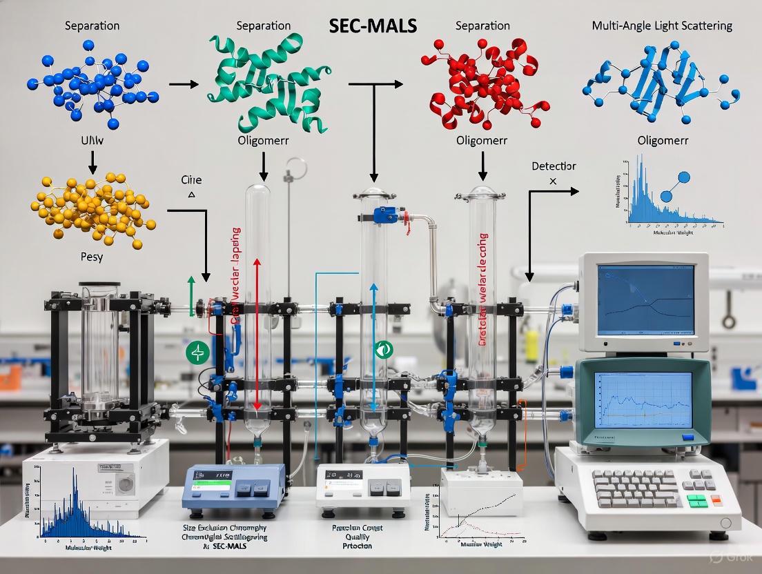
The following diagram outlines the standard end-to-end workflow for characterizing protein oligomeric states using SEC-MALS, from sample preparation to data analysis.
Application Notes and Protocols
Detailed Protocol: SEC-MALS Analysis of a Protein Oligomer
This protocol describes the steps for determining the oligomeric state and molecular weight of a protein sample using SEC-MALS, based on established methodologies [8] [7].
I. Materials and Reagents
Table 3: Research Reagent Solutions and Essential Materials
| Item | Function / Description | Example / Specification |
|---|---|---|
| SEC-MALS System | Core instrumentation. | FPLC/HPLC, MALS detector (e.g., Wyatt DAWN), dRI detector (e.g., Wyatt Optilab), UV detector [7]. |
| SEC Column | Separates molecules by hydrodynamic size. | Silica-based (e.g., SEPAX SRT SEC-300) or agarose-based (e.g., Superdex 200 Increase) [7]. |
| Running Buffer | Mobile phase compatible with the protein and column. | Filtered (0.1 µm) PBS or 25 mM HEPES, pH 7-7.5, 150 mM NaCl [7]. |
| Protein Sample | Analytic of interest. | High-purity protein in running buffer, filtered (0.02-0.2 µm) or centrifuged to remove aggregates [7]. |
| Sample Vials | Holds sample for injection. | Glass autosampler vials with low-volume inserts [7]. |
| Syringe Filters | Removes particulate matter that can damage column or create noise. | 0.02-0.2 µm pore size, compatible with protein sample [7]. |
II. Step-by-Step Procedure
System Preparation and Equilibration
- Prepare at least 1 liter of running buffer using HPLC-grade water and reagents. Filter the buffer through a 0.1 µm bottle-top polyether sulfone filter into a clean, sterile bottle [8].
- Connect the SEC column to the FPLC/HPLC system and flush it overnight at a low flow rate (e.g., 0.5 mL/min) to equilibrate the column and remove particulates. Ensure the flow does not stop until all runs are complete to maintain stability [8].
- Prime the dRI detector and purge its flow cell according to the manufacturer's instructions. Turn off the purge before starting sample runs [8].
System Cleanliness Check
- Verify the system's baseline noise by observing the signal from the 90-degree detector on the MALS instrument. The peak-to-peak noise should be no more than 50-100 microvolts. The refractive index signal should be stable to less than 1 x 10⁻⁷ refractive index units [8].
- Perform a "blank" injection of 100 µL of running buffer to check for particle contamination from the injector. If the resulting particle peak is larger than 1 mL in volume or 5 mV above baseline, perform additional blank injections or system maintenance until the signal is clean [8].
Sample Preparation
- Dialyze or dilute the protein sample into the running buffer to minimize the refractive index peak from the sample solvent.
- Determine the protein concentration accurately using a method such as UV absorbance.
- Filter the protein sample through a 0.025-0.2 µm syringe-tip filter or centrifuge at 10,000 x g for 15 minutes to precipitate insoluble aggregates [8]. A general guideline for injection concentration is 5–500 µg total, with a rule of thumb being ~10/mass (kDa) mg/mL. For example, BSA (67 kDa) at 2 mg/mL in a 100 µL injection provides a good signal [7].
- Load at least 110 µL of the prepared sample into a glass autosampler vial. The maximum injection volume is typically 100 µL [7].
SEC-MALS Data Acquisition
- In the control software (e.g., ASTRA), create a new method to acquire data from the MALS, UV, and dRI detectors. Synchronize the injection with the start of data collection, typically via an analog signal from the auto-injector [5].
- Inject the recommended volume of your protein sample (e.g., 100 µL) onto the column. The standard flow rate is 0.5-1.0 mL/min, depending on the column specifications.
- Allow the separation and data acquisition to run until the entire sample has eluted and all signals have returned to baseline.
Data Analysis
- Process the collected data using the appropriate software (e.g., ASTRA). The software will combine the light scattering and concentration data to calculate the absolute molar mass across the entire chromatogram.
- For each peak in the chromatogram, the weight-average molar mass (Mw) will be calculated. The measured Mw is compared to the theoretical mass of the monomer to determine the oligomeric state (e.g., a measured Mw twice the monomeric mass indicates a dimer).
Troubleshooting and Best Practices
- Abnormal Molar Mass Trends: If the calculated molar mass does not decrease monotonically with elution volume, this may indicate non-ideal SEC separation due to interactions between the analyte and the column matrix [6]. Mitigate this by adjusting buffer pH, ionic strength, or using a different column chemistry.
- Poor Recovery or Broad Peaks: This can be caused by protein aggregation or adherence to the column/injector. Ensure samples are free of aggregates by pre-purification with SEC and use recommended buffer conditions to maintain protein solubility [7].
- Accuracy of Molar Mass: For conjugate analysis (e.g., glycoproteins, protein-detergent complexes), accurate determination requires input of the specific refractive index increment (dn/dc) for each component. For proteins alone, a standard value of ~0.185 mL/g is often used, but direct measurement with the dRI detector is more accurate [5].
The oligomeric state of a protein is a fundamental determinant of its function, regulation, and role in disease. The ability to accurately characterize this state is therefore critical in biological research and biopharmaceutical development. SEC-MALS has emerged as an indispensable tool in this endeavor, providing an absolute, first-principles method for determining molar mass and oligomeric state independent of the pitfalls associated with standard SEC. By integrating the protocols and application notes outlined in this document, researchers can confidently employ SEC-MALS to elucidate the complex interplay between protein structure, function, and dysfunction, ultimately driving advances in both basic science and therapeutic innovation.
Size-exclusion chromatography (SEC) is a foundational technique for separating biomolecules like proteins based on their hydrodynamic volume. However, conventional analytical SEC relies on column calibration with reference standards to relate elution time to molar mass, introducing significant assumptions about the analyte's conformation and behavior. These assumptions frequently lead to inaccuracies, especially for non-globular proteins, conjugated species, or any molecule that does not mirror the properties of the standards used for calibration [5].
The combination of SEC with Multi-Angle Light Scattering (MALS) overcomes these limitations by providing an absolute determination of molar mass independent of elution time or reference standards [5] [9]. This application note details the core principles, experimental protocols, and key applications of SEC-MALS, framing it within the context of characterizing protein oligomeric states for drug development.
Core Principle: Absolute Molar Mass via First-Principles Measurement
The absolute nature of SEC-MALS stems from its direct measurement of fundamental physical properties using first-principles analysis. Unlike calibration-dependent methods, it does not assume a specific molecular conformation and is robust against non-ideal column interactions [5].
The Fundamental Relationship of Light Scattering
In a SEC-MALS experiment, as a sample elutes from the SEC column, it passes through the MALS detector where it is illuminated by a laser. The intensity of the scattered light is measured simultaneously at multiple angles. For a macromolecule in solution, the key relationship connecting the scattered light to its molar mass is derived from Rayleigh's theory, and is given by [10]:
K*·c / R(θ) = 1 / Mw · P(θ) + 2A₂c
Here:
- K* is an optical constant that depends on the solvent refractive index, laser wavelength, and the analyte's specific refractive index increment (dn/dc).
- c is the analyte concentration (determined by an in-line UV or dRI detector).
- R(θ) is the excess Rayleigh ratio (the scattered light intensity) measured at angle θ.
- Mw is the weight-average molar mass.
- P(θ) is a form factor that describes the angular dependence of scattering and relates to the molecule's size (root mean square radius, Rg).
- A₂ is the second virial coefficient, which is typically negligible at the low concentrations used in SEC [10].
Deconvoluting Molar Mass and Size
The multi-angle capability is crucial. The plot of K*c/R(θ) versus sin²(θ/2) (a Debye plot) allows for the extrapolation of scattered light intensity to zero angle. The y-intercept yields 1/Mw, while the slope provides the root mean square radius (Rg), given the molecule is sufficiently large (typically > 10 nm) [5] [10]. This process happens at each data slice across the chromatographic peak, providing a continuous, absolute measurement of molar mass and size throughout the entire elution profile.
Figure 1. SEC-MALS Instrumental Workflow. The sample is first separated by hydrodynamic volume in the SEC column. The eluent flows sequentially through the MALS detector, where light scattering is measured at multiple angles, and then through a concentration detector (UV or dRI), before data is collected and analyzed.
Experimental Protocol for Protein Oligomeric State Analysis
This protocol provides a detailed methodology for determining the absolute molar mass and oligomeric state of a protein therapeutic using SEC-MALS.
Research Reagent Solutions and Essential Materials
Table 1. Essential materials and reagents for a typical SEC-MALS experiment.
| Item | Function | Example for Proteins |
|---|---|---|
| HPLC/FPLC System | Provides solvent delivery, sample injection, and automated separation. | Any standard system (e.g., Waters ACQUITY, Wyatt MicroCal) [11]. |
| SEC Column | Separates protein species based on hydrodynamic size. | GTxResolve Premier SEC 1000 Å 3 µm column for mRNAs and larger proteins [11]. |
| MALS Detector | Measures scattered light intensity at multiple angles to determine Mw and Rg. | Wyatt DAWN (18-angle) or miniDAWN (3-angle) [5] [11]. |
| Concentration Detector | Quantifies protein concentration at each elution volume. | UV detector (for proteins with known extinction coefficient) or dRI detector (Optilab) [5]. |
| Mobile Phase | Dissolves and elutes the sample without interacting with it. | Phosphate-buffered saline (PBS), pH 7.4, 0.2 µm filtered [11]. |
| System Suitability Standard | Verifies system performance and determines instrument constants. | Bovine Serum Albumin (BSA) monomer (66.4 kDa) [11]. |
Step-by-Step Methodology
System Preparation and Equilibration
- Prepare the mobile phase (e.g., PBS, pH 7.4) and filter through a 0.2 µm membrane to remove particulate matter that would contribute to background light scattering noise [11].
- Connect and power on all instruments: HPLC, MALS, UV, and dRI detectors. Prime the system with mobile phase and install the SEC column.
- Equilibrate the entire system at the operational flow rate (e.g., 0.5-1.0 mL/min for analytical columns). Flush the system until a stable MALS baseline is achieved. This may require flushing with 20-40 column volumes; low background noise is critical for reliable results [11].
Determination of System Constants and Suitability
- Prepare a solution of a well-characterized standard, such as BSA monomer, at a known concentration (e.g., 5 mg/mL).
- Inject the standard and run the SEC-MALS method. The data analysis software (e.g., ASTRA) will use this peak to perform:
- Normalization: Relates the response of all light scattering detectors to the 90° detector.
- Alignment: Corrects for the delay volume between the MALS and concentration detectors.
- Band Broadening Correction: Accounts for peak broadening between detectors.
- Verify that the calculated molar mass of the BSA monomer is within expected error (66.4 kDa). This confirms the system is suitable for sample analysis [11].
Sample Analysis and Data Collection
- Prepare the protein sample at an appropriate concentration (typically 0.1-5 mg/mL). For oligomeric state analysis, a concentration series may be run to detect concentration-dependent aggregation or dissociation.
- Inject the sample and begin data collection. Synchronize the injection with the start of data acquisition in the ASTRA software.
- The software will collect light scattering (LS) and concentration (UV/dRI) data in real-time across the entire elution profile.
Data Analysis and Interpretation
- The ASTRA software uses the LS and concentration data at each elution slice to calculate the absolute molar mass, independent of retention time.
- Identify the molar mass of the main peak to confirm the native oligomeric state (e.g., monomer, dimer, trimer).
- Identify and quantify the molar mass of any high-mass species (aggregates) or low-mass species (fragments).
Table 2. Quantitative data from a representative SEC-MALS analysis of an mRNA sample, demonstrating the precision of the technique [11].
| Analyte Species | Theoretical Mass (kDa) | Measured Mass (kDa) - GTxResolve Column | Measured Mass (kDa) - Manufacturer A Column |
|---|---|---|---|
| Cas9 mRNA Monomer | ~ 4471 nt (Predicted) | Within 15% of predicted | Up to 50% different from predicted |
| Cas9 mRNA Dimer | ~ 8942 nt (Predicted) | Single-digit % precision | Lower confidence measurements |
Critical Experimental Considerations
The Essential Role of ∂n/∂c
The specific refractive index increment (∂n/∂c) is a critical parameter that relates the change in solution refractive index to analyte concentration. An error in ∂n/∂c translates directly into an equivalent error in the calculated molar mass [12]. For proteins, a ∂n/∂c value of ~0.185 mL/g is commonly used and is generally consistent across most proteins in aqueous buffers [5] [12]. For conjugated proteins (e.g., PEGylated proteins, glycoproteins) or polymers, ∂n/∂c must be measured experimentally or calculated for each component.
Addressing Column Interactions and Mobile Phase Composition
SEC-MALS is powerful because it reveals when SEC separation is non-ideal. If a molecule interacts with the column matrix (e.g., via electrostatic or hydrophobic interactions), its elution volume will not correlate with its size. However, since MALS determines mass independently of elution volume, the true molar mass is still reported accurately [5] [9]. In mixed solvents, preferential solvation can occur, where the local solvent composition around the polymer differs from the bulk mobile phase, potentially leading to inaccuracies in ∂n/∂c and thus molar mass if not properly accounted for [12].
Applications in Protein Therapeutics Development
SEC-MALS is indispensable throughout the biopharmaceutical development lifecycle.
- Oligomeric State and Conjugate Analysis: SEC-MALS is the gold standard for determining the native oligomeric state of proteins and the stoichiometry of complexes [13] [9]. For conjugated proteins (e.g., PEGylated proteins, antibody-drug conjugates, glycoproteins), combining MALS, UV, and dRI detection allows for the determination of the conjugation ratio and the individual molar masses of each component [5].
- Aggregation and Fragmentation Analysis: MALS is exceptionally sensitive to high-molar-mass aggregates, as the light scattering signal is proportional to Mw * c. This allows for precise quantification of aggregate levels and identification of their size, which is a critical quality attribute for therapeutic proteins [14] [9].
- Process Analytical Technology (PAT): MALS can be used as an in-line PAT tool during purification processes (e.g., HIC chromatography) to monitor aggregate levels in real-time. This enables automated fractionation based on preset molar mass criteria, improving yield and product quality [14].
Figure 2. SEC-MALS Applications in the Protein Therapeutic Lifecycle. The utility of SEC-MALS evolves from comprehensive characterization in early discovery to targeted release assays in Quality Assurance/Quality Control (QA/QC).
Size-exclusion chromatography (SEC) is a foundational technique for protein analysis, yet its conventional reliance on column calibration for molecular weight determination introduces significant inaccuracies for non-ideal or complex proteins. This application note details the inherent limitations of calibration-based SEC and establishes SEC coupled with multi-angle light scattering (SEC-MALS) as an absolute method for characterizing protein oligomeric states, aggregates, and complexes. We present validated protocols and experimental data demonstrating how SEC-MALS overcomes calibration dependencies to provide accurate molar mass and size measurements independent of protein conformation, column interactions, or structural modifications—enabling reliable characterization critical for biopharmaceutical development and basic research.
Analytical size-exclusion chromatography is widely employed for protein characterization, primarily for assessing purity, aggregation status, and oligomeric state. Traditional SEC operates on a simple premise: molecules separate based on their hydrodynamic volume as they pass through a porous stationary phase, with larger molecules eluting before smaller ones. The established practice involves calibrating the column with globular protein standards of known molecular weight to create a retention time-versus-molecular weight relationship [15] [16].
This approach suffers from critical foundational assumptions that frequently fail for complex proteins:
- Identical Molecular Conformation: All proteins are assumed to be perfectly globular, with the same shape and density as the calibration standards [5].
- Absence of Column Interactions: Separation is assumed to occur purely by size, without any enthalpic interactions (e.g., electrostatic or hydrophobic) between the protein and the stationary phase [5].
Deviations from these assumptions—commonplace with extended, denatured, glycosylated, or membrane-associated proteins—render the calibration curve invalid and lead to highly inaccurate molecular weight determinations [16]. Furthermore, the column itself can alter the sample through dilution or selective adsorption, changing the nature of protein aggregates before detection [17]. These limitations necessitate an absolute characterization method that does not depend on reference standards or elution time.
SEC-MALS: An Absolute Method for Protein Characterization
Core Principles of SEC-MALS
SEC-MALS overcomes the limitations of calibration-dependent SEC by combining the separation power of size-exclusion chromatography with the absolute detection capabilities of multi-angle light scattering. In SEC-MALS, the column serves only to separate molecules by hydrodynamic size; retention time is not used to determine molecular weight [5]. As separated components elute, they pass through a MALS detector, which measures light scattering intensity, and a concentration detector (typically UV or differential refractive index, dRI).
The molecular weight (M) is calculated at each elution volume using the fundamental relationship derived from Rayleigh scattering:
[ M = \frac{R(0)}{K \cdot c \cdot (dn/dc)^2} ]
Where:
- ( R(0) ) is the reduced Rayleigh ratio (scattered light intensity extrapolated to zero angle)
- ( K ) is an optical constant
- ( c ) is the solute concentration
- ( dn/dc ) is the refractive index increment [16]
This first-principles approach determines molar mass from 200 g/mol to 1 billion g/mol and size (radius of gyration, Rg) from 10 nm to 500 nm, independent of molecular shape, conformation, or column interactions [5].
Comparative Analysis: SEC vs. SEC-MALS
Table 1: Key differences between conventional SEC and SEC-MALS
| Parameter | Conventional SEC | SEC-MALS |
|---|---|---|
| Molecular Weight Basis | Relative to globular protein standards | Absolute, from first principles |
| Key Assumptions | Identical conformation & specific volume; no column interactions | No molecular shape or interaction assumptions required |
| Accuracy for Non-Globular Proteins | Low (e.g., intrinsically disordered, fibrous) | High |
| Accuracy for Modified Proteins | Low (e.g., glycoproteins, PEGylated) | High |
| Aggregate Detection & Quantification | Semi-quantitative, relies on separation | Quantitative, even for poorly-resolved peaks |
| Information on Conformation | Indirect, inferred from elution volume | Direct, via size (Rg) vs. mass relationship |
| Column Calibration | Required frequently | Not required |
Advantages of SEC-MALS for Complex Protein Systems
SEC-MALS provides particular advantages for characterizing challenging protein samples that confound traditional SEC analysis:
- Glycoproteins and PEGylated Proteins: These conjugated proteins exhibit different hydrodynamic volumes per unit mass compared to globular standards. SEC-MALS directly determines the absolute molar mass and can deconvolute the conjugation ratio when coupled with UV and RI detection [15].
- Membrane Proteins in Detergents: The detergent micelle contributes significantly to hydrodynamic volume. SEC-MALS can determine the true protein molar mass and oligomeric state, independent of the bound detergent [16].
- Intrinsically Disordered Proteins: With extended conformations, these proteins elute earlier than globular proteins of the same mass in SEC, leading to overestimated molecular weights. SEC-MALS provides accurate mass regardless of conformation [16].
- Oligomeric Complexes and Aggregates: SEC-MALS distinguishes between specific oligomers (dimers, trimers) and non-specific aggregates, and can identify heterogeneous populations or dynamic equilibria within a single peak [17] [15].
Experimental Protocols
Core SEC-MALS Protocol for Protein Characterization
Table 2: Essential reagents and equipment for SEC-MALS
| Category | Item | Specification/Function |
|---|---|---|
| Instrumentation | HPLC/FPLC System | Standard system with pump, autosampler, and column oven |
| SEC Column | Appropriate pore size for target protein (e.g., 150 Å for monomers, 300 Å for mAbs) | |
| MALS Detector | Measures light scattering at multiple angles (e.g., DAWN, miniDAWN) | |
| Concentration Detector | UV (for proteins with chromophores) and/or dRI (universal) | |
| Consumables | Mobile Phase Buffer | PBS or other compatible buffer, 0.1 µm filtered |
| Sample Filters | 0.1 µm or 0.22 µm syringe filters (e.g., PES) | |
| Standard Proteins | BSA (66.5 kDa) for system suitability testing |
Procedure:
- System Preparation: Filter 1 L of mobile phase (e.g., PBS) through a 0.1 µm filter. Connect the SEC column to the FPLC/HPLC system and equilibrate overnight at a low flow rate (e.g., 0.5 mL/min) to remove particulates and stabilize the system [18].
- Buffer Cleanliness Verification: Check the baseline signals of the MALS (90° detector noise should be ≤ 100 µV) and dRI detectors to ensure the system is free of particulate and refractive index contaminants [18].
- Sample Preparation: Prepare protein samples at 1-2 mg/mL in the mobile phase. Centrifuge at 10,000 × g for 15 minutes or filter through a 0.1 µm or 0.22 µm syringe filter to remove insoluble aggregates and particulates [18].
- System Suitability Test: Inject 100 µL of a BSA standard (1-2 mg/mL). Perform an analysis run to verify proper separation and molecular weight calculation (BSA monomer should yield approximately 66.5 kDa) [18].
- Sample Analysis:
- In the MALS software, create a new method. Set parameters: sample name, dn/dc (0.185 mL/g for most proteins in aqueous buffer [16]), UV extinction coefficient (if using UV), and mobile phase composition.
- Set the data collection duration to encompass the entire elution profile, typically until the total permeation volume is reached.
- In the FPLC software, program the method with the appropriate flow rate (e.g., 0.5-1.0 mL/min for analytical columns) and injection volume.
- Zero the dRI detector immediately before injection.
- Inject the sample. The MALS software should be triggered automatically by the injector signal [18].
- Data Analysis:
- In the MALS software, define baselines for all detector signals.
- Identify peaks by selecting the central 50-70% of each UV or RI peak.
- Align the signals from different detectors (UV, MALS, dRI) temporally if necessary.
- Apply a band-broadening correction if needed, using the monomer peak as a reference.
- Review the calculated molar mass across the peak. A monodisperse species will show a constant molar mass across the peak apex [18].
Advanced Application: IEX-MALS for Charge-Based Separation
When SEC fails to resolve proteins of similar size but different charge, IEX-MALS provides a powerful orthogonal approach.
Workflow:
IEX-MALS Separation Workflow
Procedure:
- Column Selection: Choose an anion-exchange (AIEX) or cation-exchange (CIEX) column based on the protein's isoelectric point.
- Method Development: Optimize the salt gradient (typically NaCl) or pH gradient to achieve resolution between species. A shallower gradient typically improves separation [19].
- System Setup: Connect the IEX column to the FPLC/HPLC system, followed by the UV, MALS, and dRI detectors.
- Buffer Preparation: Prepare starting buffer (low salt for binding) and elution buffer (high salt for elution). Filter all buffers through 0.1 µm filters.
- Analysis: Inject the sample and run with the optimized gradient. The MALS analysis will determine the molar mass of each eluting peak independently of the salt concentration [19].
- Data Interpretation: Note that in IEX, oligomers often elute after monomers at higher conductivity due to their stronger interaction with the matrix, contrary to SEC behavior [19].
Results and Data Interpretation
Representative SEC-MALS Data for Common Proteins
Table 3: Experimental SEC-MALS results for model proteins
| Protein | Theoretical Mass (kDa) | SEC-MALS Mass (kDa) | Oligomeric State | Key Observation |
|---|---|---|---|---|
| Bovine Serum Albumin (BSA) | 66.5 | 66-68 (Monomer) [19] 132-140 (Dimer) [19] | Monomer + Dimer | Demonstrates accurate mass determination of multiple species |
| Fibronectin | 263 | 278 ± 8 (Monomer) [19] ~500 (Oligomer) [19] | Monomer + Higher Oligomers | IEX-MALS provided superior separation of oligomers compared to SEC-MALS |
| Monoclonal Antibody | ~150 | ~150 (Monomer) [20] ~300 (Dimer) [20] | Monomer + Aggregate | Confirmed constant dn/dc in RP-UPLC-MALS for mAb analysis |
Case Study: Overcoming SEC Limitations with MALS
Challenge: Characterizing fibronectin oligomers, which co-elute as an asymmetric, poorly-resolved peak in SEC, preventing accurate quantification of monomers versus oligomers [19].
SEC-MALS Result: The molar mass across the heterogeneous peak varied continuously, confirming the presence of multiple, unresolved species but preventing precise determination of individual oligomeric states [19].
IEX-MALS Solution: Using anion-exchange chromatography with MALS detection, fibronectin monomers (278 ± 8 kDa) were clearly separated from higher molecular weight species (~500 kDa) and aggregated fractions. This high-resolution separation enabled accurate molar mass determination and quantification of each distinct population [19].
Discussion
Expanding the Toolkit: Complementary MALS Approaches
While SEC-MALS serves as the primary workhorse for protein characterization, other separation techniques coupled with MALS address specific challenges:
- IEX-MALS: Ideal for separating and characterizing protein isoforms, charge variants, and oligomers with poor SEC resolution [19].
- RP-UPLC-MALS: Provides high-resolution analysis of hydrophobic proteins, fragments, and chemically modified species under denaturing conditions [20].
- FFF-MALS: Suitable for very large complexes, nanoparticles, and extremely polydisperse systems that exceed the size range of SEC columns [5].
Troubleshooting and Best Practices
- Sample Preparation: Always filter or centrifuge samples to remove dust and insoluble aggregates, which cause significant light scattering artifacts [18].
- Buffer Compatibility: Ensure the mobile phase is free of fluorescent contaminants and has minimal absorbance at the laser wavelength.
- Concentration Optimization: Use sufficient protein concentration for adequate light scattering signal but avoid overloading the column. For most proteins, 1-2 mg/mL is appropriate.
- dn/dc Verification: While 0.185 mL/g is standard for most proteins in aqueous buffers, confirm this value for conjugated proteins or those in unusual solvents [16].
Conventional SEC, with its dependence on column calibration using globular standards, provides unreliable molecular weight data for a wide range of biologically and therapeutically relevant proteins. SEC-MALS eliminates this dependency by providing absolute molar mass and size determination directly from first principles, regardless of molecular conformation, column interactions, or post-translational modifications. The detailed protocols and case studies presented herein establish SEC-MALS as an essential, robust methodology for accurate protein characterization in basic research and biopharmaceutical development.
Within biophysical characterization and therapeutic drug development, determining the absolute molar mass, size, and oligomeric state of proteins is critical for understanding function, stability, and efficacy. Size-exclusion chromatography coupled with multi-angle light scattering (SEC-MALS) has emerged as a powerful, absolute technique for these measurements, independent of the limitations posed by column calibration standards [5] [21]. This Application Note details the core principles, experimental protocols, and key measurements of SEC-MALS, framing them within the context of protein oligomeric state characterization for research and development scientists.
SEC-MALS overcomes the fundamental assumptions of analytical SEC, which requires that analytes share the same conformation and hydrodynamic density as the standards used for calibration [5]. For complex molecules like glycoproteins, PEGylated proteins, protein complexes, and intrinsically disordered proteins, these assumptions often fail, leading to inaccurate molar mass determinations [5]. In contrast, SEC-MALS provides an absolute measurement by combining the separation power of SEC with first-principles light scattering analysis, enabling accurate characterization of monomers, oligomers, and aggregates under native solution conditions [5] [22] [23].
Key Principles and Advantages of SEC-MALS
Absolute Molar Mass Determination
In SEC-MALS, the molar mass (M) is determined directly at each elution volume using the fundamental relationship between the scattered light intensity at zero angle (R(0)), measured by the MALS detector, and the analyte concentration (c), measured by a UV or refractive index (dRI) detector [5] [24] [10]. The governing equation is derived from Rayleigh scattering theory:
K*c / R(0) = 1 / M
where K* is an optical constant that includes the refractive index increment (dn/dc) of the analyte-solvent system [10] [25]. This calculation is independent of elution volume and does not rely on comparison to molecular standards, making it an absolute technique [5] [24]. SEC-MALS can determine molar masses from 200 g/mol to over 1 billion g/mol [5] [26].
Size Measurement via Radius of Gyration (Rg)
For molecules larger than approximately 10-15 nm, the angular dependence of the scattered light can be analyzed to determine the root mean square radius, or radius of gyration (Rg) [5] [27] [26]. Rg represents the root mean square distance of the molecule's mass elements from its center of mass [27]. The slope of the plot of scattered light intensity versus the square of the scattering angle is used to calculate Rg, providing insight into the molecule's conformation and compactness [5] [25]. For smaller molecules like most monomeric proteins that scatter light isotropically, Rg cannot be determined via light scattering and requires alternative methods [27] [26].
Oligomeric State Characterization
A primary application of SEC-MALS in protein research is the identification and quantification of oligomeric states [22] [23] [21]. Because MALS measures the absolute molar mass of each separated species in a mixture, it can directly distinguish monomers, dimers, trimers, hexamers, and higher-order aggregates based on their measured mass [22] [7]. This is crucial for characterizing therapeutic proteins, where aggregation can impact safety and efficacy [23], and for studying biological systems where oligomerization is a key regulatory mechanism [21].
Table 1: Key Parameters Measured by SEC-MALS and Their Significance
| Parameter | Definition | Typical SEC-MALS Range | Significance in Protein Characterization |
|---|---|---|---|
| Absolute Molar Mass | Weight-average mass (Mw) determined from first principles [5] [24] | 200 Da – 1 GDa [5] [26] | Defines native oligomeric state (monomer, dimer, etc.); detects aggregates [22] [21] |
| Radius of Gyration (Rg) | Root-mean-square radius from mass center [5] [27] | 10 nm – 500 nm [5] [26] | Indicates molecular compactness and conformation; distinguishes globular from extended structures [5] [27] |
| Hydrodynamic Radius (Rh) | Radius of a hypothetical hard sphere that diffuses at the same rate [27] [26] | 0.5 nm – 100 nm (via DLS) [26] | Assesses hydrodynamic size; Rg/Rh ratio provides conformation insight [27] |
Comparison with Conventional SEC
Table 2: SEC-MALS vs. Conventional Calibration-Based SEC
| Aspect | SEC-MALS | Conventional SEC |
|---|---|---|
| Basis of Molar Mass | First-principles light scattering and concentration measurements [5] [25] | Calibration curve derived from protein standards [5] [21] |
| Dependence on Standards | Independent; no standards required [5] | Fully reliant; accuracy depends on standard suitability [5] [24] |
| Impact of Molecular Shape | Accurate for any conformation (globular, extended, random coil) [5] [21] | Incorrect for non-globular or denatured proteins [5] |
| Impact of Column Interactions | Unaffected; molar mass is determined independently of elution volume [5] [25] | Elution volume is shifted, leading to erroneous molar mass [5] |
| Information Obtained | Absolute molar mass, Rg, and (with DLS) Rh [5] [26] | Apparent molar mass only [5] |
Experimental Protocols
SEC-MALS Workflow for Protein Analysis
The following diagram illustrates the key stages of a standard SEC-MALS experiment:
Detailed Methodology
Sample and Buffer Preparation
- Buffer Selection and Matching: The running buffer must be compatible with both the SEC column and the protein sample. A common recommendation is 25 mM HEPES pH 7-7.5, 150 mM NaCl [7]. The sample must be in the running buffer or dialyzed against it to avoid refractive index (RI) peaks from buffer mismatch [26] [7].
- Sample Filtration/Centrifugation: To protect the column and ensure clear data, samples must be free of particulates and large aggregates. Filter samples using a 0.02 µm – 0.2 µm syringe filter or perform high-speed centrifugation immediately before injection [26] [7].
- Sample Concentration: The optimal concentration depends on the protein's molar mass. As a general guideline, for a protein like BSA (67 kDa), an injection of 100 µL at 2 mg/mL provides a strong signal [7]. Scattering intensity is proportional to the product of molar mass and concentration, so smaller proteins require higher concentrations. A useful rule of thumb is to aim for a total mass of ~5 – 500 µg per injection [7].
System Configuration and Execution
- Instrument Setup: A standard SEC-MALS system includes an FPLC/HPLC, an SEC column, a MALS detector (e.g., Wyatt DAWN), and a concentration detector (UV and/or dRI, e.g., Wyatt Optilab) [5] [26]. The MALS detector is typically placed after the UV detector and before the dRI detector [5].
- Column Selection: Choose an SEC column appropriate for the protein's size range. For most proteins and their oligomers, a column like a Superdex 200 Increase or TSKgel G3000SWxl is suitable [26] [7]. Ensure the column's pH tolerance matches the buffer.
- Data Collection: The system is equilibrated with at least 2-3 column volumes of filtered and degassed running buffer. Data from the MALS, UV, and dRI detectors are collected and synchronized in specialized software (e.g., Wyatt ASTRA) for analysis [5] [7].
Data Analysis for Absolute Parameters
- Molar Mass Calculation: The software uses the combined MALS and concentration data at each data slice across the peak to calculate the absolute molar mass. The key parameters that must be correctly set are the dn/dc value (typically 0.185 mL/g for proteins in aqueous buffer) and the UV extinction coefficient for the specific protein [5] [7].
- Rg Calculation: For molecules displaying angular dependence, the software fits the angular scattering data to determine Rg [5] [27]. If Rg cannot be determined directly via light scattering (for small proteins), it can sometimes be estimated using intrinsic viscosity data and the Flory-Fox equation, though this introduces approximation [27].
- Oligomeric State Assignment: The measured molar mass of each peak is compared to the theoretical monomer mass. A measured mass of ~2x the monomer mass indicates a dimer, ~3x a trimer, and so on [22] [21]. The relative peak areas from the concentration detector provide the quantification of each oligomeric species.
The Scientist's Toolkit: Essential Research Reagents and Materials
Table 3: Key Research Reagent Solutions for SEC-MALS
| Item | Function/Purpose | Examples/Recommendations |
|---|---|---|
| SEC-MALS System | Core instrumentation for separation and absolute characterization. | Wyatt DAWN (MALS) + Optilab (dRI) + Agilent/Shimadzu HPLC [21] [26] [7] |
| SEC Columns | Separates protein species by hydrodynamic volume. | Superdex 200 Increase, TSKgel G3000SWxl, SEPAX SRT SEC-300 [26] [7] |
| Running Buffer | Provides the solvent environment for separation and analysis. | 25 mM HEPES, 150 mM NaCl, pH 7.5; or PBS pH 7.4 [26] [7] |
| Syringe Filters | Removes particulates and large aggregates to protect the column. | 0.02 µm or 0.22 µm pore size (e.g., Whatman Anotop) [26] [7] |
| Autosampler Vials | Holds sample for injection. | Low-volume insert vials to minimize dead volume [7] |
| Data Analysis Software | Acquires and analyzes data to determine molar mass, size, and oligomeric state. | Wyatt ASTRA software [5] [7] |
Advanced Applications and Considerations
Analysis of Protein Conjugates and Complexes
SEC-MALS with multiple concentration detectors (UV and dRI) is uniquely powerful for characterizing conjugated proteins. By analyzing the differential response of UV and dRI to the protein and modifier components, SEC-MALS-UV-RI can determine the molar mass of each component and the conjugation ratio in glycoproteins, PEGylated proteins, AAVs, and protein-detergent complexes [5] [26] [7]. For example, it can quantify the ratio of empty to full capsids in AAV preparations [5].
Handling Challenging Samples: The Case of [Fe-S] Proteins
Proteins that absorb light at the MALS laser wavelength (e.g., 658 nm), such as iron-sulfur ([Fe-S]) cluster-containing proteins, require a correction to the light scattering data [21]. Without this correction, the measured molar mass will be inaccurate. The absorption correction can be applied within the analysis software once the protein's absorbance at the laser wavelength is known [21]. This principle applies to any light-absorbing or fluorescent sample.
When SEC-MALS is Not Sufficient: Alternative MALS Couplings
If an analyte interacts strongly with the SEC column's stationary phase or is beyond the column's size separation range, the separation will fail. In such cases, an alternative separation technique, Field-Flow Fractionation (FFF), can be coupled with MALS (FFF-MALS) [5] [26]. FFF-MALS is ideal for very large or broadly distributed samples, such as viruses, gene vectors, and large protein aggregates, covering a size range from 1 nm to 1000 nm [26] [25].
A Practical SEC-MALS Workflow: From System Setup to Diverse Applications
The precise determination of a protein's oligomeric state—whether it exists as a monomer, dimer, or higher-order complex in its native solution—is a critical step in biophysical characterization. This is paramount in biomedical research and biopharmaceutical development, as oligomeric state directly influences biological activity, stability, and efficacy [16]. While analytical Size-Exclusion Chromatography (SEC) has been widely used for this purpose, it is a relative technique that relies on comparison to standardized proteins, making it prone to error when analyzing non-globular proteins, conjugates, or complexes that elute anomalously [5] [16].
The integration of Multi-Angle Light Scattering (MALS) with SEC transforms this approach into an absolute method. SEC-MALS overcomes the limitations of calibration-based SEC by determining molar mass directly from first principles at each point in the chromatogram, independent of elution volume or molecular conformation [5]. This technique is indispensable for confirming the native oligomeric state of protein complexes, quantifying aggregates and fragments, and characterizing challenging samples like glycoproteins, PEGylated therapeutics, and detergent-solubilized membrane proteins [5] [28] [16]. This application note details the core configuration and protocols for a robust SEC-MALS system incorporating HPLC/FPLC, MALS, UV, and Refractive Index (RI) detectors, providing researchers with a definitive tool for protein characterization.
System Configuration and Principles of Operation
Core System Components
A complete SEC-MALS system is built around a standard HPLC or FPLC system, to which specialized detectors are added. The core components and their functions are summarized in the table below.
Table 1: Core Components of an SEC-MALS System
| System Component | Primary Function | Key Considerations |
|---|---|---|
| HPLC/FPLC System | Solvent delivery, sample injection, and separation. | Provides pump, autosampler, degasser, and column oven. |
| SEC Column | Separates molecules by hydrodynamic size (volume). | Choice of column resin and pore size depends on the protein size range. |
| UV/Vis Detector | Measures concentration based on light absorption. | Requires knowledge of the protein's extinction coefficient (ε). |
| MALS Detector | Measures light scattered by the analyte at multiple angles. | Determines absolute molar mass (M) and size (Rg). |
| dRI Detector | Measures concentration based on refractive index change. | Provides a universal concentration signal; requires known dn/dc. |
| Software | Data acquisition from all detectors and analysis. | Synchronizes data streams and performs first-principles calculations. |
Detector Integration and Flow Path
The optimal placement of detectors in the flow path is critical for accurate data collection and analysis. The typical order is: SEC Column → UV Detector → MALS Detector → dRI Detector [5]. This sequence is recommended because the UV flow cell is generally more robust and can tolerate small particles that might break away from the column. Placing the MALS detector upstream of the dRI detector ensures that the sample enters the MALS flow cell without any prior dilution or mixing that could occur in the dRI detector's larger volume. It also protects the sensitive dRI cell from potential pressure spikes.
Principle of Absolute Molar Mass Determination
SEC-MALS is considered an absolute technique because it calculates molar mass from fundamental physical relationships, without relying on calibration standards. The key equation is:
[ M = \frac{R(0)}{K \times c \times (dn/dc)^2} ]
Where:
- M is the molar mass of the analyte.
- R(0) is the reduced Rayleigh ratio (the light scattered by the analyte) extrapolated to zero angle, as measured by the MALS detector.
- K is an optical constant dependent on the instrument and solvent.
- c is the mass concentration of the analyte, measured by the UV or dRI detector.
- dn/dc is the refractive index increment of the analyte in the mobile phase [5] [16].
For proteins, the dn/dc value is remarkably consistent at approximately 0.185 mL/g for most pure proteins in aqueous buffers, simplifying concentration determination via dRI [16]. The angular dependence of the scattered light also allows for the determination of the root-mean-square radius (Rg, or radius of gyration) for molecules larger than ~10 nm [5].
The following workflow diagram illustrates the integration of these components and the data analysis process.
Detailed Experimental Protocol
Materials and Reagents
Table 2: Essential Research Reagents and Materials
| Item | Specification / Function |
|---|---|
| SEC Column | Size-exclusion column suitable for the target protein size range (e.g., Superdex 200 Increase, Superose 6). |
| Mobile Phase Buffer | A filtered (0.1 µm) and degassed buffer that preserves protein native state and minimizes column interactions (e.g., PBS, HEPES, Tris). |
| Protein Standards | Monodisperse, stable proteins (e.g., BSA, thyroglobulin) for system performance qualification, not calibration. |
| Sample | Purified protein sample, clarified by centrifugation (e.g., 15,000 x g) and filtration (0.22 µm) prior to injection. |
Step-by-Step Method
System Preparation and Equilibration:
- Install and flush the SEC column with the chosen mobile phase according to the manufacturer's instructions.
- Connect the detectors in the recommended order and ensure all data lines are properly connected to the ASTRA software.
- Equilibrate the entire system with at least 2-3 column volumes of mobile phase until a stable dRI and light scattering baseline is achieved.
Detector Calibration and Normalization:
- MALS Detector: Perform normalization using an isotropic scatterer such as toluene or a protein monomer standard with known Rayleigh ratio to define the response of each photodiode [5].
- dRI Detector: Calibrate using the known dn/dc value of the solvent or a standard with a known concentration and dn/dc.
- UV Detector: Verify wavelength accuracy if concentration from UV will be used.
Sample Analysis:
- Prepare the protein sample at an appropriate concentration (typically 0.5-2 mg/mL for most proteins, depending on molar mass).
- Inject a defined volume (e.g., 10-100 µL) onto the column.
- Initiate the method with a constant flow rate (e.g., 0.5-1.0 mL/min for analytical columns), starting data acquisition in ASTRA software.
- The software will collect synchronized data from the UV, MALS, and dRI detectors throughout the run.
Data Analysis in ASTRA Software:
- After the run, define the peaks corresponding to the protein species in the chromatogram.
- For each peak (or for every data slice across the peak), the software will use the signals from the concentration detector (UV or dRI) and the MALS detector to calculate the absolute molar mass using the fundamental equation.
- The calculated molar mass across the peak is used to assess homogeneity. A constant molar mass across the peak indicates a monodisperse species, while a slope indicates heterogeneity or poor separation.
Key Applications in Protein Characterization
The SEC-MALS system configured above enables several advanced applications critical for modern protein research.
Confirmation of Native Oligomeric State and Purity: SEC-MALS directly determines the molar mass of the eluting species, distinguishing monomers from dimers, trimers, and higher-order native oligomers without assumptions about shape [16]. It simultaneously assesses sample homogeneity and quantifies the levels of soluble aggregates and fragments, which is a regulatory requirement for biotherapeutic characterization [16].
Characterization of Conjugated Proteins: For glycoproteins, PEGylated proteins, or surfactant-solubilized membrane proteins, the relationship between hydrodynamic size and mass differs from globular standards. SEC-MALS accurately determines the total molar mass of the conjugate and, by combining UV and dRI signals, can deconvolute the contribution of each component (e.g., protein and carbohydrate) to determine the conjugation ratio [5] [28].
Stoichiometry Analysis of Non-covalent Complexes: The absolute molar mass measurement allows for the precise determination of the stoichiometry of protein-protein or protein-nucleic acid complexes. By comparing the measured mass to the theoretical mass of the individual components, the exact composition of the complex (e.g., 1:1, 2:1, 2:2) can be determined [5] [16].
Formulation and Stability Studies: SEC-MALS is a powerful tool for screening buffer conditions, excipients, and stresses (e.g., temperature, pH) that influence protein oligomerization and aggregation propensity [28]. It can be used to measure the second virial coefficient (A2), a parameter that quantifies protein-protein interactions in solution and helps in identifying conditions that minimize aggregation [28].
The following diagram illustrates the logical decision process for interpreting SEC-MALS data to characterize protein oligomerization.
Size Exclusion Chromatography with Multi-Angle Light Scattering (SEC-MALS) is an absolute technique for determining the molar mass and size of macromolecules in solution, overcoming the significant limitations of conventional SEC which relies on column calibration with molecular standards [5]. This technique is particularly valuable for characterizing protein oligomeric states—including monomers, dimers, hexamers, and higher-order aggregates—without assumptions about molecular conformation or shape [29]. SEC-MALS provides first-principles analysis by physically separating molecules via SEC and then directly measuring their light-scattering properties, enabling accurate determination of native oligomeric states, stoichiometry of complexes, and assessment of sample homogeneity [5] [30].
The fundamental advantage of SEC-MALS lies in its independence from elution volume for molecular weight determination. Whereas analytical SEC assumes molecules elute according to a calibration curve and share identical conformational properties with standards, SEC-MALS directly measures molar mass at each elution point, making it uniquely suited for characterizing complex biomolecules like glycoproteins, PEGylated proteins, protein-protein complexes, and membrane proteins that often deviate from ideal globular behavior [5]. This technical note provides a comprehensive protocol for implementing SEC-MALS specifically for protein oligomeric state characterization.
Theoretical Principles of SEC-MALS Analysis
Fundamental Light Scattering Equations
In SEC-MALS analysis, the key measured parameters are the light scattering intensity at multiple angles and the sample concentration at each elution volume. The fundamental equation connecting these measurements to molar mass is derived from the relationship between scattered light intensity and molecular properties:
The light scattering data is analyzed using the following equation:
[\frac{K^*c}{R(θ)} = \frac{1}{Mw} \left( \frac{1}{P(θ)} \right) + 2A2c]
Where:
- (K^) is an optical constant containing Avogadro's number (N_A), the wavelength of light (λ_0), the refractive index of the solvent (n_0), and the refractive index increment (dn/dc) of the solute: (K^ = \frac{4π^2n0^2(dn/dc)^2}{NAλ_0^4})
- (c) is the solute concentration (determined by UV or dRI detection)
- (R(θ)) is the excess Rayleigh ratio (scattered light intensity) at angle θ
- (M_w) is the weight-average molar mass
- (P(θ)) is the form factor describing the angular dependence of scattering
- (A_2) is the second virial coefficient (typically negligible in SEC due to low concentrations)
For monodisperse proteins, the form factor (P(θ)) can be related to the root mean square (rms) radius (R_g) (also known as the radius of gyration) through the relationship:
[P(θ) ≈ 1 - \frac{(16π^2n0^2)}{3λ0^2} R_g^2 sin^2(θ/2)]
In practice, the Zimm plot method is used, where (K^*c/R(θ)) is plotted against (sin^2(θ/2)) at each elution slice, enabling simultaneous determination of (Mw) (from the intercept at zero angle) and (Rg) (from the initial slope) [5].
Key Parameters for Protein Analysis
Table 1: Essential Parameters for SEC-MALS Analysis of Proteins
| Parameter | Symbol | Typical Protein Value | Measurement Method |
|---|---|---|---|
| Refractive Index Increment | (dn/dc) | 0.185 mL/g | Typically assumed for most proteins; can be measured experimentally using dRI detector |
| UV Extinction Coefficient | (ε) | Variable (protein-specific) | Determined from amino acid composition or measured experimentally |
| Second Virial Coefficient | (A_2) | ~0 in SEC conditions | Assumed negligible due to separation conditions |
| Molar Mass Range | (M) | 200 - 10^9 g/mol | Accessible with modern MALS detectors [5] |
| Size Range (Rg) | (R_g) | 10-500 nm | Measurable with 18-angle MALS instruments |
Materials and Equipment
Research Reagent Solutions
Table 2: Essential Materials for SEC-MALS Experiments
| Category | Item | Specifications | Function/Purpose |
|---|---|---|---|
| Chromatography System | HPLC/FPLC System | Standard HPLC or FPLC with pump, degasser, autosampler | Fluid delivery and sample introduction |
| Separation Column | SEC Column | Pore size appropriate for target protein size range | Hydrodynamic size-based separation |
| Primary Detectors | MALS Instrument | DAWN (18 angles), miniDAWN (3 angles), or microDAWN (UHP-SEC) | Measures light scattering at multiple angles for molar mass determination |
| UV/Vis Detector | Standard HPLC UV detector (e.g., 280 nm for proteins) | Measures protein concentration via absorbance | |
| dRI Detector | Optilab or microOptilab | Measures refractive index for concentration determination | |
| Software | Analysis Software | ASTRA or equivalent | Data acquisition and analysis |
| Buffers & Solutions | Mobile Phase | Protein-compatible buffer with preservatives | Sample separation and transport |
| Protein Standards | Monodisperse proteins of known molar mass | System validation and quality control | |
| Sample Preparation | Filtration Units | 0.1 μm or 0.22 μm pore size | Mobile phase and sample clarification |
Recommended System Configuration
A complete SEC-MALS system typically includes [5]:
- Basic Configuration: HPLC/FPLC system with UV detector, SEC column, MALS instrument, and computer with ASTRA software
- Standard Configuration: Adds Optilab dRI detector for direct concentration measurement independent of extinction coefficients
- Extended Configuration: Incorporates additional detectors such as WyattQELS for hydrodynamic radius or ViscoStar for intrinsic viscosity measurements
For protein analysis specifically, the miniDAWN MALS detector is often combined with popular FPLC systems and an Optilab dRI detector, which is particularly valuable as nearly all proteins have similar dRI response ((dn/dc)), eliminating the need to know extinction coefficients for each peak [5].
Experimental Protocol
Mobile Phase Preparation and Column Equilibration
Mobile Phase Selection and Preparation
Select appropriate buffer: Choose a buffer system compatible with your protein and SEC column. Common choices include:
- Phosphate Buffered Saline (PBS), pH 7.4
- Tris-buffered saline, pH 7.0-8.0
- HEPES buffer, pH 7.0-7.5
- Avoid buffers with high ultraviolet absorbance or high light scattering properties
Buffer preparation protocol:
- Use high-purity water (HPLC-grade) and analytical-grade salts
- Filter through 0.1 μm or 0.22 μm membrane filter to remove particulate matter
- Degas for 20-30 minutes using sonication, helium sparging, or vacuum filtration to prevent bubble formation in detectors
Additive considerations:
- Include 100-200 mM salt (e.g., NaCl) to minimize electrostatic interactions with column matrix
- For membrane proteins, include appropriate detergents at concentrations above critical micelle concentration
- Consider adding 1-5% glycerol or other stabilizers for fragile proteins
Column Equilibration
- Install SEC column following manufacturer's instructions, noting flow direction
- Connect to system with MALS detector positioned downstream of UV detector and upstream of dRI detector
- Equilibrate column with at least 5 column volumes (typically 50-100 mL) of mobile phase at the intended flow rate
- Monitor baseline signals from all detectors (UV, MALS, dRI) until stable (typically < 1% variation over 10 minutes)
- Verify system performance using protein standards if conducting quantitative analysis
Sample Preparation Guidelines
Protein Sample Requirements
- Purity: Samples should be at least 90% pure to avoid interference from contaminants
- Concentration: Optimize concentration based on expected molar mass:
- For proteins < 100 kDa: 1-5 mg/mL
- For proteins 100-500 kDa: 0.5-2 mg/mL
- For large complexes > 500 kDa: 0.1-1 mg/mL
- Volume: Typical injection volumes are 10-100 μL, depending on column size
Sample Preparation Protocol
- Buffer exchange: Transfer protein into mobile phase using:
- Dialysis (≥ 4 hours with 2-3 buffer changes)
- Size exclusion chromatography (desalting columns)
- Centrifugal filtration devices with appropriate molecular weight cutoff
- Clarification: Centrifuge at 10,000-15,000 × g for 10 minutes or filter through 0.1 μm or 0.22 μm centrifugal filters
- Storage: Keep samples on ice until injection to maintain stability
- Documentation: Record precise concentration, buffer composition, and preparation details
System Setup and Method Configuration
Detector Configuration and Calibration
MALS detector preparation:
- Power on MALS instrument and allow laser to stabilize (typically 30 minutes)
- Perform normalization using pure mobile phase or a monodisperse protein standard (e.g., bovine serum albumin)
- Verify detector alignment and responsivity according to manufacturer protocols
Concentration detector setup:
- UV detector: Set appropriate wavelength (typically 280 nm for proteins)
- dRI detector: Allow temperature equilibration (≥ 1 hour), set mobile phase refractive index
System synchronization:
- Connect analog outputs from concentration detectors to MALS instrument
- Configure analog input signals for injection synchronization
- Set data collection rate to 1-2 Hz (standard SEC) or up to 10 Hz (UHP-SEC)
SEC Method Parameters
- Flow rate: Set according to column specifications (typically 0.5-1.0 mL/min for analytical columns)
- Run time: Allow sufficient time for elution of all components (typically 30-60 minutes)
- Temperature: Maintain consistent temperature (typically 20-25°C) using column heater if available
- Detection parameters: Configure data collection for all detectors with appropriate ranges
Data Collection and Processing
Sample Run Procedure
- Blank injection: Perform injection of mobile phase to establish baseline and identify system peaks
- Standard injection (optional): Run protein standard for system validation
- Sample injection:
- Load appropriate volume of prepared sample into injection loop
- Initiate run and ensure data collection is synchronized across all detectors
- Monitor real-time signals to verify proper separation and detection
- Column cleaning: After sample run, flush with 1-2 column volumes of mobile phase
Data Analysis Workflow
- Define peak regions: Identify sample peaks in chromatogram, excluding void volume and salt peaks
- Set baselines: Establish proper baselines for each peak in all detector signals
- Input analysis parameters:
- (dn/dc) value: Use 0.185 mL/g for proteins or measure experimentally
- UV extinction coefficient: Enter protein-specific value if using UV for concentration
- Perform molar mass calculation: Software automatically determines molar mass at each elution slice
- Review results: Examine molar mass distributions across peaks and validate with expected values
Workflow Visualization
SEC-MALS Experimental Workflow: This diagram illustrates the sequential steps in SEC-MALS analysis from sample preparation through data interpretation.
Expected Results and Data Interpretation
Representative SEC-MALS Data for Insulin Oligomeric States
Table 3: Expected SEC-MALS Results for Insulin Oligomeric States [29]
| Oligomeric State | Theoretical Molar Mass (kDa) | Expected SEC-MALS Result (kDa) | Elution Volume | Notes |
|---|---|---|---|---|
| Monomer | 5.8 | 5.8 ± 0.3 | Latest | Predominant form under denaturing conditions |
| Dimer | 11.6 | 11.6 ± 0.6 | Intermediate | Common in pharmaceutical formulations |
| Hexamer | 34.8 | 34.8 ± 1.7 | Earliest | Zinc-stabilized form in physiological conditions |
| Higher Aggregates | Variable | > 40 | Varies | Indication of protein instability or misfolding |
Data Interpretation Guidelines
- Monodisperse peaks: A constant molar mass across a chromatographic peak indicates a monodisperse species with homogeneous oligomeric state
- Mass gradients: Decreasing molar mass across a peak suggests non-ideal column interactions or protein self-association
- Multiple peaks: Distinct peaks with different molar masses indicate stable oligomeric states or aggregates
- Validation: Compare measured molar mass with expected values based on sequence and known oligomerization
The example of insulin characterization demonstrates how SEC-MALS can identify and quantify multiple oligomeric states in a single experiment, providing crucial information for formulation development and quality control of therapeutic proteins [29].
Troubleshooting and Quality Control
Common Issues and Solutions
Table 4: SEC-MALS Troubleshooting Guide
| Problem | Potential Causes | Solutions |
|---|---|---|
| No light scattering signal | Protein concentration too low, air bubbles, detector issue | Increase concentration, purge flow cell, check detector alignment |
| High baseline noise | Dirty flow cell, contaminated mobile phase, air bubbles | Clean flow cell, prepare fresh mobile phase, degas buffers |
| Abnormal molar mass values | Incorrect dn/dc, poor baseline selection, protein aggregation | Verify dn/dc value, adjust baselines, check sample preparation |
| Poor chromatographic separation | Column degradation, incorrect flow rate, sample overload | Replace column, adjust flow rate, reduce injection volume |
| Mismatch between UV and LS peaks | Non-uniform conjugation, different components co-eluting | Review conjugation homogeneity, improve separation conditions |
Quality Control Measures
- Regular system validation: Perform periodic runs with protein standards of known molar mass
- Mobile phase consistency: Use the same batch of mobile phase for all experiments in a series
- Replicate injections: Perform duplicate or triplicate injections to ensure reproducibility
- Negative controls: Include buffer-only injections to identify system artifacts
- Positive controls: Run well-characterized proteins to verify system performance
Applications in Protein Therapeutics Development
SEC-MALS provides critical data for biopharmaceutical development, particularly for:
- Biosimilar characterization: Verifying similarity of oligomeric state distribution to reference products
- Formulation optimization: Assessing stability and aggregation propensity under different conditions
- Quality control: Monitoring batch-to-batch consistency of therapeutic proteins
- Conjugate analysis: Determining drug-to-antibody ratio for antibody-drug conjugates
- Vaccine development: Characterizing virus-like particles and protein complexes
The case study with insulin demonstrates how SEC-MALS enables identification and quantification of multiple oligomeric states that directly impact product safety, efficacy, and stability [29]. Similarly, research on tyrosine hydroxylase (TH) and its interaction with DNAJC12 utilized SEC-MALS to confirm the monomeric state of DNAJC12 (22.7 ± 0.14 kDa), providing crucial information for understanding the chaperone-client relationship relevant to Parkinson's disease [30].
Within the framework of size exclusion chromatography coupled with multi-angle light scattering (SEC-MALS) research, the characterization of protein oligomeric states is fundamental for understanding biological function and developing therapeutic formulations. Insulin, a critical therapeutic hormone for diabetes treatment, serves as a prime model system for such studies due to its well-known concentration-dependent and zinc-mediated association behavior [31] [32]. Under physiological conditions, insulin self-associates, forming dimers and, in the presence of zinc ions, stable hexamer complexes, which is also the form in which it is stored in the pancreas [32]. The precise distribution of these oligomeric states—monomers, dimers, hexamers, and higher-order aggregates—directly influences the stability, efficacy, and safety of insulin pharmaceutical preparations [31] [32]. This application note details the use of SEC-MALS as an absolute, quantitative method to identify these states, providing researchers with a robust protocol to determine optimal formulation, storage, and administration conditions.
Experimental Setup and Research Reagent Solutions
Key Research Reagent Solutions
The following table itemizes the essential materials and reagents required to perform the SEC-MALS analysis of insulin oligomers.
- Table 1: Essential Research Reagents and Materials
Item Name Function/Description Superose 12 300/10 Column Size-exclusion chromatography column for separation of insulin oligomers based on hydrodynamic volume [31]. DAWN HELEOS II Light Scattering Detector 18-angle MALS detector for measuring absolute molar mass of eluting species [31]. Optilab rEX Refractive Index Detector In-line differential refractometer for measuring concentration (dn/dc) of eluting samples [31]. Agilent 1260 HPLC System Liquid chromatography system comprising degasser, auto sampler, and isocratic pump [31]. ASTRA Software Specialized software for data collection and SEC-MALS analysis [31]. Mobile Phase Buffer 10 mM Tris, 140 mM NaCl, 2 mM phenol, 200 ppm NaN³, pH 7.7. Provides the solvent environment for separation [31]. Insulin Preparations Samples with varying zinc content: Zinc-free analogue, human insulin with 0.1 mM Zn²⁺, human insulin with 0.3 mM Zn²⁺ [31].
Instrument Configuration and Workflow
The instrumental setup for SEC-MALS analysis involves a specific sequence where the HPLC system, separation column, and detectors are connected to enable simultaneous separation and characterization.
Figure 1: SEC-MALS Instrument Workflow. The sample is injected by the HPLC system, separated by the SEC column, and then characterized simultaneously by the MALS, UV, and refractive index (dRI) detectors before data is collected and analyzed in the ASTRA software. [31]
Results and Data Interpretation
SEC-MALS Analysis of Insulin Oligomers
The application of SEC-MALS allows for the direct observation and quantification of insulin's oligomeric states. Figure 2 below illustrates the self-association pathway of insulin, which is influenced by both zinc ions and protein concentration.
Figure 2: Insulin Oligomerization Pathway. Monomers associate into dimers in a concentration-dependent manner. The presence of zinc ions then drives the formation of hexamers, which can further self-associate into dodecamers. [31]
The power of SEC-MALS is its ability to absolutely quantify these states without relying on column calibration standards. The key results from the analysis of different insulin preparations are summarized in Table 2.
- Table 2: Quantified Oligomeric States of Insulin Preparations via SEC-MALS
Sample Description Injected Volume Identified Oligomeric States (Molar Mass) Key Observation Sample 1: Zinc-free analogue 50 µL Monomer (5.8 kDa), Hexamer (~35 kDa) Major monomer peak well-resolved from a minor hexamer fraction. LS revealed a range of oligomers in the secondary peak [31]. Sample 2: Human insulin + 0.1 mM Zn²⁺ 50 µL Monomer-Dimer equilibrium (~6-12 kDa), Hexamer (~35 kDa) Coexistence of hexamer and a lower MW peak in dynamic equilibrium [31]. Sample 2: Human insulin + 0.1 mM Zn²⁺ 200 µL Dimer (~12 kDa), Hexamer (~35 kDa) Increased concentration shifted monomer-dimer equilibrium towards dimer and higher oligomers [31]. Sample 3: Human insulin + 0.3 mM Zn²⁺ 50 & 200 µL Hexamer (~35 kDa), Dodecamer (~70 kDa) System "locked" in hexameric state; trace amounts of dodecamer detected by LS. No shift with increased concentration [31].
The Critical Role of Zinc and Concentration
The data clearly demonstrates two central factors governing insulin oligomerization:
- Zinc Ions: The presence of zinc is the primary driver for the formation of stable hexamers. In its absence (Sample 1), the monomer is the dominant species. As zinc is introduced and its concentration increased (Samples 2 and 3), the equilibrium shifts almost entirely to the hexamer [31].
- Protein Concentration: For systems not locked into a hexamer by zinc, the self-association is reversible and concentration-dependent. This is vividly shown in Sample 2, where a higher injection volume (and thus higher on-column concentration) shifts the monomer-dimer equilibrium toward the dimer [31]. This quantitative assessment of equilibrium dynamics is a key strength of the MALS approach.
Detailed Experimental Protocol
Sample and Mobile Phase Preparation
- Mobile Phase: Prepare the eluent buffer containing 10 mM Tris, 140 mM NaCl, 2 mM phenol, and 200 ppm NaN³. Adjust the pH to 7.7 using HCl or NaOH. Filter the buffer through a 0.22 µm or 0.1 µm membrane and degas thoroughly before use [31].
- Insulin Samples: Prepare insulin samples at a concentration of approximately 0.6 mM (roughly 3.5 mg/mL) in the mobile phase buffer. To investigate the effects of zinc, use:
- A zinc-free insulin analogue.
- Human insulin supplemented with 0.1 mM zinc chloride.
- Human insulin supplemented with 0.3 mM zinc chloride [31].
- Sample Filtration: Centrifuge the samples or filter them through a 0.22 µm centrifugal filter to remove any particulate matter or large aggregates that could interfere with the light scattering detection.
SEC-MALS Execution and Data Acquisition
- System Equilibration: Install the Superose 12 300/10 column (or equivalent) in the HPLC system. Pump the mobile phase through the entire system, including the detectors, at the recommended flow rate (e.g., 0.5 mL/min) until a stable baseline is achieved on all detectors (MALS, UV, and dRI) [31].
- Instrument Calibration: Perform a system normalization for the MALS detector using a monodisperse protein standard or a toluene standard according to the manufacturer's instructions. Determine the inter-detector delay volume and band broadening coefficients by injecting a narrow-molecular-weight distribution standard [31].
- Sample Injection and Separation: Using the autosampler, inject the insulin samples. The cited study used injection volumes of 50 µL and 200 µL to probe concentration-dependent effects [31]. Begin data collection in the ASTRA software simultaneously with the injection.
- Chromatographic Separation: The isocratic pump delivers the mobile phase, separating the insulin oligomers as they pass through the SEC column based on their hydrodynamic size.
Data Analysis and Interpretation
- Data Processing: In ASTRA software, the signals from the UV (280 nm), MALS, and dRI detectors are combined for analysis. The specific refractive index increment (dn/dc) for insulin should be set to a standard value for proteins (0.185 mL/g) [31].
- Molar Mass Calculation: The software calculates the absolute molar mass across the entire chromatogram. For each data slice, the light scattering signal (proportional to weight-average molar mass Mw × concentration) and the dRI signal (proportional to concentration) are used to determine Mw directly, independent of elution volume [31].
- Peak Identification: Identify the oligomeric states by comparing the calculated molar mass at the peak apex and across the peak to the theoretical masses of the insulin monomer (5.8 kDa), dimer (~11.6 kDa), and hexamer (~34.8 kDa). The appearance of a peak with a mass of ~70 kDa indicates a dodecamer [31].
- Equilibrium Assessment: To study self-association, inject the same sample at different concentrations (e.g., 50 µL vs. 200 µL). A shift in the apparent molar mass of the oligomeric peaks indicates a reversible equilibrium, while unchanged masses suggest a stable, non-dissociating complex [31].
The characterization of therapeutic biomolecules is a critical pillar in biopharmaceutical development. For advanced modalities like mRNA therapeutics, adeno-associated virus (AAV) vectors, and PEGylated proteins, a key challenge lies in accurately determining complex molecular attributes such as size, molar mass, and oligomeric state under native solution conditions. Size Exclusion Chromatography coupled with Multi-Angle Light Scattering (SEC-MALS) has emerged as a powerful, absolute technique that addresses this challenge without relying on column calibration standards [33]. This application note details specific protocols and data for employing SEC-MALS within a broader research context focused on protein oligomeric state characterization, providing researchers and drug development professionals with detailed methodologies for these critical analyses.
Application Note: mRNA Therapeutics and LNP Delivery Systems
Analysis of LNP-mRNA Therapeutics by FFF-MALS
The analysis of Lipid Nanoparticle (LNP)-mRNA therapeutics presents unique challenges due to their inherent polydispersity and complex structure. While SEC-MALS is a gold standard for many biologics, Field-Flow Fractionation with MALS (FFF-MALS) is often better suited for characterizing such large, fragile complexes over a broad size range [34].
A study characterizing the Comirnaty and Spikevax COVID-19 vaccines demonstrated FFF-MALS for high-resolution multi-attribute quantification (MAQ). The technique simultaneously determined LNP size, particle concentration, molar mass, and mRNA payload distribution [34].
Table 1: FFF-MALS Data for Bivalent LNP-mRNA COVID-19 Vaccines
| Attribute | Comirnaty | Spikevax |
|---|---|---|
| Weight-Average Molar Mass (Mw) | 95.4 ± 2.3 MDa | 269.8 ± 5.1 MDa |
| Dispersity (Đ = Mw/Mn) | 2.58 ± 0.08 | 5.01 ± 0.11 |
| mRNA Concentration | 0.106 ± 0.002 mg/mL | 0.086 ± 0.001 mg/mL |
| Particles with Radius > 45 nm | 12% (w/w) | 50% (w/w) |
Protocol: FFF-MALS Analysis of LNP-mRNA Therapeutics
Materials:
- Eclipse FFF system (350 µm short channel)
- DAWN MALS detector
- Optilab dRI detector
- UV detector
- ASTRA software (with LNP Analysis Module)
- PBS (mobile phase)
- LNP-mRNA sample (e.g., Comirnaty, Spikevax)
Method:
- Sample Preparation: Thaw vaccine samples according to manufacturer guidelines. Do not dilute samples; inject neat for analysis.
- FFF Separation: Utilize an optimized FFF method with PBS as the mobile phase to gently separate LNP-mRNAs based on hydrodynamic size.
- Online Detection: Direct the FFF eluent through a sequential setup of UV (260 nm), MALS, and dRI detectors.
- UV Scattering Correction: Analyze an empty LNP sample (with identical lipid composition and concentration) using the same FFF method. Use this data within the ASTRA LNP Analysis Module to create a scattering correction profile for accurate mRNA concentration determination.
- Data Analysis: In ASTRA software, integrate signals from all detectors. The MALS data provides the absolute molar mass and size (radius of gyration, Rg), the dRI signal quantifies the total LNP mass, and the corrected UV signal quantifies the mRNA payload.
Research Reagent Solutions
Table 2: Essential Reagents for LNP-mRNA Characterization
| Reagent / Solution | Function | Example / Note |
|---|---|---|
| Phosphate Buffered Saline (PBS) | Mobile phase for FFF separation | Provides a physiologically relevant ionic strength and pH. |
| Empty LNPs | Control for UV scattering correction | Must match the lipid composition and concentration of the therapeutic LNP. |
| n-Dodecyl-β-D-maltopyranoside (DDM) | Membrane solubilization & purification | Used in the purification of membrane protein complexes for analysis [35]. |
Figure 1: FFF-MALS Workflow for LNP-mRNA Characterization
Application Note: AAV Vectors for Gene Therapy
SEC-MALS for AAV Capsid Characterization
Recombinant AAV (rAAV) is a leading viral vector for in vivo gene therapy due to its non-pathogenic nature and long-term transgene expression [36]. A critical quality attribute for rAAV products is the characterization of the viral capsid and the integrity of the packaged genome.
SEC-MALS provides an absolute measurement of the molar mass of the AAV capsid, confirming correct assembly and identifying the presence of empty, partial, or overfilled capsids. This is crucial for ensuring product potency and lot-to-lot consistency. The capsid is an icosahedral assembly of VP1, VP2, and VP3 proteins in a ~1:1:10 ratio, and the total genome packaging capacity is limited to about 4.7 kb [36].
Protocol: SEC-MALS Analysis of rAAV Capsids
Materials:
- HPLC system with UV detector
- Size Exclusion Chromatography column (e.g., for viruses or large proteins)
- DAWN MALS detector
- Optilab dRI detector
- ASTRA software
- Suitable SEC buffer (e.g., PBS with 200 mM NaCl)
Method:
- System Equilibration: Equilibrate the SEC column with at least two column volumes of filtered and degassed mobile phase.
- Sample Preparation: Dialyze or dilute the rAAV sample into the mobile phase to a final concentration suitable for MALS detection (typically an A260 absorbance of ~0.5-1).
- Chromatography: Inject the sample and run the SEC method at a flow rate of 0.5-0.75 mL/min to separate the main rAAV peak from potential aggregates and fragments.
- Online Detection: The eluent passes through the UV, MALS, and dRI detectors in series.
- Data Analysis: The MALS detector measures the absolute molar mass of the eluting peaks. A properly assembled, genome-filled capsid will report a molar mass consistent with the theoretical calculation for the capsid proteins plus the packaged DNA. The UV260/280 ratio can also help indicate the relative amount of packaged DNA.
Application Note: PEGylated Proteins
Confirming Oligomeric State and Conjugation Efficiency
PEGylation—the covalent attachment of polyethylene glycol (PEG) chains to therapeutic proteins—is a common strategy to improve pharmacokinetics and stability. SEC-MALS is indispensable for characterizing these conjugates, as the hydrodynamic size shift observed by SEC alone does not reveal the absolute molar mass or conjugation stoichiometry.
SEC-MALS directly determines the molar mass of the PEGylated protein, confirming the number of PEG chains attached and verifying that the native oligomeric state of the protein core is maintained post-modification. This analysis can distinguish between mono-, di-, and multi-PEGylated species, as well as detect unwanted aggregates.
Protocol: SEC-MALS Analysis of PEGylated Proteins
Materials:
- HPLC system with UV detector
- Size Exclusion Chromatography column (appropriate for the native protein size)
- DAWN MALS detector
- Optilab dRI detector
- ASTRA software
- Suitable SEC buffer (e.g., PBS)
Method:
- System Equilibration: Equilibrate the SEC column with filtered and degassed mobile phase.
- Sample Preparation: Buffer exchange the PEGylated protein sample into the mobile phase.
- Chromatography: Inject the sample. The SEC will separate species by their hydrodynamic size.
- Online Detection: The eluent is analyzed by UV, MALS, and dRI.
- Data Analysis: For each eluting peak, the MALS and dRI signals are used to calculate the absolute molar mass. Compare the measured molar mass of the main product peak to the theoretical mass of the native protein oligomer plus the mass of the attached PEG chain(s). This confirms the conjugation efficiency and oligomeric integrity.
Figure 2: SEC-MALS Workflow for Protein Oligomeric State Analysis
The Scientist's Toolkit: Research Reagent Solutions
Table 3: Essential Tools for Biomolecular Characterization
| Category | Item | Function / Application |
|---|---|---|
| Chromatography | SEC Columns | Separation of biomolecules by hydrodynamic size for native state analysis. |
| FFF Systems | Gentle, broad-range size-based separation for large, fragile complexes like LNPs and viruses. | |
| Detection | Multi-Angle Light Scattering (MALS) | Absolute measurement of molar mass and size (Rg) in solution. |
| Differential Refractometer (dRI) | Measures concentration of biomolecules based on refractive index increment (dn/dc). | |
| UV/Vis Detector | Provides protein (280 nm) or nucleic acid (260 nm) specific detection and quantification. | |
| Software & Reagents | ASTRA Software | Primary software for data acquisition and analysis from Wyatt MALS and dRI detectors. |
| PBS Buffer | Common isotonic mobile phase for SEC and FFF to maintain biomolecule stability. | |
| Detergents (e.g., DDM) | Solubilizes membrane proteins for analysis in their native oligomeric state [35]. |
Within the framework of characterizing protein oligomeric states, Size-Exclusion Chromatography with Multi-Angle Light Scattering (SEC-MALS) has established itself as an indispensable technique for determining absolute molar mass and size distributions [37]. However, the integration of Dynamic Light Scattering (DLS) with established SEC-MALS methodologies creates a powerful orthogonal approach, providing complementary hydrodynamic parameters that significantly expand capabilities for conformational analysis [38] [39]. This application note details protocols for incorporating DLS to measure the hydrodynamic radius (Rh) and, in conjunction with the radius of gyration (Rg) from MALS, elucidate molecular conformation and oligomeric state transitions for proteins and protein complexes.
Theoretical Background
Static vs. Dynamic Light Scattering
Light scattering techniques probe macromolecular properties by analyzing laser light scattered by a sample. Static Light Scattering (SLS), as employed in MALS, measures the time-averaged intensity of scattered light [40] [41]. The absolute molar mass (Mw) is derived directly from this intensity, while the angular dependence of scattering at different angles provides the radius of gyration (Rg), a measure of the root-mean-square distance from the molecule's center of mass, weighted by mass distribution [41].
In contrast, Dynamic Light Scattering (DLS) analyzes the fluctuations in scattered light intensity caused by the Brownian motion of molecules in solution [42] [38]. These fluctuations, quantified via an autocorrelation function, yield the translational diffusion coefficient (Dt) [38] [43]. This coefficient is then used to calculate the hydrodynamic radius (Rh) through the Stokes-Einstein relationship [38]:
[ Rh = \frac{kT}{6\pi\eta Dt} ]
where k is Boltzmann's constant, T is temperature, and η is solvent viscosity [38]. The Rh represents the radius of a hypothetical hard sphere that diffuses at the same rate as the molecule, incorporating any hydrating solvent molecules [43].
The Power of Rg/RhRatios for Conformation
The key advantage of combining SEC-MALS and DLS lies in the simultaneous measurement of Rg and Rh across a chromatographic peak. The ratio of these two parameters is highly sensitive to molecular conformation and internal architecture [38]. For a given molar mass, the Rg/Rh ratio varies predictably for different molecular shapes, providing a powerful tool for distinguishing between compact globular structures, elongated chains, or hollow shells.
Experimental Setup and Workflow
Instrument Configuration
The integrated SEC-MALS-DLS system involves coupling a size-exclusion chromatography system to a MALS detector, which is itself coupled with a DLS module. Two primary configurations exist:
- Embedded DLS Module: A WyattQELS or similar module is embedded within the MALS instrument's flow cell, sharing the same laser and scattering volume [39].
- External DLS Instrument: A standalone DLS instrument (e.g., DynaPro NanoStar) is coupled to the MALS detector via an optical fiber [39].
Both configurations are controlled by software (e.g., ASTRA) that acquires data from all detectors simultaneously, ensuring precise alignment of molar mass, Rg, and Rh data for each elution volume slice [39].
Workflow Diagram
The following diagram illustrates the logical workflow and data relationships in a combined SEC-MALS-DLS experiment:
Key Research Reagent Solutions
The table below lists essential materials and reagents required for successful SEC-MALS-DLS analysis.
Table 1: Essential Research Reagents and Materials for SEC-MALS-DLS
| Item | Function/Purpose | Critical Notes |
|---|---|---|
| SEC Columns | Separates protein oligomers by hydrodynamic volume. | Choice of pore size (e.g., for 10-500 kDa proteins) is critical for resolution [22]. |
| Chromatography Buffer | Dissolves and elutes sample; must be compatible with all detectors. | Must be particle-free, 0.02-0.1 µm filtered. Known viscosity (η) and refractive index (n₀) are essential for Dt and Rh calculation [38] [43]. |
| Protein Standards | System calibration and quality control for SEC and MALS. | Monodisperse, stable proteins of known molar mass and Rh (e.g., BSA, thyroglobulin). |
| Differential Refractometer | Measures the specific refractive index increment (dn/dc). | Provides concentration for ASTRA calculation of Mw and A₂; essential for accurate molar mass determination [41]. |
Detailed Protocol: Insulin Oligomeric State Analysis
This protocol is adapted from an application note on identifying insulin oligomeric states [22] and expanded to include DLS capabilities.
Sample and Buffer Preparation
- Buffer: Prepare 20 mM sodium phosphate, 100 mM sodium sulfate, 0.02% sodium azide, pH 7.4. Filter through a 0.1 µm membrane and degas.
- Insulin Sample: Dissolve human or bovine insulin in the buffer to a final concentration of 2-5 mg/mL. Centrifuge at 14,000 × g for 10 minutes to remove any large aggregates or insoluble material.
Instrument Setup and Calibration
- SEC-MALS-DLS System: Assemble the system with an appropriate SEC column (e.g., 7.8 × 300 mm, suitable for 1-300 kDa), MALS detector, DLS module, and refractive index (RI) detector.
- System Calibration: Normalize MALS detectors using a monomeric protein standard (e.g., BSA). Confirm the delay volumes between all detectors. Verify DLS alignment and correlation function with a standard of known Rh.
Data Acquisition Parameters
- Flow Rate: 0.5 - 1.0 mL/min (ensure stable flow for DLS correlation).
- Sample Injection Volume: 50 - 100 µL.
- Data Collection Frequency: Set to 1-2 seconds per data point for adequate definition of chromatographic peaks.
- DLS Acquisition: Set the autocorrelation function acquisition time to be short enough to track the eluting peak but long enough for a good signal-to-noise ratio (typically 1-5 seconds per measurement).
Data Analysis Procedure
- SEC-MALS Analysis: In ASTRA or similar software, the molar mass (Mw) is calculated at each elution slice using the combined data from the RI (concentration) and MALS (scattering intensity) detectors [41].
- Rg Determination: For oligomers larger than ~10-15 nm, the angular dependence of the scattered light is fit to determine Rg for each slice [41] [44].
- DLS Analysis: The autocorrelation function from the DLS module is analyzed using Cumulants (for monomodal populations) or Regularization (for polydisperse samples) algorithms to extract Dt and calculate Rh for each slice [38] [39].
- Co-plotting Data: Create an overlay plot of the UV/RI chromatogram, molar mass, Rg, and Rh across the entire elution profile.
Data Interpretation and Representative Results
The expected results for insulin analysis are summarized in the table below, illustrating how the combined data distinguishes oligomeric states.
Table 2: Expected SEC-MALS-DLS Results for Insulin Oligomeric States
| Oligomeric State | Theoretical Mw (kDa) | Measured Mw (kDa) | Rg (nm) | Rh (nm) | Rg/Rh Ratio |
|---|---|---|---|---|---|
| Monomer | 5.8 | 5.8 ± 0.3 | ~1.5 | ~1.8 | ~0.83 |
| Dimer | 11.6 | 11.6 ± 0.5 | ~2.0 | ~2.4 | ~0.83 |
| Hexamer | 34.8 | 34.8 ± 1.5 | ~2.8 | ~3.5 | ~0.80 |
| High Aggregate | >100 | Variable | >10 | >10 | Variable |
Conformational Analysis Diagram
The relationship between Rg/Rh and molecular conformation can be visualized as follows:
For insulin, the consistent Rg/Rh ratio of approximately 0.8 across the monomer, dimer, and hexamer states is a clear indicator that all these oligomers adopt a compact, globular conformation [22]. A significant deviation from this value for a higher aggregate would suggest a more open or extended structure.
Advanced Applications
Characterizing Biomolecular Interactions
Beyond sizing, the integrated system can quantify intermolecular interactions. The diffusion interaction parameter (kD) is derived by measuring Dt as a function of protein concentration via DLS, reporting on colloidal stability [38] [39]. Simultaneously, MALS provides the second virial coefficient (A₂), a complementary thermodynamic measure of solute-solute interactions [38] [41]. This is vital for ranking biotherapeutic formulations.
Conformational Change vs. Aggregation
A primary challenge in protein science is distinguishing between simple self-association (aggregation) and conformational changes. SEC-MALS-DLS is uniquely positioned for this:
- Simple Aggregation: Shows an increase in Mw, Rg, and Rh with a constant or slightly decreasing Rg/Rh ratio.
- Conformational Unfolding: May show a significant increase in Rg and Rg/Rh with a minimal change in Mw, indicating a more expanded structure without oligomerization.
Solving Common SEC-MALS Challenges: A Troubleshooting and Optimization Guide
In the characterization of protein oligomeric states using Size-Exclusion Chromatography coupled with Multi-Angle Light Scattering (SEC-MALS), data quality is paramount. The determination of molecular weight and complex stoichiometry relies on achieving high signal-to-noise ratios, particularly from the highly sensitive light scattering detector. This application note details established protocols for two critical aspects of method development: mobile phase filtration and column equilibration. Proper implementation of these strategies is essential for obtaining publication-quality data, enabling the accurate identification of monomers, dimers, and higher-order aggregates in therapeutic proteins and complexes like histone chaperones [45] [46].
The Critical Role of Mobile Phase Filtration
Filtration as a Primary Defense Against Noise
Light scattering detectors are exceptionally sensitive to particulate matter in the mobile phase. These particles, which can include dust, microbial contaminants, or undissolved buffer crystals, scatter light and cause significant baseline noise, obscuring the signal from the analyte of interest [47]. Inline filters placed between the eluent reservoir and the pump are standard in HPLC systems. However, these typically have a porosity of 10-20 μm and are designed to protect the pump from gross particulates; they are insufficient for protecting the more sensitive MALS detector [48] [47]. Therefore, a rigorous pre-filtration protocol is mandatory.
Vacuum filtration of the mobile phase through a 0.1 μm or 0.2 μm membrane immediately before use is the most effective strategy for noise reduction [47] [45]. While "HPLC-grade" solvents are filtered during manufacturing, they can become contaminated during laboratory handling or develop microbial growth upon storage [48]. This is especially true for aqueous buffers. Filtration also serves to remove particulates introduced from non-HPLC-grade salt additives and buffers [48].
Table 1: Mobile Phase Filtration Guidelines for SEC-MALS
| Mobile Phase Type | Recommended Filtration | Primary Rationale | Additional Considerations |
|---|---|---|---|
| Pure Organic Solvents | Often unnecessary [48] | HPLC-grade solvents are typically clean. | Ensure the solvent bottle is dedicated and free of dust. |
| Aqueous Buffers (with salts) | Mandatory 0.1 μm or 0.2 μm filtration [47] [45] | Removes salt crystals, dust, and microbial contaminants. | For MALS, 0.1 μm is recommended for lowest noise [47]. |
| Water (HPLC-grade) | Recommended, especially if stored [48] | Prevents bacterial growth from causing noise. | Use fresh, date-stocked water or add antibacterial agents (e.g., 0.02% sodium azide) [47]. |
Protocol: Mobile Phase Preparation and Filtration
Materials:
- HPLC-grade water and solvents
- Analytical grade salts and buffer components
- Vacuum filtration apparatus
- Membrane filters, 0.1 μm or 0.2 μm pore size (e.g., PES, Nylon, or PTFE based on solvent compatibility) [49]
- Clean, dedicated glass solvent bottles
Procedure:
- Prepare Mobile Phase: Dissolve all buffer salts completely in HPLC-grade water. Adjust the pH as required. For mixed organic/aqueous phases, mix the components thoroughly.
- Select Filter Membrane: Choose a membrane compatible with your solvent. Hydrophilic membranes (e.g., Mixed Cellulose Esters, PES) are for aqueous buffers. Hydrophobic PTFE is required for organic solvents [49].
- Filter: Assemble the filtration apparatus with the selected membrane. Apply a vacuum and filter the entire volume of mobile phase into a clean, scrupulously washed glass bottle.
- Quality Control: Visually inspect the filtrate. For a more sensitive check, use a laser pointer; a clean mobile phase will show the beam only at the glass interface, while a contaminated one will show scattering throughout the solution [47].
The Necessity of Extensive Column Equilibration
Ensuring Stable Baselines and Reproducible Retention
A thoroughly equilibrated SEC column is the foundation of reproducible separations and stable MALS/UV baselines. Inadequate equilibration leads to drifting baselines and shifting retention times, which compromises the accuracy of molecular weight calculations [47]. The process ensures that the stationary phase is fully solvated and that the mobile phase composition is consistent throughout the entire column bed. This is particularly crucial when a column is new, has been stored, or when the mobile phase has been changed.
Equilibration is measured by observing the baseline signal and system pressure until they stabilize. For sensitive MALS detection, this often takes longer than standard HPLC practice dictates. It is recommended to bypass all detectors during the initial, vigorous flushing of a new column, plumbed directly to waste, to prevent particles shed from the column packing from contaminating and damaging the flow cells [47].
Protocol: Column Storage, Installation, and Equilibration
Materials:
- SEC column (e.g., TSKgel UP-SW3000-LS, Superdex 200 Increase)
- HPLC system with MALS, UV, and dRI detectors
- Prepared and filtered mobile phase
Procedure:
- Column Installation: Connect the column to the HPLC system according to the manufacturer's instructions. Ensure all fittings are tight to prevent leaks.
- Initial Flushing (for new or stored columns):
- Disconnect the column outlet from the detector flow cells and plumb it directly to waste.
- Flush the column with at least 3-5 column volumes (CV) of filtered mobile phase at a slow flow rate (e.g., 0.1-0.2 mL/min), then gradually increase to the method's standard flow rate for another 2-3 CV [47]. This procedure removes storage solvent and packing particles.
- System Equilibration:
- Reconnect the column outlet to the detector flow path.
- Continue flowing mobile phase through the entire system (pump, column, and all detectors) until a stable baseline is achieved on all detectors (UV, MALS, dRI).
- For SEC-MALS, equilibration for 12-24 hours is recommended for optimal baseline stability [47]. Monitor the system backpressure and baseline; stability indicates full equilibration.
The following workflow diagrams the complete process from mobile phase preparation to system readiness.
Integrated Workflow for SEC-MALS Analysis
Implementing the above protocols positions the system for a successful SEC-MALS experiment. The subsequent steps involve sample analysis and data interpretation, which are equally critical.
- Sample Preparation: The protein sample should be highly pure (>90% recommended) and in a solution compatible with the mobile phase to avoid baseline artifacts [46]. Centrifugation or filtration of the sample through a 0.1 μm spin filter is advised to remove any insoluble aggregates or particles immediately before injection.
- Inline Filtration: As a final protective measure, a Wyatt inline filter (e.g., 0.1 μm) can be installed between the pump and the injector. This filter captures particles shed by the pump seals or the column, further polishing the eluent before it enters the MALS detector [47]. Note that these are not a substitute for mobile phase pre-filtration.
- Data Collection and Analysis: With a clean mobile phase and a fully equilibrated column, molecular weights can be determined with high accuracy. The molar mass is calculated across the entire elution peak, providing a direct measurement of the oligomeric state without reliance on calibration curves [46].
Table 2: The Scientist's Toolkit: Essential Research Reagent Solutions
| Item | Function/Application | Technical Notes |
|---|---|---|
| 0.1 µm PES Membrane Filter | Primary mobile phase filtration for lowest MALS noise [47] [45]. | Preferred over 0.2 µm for most critical MALS applications. Check solvent compatibility. |
| SEC-MALS Column (e.g., TSKgel UP-SW3000-LS) | High-resolution size-based separation with minimal column shedding [45]. | Selected for lowest background noise in light scattering detection. |
| Wyatt Inline Filter (0.1 µm) | Polishes mobile phase in-flow, capturing particles from pump or column [47]. | Installed between pump and autosampler. Not for UHPLC-level pressures. |
| Sodium Azide | Antibacterial agent for aqueous mobile phases to prevent microbial growth [47]. | Typically used at 0.02% w/v. Handle with care as it is highly toxic. |
| Helium or On-line Degasser | Removes dissolved air to prevent bubble formation in pumps and detectors [48]. | Bubbles cause spike noise and unstable baselines. Helium sparging is highly effective. |
Robust and reproducible SEC-MALS data for protein oligomeric state characterization is contingent upon meticulous attention to mobile phase cleanliness and column equilibration. The protocols outlined herein—specifically, vacuum filtration through a 0.1 μm filter and a prolonged, monitored equilibration period of 12-24 hours—are proven strategies to minimize system noise and maximize analytical performance. By integrating these practices into standard operating procedures, researchers can ensure the integrity of their biophysical characterizations, which is essential for advancing therapeutic protein development and fundamental research into protein complexes.
Size-exclusion chromatography (SEC) is a powerful, non-denaturing technique that separates molecules in solution based on their hydrodynamic volume [50]. Within the context of SEC-MALS (multi-angle light scattering) research, the primary role of the SEC column is to separate solution components by hydrodynamic size before they undergo absolute characterization by light scattering detectors [5]. This separation is crucial for accurately determining protein oligomeric state, complex stoichiometry, and sample homogeneity, as the quality of SEC separation directly impacts the reliability of MALS data [15] [5]. Proper column selection is therefore fundamental to overcoming the limitations of conventional analytical SEC, which relies on column calibration curves that assume standard molecular conformation and negligible column interactions [5].
Critical Column Parameters for SEC-MALS
Pore Size and Separation Range
The pore size of the SEC packing material dictates the separation range by controlling the accessible pore volume for analytes of different sizes.
Table 1: SEC Pore Size Selection Guide for Proteins
| Agarose Bead Percentage | Typical Pore Size | Recommended Protein Size Range | Primary Application in SEC-MALS |
|---|---|---|---|
| 4% Agarose | Large Pores | > 30 kilodaltons [51] | Large proteins, protein complexes, and high-order oligomers [51] |
| 6% Agarose | Small Pores | < 10 kilodaltons [51] | Small proteins, peptides, and protein fragments [51] |
| Pore Size Gradient Columns | Mixed Pores | Wide hydrodynamic size distribution [52] | Simultaneous separation of very small and very large analytes (e.g., mAb variants, DNA ladders) [52] |
The thermodynamic retention in SEC is governed by entropy, with the retention factor (KD) representing the fraction of intraparticle pore volume accessible to the analyte [50]. A KD of 0 indicates total exclusion, while a KD of 1 indicates full pore access [50]. The selection of a column with an appropriate pore size ensures that target analytes elute within the linear range of this separation curve for optimal resolution.
Particle Shedding and Column Base Material
The physical and chemical properties of the column packing material significantly impact column lifetime, detection fidelity, and compatibility with MALS instrumentation.
Table 2: Comparison of SEC Column Base Materials
| Column Base Material | Particle Shedding | pH Range | Pressure Tolerance | Key Considerations for SEC-MALS |
|---|---|---|---|---|
| Silica-Based (e.g., SEPAX SRT) | Low shedding [7] | Maximum pH ~7.5 [7] | High [50] | Excellent performance; requires mobile phase pH ≤ 7.5 [7] |
| Agarose-Based (e.g., Superdex) | More shedding; requires longer equilibration [7] | Wide range [7] | Low to Moderate | Crosslinked beads recommended for pressure applications [51] |
| Hybrid Particle (e.g., BEH) | Low shedding | Wide range [50] | High | Reduced silanol activity; lesser salt additives needed [50] |
Particle shedding is a critical factor in SEC-MALS, as shed particles can cause elevated background noise in light scattering detectors. Silica columns generally provide lower shedding, whereas agarose-based columns may require longer equilibration times but offer a wider pH operating range [7]. For applications requiring frequent cleaning with sodium hydroxide or exposure to high temperatures, crosslinked agarose beads are essential, as plain agarose beads are irreparably damaged by these conditions [51].
Figure 1: Decision workflow for selecting SEC columns for protein characterization. This chart guides researchers through key considerations including separation goals, operating conditions, and detector compatibility.
Experimental Protocols for Column Evaluation
Protocol 1: Assessing Separation Performance for Oligomeric State Analysis
This protocol outlines a standardized method to evaluate the performance of an SEC column for resolving protein oligomers within an SEC-MALS workflow.
- Sample Preparation: Prepare the protein sample in the running buffer to minimize refractive index (RI) discrepancies. For initial evaluation, use a standard protein with known oligomeric behavior (e.g., Bovine Serum Albumin). Know the protein's concentration (in mg/mL) and UV extinction coefficient (in mL/(mg·cm)) [7].
- Buffer Conditions: Employ a recommended buffer such as 25 mM HEPES, pH 7.5, with 150 mM NaCl. Ensure the buffer is compatible with both the column and the protein, and is filtered through a 0.02-0.2 μm filter [7]. Include sufficient salt (generally 0.15-0.2 M NaCl) to minimize non-specific ionic interactions with the stationary phase [51] [50].
- System Setup: Connect the SEC column to the FPLC/HPLC system. Plumb the MALS detector downstream of the UV detector and upstream of the RI detector [5]. For a basic SEC-MALS system, the minimal setup includes the SEC column, a UV detector, and a MALS instrument [5].
- Chromatographic Run: Inject a typical protein mass of 5-500 μg. A useful guideline is to inject BSA (67 kDa) at 100 μL of a 2 mg/mL solution. Do not overload the column; the loaded sample volume should generally be 1-5% of the total column volume [51] [7].
- Data Collection and Analysis: In the ASTRA software (or equivalent), synchronize data collection with the injection. The MALS detector will measure the light scattering intensity at multiple angles, while the UV and/or RI detectors measure concentration [5]. Analyze the data to determine the absolute molar mass across the elution peak. A monodisperse protein will show a consistent molar mass across the peak, while resolved oligomers will display distinct, constant molar mass values at their respective elution volumes [5].
Protocol 2: Testing for Particle Shedding and Column Inertness
This procedure evaluates the column's suitability for sensitive detection systems like MALS by monitoring baseline stability and analyte recovery.
- Baseline Stability Test: Equilibrate the column with at least 5 column volumes of running buffer at the operational flow rate. Monitor the baseline signals of the UV, MALS, and RI detectors. A stable, low-noise baseline in the MALS detector indicates minimal particle shedding [7].
- Analyte Recovery for Metal-Sensitive Proteins: Prepare a sample of a metal-sensitive or phosphorylated compound. Compare the chromatogram (peak shape and area) obtained on a standard column versus one marketed with "inert" or "bio-inert" hardware. Inert columns incorporate passivated hardware to prevent analyte adsorption to metal surfaces, thereby enhancing peak shape and improving analyte recovery [53].
- Column Cleaning and Re-testing: After the initial test, clean the column according to the manufacturer's instructions (e.g., with a sodium hydroxide wash for crosslinked agarose or hybrid columns [51]). Repeat the baseline stability test to confirm that the cleaning process did not degrade the column's performance or increase shedding.
The Scientist's Toolkit: Essential Research Reagent Solutions
Table 3: Key Materials for SEC-MALS Experiments
| Item | Function/Description | Application Note |
|---|---|---|
| Analytical SEC Columns (e.g., Silica, Agarose, Hybrid) | Separates protein complexes by hydrodynamic size [7]. | Choose base material and pore size based on protein properties and operating conditions (see Tables 1 & 2). |
| Inert/Sec-MALS Guard Columns | Protects the analytical column from contaminants; bioinert versions enhance recovery [53]. | Essential for extending analytical column life; use guard cartridges with inert hardware for metal-sensitive analytes [53]. |
| Sample Filters (0.02-0.2 μm) | Removes particulates and aggregates from the sample prior to injection [7]. | Critical for preventing column clogging and reducing background light scattering noise. |
| HEPES Buffer with NaCl (e.g., 25 mM HEPES, 150 mM NaCl, pH 7.5) | A common SEC running buffer that provides ionic strength to minimize protein-stationary phase interactions [7]. | Filter through a 0.02-0.2 μm filter and degas before use. Ensure compatibility with the column's pH limits. |
| Standard Proteins (e.g., BSA) | Well-characterized proteins used for system suitability testing and performance validation. | BSA at 2 mg/mL (100 μL injection) provides a good light scattering signal for system checks [7]. |
Advanced Considerations: Pore Size Gradient and UHPLC-SEC
Recent innovations in SEC technology include the development of pore size gradient columns, which contain a stationary phase with a gradient of pore sizes along the column length. Simulation studies have demonstrated that these columns substantially improve selectivity by enabling the simultaneous separation of very small and very large analytes, such as complex mixtures of monoclonal antibody size variants and DNA ladders, while maintaining peak widths comparable to well-matched uniform columns [52]. This makes them a promising tool for analyzing complex biotherapeutic products with wide hydrodynamic size distributions.
Furthermore, the adoption of UHPLC-based SEC with smaller particle sizes (e.g., 1.7 μm) offers faster separation with better resolution and reduced consumption of samples and solvents [15] [50]. This advancement requires MALS detectors with ultra-low dispersion and high data acquisition rates to match the narrower eluting peaks without loss of sensitivity [15].
Figure 2: Integrated SEC-MALS workflow for absolute protein characterization. The sequential detector setup enables independent measurement of molecular size (SEC), concentration (UV/dRI), and absolute molar mass (MALS) for each eluting fraction.
Within the framework of thesis research focused on precise protein oligomeric state characterization using Size-Exclusion Chromatography coupled with Multi-Angle Light Scattering (SEC-MALS), managing non-ideal interactions presents a fundamental challenge. Ideal SEC operates purely on a size-based separation mechanism, where separation arises from differences in the physical limitations that analytes of certain sizes experience, preventing them from exploring the entire pore network of the porous particles [54]. However, the presence of electrostatic and hydrophobic interactions between the protein analyte and the stationary phase violates this core principle, leading to skewed retention volumes, altered peak shapes, low mass recovery, and ultimately, erroneous molar mass and oligomerization state determinations [50] [16]. This Application Note provides a detailed experimental framework for identifying, troubleshooting, and mitigating these detrimental interactions to ensure the accuracy and reliability of SEC-MALS data in protein characterization.
Theoretical Background: The Basis of Non-Ideal Behavior
In an ideal SEC separation, the partitioning of an analyte is driven entirely by entropic processes, with no enthalpy of adsorption (ΔH = 0) [50]. The retention volume (VR) is thus determined solely by the accessible pore volume, described by VR = V0 + KDVi, where KD is the thermodynamic retention factor ranging from 0 (fully excluded) to 1 (fully accesses pores) [50].
Non-ideal interactions introduce an enthalpic component (ΔH ≠ 0) that disrupts this model. Electrostatic interactions often occur with stationary phases that possess charged surface groups, such as residual acidic silanols on underivatized or incompletely coated silica-based columns [50]. These can attract or repel proteins depending on the mobile phase pH relative to the protein's isoelectric point (pI). Hydrophobic interactions occur when non-polar regions on the protein surface interact with non-functionalized or inadequately hydrophilic stationary phase surfaces [54] [5]. These interactions cause increased retention, peak tailing, and even irreversible protein adsorption, compromising the quantitative accuracy of the analysis.
Table 1: Characteristics of Ideal and Non-Ideal SEC Separations
| Feature | Ideal SEC Separation | SEC with Electrostatic Interactions | SEC with Hydrophobic Interactions |
|---|---|---|---|
| Retention Mechanism | Entropic (size-based) | Enthalpic (charge-based) + Entropic | Enthalpic (hydrophobicity-based) + Entropic |
| Effect on Retention Volume | Predictable from size | Increased (attraction) or Decreased (repulsion) | Increased |
| Typical Peak Shape | Symmetric | Tailing or Fronting | Severe tailing or broadening |
| Impact on MALS Analysis | Accurate molar mass | Inaccurate molar mass and oligomeric state | Inaccurate molar mass and oligomeric state; low recovery |
Diagnostic Workflow and Mitigation Strategies
A systematic approach is required to diagnose the source of non-ideal interactions and apply the correct mitigation strategy. The following workflow and subsequent detailed protocols guide this process.
Addressing Electrostatic Interactions
Electrostatic interactions occur between charged groups on the protein and ionic sites on the stationary phase. For silica-based columns, even with diol modifications, a significant concentration of surface silanols often remains and can interact ionically with basic analytes [50].
Protocol 3.1.1: Systematic Mitigation of Electrostatic Interactions
Mobile Phase Selection:
- Prepare a stock buffer (e.g., 20-50 mM) that provides adequate buffering capacity. Common choices are Sodium Phosphate, HEPES, or Tris-HCl.
- Systematically add a salt such as Sodium Chloride (NaCl) or Potassium Chloride (KCl) to this buffer. A starting concentration of 150 mM is typically effective, but a range from 50 mM to 500 mM may need to be evaluated [50] [54].
- Filter the final mobile phase through a 0.22 µm membrane and degass thoroughly before use.
pH Scouting:
- The optimal pH is one where the protein and stationary phase have minimal net opposite charge.
- Test a pH range around the protein's pI, but avoid conditions that compromise protein stability. For basic proteins, using a mobile phase pH slightly above the pI can render the protein net negatively charged, reducing attraction to residual acidic silanols.
Column Selection:
- If electrostatic interactions persist despite mobile phase optimization, consider switching to a column chemistry with lower surface charge.
- Modern hybrid organic/inorganic particles (e.g., BEH particles with diol groups) provide a significant reduction in silanol activity compared to traditional silica [50]. These may require lower concentrations of salt additives to achieve effective shielding.
Addressing Hydrophobic Interactions
Hydrophobic interactions manifest as increased retention and peak tailing due to non-polar attractions between the protein and the stationary phase.
Protocol 3.2.1: Systematic Mitigation of Hydrophobic Interactions
Mobile Phase Modifiers:
- Introduce a small percentage of a water-miscible organic solvent such as acetonitrile or isopropanol. Start with 2-5% v/v and do not exceed levels that risk protein denaturation or precipitation.
- Add a non-ionic surfactant like Polysorbate 20 (Tween-20) to the mobile phase. A concentration of 0.01% to 0.05% v/v is typically sufficient to passivate hydrophobic sites without interfering with detection. Ensure the surfactant is compatible with MALS and dRI detectors.
pH and Salt Adjustment:
- Contrary to electrostatic issues, reducing the ionic strength can sometimes weaken hydrophobic interactions by enhancing the solvation of hydrophobic patches.
- Adjusting the mobile phase pH can alter the surface hydrophobicity of the protein, potentially mitigating these interactions.
Column Selection:
Table 2: Troubleshooting Guide for Non-Ideal Interactions
| Observation | Potential Cause | Recommended Experiments for Confirmation | Primary Mitigation Strategy |
|---|---|---|---|
| Retention increases with higher ionic strength | Hydrophobic Interactions | Run with 0-100 mM NaCl. If VR increases, hydrophobicity is likely. | Add 2-5% organic modifier (e.g., acetonitrile) or 0.01% non-ionic surfactant. |
| Retention decreases with higher ionic strength | Electrostatic Interactions (Attraction) | Run with 0-500 mM NaCl. If VR decreases, ionic attraction is confirmed. | Increase salt concentration to 150-500 mM. |
| Peak tailing and low protein recovery | Strong Adsorption (Hydrophobic/Ionic) | Perform a blank run after sample; look for ghost peaks. Check mass balance. | Combine pH adjustment, moderate salt (50-150 mM), and 1-2% organic modifier. |
| Retention volume earlier than total column porosity | Electrostatic Interactions (Repulsion) | Run at different pH values; VR will be highly sensitive to pH. | Adjust mobile phase pH. Use a column with lower surface charge. |
Experimental Protocol: Verification via SEC-MALS
This integrated protocol ensures that mitigation strategies successfully restore the accuracy of SEC-MALS analyses.
Protocol 4.1: Integrated SEC-MALS Analysis with Interaction Control
Sample Preparation:
- Dialyze or desalt the protein sample into the final, optimized mobile phase to pre-equilibrate it and avoid buffer mismatch artifacts.
- Centrifuge at >15,000 × g for 10 minutes to remove any insoluble aggregates or particulates.
- Use a protein concentration appropriate for the MALS detector, typically 0.5-3 mg/mL for a 100 µL injection.
System Setup and Calibration:
- Assemble the HPLC/FPLC system with the chosen SEC column, MALS detector, and UV (and optionally dRI) detectors.
- Detector Flow Cell Order: UV → MALS → dRI. Placing the dRI last is crucial as it is highly sensitive to pressure fluctuations.
- Calibrate the MALS detector according to the manufacturer's instructions using a toluene standard or an absolute reference material. Calibrate the dRI detector (Optilab) with the mobile phase.
System Equilibration:
- Equilibrate the entire system with the optimized mobile phase at the operational flow rate (e.g., 0.5-1.0 mL/min for a 4.6-7.8 mm ID column) until a stable MALS and dRI baseline is achieved. This may require 5-10 column volumes.
Data Acquisition and Analysis:
- Inject the prepared sample.
- In the ASTRA software (or equivalent), configure the method to acquire data from all detectors.
- For analysis, the software uses the following fundamental equation to calculate the absolute molar mass (M) at each elution slice: ( M = \frac{R(0)}{K * c * (dn/dc)^2} ) where R(0) is the light scattering intensity extrapolated to zero angle, c is the concentration from UV/dRI, and dn/dc is the refractive index increment of the analyte [16] [5].
- A successful experiment with mitigated interactions will show constant molar mass across the peak profile for a monodisperse species, confirming an accurate oligomeric state assignment.
The Scientist's Toolkit: Essential Research Reagents and Materials
Table 3: Key Research Reagent Solutions for Managing SEC Interactions
| Item | Function/Application | Example Products / Components |
|---|---|---|
| SEC Columns (Diol) | Primary separation matrix with hydrophilic coating to minimize hydrophobic interactions. | Acquity UPLC BEH SEC (200Å, 1.7µm), TSKgel UP-SW3000, Superdex Increase [50] [5]. |
| Buffering Salts | Provide ionic strength and pH control to manage electrostatic interactions and protein stability. | Sodium Phosphate, HEPES, Tris-HCl [54]. |
| Inert Salts | Shield electrostatic interactions between protein and stationary phase. | Sodium Chloride (NaCl), Potassium Chloride (KCl) [50] [54]. |
| Organic Modifiers | Disrupt hydrophobic interactions by altering solvent polarity. | Acetonitrile, Isopropanol (1-5% v/v). |
| Non-Ionic Surfactants | Passivate hydrophobic sites on the stationary phase to prevent adsorption. | Polysorbate 20 (Tween-20), 0.01-0.05% v/v. |
| MALS Detector | Absolute determination of molar mass independent of elution volume, confirming separation is based on size. | DAWN, miniDAWN, microDAWN (Wyatt Technology) [16] [5]. |
| dRI Detector | Universal concentration detection; provides concentration (c) for MALS calculations without needing extinction coefficients. | Optilab, microOptilab (Wyatt Technology) [5]. |
Managing non-ideal interactions is not merely a troubleshooting exercise but a critical prerequisite for obtaining definitive data on protein oligomeric states using SEC-MALS. By systematically diagnosing the nature of the interaction—electrostatic, hydrophobic, or mixed-mode—and applying the targeted protocols and mitigation strategies outlined in this Application Note, researchers can transform a compromised separation into a robust, size-based analysis. This ensures that the powerful combination of SEC for separation and MALS for absolute molar mass determination delivers on its promise of reliable, accurate characterization for critical research and development applications.
Accurate determination of protein oligomeric state and molecular composition is crucial in biopharmaceutical development, yet researchers frequently face the challenge of limited sample availability. This application note details optimized strategies for Size Exclusion Chromatography coupled with Multi-Angle Light Scattering (SEC-MALS) to achieve high-sensitivity analysis under sample-limited conditions. We provide validated protocols and system configuration guidelines that enable comprehensive protein characterization—including absolute molar mass determination, oligomeric state analysis, and detection of weak interactions—while minimizing sample consumption [5] [15].
Critical Parameters for Sample-Limited Analysis
System Configuration for Enhanced Sensitivity
Optimizing SEC-MALS for sample-limited scenarios requires careful attention to both separation and detection components. Key parameters influencing sensitivity are summarized in Table 1.
Table 1: Key Optimization Parameters for Sample-Limited SEC-MALS
| Parameter | Recommended Specification | Impact on Sensitivity |
|---|---|---|
| Sample Volume | 5–10% of total column volume [55] | Prevents peak broadening, maintains resolution |
| Flow Rate | Reduced flow rates for improved resolution [55] | Enhances separation efficiency at expense of run time |
| Autosampler | Temperature-controlled (4°C), zero overhead injection [56] | Preserves sample integrity, eliminates waste |
| Degasser | Low-volume unit [56] | Quicker buffer switchover, stable baseline |
| SEC Column | Minimal column shedding [56] | Cleaner baseline for light scattering detection |
| Inter-detector Tubing | Minimal length, integrated systems preferred [56] | Reduces band broadening, maintains resolution |
Detector Selection and Configuration
Detector choice profoundly impacts information quality in sample-limited analyses. Light scattering detectors enable absolute molecular weight determination without column calibration, providing significant advantages for novel proteins where standards are unavailable [5] [56]. Table 2 compares detector options and their applications.
Table 2: Detector Configurations for Sensitive SEC-MALS Analysis
| Detector Type | Optimal Application | Benefits for Sample-Limited Studies |
|---|---|---|
| RALS (Right-Angle Light Scattering) | Proteins < 15 nm radius [56] | Highest signal-to-noise for most proteins |
| MALS (Multi-Angle Light Scattering) | Broad size range (200-10⁹ g/mol) [5] | Industry standard, provides size (Rg) for larger species |
| LALS (Low-Angle Light Scattering) | Larger molecules and aggregates [56] | Most accurate for aggregates, avoids extrapolation |
| UV/RI Concentration Detection | Protein quantification [5] | RI uses constant dn/dc, requires no prior extinction coefficient knowledge |
| Online DLS | Hydrodynamic radius measurement [5] | Extends size measurement to sub-10-nm range |
| Viscometer | Structural/conformational assessment [56] | Detects folding changes, measures hydrodynamic radius |
Optimized Experimental Protocols
Standard Operating Procedure for High-Sensitivity SEC-MALS
Principle: This protocol describes an optimized SEC-MALS procedure for determining the absolute molar mass and oligomeric state of proteins under sample-limited conditions (1-100 µg) [5] [15].
Equipment and Reagents:
- UHPLC/FPLC system with temperature-controlled autosampler (4°C)
- MALS detector (e.g., Wyatt microDAWN or DAWN)
- Concentration detector (UV and/or RI, e.g., Wyatt Optilab)
- SEC column with minimal shedding (e.g., silica-based for proteins)
- Mobile phase: Appropriate buffer with optimized ionic strength (e.g., PBS or Tris with 100 mM NaCl to minimize electrostatic interactions) [55]
Procedure:
- System Equilibration: Equilibrate entire system (including detectors) with mobile phase until stable light scattering baseline achieved (typically 30-60 minutes) [5].
- Sample Preparation:
- Concentrate protein to 2-5 mg/mL using appropriate concentrator [6].
- Centrifuge at 14,000 × g for 10 minutes to remove particulates.
- Maintain samples at 4°C throughout preparation.
- System Configuration:
- Set flow rate to 0.2-0.5 mL/min for analytical columns (reduced for improved resolution) [55].
- Maintain temperature throughout system at 4-25°C (consistent temperature critical).
- Prime RI detector with mobile phase to eliminate air bubbles.
- Sample Injection:
- Use zero-overhead autosampler with minimal injection volume (10-20 µL) [56].
- For manual injection, use fixed-volume loop slightly larger than injection volume.
- Data Collection:
- Synchronize data collection across all detectors (MALS, UV, RI).
- Collect data at 1-10 points/second depending on peak width (higher for UHPLC) [5].
- Data Analysis:
- Process data using appropriate software (e.g., ASTRA).
- Determine molar mass at each elution slice using combined MALS and concentration data.
- Calculate molar mass averages and distributions across chromatogram.
Troubleshooting:
- High light scattering baseline: Ensure mobile phase is thoroughly filtered and degassed; check for column shedding [56].
- Poor recovery: Add arginine to mobile phase to minimize hydrophobic interactions [55].
- Peak tailing: Increase ionic strength to 100-150 mM NaCl to shield electrostatic interactions [55].
Alternative Protocol: IEX-MALS for Challenging Separations
Principle: For samples where SEC separation is inadequate (e.g., similar-sized oligomers), Ion Exchange Chromatography with MALS (IEX-MALS) provides orthogonal separation based on surface charge [19].
Procedure:
- Column Selection: Choose AIEX or CIEX based on protein isoelectric point.
- Method Development: Optimize salt gradient (typically 0-500 mM NaCl) for resolution of oligomeric species.
- MALS Analysis: Account for changing refractive index due to salt gradient using appropriate dn/dc corrections [19].
Applications: Particularly valuable for separating oligomers with poor SEC resolution and analyzing protein isoforms with identical size but different charge [19].
Research Reagent Solutions
Table 3: Essential Materials for High-Sensitivity SEC-MALS
| Item | Function | Example Products/Specifications |
|---|---|---|
| SEC Columns | Size-based separation | Silica-based columns with minimal shedding [56] |
| Mobile Phase Additives | Minimize non-ideal interactions | 100 mM NaCl (reduces electrostatic interactions), arginine (reduces hydrophobic interactions) [55] |
| MALS Detector | Absolute molar mass determination | DAWN (18 angles, highest sensitivity), miniDAWN (3 angles, standard protein work) [5] |
| Concentration Detectors | Quantify analyte concentration | UV detector (proteins with chromophores), RI detector (universal, requires known dn/dc) [5] [56] |
| Online DLS | Hydrodynamic radius measurement | WyattQELS module embedded in MALS flow cell [5] |
| Software | Data acquisition and analysis | ASTRA software for comprehensive data processing [5] |
Advanced Applications
Membrane Protein Characterization in Detergents
Membrane proteins present particular challenges in oligomeric state determination due to their association with detergent molecules. SEC-MALS can characterize protein-detergent complexes (PDCs) to determine the true oligomeric state of the membrane protein component [57].
Key Considerations:
- Use MALS with UV and RI detection to determine molecular weights of both protein and detergent components [5].
- Account for detergent contribution to overall complex mass [57].
- Recent studies demonstrate detection of reversible monomer/dimer equilibria influenced by detergent concentration [57].
Engineered Antibody Fragments
The analysis of engineered antibody constructs (e.g., scFv, bispecific antibodies) benefits greatly from high-sensitivity SEC-MALS due to their propensity for aggregation and instability [58].
Key Considerations:
- SEC-MALS detects increased aggregation propensity in engineered fragments compared to full-length antibodies [58].
- Enables identification of monodisperse vs. polydisperse peaks, providing insight into exact sample composition [15].
Workflow Visualization
Troubleshooting Guide
Table 4: Troubleshooting Common Issues in Sample-Limited SEC-MALS
| Issue | Potential Causes | Solutions |
|---|---|---|
| Poor recovery | Hydrophobic/electrostatic interactions with column | Add arginine or 100 mM NaCl to mobile phase [55] |
| Peak broadening | Sample overload, excessive injection volume | Limit injection to 5-10% of column volume [55] |
| High baseline noise | Mobile phase contaminants, column shedding | Filter and degas mobile phase thoroughly [56] |
| Inaccurate molar mass | Incorrect dn/dc or UV extinction coefficient | Use RI concentration detection for unknown proteins [56] |
| Aggregate detection | Sample instability, improper handling | Maintain low temperature, include stabilizing additives |
The optimized strategies presented herein enable researchers to extract maximum information from precious protein samples using SEC-MALS. Through careful system configuration, appropriate detector selection, and optimized protocols, comprehensive characterization of oligomeric state, molar mass, and size can be achieved even with sample amounts below 100 µg. These approaches support critical decisions in biopharmaceutical development where material is often limited during early-stage research.
System suitability testing is a critical component of size exclusion chromatography coupled with multi-angle light scattering (SEC-MALS) for accurate protein oligomeric state characterization. This application note details robust protocols using Bovine Serum Albumin (BSA) and other protein standards to validate instrument performance, column integrity, and data quality. We provide comprehensive methodologies for establishing acceptance criteria, performing routine quality checks, and troubleshooting common issues to ensure reliable determination of molecular weights and oligomeric distributions in pharmaceutical development and basic research contexts. The procedures outlined herein align with regulatory guidelines for chromatographic methods and provide researchers with a framework for generating reproducible, high-quality analytical data.
System suitability tests verify that the entire SEC-MALS system—comprising chromatographic components, light scattering detectors, and concentration detectors—is performing within specified parameters for its intended use. In protein oligomeric state characterization, even minor deviations in flow rate, temperature, detector response, or column performance can significantly impact molecular weight determinations and lead to erroneous conclusions about protein aggregation or complex formation. Proper system suitability testing using well-characterized standards like BSA provides assurance that experimental data accurately reflect the sample's true properties rather than analytical artifacts.
The fundamental principle of SEC-MALS combines the separation capabilities of size exclusion chromatography with the absolute molecular weight determination capabilities of multi-angle light scattering. Unlike conventional SEC which relies on calibration standards with similar hydrodynamic volumes, MALS determines molecular weight directly from the light scattering signal using the relationship between scattered light intensity and molecular mass. This orthogonal approach makes SEC-MALS particularly valuable for characterizing proteins with atypical shapes or modified proteins whose hydrodynamic volumes differ significantly from globular standards. However, this increased sophistication necessitates rigorous quality control to ensure both separation and detection components are functioning optimally.
Experimental Design and Workflows
System Suitability Testing Strategy
A comprehensive system suitability protocol for SEC-MALS should evaluate both the chromatographic separation performance and the detection/analysis system. The chromatographic assessment examines column efficiency, resolution, and peak symmetry, while the detection system verification ensures proper alignment of UV, MALS, and refractive index signals and accurate molecular weight calculations. BSA serves as an ideal primary standard for this purpose due to its well-characterized monomer-dimer equilibrium, stability, and predictable behavior under various buffer conditions.
The testing frequency should be established based on risk assessment, with typical protocols requiring system suitability verification at the beginning of each analytical sequence, after any significant maintenance or troubleshooting, and when analyzing critical samples. For regulatory studies, compliance with pharmacopeial standards such as USP <621> Chromatography is essential, which specifies requirements for parameters including plate count, tailing factor, and resolution [59].
Visualizing the SEC-MALS Workflow and Data Quality Logic
The following diagrams illustrate the integrated SEC-MALS workflow and the logical framework for establishing system suitability.
SEC-MALS Experimental Workflow
System Suitability Assessment Logic
Materials and Reagents
Research Reagent Solutions
Table 1: Essential Materials for SEC-MALS System Suitability Testing
| Item | Function/Application | Specifications |
|---|---|---|
| Bovine Serum Albumin (BSA) | Primary system suitability standard for molecular weight verification | ≥98% purity, lyophilized, protease-free |
| Monoclonal antibody standard | Validation for large protein analysis | Intact IgG, well-characterized |
| Thyroglobulin | High molecular weight standard for aggregate detection | Suitable for determining resolution of oligomeric species |
| SEC Buffer | Mobile phase for chromatography | Compatible with proteins and SEC columns; typically PBS or similar buffer with 150-200 mM NaCl |
| SEC Column | Separation of protein oligomers | Appropriate separation range for target proteins (e.g., 10-600 kDa for most proteins) |
| 0.1 µm Filter | Mobile phase and sample filtration | Removal of particulate matter that interferes with light scattering |
| MALS Instrument | Absolute molecular weight determination | Calibrated according to manufacturer specifications |
| Refractive Index Detector | Protein concentration measurement | Required for molecular weight calculation in MALS |
| UV Detector | Protein detection and concentration measurement | Typically monitored at 280 nm for proteins |
Preparation of Standards and Mobile Phase
BSA Standard Solution: Prepare BSA at 2-5 mg/mL in the SEC mobile phase. Centrifuge at 10,000-15,000 × g for 10 minutes to remove any insoluble material or microaggregates that could interfere with light scattering measurements. Prepare fresh for each system suitability test or aliquot and store at -80°C for up to 3 months, avoiding repeated freeze-thaw cycles.
Mobile Phase Preparation: Use high-purity reagents and water (HPLC grade or better). Filter through 0.1 µm membrane and degas thoroughly before use. The mobile phase should match the sample buffer as closely as possible to avoid baseline shifts and viscosity mismatches. For stability studies, include appropriate additives to prevent protein adsorption or degradation.
Protocols for System Suitability Assessment
BSA-Based System Suitability Protocol
Procedure:
- Equilibrate the SEC-MALS system with mobile phase until a stable baseline is achieved on all detectors (typically 30-60 minutes).
- Inject the prepared BSA standard (typically 50-100 µL of 2-5 mg/mL solution) and begin data collection.
- Process the data to determine the following key parameters:
- Theoretical plates: Calculate using the BSA monomer peak: N = 16(tR/w)^2, where tR is retention time and w is peak width at baseline.
- Tailing factor: Calculate for the BSA monomer peak: T = w0.05/2f, where w0.05 is the width at 5% peak height and f is the distance from peak front to retention time.
- Molecular weight accuracy: Determine the weight-average molecular weight across the BSA monomer peak.
- Peak symmetry: Assess across the monomer peak; should fall between 0.8-1.8 [59].
- UV-MALS delay volume: Verify proper alignment of detector signals.
Acceptance Criteria:
- BSA monomer molecular weight: 66-67 kDa (typically yields 66.5 kDa)
- Polydispersity index (Mw/Mn): ≤1.02 for monomer peak
- Tailing factor: 0.8-1.8
- Theoretical plates: As specified for column type (typically >10,000 plates/meter)
- Resolution between monomer and dimer: ≥1.5
Extended System Suitability with Multiple Standards
For comprehensive qualification, especially for regulatory studies, include additional standards to assess different molecular weight ranges and detect potential non-ideal column interactions.
Procedure:
- Perform BSA analysis as described in section 4.1.
- Analyze an additional standard with different molecular characteristics (e.g., thyroglobulin for high MW verification, monoclonal antibody for complex protein assessment).
- Calculate resolution between different oligomeric states present in the standards.
- Verify signal-to-noise ratio for all detectors according to defined thresholds [59].
Table 2: System Suitability Acceptance Criteria for SEC-MALS
| Parameter | Acceptance Criteria | Assessment Frequency | Corrective Action if Outside Limits |
|---|---|---|---|
| BSA Monomer MW | 66.0-67.0 kDa | Each sequence | Check MALS calibration, delay volume alignment |
| BSA Dimer MW | 132-134 kDa | Each sequence | Verify non-specific aggregation not occurring |
| Polydispersity (BSA monomer) | ≤1.02 | Each sequence | Check for peak broadening, column degradation |
| Tailing Factor | 0.8-1.8 | Each sequence | Check column performance, injection volume |
| Theoretical Plates | ≥Column mfr. specification | Weekly | Replace column if degraded |
| Retention Time RSD | ≤2% | Each sequence | Check flow rate accuracy, column temperature |
| Normalized RMS | <10% of monomer MW | Daily | Clean MALS flow cell, check for particulates |
Data Analysis and Interpretation
Quantitative Assessment of System Performance
SEC-MALS analysis of BSA typically reveals a monomer peak with a calculated molecular weight of approximately 66.5 kDa and a smaller dimer peak around 133 kDa, with the relative proportions dependent on concentration and buffer conditions [60]. The monomer peak should display low polydispersity (approaching 1.000), indicating a homogeneous population. The following table presents expected results for BSA and additional standards under optimal conditions:
Table 3: Expected SEC-MALS Results for Protein Standards
| Protein Standard | Theoretical MW (kDa) | Expected SEC-MALS MW (kDa) | Typical Oligomeric States Observed | Acceptable MW Range (kDa) |
|---|---|---|---|---|
| BSA (monomer) | 66.3 | 66.5 | Monomer, dimer | 66-67 |
| BSA (dimer) | 132.6 | 133 | Dimer | 132-134 |
| Thyroglobulin | 670 | 669 | Monomer | 660-680 |
| Monoclonal antibody | 150 | 147-149 | Monomer | 145-150 |
Data from [60] demonstrates that SEC-MALS provides accurate molecular weight determinations independent of retention time, protein conformation, or column interactions, unlike conventional SEC which relies on comparison with calibration standards. This is particularly evident with modified proteins, where SEC-MALS correctly identified the molecular weight of glycosylated Fc-fusion proteins that were significantly mischaracterized by SEC-HPLC and DLS [60].
Troubleshooting Common Issues
Abnormally High Molecular Weight Readings:
- Potential causes: Protein aggregation, contamination from column shedding, or particle interference in the MALS detector.
- Solutions: Filter samples, clean MALS flow cell, replace column if necessary.
Poor Resolution Between Oligomeric States:
- Potential causes: Column degradation, excessive injection volume, inappropriate flow rate.
- Solutions: Reduce injection volume, optimize flow rate, replace aged column.
Discrepancy Between Expected and Observed Molecular Weights:
- Potential causes: Incorrect dn/dc value, improper detector alignment, insufficient buffer equilibration.
- Solutions: Verify dn/dc value for specific protein and buffer, check delay volume between detectors, ensure complete system equilibration.
Application Examples in Protein Characterization
Case Study: Characterization of Fc-Fusion Proteins
Research has demonstrated that Fc-fusion proteins are frequently mischaracterized by conventional SEC-HPLC due to their elongated conformation and glycosylation. In one example, rhB7-H1/Fc with a predicted molecular weight of 52 kDa per monomer showed an apparent molecular weight of 264 kDa by SEC-HPLC and 265 kDa by DLS, suggesting extensive oligomerization. However, SEC-MALS analysis correctly identified the molecular weight as 146 kDa, consistent with a glycosylated dimer, which was the expected oligomeric state [60]. This case highlights the critical importance of absolute molecular weight determination for accurately assessing oligomeric states of modified proteins.
Case Study: Verification of Active Oligomeric State
In another application, rhFAP/His protein with a predicted monomer molecular weight of 86 kDa showed apparent molecular weights of 117 kDa by SEC-HPLC and 130 kDa by DLS, suggesting the protein was monomeric. However, since the protein displayed biological activity known to require dimerization, this created a discrepancy. SEC-MALS analysis confirmed the protein was indeed a homodimer with a molecular weight of 182 kDa, resolving the conflict between structural and functional data [60]. This example illustrates how SEC-MALS with proper system suitability testing provides definitive oligomeric state characterization that aligns with functional activity.
Regulatory Considerations
For pharmaceutical applications, SEC-MALS methods should comply with relevant pharmacopeial standards. USP General Chapter <621> Chromatography provides guidance on allowed adjustments to chromatographic systems, including changes to particle size in gradient elution and injection volume [59]. The chapter emphasizes system suitability assessment, including peak symmetry requirements between 0.8-1.8 and proper calculation of resolution and signal-to-noise ratios. Documentation of system suitability testing is essential for regulatory submissions and should include all relevant parameters outlined in Section 4 of this document.
Ensuring Data Accuracy: Validation, Comparative Analysis, and Orthogonal Methods
In the characterization of protein oligomeric states, Size-Exclusion Chromatography with Multi-Angle Light Scattering (SEC-MALS) has emerged as a powerful analytical technique that provides absolute molecular weight measurements in a native solution state. [13] For researchers and drug development professionals, demonstrating that this technique is fit-for-purpose through rigorous method validation is not merely a regulatory formality—it is a scientific necessity that forms the foundation of data integrity and product quality. The International Council for Harmonisation (ICH) guidelines, particularly ICH Q2(R2), provide the framework for validating analytical procedures, emphasizing a science- and risk-based approach. [61] This application note details the experimental protocols and acceptance criteria for establishing four critical validation parameters—specificity, precision, accuracy, and linearity—within the context of SEC-MALS for protein oligomeric state characterization.
Regulatory and Scientific Framework
Method validation provides objective evidence that an analytical procedure is suitable for its intended use. According to ICH and FDA guidelines, validation constitutes a lifecycle approach, beginning with proactive procedure development defined by an Analytical Target Profile (ATP) and continuing through post-approval monitoring. [61] For the quantification of protein oligomers and aggregates—Critical Quality Attributes (CQAs) for most biopharmaceuticals—SEC-MALS offers a versatile platform for multiple quality attributes in a single measurement. [13]
The core validation parameters outlined in ICH Q2(R2) ensure reliability and reproducibility. Specificity confirms the method can distinguish the analyte from interfering components. Precision quantifies the degree of scatter in repeated measurements. Accuracy establishes the closeness of results to the true value, and Linearity evaluates the proportional relationship of the analytical response to analyte concentration. [61] [62] For SEC-MALS analysis of protein oligomers, these parameters directly support the development and quality control of biologics, biosimilars, and advanced therapies.
Experimental Protocols for Validation Parameters
Specificity
Objective: To demonstrate that the SEC-MALS method can resolve and accurately quantify the target protein monomer from its oligomeric states and other potential impurities, such as aggregates or buffer components.
Detailed Protocol:
- System Preparation: Equilibrate the SEC column (e.g., Protein KW-804) with a suitable filtered (0.1 µm) and degassed mobile phase (e.g., phosphate-buffered saline) overnight at a low flow rate (e.g., 0.5 mL/min) to remove particulates and stabilize the system. [8]
- Blank Injection: Inject the SEC running buffer to establish a baseline and confirm the system is free of interfering particles. The particle peak should be no more than 1 mL in volume and 5 mV above baseline. [8]
- Sample Analysis:
- Inject a reference standard of the target protein (e.g., 1-2 mg/mL BSA, filtered at 0.025 µm or centrifuged to remove aggregates). [8]
- Separately, analyze a stressed sample (e.g., heat-treated or mechanically agitated) to induce degradation and aggregate formation.
- Data Acquisition and Evaluation: Monitor the UV (e.g., 280 nm), MALS, and refractive index (RI) signals simultaneously. Specificity is confirmed when:
- The blank chromatogram shows no interfering peaks within the integration interval of the monomer and oligomer peaks. [63]
- The resolution between the monomer and aggregate peaks is ≥ 0.5. [63]
- The MALS-derived molecular weights of the separated peaks correspond to the expected masses of the monomer and oligomers.
Precision
Objective: To determine the variability of the SEC-MALS method under normal operating conditions, encompassing repeatability (intra-assay precision) and intermediate precision (inter-day, inter-analyst).
Detailed Protocol:
- Sample Preparation: Prepare a homogenous sample of the target protein at a concentration within the intended working range (e.g., 2-3 × 10^12 VP/mL for rAAV or 1-2 mg/mL for standard proteins). [63] [8]
- Repeatability (Multiple Injections): Using a single preparation, perform at least six consecutive injections of the sample on the same system by the same analyst on the same day.
- Intermediate Precision: Repeat the experiment on a different day, using a different instrument and/or a different analyst, if possible.
- Data Analysis: For each injection, record the retention time and the calculated analyte concentration (e.g., monomer particle titer or protein concentration). Calculate the Relative Standard Deviation (RSD) for both the retention time and the concentration across all injections.
Table 1: Example Precision Data for an rAAV Particle Titer Assay via SEC-MALS [63]
| Precision Type | Measured Parameter | Average Value | RSD (%) | 95% Confidence Interval |
|---|---|---|---|---|
| Repeatability (n=6) | Monomer Peak Retention Time | 9.58 min | 0.16% | -- |
| Monomer Particle Titer | 3.09 × 10¹² VP/mL | 2.97% | 2.99 – 3.18 × 10¹² VP/mL | |
| Intermediate Precision (n=6) | Monomer Particle Titer | 2.93 × 10¹² VP/mL | 3.40% | 2.83 – 3.04 × 10¹² VP/mL |
Acceptance Criteria: RSD for retention time is typically <1-2%. RSD for concentration/amount should be defined based on the method's intended use and product specifications; an RSD of <5% is often acceptable for biopharmaceutical analysis. [63]
Accuracy
Objective: To verify that the SEC-MALS method yields results that are close to the true or accepted reference value.
Detailed Protocol (Spiking Method):
- Reference Material: Use a well-characterized reference standard of the target protein with a known concentration.
- Sample Preparation: Prepare a series of samples at different concentrations (e.g., 50%, 75%, 100%, 125%, 150%) of the target concentration by spiking the reference standard into a placebo or buffer matrix. Analyze each sample in triplicate. [63]
- Analysis and Calculation: Inject each sample and record the measured concentration (or peak area) from the SEC-MALS system. Calculate the recovery percentage for each level using the formula:
- Recovery (%) = (Measured Concentration / Expected Concentration) × 100
Table 2: Example Accuracy Data from an rAAV SEC-MALS Study [63]
| Sample Concentration | Expected Value (VP·mL⁻¹) | Average Measured Value (VP·mL⁻¹) | Average Recovery (%) |
|---|---|---|---|
| 50% | 1.43 × 10¹² | 1.43 × 10¹² | 99.99% |
| 75% | 2.15 × 10¹² | 2.26 × 10¹² | 104.94% |
| 100% | 2.87 × 10¹² | 3.09 × 10¹² | 107.70% |
| 125% | 3.58 × 10¹² | 3.95 × 10¹² | 110.25% |
| 150% | 4.30 × 10¹² | 4.83 × 10¹² | 112.39% |
Acceptance Criteria: Recovery rates are typically expected to be within 90-110%, though the specific acceptable range should be justified based on the analyte and product stage. [61]
Linearity
Objective: To demonstrate that the analytical response is directly proportional to the concentration of the analyte across a specified range.
Detailed Protocol:
- Calibration Standards: Prepare at least five standard solutions of the protein at different concentrations across the claimed range (e.g., from 5 to 30 µg/mL). [64]
- Analysis: Inject each standard in replicate (n=3) in a randomized order to avoid systematic drift.
- Data Analysis: Plot the analytical response (e.g., peak area from the UV or RI detector) against the known concentration of the standards. Perform a linear regression analysis to calculate the slope, y-intercept, and coefficient of determination (R²).
Table 3: Example Linearity Data for a Bevacizumab SEC Assay [64]
| Parameter | Result | Acceptance Criteria |
|---|---|---|
| Concentration Range | 5 - 30 µg/mL | -- |
| Regression Equation | y = (Slope)x + (Intercept) | -- |
| Coefficient of Determination (R²) | > 0.99 | Typically R² ≥ 0.99 |
| Y-Intercept | Statistically not significant | Should be a small percentage of the response at the target level |
Acceptance Criteria: The correlation coefficient (R²) is typically required to be ≥ 0.99. The y-intercept should be statistically insignificant relative to the response at the target concentration level. [61] [64]
The Scientist's Toolkit: Research Reagent Solutions
Table 4: Essential Materials and Equipment for SEC-MALS Validation
| Item | Function/Description | Example |
|---|---|---|
| SEC Columns | Separates proteins based on hydrodynamic radius. | Protein KW-804 column (Waters). [64] |
| MALS Detector | Measures absolute molecular weight of eluting particles independent of retention time. | Wyatt Technology instruments. [13] |
| Mobile Phase Buffer | Maintains protein in native state during separation. | Phosphate-buffered saline (PBS) with 50-100 mM NaCl, filtered to 0.1 µm. [8] [64] |
| Protein Standards | System suitability testing and method calibration. | Monomeric protein standards (e.g., BSA) for oligomeric state analysis. [8] |
| Syringe-Tip Filters | Removes particulates from protein samples to prevent column damage and signal noise. | 0.025 µm or 0.1 µm pore size filters. [8] |
SEC-MALS Workflow and Validation Logic
The following diagrams illustrate the integrated SEC-MALS workflow and the logical sequence of the validation process.
Diagram 1: SEC-MALS analysis workflow
Diagram 2: Core validation parameter sequence
Rigorous method validation is indispensable for generating reliable and regulatory-compliant data from SEC-MALS analyses. By systematically establishing specificity, precision, accuracy, and linearity according to the detailed protocols herein, researchers can confidently employ SEC-MALS to characterize protein oligomeric states, support biosimilar development, and ensure the quality of biopharmaceutical products. The modern validation paradigm, underscored by ICH Q2(R2) and Q14, champions a proactive, lifecycle approach, ensuring that analytical methods remain scientifically sound and fit-for-purpose throughout the product lifecycle. [61]
Size-exclusion chromatography (SEC) has long been utilized for protein oligomeric state characterization, but its conventional form relies on calibration standards that introduce significant assumptions and potential inaccuracies. The coupling of multi-angle light scattering (MALS) detection with SEC separation represents a transformative advancement for absolute characterization of macromolecules. This application note provides a direct comparison between standard SEC and SEC-MALS, demonstrating through quantitative data and detailed protocols how SEC-MALS delivers superior accuracy in oligomer identification across diverse protein systems. We present experimental evidence showcasing how SEC-MALS overcomes the limitations of standard SEC for characterizing membrane proteins, glycosylated complexes, and therapeutic biologics, enabling researchers to make informed decisions about protein characterization strategies.
Standard SEC Principles and Assumptions
Standard size-exclusion chromatography separates molecules based on their hydrodynamic volume as they pass through a column packed with porous beads [16]. Smaller molecules enter more pores and are delayed, while larger molecules elute first. Traditional SEC determines molecular weight by comparing elution volumes to a calibration curve generated from globular protein standards, relying on two critical assumptions: (1) the analyte shares the same conformation and specific volume as the standards, and (2) the analyte does not interact with the column matrix through non-ideal interactions such as electrostatic or hydrophobic effects [5] [16]. These assumptions frequently break down for non-globular proteins, glycosylated proteins, membrane proteins in detergents, or other complex biomolecules, leading to erroneous molecular weight determinations that misrepresent oligomeric states.
SEC-MALS as an Absolute Characterization Method
SEC-MALS combines the separation capability of SEC with the absolute detection power of multi-angle light scattering. In this technique, the SEC column serves solely to separate solution components, while the MALS detector determines molar mass independently from first principles using the fundamental relationship between scattered light intensity and molecular weight [5] [65]. The molecular weight (M) is calculated using the formula:
[ M = \frac{R(0)}{K \times c \times (dn/dc)^2} ]
Where R(0) is the reduced Rayleigh ratio extrapolated to zero angle, K is an optical constant, c is the concentration, and dn/dc is the refractive index increment of the analyte [16]. This approach eliminates dependence on retention time and calibration standards, providing direct measurement of molar mass regardless of molecular conformation or column interactions [5] [65].
Comparative Performance Data
Quantitative Accuracy Assessment
Table 1: Direct Comparison of Oligomeric State Determination Methods
| Protein System | Standard SEC Result | SEC-MALS Result | Orthogonal Validation | Reference |
|---|---|---|---|---|
| M. tuberculosis MscL | Based on calibration curve | Pentamer (87.4 ± 1.0 kDa protein mass) | X-ray crystallography (pentamer) | [35] |
| S. aureus MscL(CΔ26) | Based on calibration curve | Tetramer/pentamer mixture detected | Crystal structure (tetramer) | [35] |
| E. coli MscL | Based on calibration curve | Hexamer/pentamer mixture (103.0 ± 3.1 kDa) | OCAM method | [35] |
| Heavily glycosylated proteins | Incorrect mass due to different hydrodynamic properties | Accurate mass and conjugation ratio | Mass spectrometry (when feasible) | [9] [65] |
| PEGylated proteins | Cannot determine conjugate ratio | Measures both mass and composition | - | [13] [65] |
| Antibody aggregates | Relative quantification only | Absolute quantification of monomer/dimer ratios | Mass photometry verification | [33] |
Key Performance Differentiators
SEC-MALS demonstrates particular advantage over standard SEC in several critical scenarios. For membrane proteins solubilized in detergent, SEC-MALS distinguishes between the protein mass and detergent micelle mass, revealing that E. coli MscL exists as a mixture of oligomeric states (hexamers and pentamers), while standard SEC could only provide estimates based on elution volume [35]. For glycosylated proteins, which exhibit different hydrodynamic properties than non-glycosylated standards, SEC-MALS provides accurate molar mass without being misled by the altered elution profile [9]. In quality control of therapeutic exosome preparations, SEC-MALS showed higher resolution in particle size distribution analysis compared to nanoparticle tracking analysis and dynamic light scattering, while simultaneously quantifying soluble protein impurities [66].
Experimental Protocols
SEC-MALS Protocol for Oligomeric State Analysis
Materials and Reagents
- Size-exclusion chromatography column (e.g., GE Healthcare HiPrep 16/60 Sephacryl S-300HR)
- SEC-MALS system comprising FPLC, MALS detector, and UV/RI detectors
- Separation buffer: 50 mM Tris pH 7.4, 150 mM NaCl
- Protein sample (0.5-2 mg/mL in separation buffer or compatible buffer)
- Gel filtration standards for system verification (optional)
Procedure
- System Preparation: Equilibrate the SEC column with at least 2 column volumes of separation buffer at a constant flow rate (typically 0.5-1.0 mL/min for analytical columns).
- Detector Calibration: Normalize the MALS detector according to manufacturer specifications using a toluene standard or appropriate reference material. Verify the alignment and response factors.
- Concentration Detector Calibration: Calibrate the UV absorbance and/or differential refractive index (dRI) detectors. For proteins, the dRI detector is preferred as the concentration response (dn/dc) is consistently ~0.185 mL/g for most pure proteins [16].
- Sample Preparation and Injection: Clarify the protein sample by centrifugation at 14,000 × g for 10 minutes or through a 0.22 μm filter. Inject an appropriate volume (typically 50-100 μL) onto the column.
- Data Collection: Monitor the output from all detectors simultaneously (MALS, UV, dRI). Ensure proper data synchronization between detectors.
- Data Analysis: Using specialized software (e.g., ASTRA), determine the molar mass at each elution slice across the chromatogram peaks. The software calculates molecular weight directly from the light scattering and concentration data.
- Interpretation: Identify the oligomeric state by comparing the measured molecular weight to the theoretical monomer mass. Assess sample homogeneity – a constant molecular weight across a peak indicates a homogeneous species, while sloping values suggest heterogeneity [16].
Comparison Protocol: Standard SEC vs. SEC-MALS
To directly compare the methods, analyze the same protein sample using both approaches:
- Perform standard SEC with globular protein standards to create a calibration curve.
- Calculate the molecular weight of the test protein from its elution volume using the calibration curve.
- Analyze the identical sample using SEC-MALS without reference to the calibration curve.
- Compare the results, particularly for non-globular, glycosylated, or membrane protein samples.
Figure 1: Workflow comparison between standard SEC and SEC-MALS, highlighting the fundamental differences in methodology and underlying assumptions that affect accuracy.
The Scientist's Toolkit: Essential Research Reagents and Solutions
Table 2: Key Research Reagent Solutions for SEC-MALS Analysis
| Item | Function | Example Products/Composition |
|---|---|---|
| SEC Columns | Separation of protein complexes by hydrodynamic size | GE Healthcare HiPrep Sephacryl series, Bio-Rad ENrich SEC columns |
| MALS Detector | Measures light scattering at multiple angles for absolute MW determination | Wyatt DAWN, miniDAWN, microDAWN |
| Refractive Index Detector | Measures concentration of eluting species | Wyatt Optilab, microOptilab |
| Separation Buffers | Maintain native protein structure during separation | 50 mM Tris pH 7.4, 150 mM NaCl; or compatible physiological buffers |
| Protein Standards | System verification and quality control | Gel Filtration Standard (Bio-Rad 151-1901) |
| Detergents | Solubilization of membrane proteins | n-Dodecyl-β-d-maltopyranoside (DDM), Octylglucoside |
| Protease Inhibitors | Prevent protein degradation during analysis | PMSF, Roche Protease Inhibitor Cocktail |
| Crosslinkers (Optional) | Stabilize transient complexes for analysis | DSS (Disuccinimidyl suberate) |
Advanced Applications and Case Studies
Membrane Protein Oligomerization Analysis
Membrane proteins present particular challenges for oligomeric state determination due to their association with detergent micelles and lipids. SEC-MALS successfully addresses this by quantifying both the protein and modifier (detergent/lipid) masses simultaneously [65] [35]. In the analysis of mechanosensitive channels of large conductance (MscL), SEC-MALS revealed oligomeric diversity not observed by crystallography, including mixtures of tetrameric, pentameric, and hexameric forms in solution (Table 1) [35]. The protocol for membrane protein analysis includes:
- Solubilize membrane protein in appropriate detergent (e.g., DDM)
- Perform SEC-MALS with conjugate analysis to determine protein and detergent masses separately
- Calculate true oligomeric state from protein mass alone, excluding detergent contribution
Glycoprotein and Protein Conjugate Characterization
For glycosylated proteins, PEGylated therapeutics, and other conjugates, SEC-MALS determines both the molar mass and composition ratio [9] [65]. The three-detector method (MALS, UV, dRI) enables this by providing independent measurements of light scattering (proportional to mass × concentration), protein concentration (UV), and total macromolecular concentration (dRI). This approach has been successfully applied to characterize:
- Glycoproteins with varying carbohydrate content
- PEGylated proteins with different conjugation ratios
- Antibody-drug conjugates
- Protein-nucleic acid complexes
Quality Control of Biopharmaceuticals
SEC-MALS serves as a powerful quality control tool for therapeutic proteins, enabling absolute quantification of aggregates and fragments without reference standards [13] [66] [33]. Recent applications extend to complex biologics including extracellular vesicle (exosome) therapeutics, where SEC-MALS simultaneously characterizes particle composition and quantifies protein impurities [66]. Validation studies demonstrate strong correlation between SEC-MALS and emerging techniques like mass photometry for antibody aggregation analysis [33].
Figure 2: SEC-MALS conjugate analysis workflow utilizing multiple detectors to characterize complex biomolecules, enabling determination of individual component masses and ratios in conjugated systems.
SEC-MALS represents a significant advancement over standard SEC for oligomeric state identification, providing absolute molecular weight determination independent of molecular conformation, shape, or column interactions. Through direct comparison studies and diverse applications across protein science, SEC-MALS consistently demonstrates superior accuracy for characterizing membrane proteins, glycoproteins, protein conjugates, and therapeutic biologics. The detailed protocols and case studies presented herein provide researchers with a framework for implementing this powerful characterization technique to advance their protein research programs and ensure accurate oligomeric state determination.
The accurate characterization of a protein's oligomeric state and molar mass is a critical step in biopharmaceutical development and basic research. Size-exclusion chromatography coupled with multi-angle light scattering (SEC-MALS) has emerged as a powerful, absolute method for this purpose. However, no single technique is infallible. This application note frames the use of SEC-MALS within a broader research thesis, positioning it against three orthogonal techniques: Flow-Induced Dispersion Analysis (FIDA), Two-Dimensional Liquid Chromatography (2D-LC), and Nuclear Magnetic Resonance (NMR).
Orthogonal techniques cross-validate results by relying on different physical principles, thereby overcoming the inherent limitations of any single method. This note provides a detailed comparative analysis, supported by quantitative data and detailed protocols, to guide researchers in selecting and implementing the most appropriate strategies for robust protein characterization.
The following table summarizes the core principles, key outputs, and relative advantages of each technique discussed in this note.
Table 1: Orthogonal Techniques for Protein and Polymer Characterization: A Comparative Overview
| Technique | Core Principle | Key Measured Output(s) | Key Advantages | Typical Analysis Time |
|---|---|---|---|---|
| SEC-MALS [5] [67] | Separation by hydrodynamic volume, then absolute measurement of scattered light intensity and concentration. | Absolute molar mass (Mw), oligomeric state, radius of gyration (Rg). | Absolute measurement independent of elution time/column calibration; provides molar mass distribution. | ~30 minutes [33] |
| FIDA [68] | Measures diffusion coefficient under laminar flow in a capillary via Taylor Dispersion Analysis. | Hydrodynamic radius (Rh), inferred molar mass (requires calibration). | Rapid analysis; minimal sample preparation; no stationary phase, avoiding secondary interactions. | ~1 minute per sample [68] |
| 2D-LC-NMR [69] | Two separation dimensions coupled to NMR detection for superior resolution and structural elucidation. | Structural identification, can resolve isomeric/isobaric compounds; molecular dynamics. | Superior structural elucidation power; handles complex mixtures effectively. | Hours (includes separation and NMR acquisition) [69] |
| NMR (1D/2D) [70] | Exploits magnetic properties of nuclei in a magnetic field to probe molecular environment and structure. | Higher-order structure (HOS), fingerprint of molecular conformation, site-specific information. | Provides atomic-level resolution on structure and dynamics; can be performed under native conditions. | 10-60 minutes (1D), several hours (2D) [70] |
Key Research Reagent Solutions
Successful implementation of these techniques requires specific reagents and materials. The following table details essential items for the featured experiments.
Table 2: Essential Research Reagents and Materials
| Item | Function/Description | Example Application |
|---|---|---|
| SEC Columns | Stationary phase for size-based separation of proteins or polymers. | Core component of SEC-MALS for resolving oligomers [5]. |
| MALS Detector | Measures absolute molar mass and size (Rg) of analytes in solution. | Absolute molar mass determination in SEC-MALS [5] [67]. |
| Refractive Index (dRI) Detector | Measures analyte concentration in solution. | Essential for concentration determination in SEC-MALS analysis [5] [67]. |
| FIDA Capillary Cell | Microfluidic capillary where Taylor dispersion occurs. | Core component for FIDA measurements of hydrodynamic radius [68]. |
| Deuterated Solvents & Buffers | Solvents for NMR that minimize background signal in the 1H spectrum. | Required for all LC-NMR and NMR experiments to provide a lock signal and minimize solvent interference [69]. |
| NMR Flow Probe | NMR probe designed for online analysis from an LC system. | Enables all modes of LC-NMR operation (on-flow, stop-flow) [69]. |
| Solid-Phase Extraction (SPE) Cartridges | Traps and concentrates LC eluents for offline NMR analysis with deuterated solvent. | Used in LC-SPE-NMR to reduce deuterated solvent consumption and increase sensitivity [69]. |
Detailed Experimental Protocols
Protocol for SEC-MALS Analysis of Membrane Protein Oligomeric State
This protocol is adapted from studies investigating the detergent-induced oligomerization of membrane proteins, such as the ShuA protein in OPOE and DDM detergents [57].
Materials:
- HPLC/FPLC System: Capable of isocratic or gradient flow.
- SEC Column: Appropriate for the protein's size (e.g., Superdex 200 Increase for monomers/oligomers).
- MALS Detector: e.g., DAWN, miniDAWN, or microDAWN.
- dRI Detector: e.g., Optilab.
- Mobile Phase: Filtered (0.22 µm) and degassed buffer compatible with the protein and column, containing the desired detergent at a concentration well above its CMC.
- Protein Sample: Purified membrane protein in detergent, concentrate to ~1-5 mg/mL.
Procedure:
- System Equilibration: Flush the SEC column and the entire flow path (including detectors) with the mobile phase until a stable MALS and dRI baseline is achieved. This may take several column volumes.
- MALS/dRI Calibration: Perform calibration and normalization of the MALS detector according to the manufacturer's instructions using a toluene standard or an isotropic scatterer. The dRI detector should be calibrated for the specific solvent system.
- Sample Injection: Inject 50-100 µL of the protein sample onto the column.
- Chromatographic Separation: Run an isocratic method at a constant flow rate (e.g., 0.5-1.0 mL/min for analytical columns).
- Data Collection: The MALS, UV, and dRI detectors collect data simultaneously as the sample elutes.
- Data Analysis:
- The software (e.g., ASTRA) uses the light scattering data (proportional to molar mass × concentration) and concentration data (from dRI or UV) to calculate the absolute molar mass at each data slice across the eluting peak.
- The calculated molar mass is compared to the theoretical mass of the monomer to determine the oligomeric state (e.g., a measured mass of ~80 kDa for a 40 kDa monomer indicates a dimer).
- Analyze the entire peak to check for homogeneity or the presence of multiple oligomeric states.
Protocol for FIDA Analysis of Polydisperse Samples
This protocol is based on the application of FIDA for characterizing heterogeneous lignin samples, demonstrating its utility for polydisperse systems [68].
Materials:
- FIDA Instrument: e.g., FIDA 1.
- Fused Silica Capillary: The core component of the FIDA flow cell.
- Appropriate Solvent/Buffer: Compatible with the protein and instrument.
- Protein Sample: In the same solvent, typically at a low concentration for fluorescence detection.
Procedure:
- System Preparation: Install the capillary and prime the system with the running buffer.
- Sample Injection: Inject a small plug of the protein sample into the capillary.
- Application of Flow: Apply a controlled hydrodynamic pressure to move the sample plug through the capillary.
- Dispersion Measurement: As the plug moves, the parabolic flow profile and radial diffusion cause it to disperse. A detector records the concentration profile over time, producing a "Taylorgram."
- Data Analysis:
- For monodisperse samples, the dispersion profile is fit with a single Gaussian function to determine the diffusion coefficient (D), which is used to calculate the hydrodynamic radius (Rh) via the Stokes-Einstein equation.
- For polydisperse samples (e.g., containing monomers and aggregates), the signal is computationally fit with multiple Gaussian functions (e.g., dual Gaussian fitting).
- The Rh for each species is calculated from its respective diffusion coefficient.
- If a calibration curve with standards of known molar mass is available, Rh can be converted to an estimated molar mass.
Protocol for Oligomeric State Analysis via 2D-LC-NMR
This protocol outlines the use of stop-flow LC-SPE-NMR for analyzing complex mixtures like natural products, which can be adapted for challenging protein formulations [69].
Materials:
- HPLC System: With two distinct separation columns (e.g., reversed-phase x ion-exchange).
- SPE Cartridges: For trapping eluted peaks.
- NMR Spectrometer: Equipped with a flow probe.
- Solvents: HPLC-grade solvents for mobile phases; deuterated solvents for eluting samples from SPE cartridges into the NMR.
Procedure:
- First Dimension Separation: The sample is injected and separated on the first column (e.g., by hydrophobicity).
- Peak Transfer and Trapping: As peaks elute from the first dimension, they are transferred via a switching valve and trapped on individual SPE cartridges. The use of non-deuterated solvents is possible here.
- Second Dimension Separation: The entire effluent from the first dimension can be analyzed, or selected trapped fractions can be re-injected onto a second column with a different separation mechanism (e.g., by charge) for further resolution.
- NMR Analysis:
- After the 2D-LC separation is complete, the trapped analytes on the SPE cartridges are dried with nitrogen to remove non-deuterated solvent.
- Each cartridge is then eluted with a deuterated solvent directly into the NMR flow cell.
- The flow is stopped, and high-quality 1D and 2D NMR spectra (e.g., COSY, HSQC) are acquired for unambiguous structural identification of each oligomeric or isobaric species.
Figure 1: A strategic workflow for integrating orthogonal techniques to achieve a robust conclusion on protein oligomeric state and properties.
Application in Research: A Case Study on Membrane Proteins
The critical importance of orthogonal characterization is powerfully illustrated in a 2025 study of the integral membrane protein ShuA [57]. The research aimed to understand how detergent type and concentration influence the protein's colloidal stability and oligomeric state—a key factor in producing samples suitable for structural biology techniques like crystallography and cryo-EM.
The researchers employed SEC-MALS as a central technique to determine the absolute molar mass and thus the oligomeric state of ShuA directly in solution. Their findings revealed a stark contrast in behavior:
- In 1% OPOE detergent, SEC-MALS confirmed that ShuA existed as a monomer.
- In 0.5 mM DDM, SEC-MALS detected a reversible monomer/dimer equilibrium.
- As the DDM concentration was increased to 7.5 mM, the system shifted towards a monodisperse, monomeric state.
This study highlighted the "significant influence of detergent type and concentration on protein colloidal stability." Furthermore, it emphasized the power of SEC-MALS in "determining oligomeric or association equilibrium states, detecting weak intermolecular interactions often overlooked in conventional SEC," even in the complex environment of a protein-detergent complex. This case study underscores that SEC-MALS is not just a characterization tool but essential for optimizing purification and formulation conditions to ensure structural integrity and prevent detergent-induced artifacts.
Within a research thesis focused on protein oligomeric state characterization, SEC-MALS stands as a cornerstone technique due to its ability to provide absolute molar mass and size distributions independently of column calibration. However, as this application note demonstrates, its true power is unlocked when used in concert with orthogonal methods.
- FIDA serves as a rapid, complementary technique for sizing and assessing polydispersity without stationary-phase interactions.
- NMR provides unparalleled detail on higher-order structure and local molecular environment, critical for understanding the functional implications of oligomerization.
- 2D-LC-NMR offers a powerful solution for deconvoluting the most complex mixtures, ensuring that even minor or co-eluting species are identified.
A strategic workflow that integrates these techniques, leveraging their respective strengths, provides the most robust and defensible characterization data. This multi-faceted approach is essential for driving confident decision-making in both fundamental biological research and the development of biopharmaceutical therapeutics.
Within biopharmaceutical development, the oligomeric state of a protein-based Active Pharmaceutical Ingredient (API) is a critical quality attribute (CQA) directly impacting therapeutic efficacy, stability, and safety [71] [15]. Undesired oligomeric species, such as aggregates or fragmented variants, are classified as product-related impurities that can potentially induce immunogenic responses in patients, compromising drug safety [71] [15]. This application note, framed within broader research on Size Exclusion Chromatography coupled with Multi-Angle Light Scattering (SEC-MALS), details a comparative case study analyzing oligomeric impurities across different API batches. We demonstrate how SEC-MALS provides an absolute determination of molar mass independent of elution time, overcoming the limitations of conventional SEC methods that rely on column calibration with molecular standards [72] [5]. This technique is indispensable for characterizing complex molecules like GLP-1 receptor agonists, which are prone to aggregation and the formation of oligomeric impurities [73] [74].
Experimental Design
Materials and Instrumentation
The core SEC-MALS system consisted of an Agilent 1260 Infinity II HPLC system, a Wyatt miniDAWN MALS detector, and a Wyatt Optilab differential refractometer (dRI) for concentration measurement [72] [5]. Separation was achieved using two serially connected Waters Acquity UPLC Protein BEH SEC columns (200Å, 1.7 µm) to enhance resolution of monomeric, dimeric, and higher-order oligomeric species. The mobile phase was a formulation-relevant phosphate-buffered saline (PBS) at pH 7.4. The model API was a therapeutic monoclonal antibody, and three consecutive Good Manufacturing Practice (GMP) batches (Batches A, B, and C) were selected for analysis to assess process consistency.
Research Reagent Solutions
Table 1: Essential Materials and Reagents
| Item | Function in the Experiment |
|---|---|
| UHP-SEC Columns (e.g., BEH SEC, 200Å, 1.7 µm) | High-resolution size-based separation of monomer, dimer, and higher-order oligomers [15]. |
| MALS Detector (e.g., DAWN, miniDAWN) | Absolute measurement of molar mass and quantification of oligomeric states across the chromatogram [72] [5]. |
| Differential Refractometer (e.g., Optilab) | Measures concentration of the eluting sample for direct molar mass calculation via MALS [72] [5]. |
| UV Detector | Provides complementary concentration data and detects chromophores; essential for conjugate analysis [15] [5]. |
| ASTRA Software | Acquires and analyzes data from all detectors to determine molar mass, size, and quantify populations [72]. |
| Phosphate-Buffered Saline (PBS), pH 7.4 | Isocratic mobile phase that maintains protein stability and mimics formulation conditions [15]. |
SEC-MALS Workflow
The following workflow diagram outlines the key experimental and data analysis procedures for characterizing oligomeric impurities.
Methodology
Sample Preparation
API batches were thawed at 4°C and gently mixed to ensure homogeneity without introducing shear stress. Samples were prepared at a target concentration of 1.0 mg/mL in the mobile phase, followed by filtration through a 0.22 µm PVDF membrane filter to remove any pre-existing particulates. A constant injection volume of 10 µL was used for all batches to ensure consistent mass loading onto the column [15].
SEC-MALS Operating Conditions
- Chromatography: Isocratic elution with PBS pH 7.4 at a flow rate of 0.35 mL/min. Column temperature was maintained at 25°C.
- MALS Detection: The miniDAWN MALS detector, equipped with a 658 nm laser, was calibrated with pure toluene. The normalization of all photodetectors was performed using a monomeric bovine serum albumin (BSA) standard.
- Concentration Detection: The Optilab dRI detector was maintained at 25°C. The specific refractive index increment (dn/dc) for the protein was set to a standard value of 0.185 mL/g for the PBS mobile phase [67] [5].
- Data Collection and Analysis: Data from the UV, MALS, and dRI detectors were collected and processed by the ASTRA software. The molar mass at each data slice (one-second interval) across the chromatographic peak was calculated using the combined data from the light scattering and concentration detectors [72] [5].
Results and Data Analysis
Quantitative Oligomer Distribution
The SEC-MALS analysis provided an absolute quantification of the oligomeric species present in each API batch. The results, summarized in Table 2, demonstrate the consistency and impurity profile across the manufacturing batches.
Table 2: Oligomeric Distribution Across API Batches by SEC-MALS
| API Batch | Monomer (%) | Molar Mass (kDa) | Dimer (%) | Molar Mass (kDa) | HMWP (%) | Molar Mass (kDa) |
|---|---|---|---|---|---|---|
| Batch A | 96.8 | 148.2 | 2.5 | 295.8 | 0.7 | >450 |
| Batch B | 97.5 | 147.9 | 2.1 | 296.1 | 0.4 | >450 |
| Batch C | 95.9 | 148.1 | 3.2 | 294.9 | 0.9 | >450 |
HMWP: High Molecular Weight Products.
Representative Chromatograms and Molar Mass Deconvolution
For all batches, the primary peak eluting at approximately 8.5 minutes was identified as the monomeric species, with a measured molar mass of ~148 kDa, which aligns with the expected mass of the monoclonal antibody. A later-eluting peak was confirmed via MALS to be a covalent dimer, with a molar mass of ~295 kDa. A small, early-eluting peak was identified as high molecular weight products (HMWP). A key strength of SEC-MALS was demonstrated here: while the HMWP peak eluted first, MALS confirmed it did not possess the highest molar mass in the sample, a finding that could be misinterpreted by standard SEC [5]. The overlay of the molar mass trace over the UV chromatogram in the ASTRA software allowed for direct assignment of the absolute molar mass to each resolved species [72] [5].
Discussion
The data from the three API batches confirm a high level of process consistency. The monomeric purity for all batches exceeded 95%, and the levels of dimeric and HMWP impurities were well within the specified acceptance criteria for the product. The absolute molar mass values obtained by MALS confirmed the identity of each species unequivocally. This is critical because molecular conformation influences elution volume in SEC; a compact dimer might co-elute with a larger monomeric species if using only UV detection and calibrated retention times [5]. SEC-MALS eliminates this ambiguity, providing confidence in the identification and quantification of oligomeric impurities [15] [5]. The case of insulin HMWP characterization in the literature further underscores the importance of accurate oligomer analysis for product quality and safety [75].
This case study successfully demonstrates the application of SEC-MALS for the absolute characterization and comparative analysis of oligomeric impurities in protein API batches. The technique provides a robust, first-principles methodology that is independent of column calibration and molecular conformation, making it superior to conventional SEC for monitoring this critical quality attribute. The detailed protocol and data analysis workflow provided herein can serve as a template for researchers and drug development professionals implementing SEC-MALS in GxP environments to ensure product quality, safety, and regulatory compliance [74].
Within the framework of a broader thesis on protein oligomeric state characterization, the accuracy of Size Exclusion Chromatography coupled with Multi-Angle Light Scattering (SEC-MALS) is fundamentally dependent on the performance of the SEC column. SEC-MALS provides an absolute measurement of molar mass and size, independent of elution volume, by combining the separation power of SEC with the first-principles detection of MALS [5] [6]. However, this independence from elution time for molar mass calculation does not negate the critical need for effective chromatographic separation. The column's primary role is to resolve different oligomeric states and aggregates, ensuring that the MALS detector analyzes temporally resolved, homogenous slices of the sample [5]. This application note details protocols for benchmarking SEC column performance and confirming MALS readiness to ensure reliable characterization of protein oligomeric states for research and drug development.
Column Performance Evaluation
Key Parameters for Assessing Separation Efficiency
The efficiency of an SEC column is quantitatively assessed through several key parameters derived from the chromatogram of a well-characterized standard. The evaluation criteria and their target values for a high-performance column are summarized in Table 1.
Table 1: Key Quantitative Parameters for Benchmarking SEC Column Performance
| Parameter | Definition | Measurement Method | Target Value for High Performance |
|---|---|---|---|
| Theoretical Plates (N) | A measure of column efficiency and peak broadening. | Calculation from a monodisperse protein peak (e.g., BSA): ( N = 16 \times (tR / w)^2 ) where ( tR ) is retention time and ( w ) is peak width [55]. | >15,000 plates/meter is a common benchmark for a well-performing column. |
| Asymmetry Factor (A~s~) | Measure of peak tailing or fronting. | ( A_s = b / a ), where ( a ) and ( b ) are the widths of the front and tail of the peak at 10% of peak height [55]. | 0.8 - 1.8; ideally 1.0, indicating a symmetric peak. |
| Resolution (R~s~) | Ability to separate two closely eluting species. | ( Rs = 2 \times (t{R2} - t{R1}) / (w1 + w2) ), where ( tR ) is retention time and ( w ) is peak width [55]. | >1.5 for baseline resolution of monomer and dimer peaks. |
| Peak Width | Volume or time-based width of the eluting peak. | Measured at 5% or 10% of the peak's maximum height. | Narrower peaks indicate less band broadening and higher efficiency. |
| Height Equivalent to a Theoretical Plate (HETP) | Column efficiency normalized for column length: ( \text{HETP} = L / N ), where ( L ) is column length. | Derived from the Theoretical Plates calculation. | Lower values indicate higher efficiency. |
Protocol: Initial Column Benchmarking
This protocol establishes a baseline for any new SEC column or for monitoring the performance of an existing column over time.
1. Materials and Reagents
- HPLC or FPLC system equipped with a UV detector.
- New SEC column to be benchmarked (e.g., Superdex 200 Increase 10/300 GL or equivalent).
- Mobile phase: Appropriate buffer (e.g., 1X PBS, pH 7.4, filtered through 0.22 µm filter and degassed).
- Protein standard: A monodisperse, stable protein such as Bovine Serum Albumin (BSA) or a protein mixture with known oligomers (e.g., thyroglobulin, BSA monomer/dimer).
2. Experimental Procedure 1. Equilibrate the column with at least 2 column volumes (CV) of mobile phase at the recommended flow rate (e.g., 0.5-0.75 mL/min for analytical columns). 2. Prepare a 1-2 mg/mL solution of the protein standard in the mobile phase. Centrifuge at >10,000 x g for 10 minutes to remove any particulate matter. 3. Inject a volume corresponding to 0.5-1% of the column volume (e.g., 50-100 µL for a 10/300 column). 4. Run the isocratic method and monitor the UV signal at 280 nm. 5. Record the chromatogram and export the data for analysis.
3. Data Analysis 1. Theoretical Plates (N): Identify the peak for the monodisperse protein (e.g., BSA monomer). Measure its retention time (( tR )) and its width at the baseline (( w )) between the intersections of the baseline with tangents to the inflection points of the peak. Calculate ( N = 16 \times (tR / w)^2 ). 2. Asymmetry Factor (A~s~): For the same peak, draw a vertical line from the peak maximum to the baseline. Measure the front half (a) and the back half (b) of the peak width at 10% of the peak height. Calculate ( As = b / a ). 3. Resolution (R~s~): If using a standard with two resolvable species (e.g., BSA monomer and dimer), measure the retention times and peak widths for both. Calculate ( Rs ) using the formula above.
This baseline should be recorded and tracked over the column's lifetime. A significant drop in theoretical plates or a shift in the asymmetry factor outside the target range indicates column degradation or fouling.
MALS Readiness and System Suitability
A column that is "MALS-ready" not only separates efficiently but also minimizes non-ideal interactions that can compromise the absolute nature of MALS measurements. SEC-MALS determines molar mass from first principles using the relationship between scattered light intensity (from the MALS detector) and concentration (from a UV or Refractive Index (RI) detector) [5] [6]. Any column-induced anomalies can invalidate the results.
Validating MALS-Specific Column Behavior
1. Checking for Non-Ideal Interactions: Non-ideal interactions, such as electrostatic or hydrophobic interactions with the stationary phase, can cause abnormal elution behavior. This is a critical check, as MALS provides accurate molar mass despite these interactions, but the separation itself may be compromised [5] [6].
- Protocol: Analyze a protein sample with a known, stable oligomeric state (e.g., a well-characterized mAb monomer). The plot of molar mass versus elution volume should show a steady, monotonic decrease from high to low molar mass across the peak. A non-monotonic plot, such as one showing a molar mass minimum followed by an increase, indicates strong enthalpic interactions (see Figure 1) [6].
- Mitigation: Adjust the mobile phase ionic strength (e.g., add 100 mM NaCl) to shield electrostatic interactions, or use additives like arginine to minimize hydrophobic interactions [55].
2. Assessing Recovery and Minimizing Adsorption: High sample recovery is essential for obtaining accurate concentration measurements from the UV or RI detector, which directly impacts the calculated molar mass.
- Protocol: Inject a known amount of protein and collect the entire eluted peak. Compare the integrated peak area from the concentration detector (UV or RI) to the area from a direct injection without the column (a bypass injection). Recovery should typically be >90%.
- Mitigation: If recovery is low, optimize the mobile phase composition as described above to reduce non-specific adsorption.
Critical Factors for MALS Integration
- Sample Load: Overloading the column leads to peak broadening and co-elution, preventing the MALS detector from analyzing monodisperse slices. The injected mass and volume should be optimized, typically not exceeding 5-10% of the total column volume [55].
- Flow Rate: Slower flow rates generally improve resolution but increase analysis time and potential peak broadening due to diffusion. A balance must be struck; for example, a flow rate of 0.5 mL/min is often a good starting point for analytical SEC-MALS of proteins [55].
- Mobile Phase Compatibility: The mobile phase must be free of dust and aggregates, which can cause significant light scattering noise. All buffers must be filtered through a 0.22 µm or, ideally, a 0.1 µm filter [5].
Table 2: Troubleshooting Guide for SEC-MALS Column Performance
| Issue | Potential Cause | Solution |
|---|---|---|
| Low Theoretical Plates | Column degradation, void formation, system dispersion. | Check for system extra-column volume; replace column if degraded. |
| Peak Tailing (A~s~ > 1.8) | Strong cationic exchange interactions, column frit blockage. | Increase ionic strength of mobile phase; clean or replace column frits. |
| Peak Fronting (A~s~ < 0.8) | Channeling in column bed, overloading. | Avoid overloading; replace column if bed is compromised. |
| Abnormal Molar Mass vs. Elution Volume | Enthalpic interactions (electrostatic, hydrophobic). | Adjust mobile phase pH/ionic strength; add arginine or mild organic modifiers [55]. |
| Low Sample Recovery | Protein adsorption to stationary phase. | Optimize mobile phase composition; use a different column chemistry. |
| High Light Scattering Baseline | Dirty flow cell, contaminated buffers, or column shedding. | Clean MALS flow cell; filter all buffers rigorously; flush column thoroughly. |
Essential Workflows and Reagents
The following diagram and table summarize the core components and logical workflow for a successful SEC-MALS experiment focused on protein oligomeric state characterization.
Diagram 1: SEC-MALS Workflow for Protein Characterization. The process integrates separation (SEC) with absolute detection (MALS/UV/RI) for independent molar mass analysis.
Table 3: Research Reagent Solutions for SEC-MALS of Proteins
| Item | Function in SEC-MALS | Example Products / Specifications |
|---|---|---|
| SEC Columns | Separates protein complexes by hydrodynamic size. | Superdex 200 Increase, TSKgel SW, Acquity UPLC Protein BEH (sub-2 µm for UHP-SEC) [55]. |
| MALS Detector | Measures absolute molar mass and radius of gyration. | DAWN (18 angles), miniDAWN (3 angles), microDAWN (for UHPLC) [5]. |
| Concentration Detector | Quantifies protein concentration for molar mass calculation. | UV/VIS detector (for proteins with chromophores), Optilab dRI detector (universal) [5] [55]. |
| Mobile Phase Buffers | Dissolves and elutes samples while maintaining native state and minimizing interactions. | PBS, Tris, HEPES; filtered through 0.1 µm filter; may require 100-150 mM NaCl [55]. |
| Protein Standards | System suitability testing and column calibration verification. | BSA (monomer/dimer), thyroglobulin, gel filtration standards. |
| Data Analysis Software | Acquires and analyzes light scattering, UV, and RI data to determine molar mass. | ASTRA (Wyatt), ParSEC (Brookhaven) [6] [76]. |
Rigorous benchmarking of SEC column performance is not a preliminary step but a continuous requirement for generating reliable data in protein oligomeric state characterization via SEC-MALS. By systematically evaluating parameters such as theoretical plates, asymmetry, and resolution, and by validating the system for MALS-specific requirements like the absence of non-ideal interactions, researchers can ensure their platform is "MALS-ready." This diligence guarantees that the powerful, absolute measurements provided by MALS are built upon a foundation of high-quality separation, leading to confident conclusions in both basic research and biopharmaceutical development.
Conclusion
SEC-MALS stands as a powerful, absolute technique that has revolutionized the characterization of protein oligomeric states, moving beyond the limitations of traditional SEC. By providing direct, calibration-independent measurements of molar mass and size, it offers unparalleled insights for drug development, from ensuring the correct oligomeric state of biotherapeutics to identifying potentially immunogenic aggregates and oligomers. The future of SEC-MALS is geared toward higher throughput, lower sample consumption, and deeper integration with other analytical modalities like DLS and viscometry. As therapeutic modalities expand to include mRNA, AAVs, and complex conjugates, the role of SEC-MALS in validating critical quality attributes and ensuring product safety and efficacy will only become more central to successful biomedical and clinical research.
References
- 1. Oligomerization - (Biological Chemistry I) [https://fiveable.me/key-terms/biological-chemistry-i/oligomerization]
- 2. Characterization of Native Protein Complexes and ... - PMC [https://pmc.ncbi.nlm.nih.gov/articles/PMC3861729/]
- 3. Mechanisms of protein oligomerization, the critical role ... [https://pmc.ncbi.nlm.nih.gov/articles/PMC2996646/]
- 4. Size-Exclusion Chromatography with Multi-Angle Light ... [https://contractlaboratory.com/size-exclusion-chromatography-with-multi-angle-light-scattering-an-overview/]
- 5. SEC-MALS for Absolute Molar Mass and Size Measurements [https://www.wyatt.com/solutions/techniques/sec-mals-molar-mass-size-multi-angle-light-scattering.html]
- 6. The Benefits of Multi‐angle Light Scattering for Size‐ ... [https://www.chromatographyonline.com/view/the-benefits-of-multi-angle-light-scattering-for-size-exclusion-chromatography]
- 7. Size-Exclusion Chromatography with Multi-Angle Light ... [https://cmi.hms.harvard.edu/sec-mals]
- 8. Size-Exclusion Chromatography with Multi-Angle Light ... [https://www.jove.com/v/30543/size-exclusion-chromatography-with-multi-angle-light-scattering-for]
- 9. Analysis of Oligomeric and Glycosylated Proteins by Size ... [https://pubmed.ncbi.nlm.nih.gov/33908019/]
- 10. Absolute Molar Mass - an overview [https://www.sciencedirect.com/topics/chemistry/absolute-molar-mass]
- 11. Comparison of MALS-Based Analyses of Large mRNA ... [https://www.waters.com/nextgen/de/de/library/application-notes/2025/comparison-of-mals-based-analyses-of-large-mrna-separated-on-widepore-sec-columns.html?srsltid=AfmBOoq1fm8bsQsJnjcxG1qJZ9Qkw7agv6VU1UZ2NuQ2BRZwIOyFFtxn]
- 12. Absolute Molar Mass Determination in Mixed Solvents. 3. ... [https://link.springer.com/article/10.1007/s10337-025-04438-z]
- 13. Towards Well-Characterized Proteins and ... [https://www.wyatt.com/library/webinars/towards-well-characterized-proteins-and-protein-conjugates-using-sec-mals.html]
- 14. Multi-angle light scattering as a process analytical ... [https://pmc.ncbi.nlm.nih.gov/articles/PMC6204791/]
- 15. Analyzing Proteins Using SEC, MALS, and UHPLC [https://www.biopharminternational.com/view/analyzing-proteins-using-sec-mals-and-uhplc]
- 16. Characterization of Proteins by Size-Exclusion ... [https://www.jove.com/t/59615/characterization-proteins-size-exclusion-chromatography-coupled-to]
- 17. Challenges in the Determination of Protein Aggregates ... [https://www.chromatographyonline.com/view/challenges-determination-protein-aggregates-part-1]
- 18. Video: Characterization of Proteins by Size-Exclusion ... [https://www.jove.com/v/59615/characterization-proteins-size-exclusion-chromatography-coupled-to]
- 19. Coupling Multi Angle Light Scattering to Ion Exchange ... [https://www.nature.com/articles/s41598-018-25246-6]
- 20. Coupling Multi-Angle Light Scattering to Reverse-Phase ... [https://www.nature.com/articles/s41598-019-51233-6]
- 21. Determination of the Absolute Molar Mass of [Fe-S] [https://pmc.ncbi.nlm.nih.gov/articles/PMC8961635/]
- 22. Identification of Insulin Oligomeric States using SEC-MALS [https://www.chromatographyonline.com/view/identification-insulin-oligomeric-states-using-sec-mals-0]
- 23. AN1901: Identification of GLP-1 analog oligomeric states ... [https://www.wyatt.com/library/application-notes/an1901-identify-glp-1-analog-oligomeric-states-using-sec-mals.html]
- 24. Absolute molar mass [https://en.wikipedia.org/wiki/Absolute_molar_mass]
- 25. Introduction to Light Scattering Theory - Waters [https://www.wyatt.com/library/theory/introduction-to-laser-light-scattering-theory.html]
- 26. Multi-Angle Light Scattering (MALS) - OSTR [https://ostr.ccr.cancer.gov/mals-2/]
- 27. Rh or Rg: Which is best for my GPC/SEC samples? [https://www.malvernpanalytical.com/en/learn/knowledge-center/insights/rh-or-rg-which-is-best-for-my-gpc-sec-samples]
- 28. The Use of Light-Scattering Detection with SEC and HPLC ... [https://www.chromatographyonline.com/view/use-light-scattering-detection-sec-and-hplc-protein-and-antibody-studies-part-ii-examples-and-compar]
- 29. Identification of Insulin Oligomeric States using SEC-MALS [https://www.chromatographyonline.com/view/identification-insulin-oligomeric-states-using-sec-mals]
- 30. Structural recognition and stabilization of tyrosine ... [https://www.nature.com/articles/s41467-025-57733-6]
- 31. Characterization of Monomers, Dimers and Hexamers ... [https://www.news-medical.net/whitepaper/20140731/Characterization-of-Monomers-Dimers-and-Hexamers-of-Insulin-using-MALS-and-Column-Chromatography-(SECGPC).aspx]
- 32. Identification of Insulin Oligomeric States Using SEC-MALS [https://www.technologynetworks.com/biopharma/application-notes/identification-of-insulin-oligomeric-states-using-secmals-228712]
- 33. Antibody aggregation analysis: Mass photometry vs. SEC-UV [https://refeyn.com/mass-photometry-vs-sec-in-protein-analysis/]
- 34. Characterizing LNP-mRNA Therapeutics with FFF-MALS ... [https://www.news-medical.net/whitepaper/20241120/Advanced-characterization-of-LNP-mRNA-therapeutics-via-FFF-MALS-and-DLS-techniques.aspx]
- 35. OCAM: A new tool for studying the oligomeric diversity of ... [https://pmc.ncbi.nlm.nih.gov/articles/PMC3048416/]
- 36. Adeno-associated virus as a delivery vector for gene ... [https://www.nature.com/articles/s41392-024-01780-w]
- 37. SEC-MALS Application Notes - Waters [https://www.wyatt.com/library/application-notes/by-technique/sec-mals.html]
- 38. Understanding Dynamic Light Scattering Theory - Waters [https://www.wyatt.com/library/theory/dynamic-light-scattering-theory.html]
- 39. Dynamic Light Scattering (DLS) [https://www.wyatt.com/solutions/techniques/dynamic-light-scattering-nanoparticle-size.html]
- 40. Multiangle light scattering [https://en.wikipedia.org/wiki/Multiangle_light_scattering]
- 41. MALS Theory - Waters [https://www.wyatt.com/library/theory/multi-angle-light-scattering-theory.html]
- 42. Dynamic light scattering: a practical guide and applications in ... [https://pmc.ncbi.nlm.nih.gov/articles/PMC5425802/]
- 43. Dynamic light scattering [https://en.wikipedia.org/wiki/Dynamic_light_scattering]
- 44. Multi Angle Light Scattering - SEC-MALS [https://www.malvernpanalytical.com/en/products/technology/light-scattering/multi-angle-light-scattering]
- 45. SEC-MALS of Antibody Therapeutics—A Robust Method for In-Depth Sample Characterization | LCGC International [https://www.chromatographyonline.com/view/sec-mals-of-antibody-therapeutics-a-robust-method-for-in-depth-sample-characterization]
- 46. Comprehensive Analysis of Histone-binding Proteins with ... [https://pmc.ncbi.nlm.nih.gov/articles/PMC7381358/]
- 47. What are your recommendations for keeping a SEC-MALS ... [https://www.wyatt.com/support/ask-the-expert-what-are-your-recommendations-for-keeping-an-sec-mals-system-clean-for-as-long-as-possible.html]
- 48. Mobile Phase Filtration - Chromatography Forum [https://www.chromforum.org/viewtopic.php?t=11204]
- 49. Mobile Phase Filtration - Page 2 - Chromatography Forum [https://www.chromforum.org/viewtopic.php?t=11204&start=15]
- 50. Size-Exclusion Chromatography for the Analysis of Protein ... [https://pmc.ncbi.nlm.nih.gov/articles/PMC3556795/]
- 51. An Overview of Size Exclusion Chromatography for Protein ... [https://www.goldbio.com/blogs/articles/an-overview-of-size-exclusion-chromatography-for-protein-purification?srsltid=AfmBOop4QVnNJU1xogLbqSgU2HNVXoYfn6jDULQ2lK1BgsrmsaM7OeU8]
- 52. Band broadening in pore size gradient size exclusion ... [https://pubmed.ncbi.nlm.nih.gov/41218333/]
- 53. Innovations in Liquid Chromatography: 2025 HPLC ... [https://www.chromatographyonline.com/view/innovations-in-liquid-chromatography-2025-hplc-column-and-accessories-review]
- 54. Contemporary Separations of Proteins by Size-Exclusion ... [https://www.chromatographyonline.com/view/tips-tricks-and-troubleshooting-separations-biomolecules-part-ii-contemporary-separations-proteins-s]
- 55. Size exclusion chromatography: Fundamentals and ... [https://www.abcam.com/en-us/knowledge-center/proteins-and-protein-analysis/size-exclusion-chromatography-fundamentals-and-applications]
- 56. Optimizing SEC for Protein Characterization [https://www.chromatographyonline.com/view/optimizing-sec-protein-characterization-0]
- 57. Unraveling ShuA detergent‐induced colloidal behavior in ... [https://pmc.ncbi.nlm.nih.gov/articles/PMC12356141/]
- 58. Enhancing therapeutic antibody profiling: orthogonal ... [https://pmc.ncbi.nlm.nih.gov/articles/PMC12510943/]
- 59. <621> Chromatography [https://www.usp.org/harmonization-standards/pdg/excipients/chromatography]
- 60. Protein Characterization Utilizing SEC-MALS [https://www.bio-techne.com/reagents/proteins/sec-mals]
- 61. ICH and FDA Guidelines for Analytical Method Validation [https://www.labmanager.com/ich-and-fda-guidelines-for-analytical-method-validation-34252]
- 62. ICH Q2(R2) Validation of analytical procedures [https://www.ema.europa.eu/en/ich-q2r2-validation-analytical-procedures-scientific-guideline]
- 63. PMC - PubMed Central [https://pmc.ncbi.nlm.nih.gov/articles/PMC12114095/]
- 64. Validation of a Size-Exclusion Chromatography Method for ... [https://www.mdpi.com/2297-8739/6/3/43]
- 65. Absolute Characterization - Waters [https://www.wyatt.com/solutions/techniques/absolute-characterization.html]
- 66. Usefulness of Size-Exclusion Chromatography-Multi-Angle ... [https://pubmed.ncbi.nlm.nih.gov/39771505/]
- 67. Polymer Characterization by Size-Exclusion ... [https://pmc.ncbi.nlm.nih.gov/articles/PMC11281244/]
- 68. Lignin Molar Mass Estimation by Dispersion Analysis - PMC [https://pmc.ncbi.nlm.nih.gov/articles/PMC11925321/]
- 69. LC–NMR for Natural Product Analysis: A Journey from an ... [https://www.mdpi.com/2413-4155/3/1/6]
- 70. Comparative Analysis of 1D PROFILE and 2D 1H,13C Methyl ... [https://pmc.ncbi.nlm.nih.gov/articles/PMC8054654/]
- 71. Leveraging Mass Photometry for AAV Analytics [https://www.bioprocessintl.com/sponsored-content/challenging-the-status-quo-in-aav-analytical-methods]
- 72. Molar Mass by Multi-Angle Light Scattering SEC MALS [https://www.wyatt.com/solutions/properties/molar-mass-by-multi-angle-light-scattering-sec-mals.html]
- 73. Tools and Techniques for GLP-1 Analysis [https://www.chromatographyonline.com/view/tools-and-techniques-for-glp-1-analysis]
- 74. From Aggregation to Impurity Profiling: Analytical Best ... [https://www.chromatographyonline.com/view/from-aggregation-to-impurity-profiling-analytical-best-practices-for-glp-1ras]
- 75. Simultaneous characterization of insulin HMWP and ... [https://www.sciencedirect.com/science/article/abs/pii/S0731708521002995]
- 76. Optimizing GPC performance: Multi-Angle Static Light ... [https://www.brookhaveninstruments.com/optimizing-gpc-performance-multi-angle-static-light-scattering-detection-using-the-bi-mwa/]