Validating Protein Stability Design: From Computational Methods to Cross-Family Applications
This article provides a comprehensive overview of the current landscape, methodologies, and validation frameworks for computational protein stability design.
Validating Protein Stability Design: From Computational Methods to Cross-Family Applications
Abstract
This article provides a comprehensive overview of the current landscape, methodologies, and validation frameworks for computational protein stability design. Aimed at researchers, scientists, and drug development professionals, it explores the foundational principles of stabilizing proteins, examines cutting-edge AI-driven and multimodal design tools, and addresses critical challenges in functional preservation and prediction. By presenting comparative analyses of methods across diverse protein families and outlining robust experimental validation strategies, this review serves as a guide for reliably enhancing protein stability for therapeutic and biotechnological applications, ultimately aiming to bridge the gap between computational prediction and experimental reality.
The Stability Imperative: Why Protein Design is Crucial for Therapeutics and Research
The Impact of Marginal Stability on Heterologous Expression and Drug Development
Proteins exist in a delicate equilibrium, and their functional, folded state is often only marginally more stable than a spectrum of inactive, misfolded, or aggregated states [1]. This marginal stability is a prevalent characteristic among natural proteins, a phenomenon largely explained by evolutionary pressures that select for functional efficiency—which often requires conformational dynamics—over maximal stability [2]. While this trait is advantageous in native physiological contexts, it poses a significant bottleneck in biotechnology and pharmaceutical development, particularly during the heterologous expression of proteins in non-native host systems [1] [2].
The challenges of marginal stability are multifaceted. Marginally stable proteins are prone to low expression yields, misfolding, and aggregation when produced in heterologous hosts like bacteria or yeast, which may lack the specialized chaperone systems of the native organism [1] [3]. This directly impacts the feasibility of producing protein-based therapeutics and research reagents. Furthermore, in drug development, a protein therapeutic's stability influences its shelf-life, in vivo efficacy, and immunogenicity [2]. Consequently, developing robust methods to predict and enhance protein stability is a critical objective in modern protein science. This review examines the impact of marginal stability on heterologous expression and drug development, framing the discussion within the broader thesis of validating stability design methods across diverse protein families. We synthesize recent experimental data and compare advanced computational and experimental strategies aimed at overcoming these stability-related hurdles.
Quantitative Impact of Marginal Stability on Expression and Function
The detrimental effects of marginal stability on heterologous expression and protein function are well-documented. The table below summarizes key quantitative findings from recent studies that illustrate how stability engineering can reverse these effects.
Table 1: Experimental Data on Stability Engineering Outcomes
| Protein / System | Stability Metric | Expression & Functional Outcomes | Citation |
|---|---|---|---|
| Malaria vaccine candidate RH5 | ~15°C increase in thermal denaturation temperature | Shift from expensive insect cell to robust E. coli expression; enhanced resilience for distribution [1]. | [1] |
| Allose binding protein | ∆Tm ≥ 10 °C | 17-fold higher binding affinity; retention of functional conformational changes [4]. | [4] |
| Engineered Aspergillus niger chassis (AnN2) | N/A (Reduced background protein secretion) | Successful secretion of four diverse heterologous proteins with yields of 110.8 to 416.8 mg/L in shake-flasks [5]. | [5] |
| Endo-1,4-β-xylanase & TEM β-lactamase | ∆Tm ≥ 10 °C | Maintained or surpassed wild-type catalytic activity despite dozens of mutations [4]. | [4] |
| OXA β-lactamase | ∆Tm ≥ 10 °C | Altered substrate selectivity, demonstrating controlled functional reprogramming [4]. | [4] |
The data reveals a consistent theme: enhancing stability frequently correlates with improved expression and often enables, rather than compromises, superior function. The ability of designed proteins to not only withstand higher temperatures but also exhibit increased binding affinity or altered specificity underscores the profound link between stability and functional robustness.
Experimental Protocols for Stability Design and Validation
Evolution-Guided Atomistic Design for Stability Optimization
This protocol combines evolutionary information with physical calculations to optimize protein stability while preserving the native fold and function [1].
- Sequence Alignment and Analysis: A multiple sequence alignment (MSA) of homologous proteins is generated. The natural diversity at each amino acid position is analyzed to identify evolutionarily conserved residues.
- Filtering Design Space: Positions critical for function or folding (inferred from evolutionary conservation) are typically fixed. The remaining positions are considered for mutation, but the set of allowed amino acids is restricted to those frequently observed in the MSA. This implements a form of negative design by excluding sequences prone to misfolding or aggregation [1].
- Atomistic Design Calculations: Using software like Rosetta, sequence variants within the filtered design space are generated. These variants are evaluated based on atomistic force fields that calculate the energy of the protein in its desired folded state (positive design). The primary goal is to identify sequences predicted to have lower energy (higher stability) in the target structure.
- Experimental Validation: A small number of top-designed sequences are synthesized and expressed in a heterologous host (e.g., E. coli or yeast). Their stability is biophysically characterized (e.g., by measuring melting temperature, ∆Tm, via circular dichroism or differential scanning fluorimetry), and their functional activity is assessed to ensure preservation.
Multimodal Inverse Folding with ABACUS-T
ABACUS-T is a deep learning-based method that redesigns a protein's sequence from its backbone structure, specifically engineered to maintain function [4].
- Input Preparation: The process begins with the target protein's backbone structure. To preserve function, optional inputs can be included:
- Ligand Structures: Atomic coordinates of substrates or small molecule binders.
- Multiple Backbone Conformations: Structures representing different functional states (e.g., open/closed conformations).
- Multiple Sequence Alignment (MSA): Evolutionary information from homologous proteins.
- Sequence-Space Denoising Diffusion: ABACUS-T uses a denoising diffusion probabilistic model (DDPM). It starts from a fully "masked" sequence and iteratively refines the amino acid identities at all positions. Each step is conditioned on the input backbone and other functional constraints, and includes self-conditioning from previous steps [4].
- Sidechain Decoding: Unlike many models, at each denoising step, ABACUS-T also decodes the sidechain conformations, leading to more accurate atomic-level modeling [4].
- Sequence Selection and Testing: Designed sequences are selected and subjected to experimental testing. Notably, ABACUS-T has demonstrated the ability to generate highly stable (∆Tm ≥ 10 °C) and functional proteins by testing only a handful of designs, each containing dozens of simultaneous mutations [4].
Stability Design Workflow and Host System Engineering
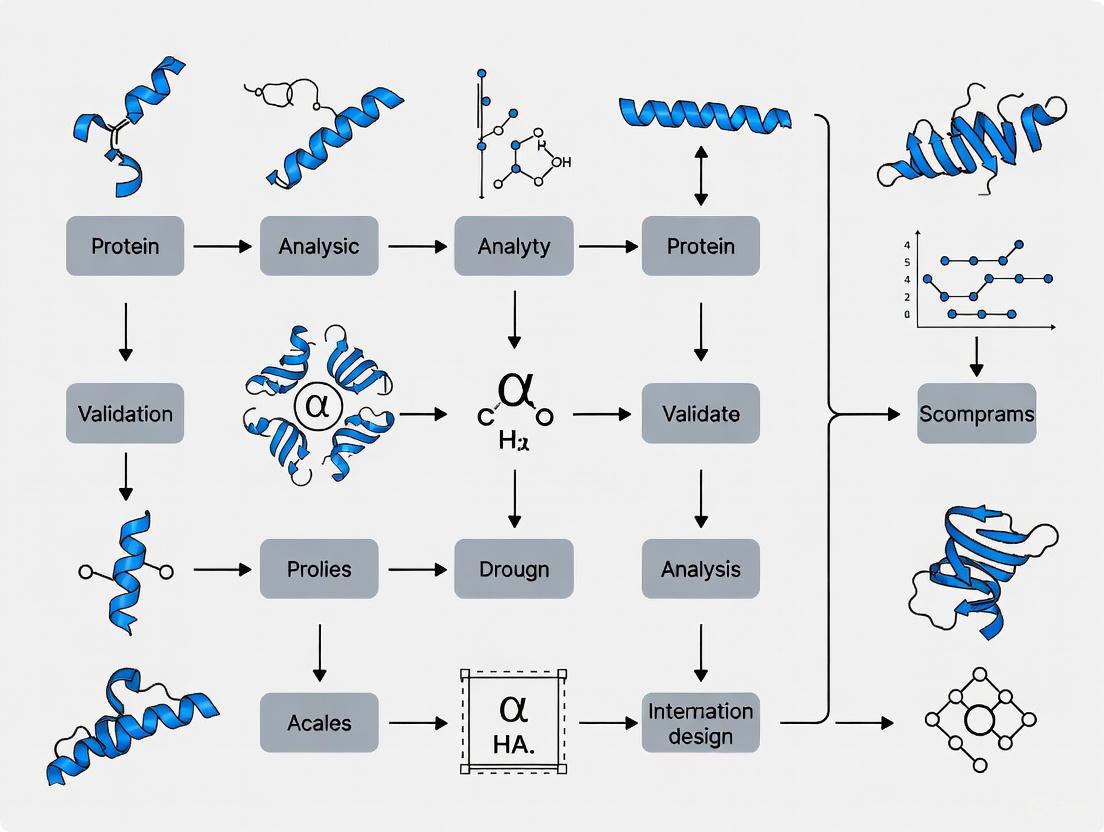
The process of overcoming marginal stability challenges involves both computational protein design and host system engineering. The following diagram illustrates the logical relationship and workflow between these key strategies.
The Scientist's Toolkit: Key Research Reagents and Solutions
Success in designing stable proteins and achieving high-yield expression relies on a suite of computational tools, experimental reagents, and host systems.
Table 2: Essential Research Reagent Solutions for Stability and Expression Research
| Tool / Reagent | Function / Application | Specific Examples / Notes |
|---|---|---|
| Computational Design Suites | In silico protein stability optimization and de novo design. | Rosetta: Physics-based modeling and design [1]. ABACUS-T: Multimodal inverse folding that integrates structure, language models, and MSA [4]. RFpeptides: Denoising diffusion-based pipeline for designing macrocyclic binders [6]. |
| Stability Assessment Kits | Experimental measurement of protein thermal stability. | Differential Scanning Fluorimetry (DSF): Uses a fluorescent dye to measure protein unfolding at different temperatures. |
| Heterologous Expression Hosts | Production of recombinant proteins. | S. cerevisiae: Eukaryotic host for proteins requiring disulfide bonds or glycosylation [3]. A. niger: Industrial filamentous fungus with high secretion capacity; chassis strains like AnN2 reduce background protease activity [5]. |
| Gene Integration Systems | Stable, high-copy number integration of target genes. | CRISPR/Cas9-assisted systems: For precise genomic editing and multi-copy integration into high-expression loci [5] [3]. |
| Secretion Pathway Components | Engineering to enhance protein folding and secretion. | COPI/COPII vesicle trafficking genes: Overexpression of components like Cvc2 (COPI) can enhance secretion yield [5]. |
Marginal stability is a fundamental property of natural proteins that presents a formidable challenge in applied biotechnology. As the data and methodologies reviewed here demonstrate, the convergence of evolution-guided principles with advanced AI-driven design is creating a powerful paradigm for overcoming this challenge. The experimental successes of methods like ABACUS-T, which can simultaneously enhance stability and function with remarkable efficiency, validate the premise that stability design rules are becoming broadly applicable across protein families. Furthermore, the synergy between computational protein design and sophisticated host system engineering is paving the way for a new era in drug development. This integrated approach enables the reliable production of previously intractable therapeutic targets, the creation of hyperstable vaccine immunogens, and the design of novel protein and peptide therapeutics with tailored properties. As these tools continue to evolve and be validated across an ever-wider range of proteins, they promise to make computational stability design a mainstream component of protein science and biopharmaceutical development.
The Inverse Folding Problem and its Evolution into the Inverse Function Problem
The inverse folding problem represents a fundamental challenge in computational biology: designing amino acid sequences that fold into a predetermined three-dimensional protein structure. This stands in direct contrast to the protein folding problem, which predicts a structure from a given sequence, a task revolutionized by tools like AlphaFold. In recent years, however, the field has witnessed a significant evolution—what we term the "inverse function problem"—which extends beyond structural compatibility to actively design sequences that fulfill specific functional roles, such as catalytic activity, molecular binding, or therapeutic potency. This paradigm shift moves computational protein design from merely replicating structural blueprints to engineering functional biomolecules with enhanced or entirely novel capabilities.
This transition is particularly critical within protein stability engineering, where the overarching goal is to design variants with improved stability while retaining, or even enhancing, biological function. Traditional directed evolution approaches, while successful, often require extensive experimental screening. The emergence of machine learning-guided inverse folding offers a transformative alternative, enabling more efficient exploration of sequence space while directly incorporating functional constraints into the design process. This guide provides a comparative analysis of leading computational methodologies driving this evolution, examining their experimental validation, and performance across diverse protein families.
Core Methodologies and Comparative Performance
The landscape of inverse folding models is diverse, encompassing various architectural approaches and training strategies. The table below compares several leading models, highlighting their key features, underlying principles, and performance on standard benchmarks.
Table 1: Comparison of Leading Inverse Folding Models and Their Capabilities
| Model Name | Core Methodology | Key Differentiating Features | Reported Sequence Recovery | Functional Consideration |
|---|---|---|---|---|
| Structure-informed Language Model [7] | Protein language model augmented with backbone coordinates. | Autoregressive; trained on inverse folding objective; generalizes to complexes. | N/A (Demonstrated ~26-fold neutralization improvement in antibodies) | Implicitly learned features of binding and epistasis. |
| ProteinMPNN [8] [9] | SE(3)-equivariant graph neural network. | Fast, robust; allows chain fixing; offers soluble-protein trained version. | ~58% (CATH benchmark, varies by version and training set) [9] | Limited; requires explicit residue fixing for function. |
| ESM-IF1 [9] | Language model trained on sequences and structures. | Uses extensive data augmentation with AlphaFold-predicted structures. | High (SOTA on CATH benchmark) [9] | Limited; primarily focused on structure. |
| ABACUS-T [4] | Sequence-space denoising diffusion model. | Unifies atomic sidechains, ligand interactions, multiple backbone states, and MSA. | High precision (specific metrics not provided) | Explicitly models functional motifs and conformational dynamics. |
| ProRefiner [9] | Global graph attention with entropy-based refinement. | Refines outputs of other models; one-shot generation; uses entropy to filter noisy residues. | Consistently improves recovery of base models (e.g., +~5% over ESM-IF1 on CATH) [9] | Can be applied to partial sequence design for functional optimization. |
A critical metric for evaluating these models is their performance on standardized structural design benchmarks. The following table summarizes key quantitative results from independent evaluations on the CATH and TS50 test sets, providing a direct comparison of their sequence design capabilities.
Table 2: Benchmark Performance of Inverse Folding Models on Standardized Datasets
| Model | Training Data | CATH 4.2 Test Set (Recovery) | TS50 Benchmark (Recovery) | Latest PDB (Recovery) |
|---|---|---|---|---|
| GVP-GNN [9] | CATH 4.2 | ~40% | N/A | N/A |
| ProteinMPNN [9] | PDB Clusters | ~52% | N/A | N/A |
| ProteinMPNN-C [9] | CATH 4.2 | ~58% | N/A | N/A |
| ESM-IF1 [9] | CATH 4.3 + 12M AF2 Structures | ~65% | N/A | N/A |
| ProRefiner (with ESM-IF1) [9] | CATH 4.2 | ~70% | N/A | N/A |
Experimental Validation and Protocols for Stability and Function
The true test of any inverse folding model lies in experimental validation. The transition to solving the "inverse function problem" requires rigorous assessment of both stability and function. The following experimental protocols are commonly used to validate computational designs.
Key Experimental Protocols
Thermal Shift Assays (TSA): This is a standard method for quantifying protein thermostability.
- Procedure: The protein is mixed with a fluorescent dye (e.g., SYPRO Orange) whose fluorescence increases upon binding hydrophobic patches exposed during unfolding. The sample is heated at a controlled rate (e.g., 1°C/min) while fluorescence is monitored. The melting temperature (Tm) is defined as the midpoint of the unfolding transition curve [4] [10].
- Application: Used to measure the increase in Tm (∆Tm) of designed variants compared to wild-type, with improvements of ≥10°C being a common target for successful stabilization [4].
Surface Plasmon Resonance (SPR) or Biolayer Interferometry (BLI): These techniques quantify binding affinity and kinetics for therapeutic antibodies or enzymes.
- Procedure: The target antigen is immobilized on a sensor chip (SPR) or biosensor tip (BLI). Purified protein designs are flowed over the surface at varying concentrations. The association and dissociation phases are monitored in real-time to calculate the equilibrium dissociation constant (KD) [7].
- Application: Validates improvements in binding affinity. For instance, a structure-informed model achieved a 37-fold improvement in KD for an antibody against SARS-CoV-2 variants [7].
Enzyme Activity Assays: These are essential for validating the functional integrity of designed enzymes.
- Procedure: The catalytic activity of wild-type and designed enzymes is measured under standardized conditions. For β-lactamases, this involves monitoring the hydrolysis of nitrocefin, a chromogenic substrate, by the increase in absorbance at 486 nm over time [4]. Specific activity (e.g., μmol substrate/min/μmol enzyme) is calculated and compared.
- Application: ABACUS-T redesigned TEM β-lactamase and endo-1,4-β-xylanase variants that maintained or surpassed wild-type activity despite dozens of mutations [4].
Performance in Functional Protein Engineering
The table below synthesizes experimental outcomes from recent studies that exemplify the "inverse function" approach, demonstrating simultaneous enhancement of stability and function.
Table 3: Experimental Validation of Inverse Folding for Functional Protein Enhancement
| Protein Target | Model Used | Stability Outcome | Functional Outcome | Experimental Throughput |
|---|---|---|---|---|
| SARS-CoV-2 mAbs (Ly-1404, SA58) [7] | Structure-informed Language Model | N/A | Up to 26-fold improvement in viral neutralization; 37-fold improved affinity. | 25-31 variants tested |
| Allose Binding Protein [4] | ABACUS-T | ∆Tm ≥ 10°C | 17-fold higher ligand affinity; retained conformational change. | A few designs tested |
| TEM β-lactamase [4] | ABACUS-T | ∆Tm ≥ 10°C | Maintained or surpassed wild-type catalytic activity. | A few designs tested |
| Endo-1,4-β-xylanase [4] | ABACUS-T | ∆Tm ≥ 10°C | Maintained or surpassed wild-type catalytic activity. | A few designs tested |
| Transposase B [9] | ProRefiner | N/A | 6 of 20 designed variants showed improved gene editing activity. | 20 variants tested |
Successful application of inverse folding requires a suite of computational and experimental tools. The following table details essential "research reagent solutions" for scientists in this field.
Table 4: Essential Research Reagents and Tools for Inverse Folding Research
| Item / Resource | Function / Purpose | Example or Note |
|---|---|---|
| Inverse Folding Models | Generates candidate sequences for a target structure. | ProteinMPNN (fast, good for scaffolding), ABACUS-T (for function-aware design). |
| Protein Folding Predictors | Validates that designed sequences fold into the target structure. | AlphaFold2, ESMFold [8] [11]. TM-Score is a key validation metric [11]. |
| Structure Visualization | Visualizes and analyzes 3D protein structures and designs. | PyMOL, UCSF Chimera. |
| Fluorescent Dye | Reports on protein unfolding in thermal shift assays. | SYPRO Orange [4]. |
| Chromogenic Substrate | Measures enzyme activity through a colorimetric change. | Nitrocefin for β-lactamase activity [4]. |
| Biosensor Instrument | Measures binding affinity and kinetics (BLI). | ForteBio Octet systems. |
Visualizing the Evolution from Inverse Folding to Inverse Function
The logical progression from a structure-centric to a function-aware design paradigm involves the integration of multiple data types and feedback loops, as illustrated in the workflow below.
Diagram 1: The evolving inverse folding workflow, integrating structural validation and functional constraints to solve the inverse function problem.
The field of computational protein design is undergoing a decisive shift from the inverse folding problem toward the more ambitious inverse function problem. As evidenced by the comparative data, models like ABACUS-T and advanced refinement strategies like ProRefiner are leading this charge by integrating evolutionary information, ligand interactions, and conformational dynamics directly into the sequence design process. The experimental successes—achieving double-digit increases in thermostability alongside significantly enhanced antibody neutralization, enzyme activity, and altered substrate specificity—demonstrate the profound potential of this approach. For researchers focused on validating stability designs across protein families, the key insight is that multimodal models that unify structural, evolutionary, and functional data are consistently outperforming older, structure-only methods. By leveraging these advanced tools and the accompanying experimental frameworks, scientists can now engineer stable, functional proteins with a efficiency and success rate that was previously unattainable.
The Vast but Constrained Protein Functional Universe Unexplored by Natural Evolution
The protein functional universe encompasses all possible amino acid sequences, their three-dimensional structures, and the biological activities they can perform. This theoretical space includes not only the folds and functions observed in nature but also every other stable protein fold and corresponding activity that could potentially exist [12]. Despite the extraordinary diversity of natural proteins, comparative analyses suggest that known functions represent only a tiny subset of the diversity that nature can produce [12]. The known natural fold space appears to be approaching saturation, with recent functional innovations predominantly arising from domain rearrangements rather than genuinely novel structural elements [12].
The exploration of this universe faces two fundamental challenges: the problem of combinatorial explosion and the constraints of natural evolution. For a mere 100-residue protein, 20^100 (≈1.27 × 10^130) possible amino acid arrangements exist, exceeding the estimated number of atoms in the observable universe (~10^80) by more than fifty orders of magnitude [12]. Meanwhile, natural proteins are products of evolutionary pressures for biological fitness, not optimized as versatile tools for human utility. This "evolutionary myopia" has constrained exploration to functional neighborhoods immediately surrounding natural sequences, leaving vast regions of possible sequence-structure-function space uncharted [12].
The Constraints of Natural Evolution
Evolutionary Barriers to Exploring Novel Protein Space
Natural evolution operates under constraints that fundamentally limit its ability to explore the full protein universe. Proteins must maintain folding stability and biological function throughout evolutionary trajectories, creating fitness barriers that obstruct paths to novel structures [13]. This phenomenon results in a cusped relationship between sequence and structure divergence—sequences can diverge up to 70% without significant structural evolution, but below 30% sequence identity, structural similarity abruptly decreases [13].
This nonlinear relationship emerges from selection for protein folding stability in divergent evolution. Fitness constraints prevent the emergence of unstable evolutionary intermediates, enforcing paths that preserve protein structure despite broad sequence divergence [13]. On longer timescales, evolution is punctuated by rare events where fitness barriers are overcome, enabling discovery of new structures [13]. The strength of selection for folding stability thus modulates a protein's capacity to evolve new structures, with less stable proteins proving more evolvable at the structural level [13].
Documented Evidence of Evolutionary Constraints
The scale of this constraint becomes evident when comparing known protein sequences to theoretical possibilities. While resources like the MGnify Protein Database catalog nearly 2.4 billion non-redundant sequences and the AlphaFold Protein Structure Database contains ~214 million predicted structures, these datasets constitute an infinitesimally small portion of the theoretical protein functional space [12]. Public datasets remain biased by evolutionary history and assay feasibility, channeling data-driven methods toward well-explored regions of sequence-structure space [12].
Table 1: Documented Evolutionary Constraints in Protein Sequence-Structure Space
| Constraint Factor | Impact on Protein Diversity | Experimental Evidence |
|---|---|---|
| Folding Stability Requirements | Limits evolutionary paths to those maintaining stability, creating fitness valleys that block access to novel folds [13] | Computational models show structure evolution traverses unstable intermediates; less stable proteins are more evolvable [13] |
| Functional Conservation Pressure | Maintains essential biological functions, restricting exploration of sequences without immediate fitness benefits [12] | Natural enzyme engineering shows catalytic efficiency optima often exceed natural levels, indicating evolutionary sub-optimization [14] |
| Domain Rearrangement Preference | Favors recombination of existing domains over de novo emergence of structural motifs [12] | Analysis of protein families shows recent innovations predominantly from domain shuffling rather than novel fold discovery [12] |
| Sequence-Structure Divergence Cusp | Creates "twilight zone" below 30% sequence identity where structural prediction from sequence becomes unreliable [13] | Bioinformatics studies of SCOP folds reveal abrupt decrease in structural similarity below 30% sequence identity [13] |
Computational Strategies to Overcome Evolutionary Constraints
Inverse Folding and De Novo Protein Design
To transcend natural evolutionary constraints, researchers have developed computational strategies that actively design proteins rather than modifying natural templates. Inverse folding approaches aim to identify amino acid sequences that fold into a given backbone structure, enabling extensive sequence changes while maintaining structural integrity [4]. However, traditional inverse folding models often produce functionally inactive proteins because they don't impose sequence restraints necessary for functions like substrate recognition or chemical catalysis [4].
The ABACUS-T model represents a multimodal inverse folding approach that unifies several critical features: detailed atomic sidechains and ligand interactions, a pre-trained protein language model, multiple backbone conformational states, and evolutionary information from multiple sequence alignment (MSA) [4]. This integration enables the generation of functionally active sequences without having to explicitly fix predetermined "functionally important" residues [4]. In experimental validations, ABACUS-T redesigned proteins showed notable improvements—an allose binding protein achieved 17-fold higher affinity while retaining conformational change, while redesigned endo-1,4-β-xylanase and TEM β-lactamase maintained or surpassed wild-type activity with substantially increased thermostability (∆Tm ≥ 10 °C) [4].
AI-Driven Exploration of Novel Protein Space
Artificial intelligence has enabled a paradigm shift from optimizing existing proteins to designing entirely novel sequences. AI-driven de novo protein design uses generative models, structure prediction tools, and iterative experimental validation to explore protein sequences beyond natural evolutionary pathways [12]. These approaches leverage statistical patterns from biological datasets to establish high-dimensional mappings between sequence, structure, and function [12].
A particularly powerful framework employs controllable generation, where models are conditioned on desired functions [15]. Researchers can specify target chemical reactions—even those without natural enzyme counterparts—and the model generates novel protein sequences predicted to catalyze them [15]. This transforms discovery from a search process into a design problem. When integrated with automated laboratory systems that enable continuous design-build-test-learn cycles, these approaches create a powerful engine for exploring the uncharted protein universe [16] [15].
Figure: Computational strategies overcome natural evolution constraints through inverse folding and AI-driven generation, validated by automated experimentation.
Experimental Validation of Designed Proteins
Methodologies for Assessing Stability and Function
Validating computationally designed proteins requires rigorous experimental assessment of stability and function. Two primary methods dominate the field: equilibrium unfolding measurements that determine thermodynamic stability, and kinetic stability assessments that measure resistance to irreversible denaturation [14].
Equilibrium unfolding typically uses urea or other chemical denaturants with spectroscopic detection (circular dichroism, intrinsic fluorescence) to monitor the folded-to-unfolded transition. Data are analyzed to calculate the Gibbs free energy change (ΔG) for unfolding, with extrapolation to zero denaturant concentration yielding ΔGH₂O [14]. Kinetic stability measurements involve heating protein samples, then cooling and measuring remaining catalytic activity. The ratio of half-lives for mutant versus wild-type proteins yields the difference in Gibbs free energy of activation for activity loss according to: ΔΔG‡ = RT ln(t½,mutant / t½,wildtype) [14].
Table 2: Experimental Validation Methods for Designed Proteins
| Method Category | Specific Techniques | Measured Parameters | Key Advantages | Limitations |
|---|---|---|---|---|
| Thermodynamic Stability | Urea-induced unfolding with CD/fluorescence detection [14] | ΔG (folding free energy), ΔGH₂O (in water) [14] | Measures reversible unfolding; provides fundamental thermodynamic parameters [14] | May not reflect stability under application conditions; requires reversible unfolding [14] |
| Kinetic Stability | Heat inactivation assays, residual activity measurement [14] | t½ (half-life), ΔΔG‡ (activation energy) [14] | Reflects practical stability under application conditions; measures irreversible denaturation [14] | Mechanism complex (unfolding + aggregation); difficult to connect to molecular changes [14] |
| High-Throughput Stability | cDNA display proteolysis [17], yeast display proteolysis [17] | Protease resistance, inferred ΔG values [17] | Enables massive scale (≈900,000 domains/week); good correlation with traditional methods [17] | Model limitations for partially folded states; reliability limits at stability extremes [17] |
| Functional Characterization | Enzyme kinetics, binding affinity measurements, biological activity assays [18] | KM, kcat, IC50, KD [18] | Directly measures protein function; most relevant for applications [18] | Low throughput; requires specific assays for each protein [18] |
Comparative Performance of Engineering Strategies
Different protein engineering strategies yield varying success rates and stabilization levels. A comparative study of five strategies applied to an α/β-hydrolase fold enzyme (salicylic acid binding protein 2, SABP2) revealed distinct performance characteristics [14]. Location-agnostic methods (e.g., random mutagenesis via error-prone PCR) yielded the highest absolute stabilization (average 3.1 ± 1.9 kcal/mol), followed by structure-based approaches (2.0 ± 1.4 kcal/mol) and sequence-based methods (1.2 ± 0.5 kcal/mol) [14].
The mutation-to-consensus approach demonstrated the best balance of success rate, degree of stabilization, and ease of implementation [14]. This strategy hypothesizes that evolution conserves important residues, and positions where the target protein differs from consensus are more likely to be stabilizing when mutated to the consensus residue [14]. Automated programs for predicting consensus substitutions are publicly available to facilitate this approach [14].
Table 3: Comparison of Protein Engineering Strategies
| Engineering Strategy | Key Principles | Stabilization Performance | Success Rate | Implementation Complexity |
|---|---|---|---|---|
| Location-Agnostic Random Mutagenesis [14] | Random mutations throughout sequence; screening of large variant libraries [14] | 3.1 ± 1.9 kcal/mol average improvement [14] | 67% of reports yielded >2 kcal/mol improvements [14] | High (requires large library construction & screening) [14] |
| Structure-Based Design [14] | Computational modeling of molecular interactions (Rosetta, FoldX) [14] | 2.0 ± 1.4 kcal/mol average improvement [14] | 37% of reports yielded >2 kcal/mol improvements [14] | High (requires structural knowledge & expertise) [14] |
| Mutation-to-Consensus [14] | Replace residues with amino acids conserved in homologs [14] | 1.2 ± 0.5 kcal/mol average improvement [14] | High success rate for identified positions [14] | Low (requires multiple sequence alignment) [14] |
| Inverse Folding (ABACUS-T) [4] | Multimodal model integrating structure, language model, MSA, multiple conformations [4] | ΔTm ≥ 10°C while maintaining or improving function [4] | High (successful with testing of only a few designed sequences) [4] | Medium (requires structural input) [4] |
| Automated Continuous Evolution [16] | Growth-coupled selection in automated laboratory systems [16] | Enabled evolution of proteins from inactive precursors to full function [16] | High for complex functions difficult to design rationally [16] | High (requires specialized automated equipment) [16] |
The Scientist's Toolkit: Research Reagent Solutions
Table 4: Essential Research Reagents and Platforms for Protein Engineering
| Tool Category | Specific Solutions | Primary Function | Key Applications |
|---|---|---|---|
| Computational Design Platforms | ABACUS-T [4], Rosetta [12], FoldX [14] | Structure-based sequence design, stability prediction, inverse folding [4] [12] [14] | De novo protein design, stability enhancement, function preservation [4] |
| High-Throughput Stability Assays | cDNA display proteolysis [17], yeast display proteolysis [17] | Massively parallel measurement of folding stability for thousands of variants [17] | Mapping stability landscapes, validating designs, training machine learning models [17] |
| Automated Evolution Systems | OrthoRep continuous evolution [16], iAutoEvoLab [16] | Automated, continuous protein evolution with growth-coupled selection [16] | Evolving complex functions, exploring adaptive landscapes, bypassing design challenges [16] |
| Sequence-Function Mapping | Deep mutational scanning [18], site-saturation mutagenesis [18] | Comprehensive characterization of sequence-performance relationships [18] | Understanding mutational effects, guiding library design, improving computational models [18] |
| Structure Prediction Databases | AlphaFold Database [19], ESM Metagenomic Atlas [12] | Access to predicted structures for millions of proteins [19] | Identifying novel folds, functional annotation, template for design [19] |
The constraints of natural evolution have limited the exploration of the protein functional universe to an infinitesimal fraction of its theoretical expanse. However, integrated computational and experimental approaches are now overcoming these constraints. Multimodal inverse folding models like ABACUS-T enable extensive sequence redesign while maintaining function [4], while AI-driven generative approaches facilitate creation of proteins with novel functions [12] [15]. Automated experimental systems enable continuous evolution and high-throughput validation of designed proteins [16] [17].
These advances are transforming protein engineering from a process of local optimization to one of global exploration. By combining computational design with massive experimental validation, researchers can now systematically illuminate the "dark matter" of the protein universe [19], discovering new folds [19], families [19], and functions [15] that natural evolution has never sampled. This expanded access to protein space promises innovations across biotechnology, medicine, and synthetic biology, enabling the design of bespoke biomolecules with tailored functionalities [12]. As these technologies mature, they will increasingly allow researchers to not just explore but actively create within the vast, untapped potential of the protein functional universe.
Within protein engineering, the pursuit of enhanced stability is a cornerstone for developing effective biotherapeutics and industrial enzymes. Achieving this requires a multi-faceted approach targeting three critical objectives: increased melting temperature (Tm), improved solubility, and enhanced resistance to aggregation. These properties are deeply interconnected; for instance, mutations that improve thermostability can sometimes inadvertently promote aggregation, thereby reducing solubility and overall functional yield [20]. This guide objectively compares the performance of modern stability-design strategies, framing the analysis within a broader research thesis on validating these methods across diverse protein families. The data and protocols summarized herein provide a roadmap for researchers and drug development professionals to select and implement the most appropriate design strategies for their specific protein engineering challenges.
Comparative Analysis of Protein Stability-Design Strategies
A comparison of key design strategies reveals distinct performance trade-offs in achieving stability objectives. The following table synthesizes quantitative data and experimental outcomes from recent studies.
Table 1: Comparison of Key Stability-Design Strategies and Their Outcomes
| Design Strategy | Key Features & Inputs | Reported ΔTm | Impact on Solubility & Aggregation | Key Experimental Validation |
|---|---|---|---|---|
| Multimodal Inverse Folding (ABACUS-T) [4] | Integrates backbone structure, atomic sidechains, ligand interactions, multiple sequence alignment (MSA), and a protein language model. | ΔTm ≥ +10°C (in redesigned allose binding protein, xylanase, β-lactamases) | Maintains or improves functional activity; designed proteins show no major solubility issues. | Activity assays, thermal shift assays, binding affinity measurements (e.g., 17-fold higher affinity for allose binding protein). |
| Consensus Design [14] [21] | Derives sequences from multiple sequence alignments (MSA), assigning the most frequent amino acid at each position. | Variable; often increases stability. | Can produce conformationally homogeneous, soluble proteins (e.g., Conserpin) [21]. | Circular Dichroism (CD) for Tm, NMR for dynamics, equilibrium unfolding experiments for ΔG. |
| Structure-Based Computational Design (e.g., Rosetta, FoldX) [14] [21] | Uses physical energy functions to optimize sequences for a given backbone structure or to predict stabilizing point mutations. | Average stabilization: ~2.0 kcal/mol reported for α/β-hydrolase fold enzymes. | Risk of poor solubility and aggregation if stabilizing mutations expose hydrophobic patches (e.g., LinB116) [20]. | Site-saturation mutagenesis combined with activity screening after thermal challenge. |
| Location-Agnostic Methods (e.g., Error-Prone PCR) [14] | Creates random mutations throughout the gene without prior structural or sequence knowledge. | Highest reported stabilization: Average of ~3.1 kcal/mol. | Success depends on screening; can identify highly stable variants without precise solubility targeting. | High-throughput screening of libraries for residual activity after heat incubation. |
Experimental Protocols for Validating Stability Designs
Rigorous experimental validation is required to confirm that designed proteins meet stability objectives. The following protocols are standard in the field.
Protocol for Measuring Thermostability (Tm)
- Objective: Quantify the melting temperature (Tm), the temperature at which 50% of the protein is unfolded.
- Method: Differential Scanning Fluorimetry (DSF, or thermal shift assay) [21].
- Sample Preparation: Mix protein sample with a fluorescent dye (e.g., SYPRO Orange) that binds to hydrophobic regions exposed upon unfolding.
- Equipment Setup: Use a real-time PCR instrument or dedicated thermal shift scanner.
- Procedure: Heat the sample from 25°C to 95°C with a gradual ramp (e.g., 1°C per minute) while monitoring fluorescence.
- Data Analysis: Plot fluorescence against temperature. The Tm is determined as the inflection point of the resulting sigmoidal curve.
- Data Interpretation: A higher Tm in a designed variant compared to the wild type indicates improved thermostability. This data can be converted to a kinetic stabilization energy (ΔΔG‡) using half-life ratios [14].
Protocol for Assessing Aggregation and Solubility
- Objective: Evaluate the propensity of a protein to aggregate and its solubility under specific conditions.
- Method 1: Accelerated Aggregation Studies [22]
- Sample Preparation: Incubate protein samples at high concentration and elevated temperatures (e.g., 40°C) for extended periods (days to weeks).
- Analysis: Periodically analyze samples using techniques like:
- Size-Exclusion Chromatography (SEC): To quantify the amount of soluble monomer versus higher-order aggregates.
- Dynamic Light Scattering (DLS): To monitor the increase in particle size over time.
- Method 2: Analysis of Aggregation-Prone Regions [20] [22]
- In-silico Prediction: Use tools to identify "cryptic aggregation-prone regions" that may become exposed during unfolding.
- Experimental Correlation: Combine with molecular dynamics (MD) simulations to identify regions exposed during unfolding that correlate with observed aggregation.
Workflow Diagram for an Integrated AI-MD Stability Design Platform
The following diagram illustrates a modern computational workflow that integrates artificial intelligence and molecular dynamics for predicting and designing protein stability, particularly against aggregation.
AI-MD Workflow for Aggregation Prediction - An integrated platform that starts from sequence, predicts structure, simulates dynamics, calculates novel surface features, and uses machine learning to predict aggregation propensity [22].
The Scientist's Toolkit: Essential Research Reagents and Computational Tools
Success in protein stability design relies on a suite of specialized reagents and software tools.
Table 2: Key Research Reagent Solutions for Protein Stability Design
| Tool/Reagent Name | Category | Primary Function in Stability Design |
|---|---|---|
| SYPRO Orange Dye [21] | Chemical Reagent | Fluorescent dye used in Differential Scanning Fluorimetry (DSF) to measure protein thermal unfolding and determine Tm. |
| Rosetta Software Suite [14] [23] [24] | Computational Tool | A comprehensive platform for protein structure prediction, design, and energy-based stability calculations (e.g., ΔΔG). |
| FoldX [14] [24] | Computational Tool | A molecular modeling algorithm for the rapid in-silico evaluation of the effect of mutations on protein stability and interactions. |
| ENDURE Web Application [23] | Computational Tool | Provides an interactive workflow for energetic analysis of protein designs, helping select optimal mutants for experimental testing. |
| RaSP (Rapid Stability Predictor) [24] | Computational Tool | A deep learning-based method for making rapid predictions of changes in protein stability (ΔΔG) upon mutation. |
| Gromacs [22] | Computational Tool | A software package for performing molecular dynamics (MD) simulations, used to study protein dynamics and unfolding pathways. |
The Computational Toolkit: AI, Multimodal Models, and Stability Design Strategies
Protein stability is a fundamental property essential for biological function, yet it presents a major challenge for protein engineers and drug developers. The stability of a protein's native fold is marginal, typically only 5–15 kcal mol⁻¹ more stable than its unfolded state, meaning even single mutations can lead to destabilization and loss of function [25]. For decades, scientists have sought reliable methods to predict and engineer protein stability, with two dominant paradigms emerging: physics-based atomistic simulations that model molecular interactions from first principles, and evolution-guided approaches that extract information from natural sequence variation across protein families. This review objectively compares these methodologies, examining their performance in predicting mutational effects and designing stable proteins, with particular focus on their validation across diverse protein families. Understanding the relative strengths and limitations of these approaches is crucial for researchers aiming to develop more effective therapeutics, enzymes, and biomaterials with enhanced stability properties.
Methodological Foundations: A Tale of Two Approaches
Evolution-Guided Design Principles
Evolution-guided protein design operates on the principle that modern protein sequences contain information about the structural and functional constraints that have shaped their evolution. These methods leverage the evolutionary record encapsulated in multiple sequence alignments (MSAs) to infer which amino acid combinations are likely to fold into stable, functional proteins [26]. The core assumption is that natural selection has already conducted a massive experiment in protein stability optimization over geological timescales, and this information can be mined to guide engineering efforts.
Key implementations of this approach include:
- EvoDesign: This server uses evolutionary profiles from structurally analogous folds or interfaces to guide replica-exchange Monte Carlo simulations. It combines both monomer structural profiles and protein-protein interaction interface profiles with physical energy terms to design stable proteins and optimize binding interactions [27].
- EVcouplings framework: This method employs maximum entropy models parameterized by site-specific (hᵢ) and pairwise (Jᵢⱼ) constraints learned from MSAs. The fitness of a sequence (σ) is defined by its evolutionary Hamiltonian (EVH), calculated as EVH(σ) = -∑hᵢ(σᵢ) - ∑Jᵢⱼ(σᵢ,σⱼ) [28].
- Ancestral Sequence Reconstruction (ASR): This "vertical" strategy reconstructs likely ancestral proteins to provide appropriate backgrounds for studying historical mutations that led to functional diversification, effectively avoiding epistatic conflicts that plague horizontal comparisons between modern sequences [26].
Physics-Based Atomistic Simulations
In contrast to data-driven evolutionary methods, physics-based approaches aim to calculate stability from fundamental physical principles, using force fields to model atomic interactions. These methods include:
- Free Energy Perturbation (FEP): A rigorous statistical thermodynamics approach for calculating free energy differences between states. QresFEP-2 represents a recent advancement implementing a hybrid-topology protocol that combines single-topology backbone representation with dual-topology side chains, balancing accuracy with computational efficiency [29].
- Rosetta Design Suite: This widely used platform employs detailed physical energy functions for protein design and stability prediction, with remarkable successes in designing novel stable folds and enzymes [25].
- FoldX and Eris: Specialized tools for predicting ΔΔG changes from mutations using empirical force fields and simplified geometric parameters [25].
Table 1: Comparison of Primary Methodologies in Protein Stability Design
| Method | Core Principle | Key Inputs | Primary Output | Methodology Category |
|---|---|---|---|---|
| EvoDesign | Evolutionary profile guidance | MSA, Structural templates | Designed sequences with enhanced stability/binding | Evolution-guided |
| EVcouplings | Statistical couplings from MSA | MSA of homologous sequences | Sequences with optimized evolutionary Hamiltonian | Evolution-guided |
| QresFEP-2 | Hybrid-topology free energy calculations | Protein structure, Force field | Predicted ΔΔG for mutations | Physics-based atomistic |
| Rosetta | Physical energy function optimization | Protein structure, Rotamer library | Designed stable sequences/structures | Physics-based atomistic |
| Ancestral Reconstruction | Historical mutation analysis | Phylogeny, Modern sequences | Ancestral proteins and mutational effects | Evolution-guided |
Experimental Protocols and Validation Frameworks
Benchmarking Standards and Performance Metrics
Robust validation is essential for assessing protein stability prediction methods. Community-standard benchmarks typically involve:
- Deep Mutational Scanning (DMS) Data: Large-scale experimental measurements of mutational effects, such as those obtained for the GRB2-SH3 domain where ΔΔGf values were inferred for nearly all possible mutations (1,056/1,064) using pooled variant synthesis, selection, and sequencing [30].
- Thermal Stability Assays: Melting temperature (Tm) measurements and denaturation midpoint determinations using techniques like circular dichroism (CD) spectroscopy and differential scanning calorimetry.
- Functional Assays: For enzymes like TEM-1 β-lactamase, activity measurements on substrates (e.g., ampicillin hydrolysis) provide functional validation of stability predictions [28].
- High-Throughput Stability Measurements: Methods like AbundancePCA enable quantitative cellular abundance measurements for thousands of variants simultaneously, providing fitness proxies for stability [30].
Performance is typically quantified using:
- Correlation coefficients (Pearson's r or Spearman's ρ) between predicted and experimental stability measurements
- Root Mean Square Error (RMSE) for ΔΔG predictions
- Variance explained (R²) in combinatorial mutant datasets
- Classification accuracy for identifying stabilizing vs. destabilizing mutations
Evolution-Guided Design Workflow
The typical workflow for evolution-guided design, as implemented in EVcouplings, involves several key stages [28]:
- MSA Construction: A multiple sequence alignment of homologous proteins is generated using tools like jackhmmer, with careful attention to alignment depth and quality.
- Model Building: A maximum entropy model is parameterized with site-specific and pairwise terms from the MSA.
- Model Validation: The model is assessed by its ability to recapitulate known structural contacts and mutation effects from deep mutational scans.
- Sequence Generation: Designed sequences are generated using sampling algorithms (Markov Chain Monte Carlo or Gibbs sampling) that optimize evolutionary fitness while satisfying sequence distance constraints.
- Experimental Testing: Designed variants are characterized for stability (thermal denaturation), structure (circular dichroism, X-ray crystallography), and function (activity assays).
Figure 1: Evolution-Guided Protein Design Workflow. This diagram illustrates the iterative process of using evolutionary information to design stable proteins, with validation checkpoints to ensure model quality.
Physics-Based Simulation Protocol
The QresFEP-2 protocol exemplifies modern physics-based approaches to stability prediction [29]:
- System Preparation: The protein structure is prepared with appropriate protonation states and solvation.
- Hybrid Topology Construction: A dual-like hybrid topology combines single-topology representation for backbone atoms with separate topologies for side-chain atoms.
- Restraint Setup: Distance restraints are applied between topologically equivalent atoms to maintain phase-space overlap during alchemical transformations.
- λ-Window Simulations: Multiple independent molecular dynamics simulations are performed at intermediate λ values between wild-type (λ=0) and mutant (λ=1) states.
- Free Energy Integration: The free energy difference is calculated by integrating over the λ pathway using Bennett Acceptance Ratio (BAR) or similar methods.
- Error Analysis: Statistical errors are estimated through bootstrapping or ensemble simulations.
This protocol has been benchmarked on comprehensive protein stability datasets encompassing nearly 600 mutations across 10 protein systems, with additional validation through domain-wide mutagenesis scans.
Performance Comparison Across Protein Families
Quantitative Accuracy Assessment
Recent large-scale studies enable direct comparison of evolution-guided and physics-based methods. A 2024 Nature study on the genetic architecture of protein stability revealed that simple additive energy models based on evolutionary data explain a remarkable proportion of fitness variance in high-dimensional sequence spaces [30]. When trained on single and double mutant data alone, an evolutionary energy model explained approximately 50% of the fitness variance in combinatorial multi-mutants of the GRB2-SH3 domain, most of which contained at least 13 amino acid substitutions.
Table 2: Performance Comparison Across Protein Systems and Mutation Types
| Method Category | Representative Tool | Accuracy (Correlation with Experiment) | Key Strengths | Key Limitations |
|---|---|---|---|---|
| Evolution-guided | EVcouplings (TEM-1 β-lactamase) | Spearman ρ = 0.71-0.72 for single mutants [28] | Excellent generalization, handles many mutations | Limited by MSA depth/quality |
| Physics-based | QresFEP-2 (T4 Lysozyme) | Excellent accuracy on comprehensive benchmark [29] | Physical interpretability, no MSA requirement | Computationally intensive, force field inaccuracies |
| Evolution-guided | EvoDesign (Various folds) | Successful design of stable novel binders [27] | Combines evolutionary and physical constraints | Primarily for structure-based design |
| Evolution-guided | Additive Energy Model (GRB2-SH3) | R² = 0.63 for combinatorial mutants [30] | Simple, interpretable, captures global epistasis | Misses specific pairwise interactions |
| Hybrid | Evolution + Solvent Accessibility | Similar accuracy to machine learning methods [31] | Balance of evolutionary and structural information | Implementation complexity |
The addition of pairwise energetic couplings to evolutionary models further improves performance. For the GRB2-SH3 domain, including second-order energetic couplings increased the variance explained from 63% to 72%, demonstrating that while additive effects dominate, specific epistatic contributions are measurable and important for accurate prediction [30]. These couplings were found to be sparse and associated with structural contacts and backbone proximity.
Case Study: TEM-1 β-Lactamase Engineering
A compelling demonstration of evolution-guided design comes from the engineering of TEM-1 β-lactamase using the EVcouplings framework [28]. Researchers generated variants with sequence identities to wild-type TEM-1 ranging from 98% down to 50%, including one design with 84 mutations from the nearest natural homolog. Remarkably, nearly all 14 experimentally characterized designs were functional, with large increases in thermostability and increased activity on multiple substrates, while maintaining nearly identical structure to the wild-type enzyme.
This achievement is particularly significant because previous studies had shown that introducing 10 random mutations to TEM-1 completely abrogates enzyme activity, highlighting the power of evolution-guided methods to enable large jumps in sequence space while maintaining or enhancing function [28].
Case Study: SH3 Domain Stability Landscapes
A massive experiment on the FYN-SH3 domain challenged conventional wisdom about protein stability [32]. Contrary to the prevailing "house of cards" metaphor where any core mutation collapses the structure, researchers found that SH3 retained its shape and function across thousands of different core and surface combinations. By testing hundreds of thousands of variants, they identified that only a few true "load-bearing" amino acids exist in the protein's core, with physical rules governing stability being more like Lego than Jenga.
Machine learning applied to this large dataset produced a tool that could accurately predict SH3 stability, correctly identifying almost all natural SH3 domains as stable even with less than 25% sequence identity to the human version used for training [32]. This demonstrates the generalizability of evolution-guided stability predictions across protein families.
Table 3: Key Research Reagents and Computational Tools for Protein Stability Studies
| Resource | Type | Primary Function | Accessibility |
|---|---|---|---|
| EvoDesign Server | Web Server | Evolution-guided protein design | Freely available online |
| EVcouplings Framework | Software Package | Evolutionary covariance analysis | Open source |
| QresFEP-2 | Software Module | Free energy perturbation calculations | Integrated with Q molecular dynamics |
| Rosetta | Software Suite | Physics-based protein design | Academic and commercial licenses |
| FoldX | Software Plugin | Rapid stability prediction | Freely available for academics |
| AbundancePCA | Experimental Method | High-throughput stability measurement | Protocol described in literature |
| ProTherm Database | Database | Curated protein stability data | Publicly accessible |
Integration Strategies and Future Directions
The most powerful approaches emerging in protein stability design combine evolutionary information with physical principles. For instance, simply combining evolutionary features with a basic structural feature—the relative solvent accessibility of mutated residues—can achieve prediction accuracy similar to supervised machine learning methods [31]. This suggests that hybrid approaches leveraging both the historical information in evolutionary records and the mechanistic understanding from physical models will provide the most robust solutions to protein stability challenges.
Future methodology development will likely focus on:
- Improved epistatic models that better capture the sparse pairwise interactions important for stability
- Machine learning hybrids that integrate evolutionary, physical, and structural features
- High-throughput experimental validation methods that enable rapid testing of computational predictions
- Generalizable force fields that accurately model diverse protein families without requiring extensive evolutionary data
For researchers and drug development professionals, the choice between evolution-guided and physics-based approaches depends on specific project requirements. Evolution-guided methods typically excel when deep multiple sequence alignments are available and when making large jumps in sequence space, while physics-based approaches provide mechanistic insights and can be applied to novel scaffolds with limited evolutionary information. The most successful engineering campaigns will likely continue to strategically combine both approaches to overcome their respective limitations and leverage their complementary strengths.
Protein engineering aims to overcome the limitations of natural proteins for biotechnological and therapeutic applications. A central challenge in this field is enhancing structural stability without compromising biological function. Traditional computational methods, particularly structure-based inverse folding, have demonstrated remarkable success in designing sequences that fold into stable target structures. However, these hyper-stable designs often suffer from loss of functional activity, as the optimization for folding can neglect residues and conformational dynamics essential for function [4]. This limitation stems from an over-reliance on single structural snapshots and insufficient incorporation of functional constraints during the sequence design process.
The emerging paradigm of multimodal inverse folding addresses this challenge by integrating diverse data types to inform the design process. This approach recognizes that functional proteins exist in conformational ensembles, interact with ligands and other molecules, and contain evolutionary information embedded in their sequences. Here, we examine ABACUS-T (A Backbone based Amino aCid Usage Survey-ligand Targeted), a multimodal inverse folding model that unifies structural, evolutionary, and biochemical information to redesign functional proteins with enhanced stability [4]. Through detailed case studies and comparative analysis, this guide evaluates ABACUS-T's performance against alternative protein design methodologies.
ABACUS-T: Technical Framework and Methodological Innovations
Core Architecture and Design Philosophy
ABACUS-T employs a sequence-space denoising diffusion probabilistic model (DDPM) that generates amino acid sequences through successive reverse diffusion steps [4]. Unlike conventional inverse folding models that focus primarily on backbone geometry and sidechain packing, ABACUS-T incorporates multiple critical features into a unified framework:
- Detailed atomic sidechains and ligand interactions: Explicit modeling of molecular interactions preserves functional sites
- Pre-trained protein language model: Leverages evolutionary knowledge from protein sequence databases
- Multiple backbone conformational states: Accounts for functional dynamics beyond single structures
- Evolutionary information from multiple sequence alignment (MSA): Incorporates natural sequence constraints
A key innovation in ABACUS-T is its self-conditioning scheme, where each denoising step is informed by the output amino acid sequence and sidechain atomic structures from the previous step [4]. This iterative refinement process enables more precise sequence selection than single-pass prediction methods.
Experimental Workflow for Protein Redesign
The ABACUS-T methodology follows a systematic workflow for functional protein redesign:
Figure 1: ABACUS-T employs a multimodal workflow that integrates diverse structural and evolutionary data to redesign functional proteins with enhanced stability.
Table 1: Essential Research Resources for Protein Redesign Studies
| Resource Category | Specific Tools | Application in Protein Design |
|---|---|---|
| Structure Prediction | AlphaFold2, RoseTTAFold, ESMFold [33] [34] | Validate fold stability of designed sequences |
| Stability Prediction | Pythia, Rosetta, FoldX [35] | Predict ΔΔG changes from mutations |
| Molecular Visualization | PyMOL, ChimeraX | Analyze structural features and binding sites |
| Experimental Validation | Circular Dichroism (CD), Surface Plasmon Resonance (SPR), Enzyme Activity Assays [4] | Measure thermostability (ΔTm), binding affinity, catalytic efficiency |
| Sequence Analysis | HMMER, PSI-BLAST, ClustalOmega [33] | Generate multiple sequence alignments for evolutionary data |
Comparative Performance Analysis: ABACUS-T Versus Alternative Approaches
Experimental Outcomes Across Protein Families
ABACUS-T has been experimentally validated across diverse protein systems, demonstrating its ability to enhance stability while maintaining or improving function. The following table summarizes key performance metrics:
Table 2: Experimental Performance of ABACUS-T Across Protein Families
| Protein System | Key Mutations | Thermostability (ΔTm) | Functional Outcomes | Experimental Validation |
|---|---|---|---|---|
| Allose Binding Protein | Dozens of simultaneous mutations [4] | ≥10°C increase [4] | 17-fold higher affinity while retaining conformational change [4] | Retained ligand-induced conformational transition |
| Endo-1,4-β-xylanase | Dozens of simultaneous mutations [4] | ≥10°C increase [4] | Maintained or surpassed wild-type activity [4] | Enzyme activity assays under harsh conditions |
| TEM β-lactamase | Dozens of simultaneous mutations [4] | ≥10°C increase [4] | Maintained or surpassed wild-type activity [4] | Antibiotic resistance profiling |
| OXA β-lactamase | Dozens of simultaneous mutations [4] | ≥10°C increase [4] | Altered substrate selectivity [4] | Specificity profiling against β-lactam antibiotics |
Comparison with Alternative Protein Design Methodologies
Table 3: ABACUS-T Performance Against Alternative Protein Design Approaches
| Design Method | Stability Enhancement | Functional Preservation | Key Limitations | Experimental Success Rate |
|---|---|---|---|---|
| ABACUS-T | High (ΔTm ≥10°C) [4] | Excellent (maintained or enhanced activity) [4] | Requires multiple structural/evolutionary inputs | High (multiple successful cases with few designs tested) [4] |
| Traditional Inverse Folding | High [21] | Often compromised [4] | Neglects functional constraints and conformational dynamics | Variable (often requires extensive screening) [4] |
| Consensus Design | Moderate to High [21] | Moderate (depends on MSA quality) | Limited to naturally occurring variations | Moderate [21] |
| Ancestral Sequence Reconstruction | Moderate to High [21] | Moderate to High [21] | Dependent on accurate phylogenetic reconstruction | Moderate [21] |
| Directed Evolution | Variable (depends on screening method) | High (screening directly for function) | Limited exploration of sequence space (few mutations) | High but resource-intensive [4] |
Advancements Over Previous Inverse Folding Models
ABACUS-T represents a significant advancement over previous inverse folding models through its multimodal approach:
- Functional Preservation: Unlike traditional inverse folding that often produces functionally inactive proteins [4], ABACUS-T maintained or enhanced activity in all tested cases [4]
- Extensive Mutations: Successful designs incorporated dozens of simultaneous mutations, far exceeding the typical scope of directed evolution campaigns [4]
- Efficient Screening: Functional variants were identified with testing of only a few designed sequences, dramatically reducing experimental burden [4]
Experimental Protocols and Validation Methodologies
Redesign Protocol for Enzyme Systems
For enzyme redesign, ABACUS-T follows a structured protocol:
Input Preparation:
- Obtain high-resolution structure with bound ligands or substrates
- Curate multiple sequence alignment from homologous families
- Identify multiple conformational states when available
Sequence Generation:
- Apply denoising diffusion process conditioned on structural and evolutionary inputs
- Generate multiple sequence variants with dozens of mutations
Experimental Validation:
- Express and purify designed variants
- Assess thermostability via circular dichroism (measuring Tm)
- Evaluate catalytic activity with enzyme-specific assays
- Determine ligand binding affinity where applicable
Key Methodological Innovations in ABACUS-T
ABACUS-T's performance advantages stem from several methodological innovations:
Figure 2: ABACUS-T's core methodological innovations integrate multiple data types and preservation mechanisms to maintain protein function while enhancing stability.
Discussion and Future Perspectives
The development of ABACUS-T represents significant progress in addressing the fundamental challenge of functional protein redesign. By unifying structural, evolutionary, and biochemical information in a single framework, ABACUS-T demonstrates that substantial stability enhancements (ΔTm ≥10°C) can be achieved without compromising biological activity [4]. This multimodal approach effectively bridges the gap between structure-based design and function preservation.
The implications for protein engineering and drug development are substantial. The ability to redesign proteins with enhanced stability while maintaining function opens new possibilities for:
- Engineering robust industrial enzymes for biotechnology
- Developing stable therapeutic proteins with extended shelf-life
- Creating specialized biosensors with tailored properties
- Designing novel biocatalysts with altered substrate specificity
Future developments in multimodal inverse folding will likely focus on incorporating dynamics more explicitly, expanding to membrane protein systems, and improving accuracy for de novo protein design. As the field progresses, the integration of advanced deep learning approaches with biophysical principles promises to further accelerate the design of functional proteins with tailored properties.
The validation of protein stability design methods across diverse protein families is a core challenge in computational biology. Two revolutionary AI approaches—Denoising Diffusion Models and Protein Language Models (exemplified by the ESM family)—offer distinct and complementary pathways for this task. Diffusion models excel in generating novel, thermodynamically stable protein structures by learning the physical principles of folding, while ESM-style PLMs leverage evolutionary information from massive sequence databases to infer protein function and stability from sequence alone. This guide provides an objective comparison of their performance, experimental protocols, and practical applications to inform researchers' choices in protein engineering campaigns.
Comparative Analysis at a Glance
The table below summarizes the core architectural and performance characteristics of Denoising Diffusion Models and ESM-based Protein Language Models.
Table 1: High-Level Comparison of Denoising Diffusion Models and ESM-based PLMs
| Feature | Denoising Diffusion Models | ESM-based Protein Language Models |
|---|---|---|
| Primary Input | 3D atomic coordinates (Structure) | Amino acid sequences (Sequence) |
| Core Methodology | Iterative denoising of random noise conditioned on constraints [36] [37] | Self-supervised learning on evolutionary-scale sequence datasets [38] [39] |
| Key Output | Novel protein backbone structures and scaffolds [36] [40] | Sequence embeddings, function predictions, variant effects [38] [41] |
| Typical Scope | De novo protein design, motif scaffolding [36] | Transfer learning, fine-tuning for specific prediction tasks [38] [42] |
| Computational Load | High (3D structure generation is resource-intensive) [40] | Variable (Medium-sized models offer efficient transfer learning) [38] |
| Experimental Validation Metric | scRMSD, pLDDT, TM-score [36] | Perplexity, Recovery Rate, Variant effect prediction accuracy [38] [43] |
Performance and Experimental Validation
Performance on Protein Design Tasks
The following table compares the quantitative performance of leading models from both paradigms against key protein design and stability metrics.
Table 2: Experimental Performance Comparison Across Key Protein Design Tasks
| Model / Task | Sequence Recovery (%) | Designability (scRMSD < 2Å) | Success in Low-Data Regimes (Variant Prediction) | Novel Protein Generation (Length) |
|---|---|---|---|---|
| SALAD (Diffusion) | Information Missing | Matching/Improving state-of-the-art for lengths up to 1,000 residues [36] | Not Directly Applicable | Up to 1,000 residues [36] |
| MapDiff (Diffusion for IPF) | Substantially outperforms state-of-the-art baselines on CATH benchmarks [43] | High-quality refolded structures (via AlphaFold2) closely match native templates [43] | Not Directly Applicable | Not Primary Focus |
| ESM-2 (PLM) | Not Primary Focus | Not Primary Focus | Competitive, but medium-sized models (e.g., 650M) perform well with limited data [38] | Not Primary Focus |
| METL (Biophysics PLM) | Not Primary Focus | Not Primary Focus | Excels; designs functional GFP variants trained on only 64 examples [42] | Not Primary Focus |
Key Experimental Protocols
To ensure the validity and reproducibility of stability design methods, researchers employ several standardized experimental workflows.
1. In-silico Validation of Designed Structures: This is the standard protocol for validating structures generated by diffusion models.
- Methodology: A generated protein backbone is first fed into a sequence design tool (e.g., ProteinMPNN) to propose a amino acid sequence. This sequence is then processed by a structure predictor (e.g., AlphaFold2 or ESMFold) to obtain a predicted 3D structure [36].
- Success Criteria: A design is deemed successful if the predicted structure has high confidence (pLDDT > 70-80) and closely matches the original design, typically with a self-consistent RMSD (scRMSD) of less than 2 Å [36]. The Template Modeling (TM) score is used to assess structural diversity and novelty compared to natural proteins [36].
2. Transfer Learning for Sequence-Function Prediction: This protocol is standard for applying ESM-style PLMs to predict stability and function from sequence data.
- Methodology:
- Embedding Extraction: Pre-trained ESM model processes protein sequences to generate vector representations (embeddings). For global protein properties, mean pooling of residue-level embeddings has been shown to consistently outperform other compression methods [38].
- Model Training: The compressed embeddings are used as input features to train a simple supervised model (e.g., LassoCV regression) to predict experimental measurements, such as thermostability or activity [38].
- Evaluation: Model performance is evaluated by measuring the variance explained ((R^2)) on a held-out test set [38]. This tests the model's ability to generalize from limited experimental data.
The application of these advanced AI models relies on a ecosystem of computational tools and databases.
Table 3: Essential Research Reagents and Resources for AI-Driven Protein Design
| Tool / Resource Name | Type | Primary Function in Research |
|---|---|---|
| RFdiffusion | Denoising Diffusion Model | Generates novel protein backbone structures; excels at motif scaffolding and conditioning on target structures [40]. |
| SALAD | Sparse Denoising Model | Efficiently generates large protein structures (up to 1,000 residues) and can be combined with "structure editing" for new tasks [36]. |
| ESM-2 & ESM-3 | Protein Language Model | Provides state-of-the-art sequence representations for transfer learning on tasks like function prediction and variant effect analysis [38]. |
| METL | Biophysics-based PLM | A PLM pre-trained on molecular simulation data, excelling at predicting stability and function from very small experimental datasets [42]. |
| ProteinMPNN | Sequence Design Algorithm | The standard tool for fixing the sequence onto a given protein backbone, a critical step after backbone generation [36]. |
| AlphaFold2 | Structure Prediction | The benchmark tool for in-silico validation of designed protein structures and for assessing foldability [36] [43]. |
| CATH Database | Protein Structure Database | Provides topology-based splits for benchmarking inverse folding and design methods, ensuring rigorous evaluation [43]. |
| EnVhogDB | Protein Family Database | An extended database of viral protein families, useful for annotating and understanding the context of designed proteins [44]. |
Within the critical context of validating protein stability across families, Denoising Diffusion Models and ESM-based PLMs are not competing technologies but rather two halves of a complete solution. Diffusion models are the premier choice for de novo design projects where the target is a novel stable fold or a precise structural scaffold, as demonstrated by SALAD's ability to handle large proteins [36]. In contrast, ESM-style PLMs are invaluable for high-throughput analysis and engineering of existing protein families, especially when experimental data is scarce, a task where biophysics-enhanced models like METL excel [42].
The most powerful future workflows will likely integrate both: using diffusion models to invent novel, stable backbones and leveraging the evolutionary wisdom embedded in PLMs to design functional, stable sequences for them. This synergistic approach will significantly accelerate the reliable design of stable proteins for therapeutic and industrial applications.
Proteins are inherently dynamic molecules that exist as ensembles of interconverting conformations, a fundamental property that single-state structural models cannot fully capture [45]. This conformational diversity is crucial for function, enabling mechanisms like allosteric regulation and ligand binding [45]. Traditional protein design approaches often optimize sequences for a single, rigid backbone structure, which can yield hyper-stable proteins that lack functional activity, particularly in enzymes where conformational flexibility is essential for catalysis [4]. The emerging paradigm in computational protein design acknowledges that representing and designing for multiple backbone conformational states is essential for creating proteins that are both stable and functional [4].
This comparison guide evaluates computational methods that address the critical challenge of designing protein sequences that accommodate conformational flexibility and maintain ligand interactions. We objectively analyze tools ranging from molecular mechanics force fields to advanced machine learning platforms, comparing their performance across key metrics including structural accuracy, functional preservation, and stability enhancement. By validating these methods against experimental data across diverse protein families, we provide researchers with evidence-based guidance for selecting appropriate tools for dynamic protein design.
Methodologies for Modeling Multiple Conformational States
Experimental Approaches for Characterizing Dynamics
Experimental structural biology methods provide the foundational data for understanding protein conformational states. Nuclear Magnetic Resonance (NMR) spectroscopy is particularly powerful for studying dynamic behavior, as it can detect conformational exchange processes across various timescales through techniques like CPMG relaxation dispersion and chemical exchange saturation transfer (CEST) [45]. These methods can characterize sparsely-populated states and provide quantitative information about exchange rates and populations [45] [46]. For example, in studies of galectin-3, NMR relaxation dispersion experiments revealed that lactose binding proceeds primarily through an induced-fit pathway despite the existence of a pre-equilibrium between conformations [46].
X-ray crystallography can also capture multiple states through different crystal forms or by modeling alternative conformations within a single electron density map [45]. Room-temperature crystallography avoids cryogenic artifacts that might trap single conformations, revealing motions crucial for function [45]. Advanced computational tools like qFit-ligand automate the identification and modeling of alternative ligand conformations in crystallographic and cryo-EM density maps [47]. By integrating RDKit's ETKDG conformer generator with mixed integer quadratic programming optimization, qFit-ligand can parsimoniously select conformational ensembles that better fit experimental electron density while reducing torsional strain compared to single-conformer models [47].
Computational Design Strategies
Computational methods for designing multiple conformational states employ diverse strategies to sample and select sequences compatible with structural flexibility:
Molecular Dynamics Simulations explore conformational landscapes through physics-based simulations. Steered molecular dynamics (SMD) applies external forces to simulate unbinding processes, though careful restraint of protein backbone atoms is necessary to prevent unrealistic drift [48]. Restraining Cα atoms beyond 1.2 nm from the ligand rather than fixing all heavy atoms produces more natural ligand release profiles [48].
Molecular Mechanics Force Fields enable precise modeling of atomic interactions. The CHARMM22 potential with generalized Born solvation can accurately reconstruct binding sites (average RMSD 0.61 Å) and reproduce ligand-induced side-chain conformational shifts when combined with high-resolution rotamer sampling (≥5449 rotamers per position) and local energy minimization [49]. This approach successfully distinguished weak (Kd > 1 mM) and tight (Kd < 10 µM) binders across 34 mutants of arabinose-binding protein [49].
Machine Learning and Inverse Folding models represent the cutting edge in protein design. ABACUS-R employs sequence-space denoising diffusion conditioned on backbone structure, optionally incorporating ligand interactions, multiple conformational states, and evolutionary information from multiple sequence alignments [4]. Unlike traditional inverse folding models, ABACUS-R decodes both residue types and sidechain conformations at each denoising step, enabling more accurate preservation of functional sites [4].
Table 1: Computational Methods for Designing Multiple Conformational States
| Method | Approach | Strengths | Limitations |
|---|---|---|---|
| Molecular Dynamics [48] | Physics-based simulation of atomic movements | Models realistic trajectories and kinetics | Computationally expensive; timescale limitations |
| Force Fields with Rotamer Sampling [49] | Molecular mechanics with extensive conformational sampling | High structural accuracy (0.61 Å RMSD); explicit modeling of protonation states | Requires significant computational resources for large-scale designs |
| qFit-ligand [47] | Experimental density fitting with stochastic conformer generation | Improved fit to electron density; reduced ligand strain | Limited to modeling flexibility within experimental resolution constraints |
| ABACUS-R Multimodal Inverse Folding [4] | Deep learning conditioned on structure, MSA, and ligands | Designs dozens of simultaneous mutations; maintains function | Requires substantial training data; complex implementation |
Comparative Performance Across Protein Families
Enzyme Redesign for Stability and Activity
Comprehensive validation across diverse protein families demonstrates the capabilities and limitations of current methods for designing multiple conformational states. In redesigning the hydrophobic core of the spectrin SH3 domain, force field-based approaches like FoldX with TriCombine successfully identified stable mutants, though accuracy depended on proper backbone rearrangement modeling [50]. The TriCombine algorithm matches residue triangles from input structures to a database of naturally occurring triplets (TriXDB), scoring variants based on substitution frequencies to preserve structural compatibility while introducing diversity [50].
For enzyme redesign, multimodal inverse folding with ABACUS-R demonstrated remarkable performance across multiple systems. When applied to TEM β-lactamase and endo-1,4-β-xylanase, ABACUS-R generated designs with 10-24°C improved thermostability (∆Tm) while maintaining or even surpassing wild-type catalytic activity, despite containing dozens of simultaneous mutations [4]. This success was attributed to the model's integration of atomic-level sidechain interactions, ligand information, and evolutionary constraints from multiple sequence alignments.
Table 2: Experimental Validation of Designed Proteins Across Families
| Protein System | Design Method | Mutations | Thermostability (∆Tm) | Functional Activity |
|---|---|---|---|---|
| TEM β-lactamase [4] | ABACUS-R (with MSA) | Dozens | +10 to +24°C | Maintained or improved vs. wild-type |
| Endo-1,4-β-xylanase [4] | ABACUS-R (with MSA) | Dozens | +15 to +21°C | 90-110% of wild-type activity |
| Allose Binding Protein [4] | ABACUS-R (multiple states) | Dozens | +18°C | 17-fold higher affinity; conformational change retained |
| SH3 Domain [50] | FoldX + TriCombine | 3-9 residues | Variable stabilization | N/A (hydrophobic core) |
| Arginine Binding Protein [49] | CHARMM22 + GB | Point mutations | N/A | Distinguished weak/strong binders (34/34 mutants) |
Ligand Binding Proteins and Conformational Selection
Designing proteins that undergo ligand-induced conformational changes presents particular challenges. In the allose binding protein, ABACUS-R was provided with multiple backbone conformational states representing both apo and holo forms [4]. The resulting designs not only achieved 18°C thermal stabilization but maintained the conformational transition upon ligand binding, with one variant exhibiting a 17-fold higher binding affinity [4]. This demonstrates the importance of explicitly considering multiple states in the design process for functional proteins that rely on dynamics.
Studies of galectin-3 binding to lactose revealed that despite the existence of a pre-equilibrium between low- and high-affinity conformations (confirming conformational selection is possible), the induced-fit pathway dominated due to ligand-induced transition state stabilization [46]. This nuanced understanding of binding mechanisms highlights the complexity that design algorithms must capture to create effective ligand-binding proteins.
Research Reagent Solutions
Table 3: Essential Research Reagents and Tools for Dynamic Protein Design
| Reagent/Tool | Function | Application Example |
|---|---|---|
| qFit-ligand [47] | Automated multiconformer modeling | Identifying alternative ligand conformations in crystallographic data |
| RDKit ETKDG [47] | Stochastic conformer generation | Sampling diverse, low-energy small molecule conformations |
| ABACUS-R [4] | Multimodal inverse folding | Redesigning enzymes with multiple conformational states |
| TriCombine with TriXDB [50] | Residue triangle matching | Identifying structurally compatible mutations for core redesign |
| CHARMM22/GB [49] | Molecular mechanics with solvation | Precise binding site reconstruction and affinity prediction |
| 13C-labeled amino acids [51] | NMR spectroscopy | Detecting protein-ligand interactions and dynamics in solution |
| NMR relaxation dispersion [45] [46] | Measuring conformational exchange | Quantifying populations and rates of invisible excited states |
Integrated Workflow for Dynamic Protein Design
The following workflow diagram illustrates a comprehensive approach to designing proteins with multiple conformational states, integrating computational and experimental methods:
Diagram 1: Integrated workflow for designing proteins with multiple conformational states, combining experimental characterization, computational modeling, and experimental validation.
The comparative analysis presented here demonstrates that successful design of multiple conformational states requires methods that explicitly account for protein flexibility, ligand interactions, and evolutionary constraints. Force field-based approaches provide atomic precision but face sampling challenges, while machine learning methods like ABACUS-R enable extensive sequence exploration but require careful integration of functional constraints [49] [4].
The most robust results come from combining complementary approaches: using molecular dynamics to sample conformational states, machine learning to explore sequence space, and force fields to refine atomic interactions [50]. Experimental validation across protein families confirms that incorporating multiple state information yields designs with improved stability without compromising function [4]. As structural biology continues recognizing the prevalence and importance of conformational heterogeneity [45], design methods that move beyond single structures will become increasingly essential for creating functional proteins for therapeutic and biotechnological applications.
This guide objectively compares the performance of a modern computational protein design method against traditional approaches, focusing on the practical validation of protein stability design across different protein families. The analysis centers on the ABACUS-T model, a multimodal inverse folding method, and evaluates its success in redesigning β-lactamase enzymes and a binding protein. Supporting experimental data on thermostability, enzymatic activity, and ligand affinity are synthesized to provide researchers with a clear comparison of capabilities and outcomes.
Computational protein design aims to create variants with enhanced stability, yet a significant challenge has been the frequent trade-off where stability improvements come at the cost of functional activity. Traditional methods, such as structure-based inverse folding, often produce hyper-thermostable proteins that are functionally inactive, while directed evolution campaigns are typically limited to exploring mutations at a few sites due to screening burdens [4]. This analysis examines how emerging computational frameworks that unify structural information, evolutionary data, and conformational dynamics are demonstrating success across diverse protein families, including the critically important β-lactamase enzymes and allose binding protein.
Comparative Performance Data
The following tables summarize key quantitative results from redesign experiments, comparing the performance of ABACUS-T-generated variants against their wild-type counterparts and other design approaches.
Table 1: Performance Summary for Redesigned β-lactamase Enzymes
| Protein / Enzyme | Design Method | Key Mutations | Thermostability (ΔTm) | Functional Activity | Key Functional Outcome |
|---|---|---|---|---|---|
| TEM β-lactamase | ABACUS-T (Multimodal Inverse Folding) | Dozens of simultaneous mutations | ≥ 10 °C increase | Maintained or surpassed wild-type activity | Preserved hydrolysis of β-lactam antibiotics [4] |
| OXA β-lactamase | ABACUS-T (Multimodal Inverse Folding) | Dozens of simultaneous mutations | ≥ 10 °C increase | Altered substrate selectivity | Successfully altered enzyme specificity profile [4] |
| Precambrian β-lactamases (Ancestral) | Ancestral Sequence Reconstruction (ASR) | N/A | N/A | Varied | Older ASRs were more flexible globally and in the catalytic pocket [21] |
Table 2: Performance Summary for a Redesigned Binding Protein
| Protein | Design Method | Key Mutations | Thermostability (ΔTm) | Functional Activity | Key Functional Outcome |
|---|---|---|---|---|---|
| Allose Binding Protein | ABACUS-T (Multimodal Inverse Folding) | Dozens of simultaneous mutations | ≥ 10 °C increase | 17-fold higher affinity | Retained critical ligand-induced conformational change [4] |
Detailed Experimental Protocols and Methodologies
Computational Redesign Workflow with ABACUS-T
The ABACUS-T model employs a sequence-space denoising diffusion probabilistic model (DDPM) conditioned on a protein backbone structure. The process integrates multiple data types to preserve function while enhancing stability [4].
- Input Preparation: The process begins with one or more input structures.
- For enzymes (TEM, OXA, Xylanase): The input was a backbone structure, sometimes with a bound ligand and a Multiple Sequence Alignment (MSA) to provide evolutionary constraints.
- For the Allose Binding Protein: The input included multiple backbone conformational states to preserve functional dynamics.
- Sequence Denoising: The model starts from a fully "noised" (masked) sequence. Over multiple reverse diffusion steps, it generates a sequence (
x(t)) and decodes both residue types and sidechain conformations at each step. - Self-Conditioning: Each step is self-conditioned using the output amino acid sequence (embedded via a protein language model) and sidechain atomic structures from the previous step. This improves accuracy.
- Output: The final output (
x(0)) is a designed amino acid sequence predicted to fold into the target backbone with high stability and retained function.
The diagram below illustrates this integrative workflow.
Experimental Validation Protocols
Designed proteins were experimentally validated using standardized protocols to quantify stability and function [4].
Thermal Stability Assay:
- Objective: Measure the melting temperature (
T_m) and calculate the change (ΔT_m) relative to wild-type. - Protocol: Use of differential scanning fluorimetry (DSF) or circular dichroism (CD) spectroscopy. The protein sample is heated gradually while monitoring signals (e.g., fluorescence of a dye like SYPRO Orange in DSF, or ellipticity at a specific wavelength in CD). The
T_mis the temperature at which 50% of the protein is unfolded. ΔT_m=T_m(variant) -T_m(wild-type).
Enzymatic Activity Assay:
- Objective: Determine catalytic activity and compare it to wild-type.
- Protocol (for β-lactamases): Use a spectrophotometric assay to monitor hydrolysis of β-lactam antibiotics (e.g., nitrocefin). The increase in absorbance at a specific wavelength (e.g., 482 nm for nitrocefin) is measured over time. Initial reaction rates are calculated and compared to wild-type to determine relative activity.
Ligand Binding Affinity Assay:
- Objective: Quantify the binding affinity (e.g., dissociation constant,
K_d) of a binding protein for its ligand. - Protocol (for Allose Binding Protein): Use a method like isothermal titration calorimetry (ITC) or surface plasmon resonance (SPR). In ITC, the ligand is titrated into the protein solution, and the heat change is measured. The data is fit to a binding model to determine the
K_d. A 17-fold higher affinity corresponds to aK_dthat is 17-fold lower than the wild-type.
The Scientist's Toolkit: Research Reagent Solutions
Table 3: Essential Research Reagents and Materials
| Reagent / Material | Function in Research | Application Example |
|---|---|---|
| Nitrocefin | Chromogenic substrate for β-lactamases; changes color upon hydrolysis. | Measuring hydrolytic activity of TEM and OXA β-lactamase variants [4]. |
| SYPRO Orange Dye | Fluorescent dye that binds hydrophobic regions of proteins exposed during unfolding. | Determining melting temperature (T_m) in Differential Scanning Fluorimetry (DSF) assays [21]. |
| Allose Sugar | Native ligand for the allose binding protein. | Validating ligand-binding affinity and confirming the protein's functional conformation [4]. |
| ESM Protein Language Model | Pre-trained deep learning model that understands evolutionary sequence constraints. | Embedded within ABACUS-T to guide sequence generation towards functional and foldable proteins [4]. |
| Multiple Sequence Alignment (MSA) | Provides evolutionary information and identifies conserved, functionally important residues. | Used as an optional input to ABACUS-T to impose functional constraints during redesign [4]. |
The comparative data demonstrates that modern, multimodal computational design strategies like ABACUS-T can successfully overcome the traditional stability-function trade-off. By integrating structural, evolutionary, and dynamic information, these methods enable the simultaneous introduction of dozens of mutations that significantly boost thermostability (ΔT_m ≥ 10 °C) while maintaining, and in some cases enhancing, functional properties like catalytic activity and ligand affinity. This represents a significant advance in the practical validation of protein stability design methods across diverse protein families.
Navigating Challenges: Functional Preservation, Dynamics, and Prediction Limitations
The pursuit of hyper-stable proteins stands as a central goal in protein engineering, driven by demands for robust therapeutics, efficient industrial enzymes, and resilient diagnostic tools. However, this pursuit is intrinsically bounded by a fundamental trade-off: the optimization of folding stability often occurs at the expense of functional activity. This guide objectively compares contemporary methods for navigating this trade-off, evaluating computational design, statistical approaches, and experimental methodologies through the critical lens of functional preservation. As protein engineering increasingly focuses on validation across diverse protein families, understanding these trade-offs becomes paramount for researchers and drug development professionals selecting appropriate strategies for their specific applications. The following sections provide a detailed comparison of these approaches, supported by experimental data and methodological protocols, to inform strategic decision-making in protein design and engineering.
Comparative Analysis of Protein Stability Methods
Table 1: Comparison of Major Protein Stability Design Approaches
| Method Category | Key Examples | Reported Stability Gains | Impact on Functional Activity | Throughput | Key Limitations |
|---|---|---|---|---|---|
| Computational Design | Rosetta-designed idealized proteins [25] | Tm > 95°C; ΔG > 60 kcal/mol [25] | Potential incompatibility with functional sites; often requires subsequent directed evolution [25] | Low to moderate (individual designs) | Difficult modeling of unfolded state and entropy; challenges with backbone flexibility [25] |
| Consensus Sequence Analysis | Co-variation filtered consensus [25] | Significant stabilization (specific metrics not provided) | High likelihood of preserving function [25] | High (library-scale) | Requires multiple sequence alignments; may average out specialized adaptations |
| Mega-Scale Experimental Screening | cDNA display proteolysis [52] | Comprehensive ΔΔG measurements for 776,298 variants [52] | Can directly assay function after stability screening | Very high (900,000 domains/week) [52] | Limited to smaller domains (<72 aa); may miss in vivo functional contexts |
| Deep Learning Prediction | RaSP [24] | Correlation: 0.57-0.79 with experimental ΔΔG [24] | Predictions guide mutations that could preserve function | Ultra-high (proteome-wide in silico) | Trained on Rosetta data; generalization to diverse functions uncertain |
Table 2: Experimental Validation of Stability-Function Trade-offs in Selected Studies
| Study Focus | Protein System | Stability Outcome | Functional Outcome | Validation Method |
|---|---|---|---|---|
| De Novo Designed Enzymes [25] | Kemp eliminase KE59 [25] | Initially too unstable to evolve | Required consensus mutations + directed evolution for activity [25] | Kinetic assays (directed evolution) |
| Idealized Core Redesign [25] | CheA four helix bundle [25] | Tm > 140°C; ΔG = 15-16 kcal/mol [25] | Functional activity not reported for most stable designs | Thermal denaturation; expressibility |
| Natural vs. Designed Stability [25] | Various natural proteins vs. designs [25] | Designed proteins often far exceed natural protein stability | Questioned compatibility with natural function dynamics | Comparative analysis |
| Mega-Scale Stability Mapping [52] | 331 natural and 148 de novo designed domains [52] | 776,298 absolute folding stability measurements | Not directly assayed; provides resource for functional correlation | cDNA display proteolysis |
Methodologies: Experimental Protocols for Stability-Function Validation
cDNA Display Proteolysis for Mega-Scale Stability Assessment
The cDNA display proteolysis method represents a breakthrough in high-throughput stability measurement, enabling the quantitative assessment of folding stability for up to 900,000 protein domains in a single experiment [52].
Experimental Workflow:
Library Construction: A DNA library is created using synthetic oligonucleotide pools, with each oligonucleotide encoding a single test protein variant.
cDNA Display: The DNA library is transcribed and translated using cell-free cDNA display, producing proteins covalently attached to their encoding cDNA at the C-terminus [52].
Protease Challenge: Protein-cDNA complexes are incubated with varying concentrations of proteases (trypsin or chymotrypsin). Folded proteins resist proteolysis while unfolded forms are preferentially cleaved [52].
Intact Protein Recovery: Protease reactions are quenched, and intact (protease-resistant) proteins are purified via pulldown of an N-terminal tag [52].
Quantification by Sequencing: The relative abundance of each protein surviving at each protease concentration is determined by deep sequencing, enabling inference of protease stability (K50) for each variant [52].
ΔG Calculation: Thermodynamic folding stability (ΔG) is derived using a kinetic model that accounts for cleavage rates in folded and unfolded states, based on the measured K50 and inferred unfolded state susceptibility (K50,U) [52].
Figure 1: cDNA Display Proteolysis Workflow for High-Throughput Stability Measurement
Computational Stability-Function Optimization Protocols
Rosetta-Based Design Protocol: [25]
- Structural Idealization: Protein structures are decomposed into local elements (ββ, βα, αβ) that are optimized for local interactions.
- Element Assembly: Idealized elements are assembled to favor a single tertiary structure with minimal backbone strain.
- Core Packing Optimization: Large hydrophobic residues are selected to strongly favor burial and optimize side chain complementarity.
- Stability Validation: Designed sequences are expressed, and stability is assessed via thermal denaturation (Tm) and chemical denaturation (ΔG).
- Functional Assays: Successful designs are subjected to functional testing relevant to the protein's intended application.
RaSP Deep Learning Protocol: [24]
- Representation Learning: A 3D convolutional neural network is trained self-supervised on high-resolution structures to learn atomic environment representations.
- Supervised Fine-Tuning: A downstream network uses the structural representations to predict ΔΔG values, trained on Rosetta-generated stability changes.
- Prediction and Analysis: The model predicts stability changes for single amino acid substitutions across protein structures.
- Functional Correlation: Stability predictions can be correlated with functional data from separate experiments.
The Scientist's Toolkit: Essential Research Reagents and Platforms
Table 3: Key Research Reagent Solutions for Stability-Function Studies
| Reagent/Platform | Primary Function | Application Context |
|---|---|---|
| Rosetta Software Suite [25] | Computational protein design and stability prediction | De novo enzyme design; stabilizing mutation prediction |
| cDNA Display Proteolysis [52] | High-throughput experimental stability measurement | Mega-scale variant screening; stability landscape mapping |
| RaSP Prediction Tool [24] | Rapid deep learning stability prediction | Proteome-wide stability change prediction; variant prioritization |
| Differential Scanning Calorimetry (DSC) [53] | Gold standard for thermal stability measurement | Biopharmaceutical formulation; domain-specific stability profiling |
| FoldX [25] [24] | Energy-function-based stability prediction | Rapid in silico mutagenesis effects |
| AlphaFold2/ColabFold [54] | Protein structure prediction | Template-based model improvement; structural basis for design |
Strategic Framework for Navigating the Trade-off
Integrated Approaches for Success
The most successful strategies for achieving hyper-stability without functional compromise increasingly involve integrated methodologies that leverage the complementary strengths of multiple approaches:
Combined Computational and Library Approaches: [25] The integration of computational design with statistical approaches (consensus sequence analysis improved by co-variation filters) and library screening technologies represents a powerful paradigm. This combination allows for both rational stabilization and functional selection, with deep sequencing and high-throughput stability measurements providing critical feedback for design iteration.
Stability-Function Mapping: The emergence of mega-scale stability datasets [52] enables researchers to identify stability "sweet spots" – regions of sequence space where adequate stability coexists with high functional activity. By analyzing comprehensive mutational scans, researchers can identify positions where mutations dramatically impact stability versus those critical for function, enabling intelligent design choices.
Template-Based Rescue Strategies: [54] For protein families with structural variability, using high-confidence predicted structures (high pLDDT) from the same family as templates in AlphaFold2 can improve models for lower-confidence members. This approach successfully "rescues" approximately one-third of structures from low to reasonable confidence [54], providing better structural models for subsequent stability-function engineering.
Future Directions in Stability-Function Optimization
The field is rapidly evolving toward more sophisticated integration of stability and function prediction. Advances in deep learning models like RaSP that can predict stability changes from sequence and structure [24] will enable pre-screening of designed variants before experimental testing. Additionally, methods that simultaneously measure stability and function in high-throughput assays will provide more direct insight into the stability-activity relationship. As structural databases expand with efforts like the AlphaFold Protein Structure Database [54], the ability to perform stability-informed designs across protein families will become increasingly routine, potentially revealing general principles for achieving both hyper-stability and maintained function across diverse protein architectures.
Limitations of Current AI in Predicting Multi-Chain Assemblies and Mutation Effects
The advent of artificial intelligence (AI) has revolutionized structural biology, enabling unprecedented accuracy in predicting single protein structures. Tools like AlphaFold2 have demonstrated performance on par with experimental methods for monomeric proteins [55]. However, this success has illuminated a new frontier of challenges. This guide objectively compares the performance of current computational methods in tackling two more complex problems: predicting the structures of multi-chain protein assemblies and accurately forecasting the functional effects of mutations. These capabilities are foundational to advancing protein engineering, therapeutic design, and our fundamental understanding of disease mechanisms. Despite rapid progress, AI models face inherent limitations related to conformational flexibility, data scarcity, and the complex energetics of biomolecular interactions, which this analysis will explore through quantitative data and experimental protocols.
Current State and Limitations in Multi-Chain Assembly Prediction
Predicting the structure of multi-chain complexes is a more challenging task than monomer prediction due to the need to model interfacial contacts, relative chain orientations, and often, large-scale conformational changes. The table below summarizes the core limitations of current state-of-the-art methods.
Table 1: Key Limitations in Predicting Multi-Chain Protein Assemblies
| Limitation Category | Specific Challenge | Impact on Prediction Performance | Supporting Evidence |
|---|---|---|---|
| Conformational Flexibility | Inability to model dynamic flexibility and induced fit upon binding [56]. | Poor accuracy for complexes requiring backbone shifts or side-chain rearrangements. | Traditional docking fails with conformational changes; MD refinements are resource-heavy [56]. |
| Dependence on Co-evolutionary Signals | Heavy reliance on Multiple Sequence Alignments (MSAs) for interfacial contacts [56]. | Performance drops for complexes with weak co-evolutionary signals or unknown stoichiometry [57]. | Limited by homologous sequence availability; struggles with transient interactions [56] [57]. |
| Modeling of Large Assemblies | Prediction accuracy declines as the number of interacting subunits increases [56]. | Escalating computational resource requirements and decreased interfacial accuracy. | Significant accuracy decline with rising subunit count [56]. |
| Intrinsically Disordered Regions (IDRs) | Difficulty modeling unstructured regions that undergo disorder-to-order transitions upon binding [56]. | Incomplete or incorrect complex structures for proteins with crucial IDRs. | IDRs lack stable 3D structure, defying current structure-based AI models [56]. |
While end-to-end deep learning systems like AlphaFold-Multimer (AF-M) and AlphaFold3 (AF3) represent significant advances, they are not a panacea. AF-M, a retrained version of AlphaFold2 specifically for complexes, still shows variable performance and a drop in accuracy compared to monomeric predictions [56]. AF3 extends capabilities by incorporating a diffusion model and can predict a broader range of biomolecular interactions, yet it remains susceptible to the general limitations listed above, particularly regarding conformational diversity and the modeling of large assemblies [56] [57].
Current State and Limitations in Predicting Mutation Effects
A core application of protein models is predicting how mutations affect stability and function, which is critical for understanding genetic diseases and engineering proteins. AI models have been developed for this task, but their limitations are pronounced.
Table 2: Key Limitations in Predicting Mutation Effects on Stability and Function
| Limitation Category | Specific Challenge | Impact on Prediction Performance | Supporting Evidence |
|---|---|---|---|
| Data Limitations | Reliance on limited, noisy, and biased experimental data (e.g., overrepresentation of alanine mutations) for training and validation [58]. | Models may not generalize well to all mutation types or protein families. | Experimental ΔΔG data is limited; ~73% of mutations in one curated set are destabilizing [58]. |
| Neglect of Higher-Order Epistasis | Most models focus on additive effects or pairwise couplings, missing complex interactions between multiple mutations [30]. | Inaccurate predictions for combinatorial mutagenesis, a common protein engineering strategy. | Energetic couplings are sparse, but incorporating them improves model performance [30]. |
| Context Dependence | Failure to fully account for genomic, cellular, and environmental context, especially in regulatory regions [59]. | Reduced accuracy for mutations in non-coding regions or traits influenced by multiple loci. | Modern sequence models aim to generalize across contexts but depend heavily on training data [59]. |
| Scoring Pathogenicity | Distinguishing driver from passenger mutations in cancer remains challenging; performance varies by gene and mutation type [60]. | Clinical misinterpretation of Variants of Unknown Significance (VUS). | AlphaMissense outperforms other methods, but sensitivity is higher for tumor suppressors than oncogenes [60]. |
The "black box" nature of complex deep learning models also limits their interpretability. In contrast, recent research suggests that the genetic architecture of protein stability can be surprisingly simple, dominated by additive energetic effects with a small contribution from sparse pairwise couplings, allowing accurate prediction with more interpretable models [30].
Experimental Protocols for Validation
Rigorous experimental validation is essential for benchmarking computational predictions. Below are detailed protocols for key assays used to generate data cited in this guide.
Deep Mutational Scanning (DMS) for Mutant Stability/Fitness
Objective: To quantitatively measure the functional effects of thousands of protein variants in a high-throughput manner [59] [61]. Workflow:
- Library Construction: Create a diverse library of protein variant genes via mutagenesis.
- Transformation: Introduce the DNA library into a host organism (e.g., yeast or bacteria) such that each cell expresses a single variant.
- Selection: Apply a functional pressure (e.g., antibiotic for stability, binding to a target for function). Cells expressing functional variants survive and proliferate.
- Sequencing & Quantification: Use high-throughput sequencing to count the frequency of each variant before and after selection.
- Fitness Score Calculation: A variant's fitness score is derived from its enrichment after selection, which can be related to biophysical properties like stability (ΔΔG) [30].
Abundance PCA (AbundancePCA) for Stability Measurement
Objective: To infer changes in Gibbs free energy of folding (ΔΔGf) for thousands of protein variants by measuring their cellular abundance [30]. Workflow:
- Fusion Protein Design: Fuse the protein domain of interest to a reporter protein fragment.
- Variant Library Creation: Generate a library of mutant genes for the domain of interest.
- Cell Sorting & Sequencing: Express the library in cells and sort them based on the abundance of the folded protein (which correlates with reporter signal). Use sequencing to count variants in different abundance bins.
- Energy Inference: The cellular abundance of a variant, relative to the wild type, serves as a proxy for its folding stability. These abundance scores are used to infer ΔΔGf values, often using a thermodynamic model that accounts for global epistasis [30].
Clinical Validation of Pathogenic Mutations
Objective: To validate AI-predicted pathogenic mutations using real-world clinical data [62] [60]. Workflow:
- Cohort Definition: Assemble a large cohort of patients with genomic and clinical data (e.g., Electronic Health Records, survival data).
- Variant Annotation: Annotate somatic mutations from tumor sequencing using AI predictors (e.g., AlphaMissense) and knowledge bases (e.g., OncoKB).
- Survival Analysis: Compare overall survival between patients harboring "pathogenic" VUSs and those with benign variants or no variants, using statistical models like Kaplan-Meier analysis and Cox regression.
- Mutual Exclusivity Analysis: Test if "pathogenic" VUSs are mutually exclusive with other known oncogenic drivers in the same pathway, supporting their role as true drivers [60].
The Scientist's Toolkit: Research Reagent Solutions
This section details key computational tools and resources essential for research in multi-chain prediction and mutation effect analysis.
Table 3: Essential Research Resources and Tools
| Tool/Resource Name | Type | Primary Function | Relevance to Limitations |
|---|---|---|---|
| AlphaFold-Multimer [56] | End-to-End Deep Learning Model | Predicts 3D structures of multi-chain protein complexes. | Subject to co-evolution dependency and flexibility issues. Benchmarking is crucial. |
| AlphaFold3 [56] | End-to-End Deep Learning Model | Predicts interactions between proteins, nucleic acids, and small molecules. | Struggles with conformational diversity and large assemblies. |
| AlphaMissense [60] | AI Variant Effect Predictor | Classifies missense mutations as likely pathogenic or benign. | Outperforms other VEPs but shows variable sensitivity across genes. |
| KORPM [58] | Knowledge-Based Scoring Function | Predicts protein stability changes (ΔΔG) upon mutation using a simple orientational potential. | Offers a less complex, less overfit alternative to large ML models for stability prediction. |
| AiCE [61] | Protein Engineering Framework | AI-informed Constraints for protein Engineering; predicts high-fitness single and combinatorial mutations. | Integrates structural/evolutionary constraints to manage negative epistasis in multi-mutant designs. |
| PDB [56] [57] | Database | Central repository for experimentally determined 3D structures of biomolecules. | Essential for training and testing models. Limited multimeric structural data is a key bottleneck. |
| ProTherm/ThermoMutDB [58] | Database | Curated databases of experimental protein stability data upon mutation. | Provides essential but limited and noisy benchmark data for stability predictor development. |
This comparison guide demonstrates that while AI has provided powerful tools for structural biology and genomics, significant limitations persist. In predicting multi-chain assemblies, challenges like conformational flexibility, dependency on co-evolutionary data, and the complexity of large assemblies remain major hurdles. For mutation effects, issues of data quality, epistasis, and context dependence limit the generalizability and interpretability of models. The experimental protocols and research tools detailed herein provide a framework for researchers to rigorously validate computational predictions. The field is advancing toward more integrated approaches that combine AI with physical principles and experimental data, aiming to move beyond static structures and single mutations to dynamic, multi-scale, and functional predictions. This progress is essential for realizing the full potential of computational protein design in therapeutic and industrial applications.
Within the cellular environment, proteins face a constant risk of misfolding and aggregation, a pathological state implicated in a growing list of human diseases, including neurodegenerative disorders and metabolic syndromes [63]. Protein stabilization can be achieved via two primary strategies: positive design, which stabilizes the native state by introducing favorable interactions, and negative design, which destabilizes competing non-native conformations by introducing unfavorable interactions into non-native states [64]. The strategic balance between these approaches is fundamental to designing novel, stable protein sequences that avoid misfolding and aggregation.
The cellular proteostasis network—an integrated system of molecular chaperones, folding enzymes, and degradation machineries—normally manages these risks [65]. However, designed proteins must intrinsically resist these failure modes, particularly when destined for therapeutic applications. Negative design specifically addresses this challenge by making non-functional states energetically unfavorable, thereby guiding the protein toward its correct native conformation.
Table 1: Core Concepts in Protein Folding Design Strategies
| Concept | Definition | Primary Objective |
|---|---|---|
| Positive Design | Introducing favorable pairwise interactions between residues in contact in the native state [64]. | Stabilize the native, functional conformation. |
| Negative Design | Introducing unfavorable pairwise interactions between residues that are in contact in non-native conformations [64]. | Destabilize misfolded states and aggregation-prone intermediates. |
| Contact-Frequency | The fraction of states in a conformational ensemble where a residue pair is in contact [64]. | Determines the optimal balance between positive and negative design. |
| Proteostasis | The cellular balance between protein synthesis, folding, trafficking, and degradation [65]. | Maintain a functional proteome and prevent dysproteostasis. |
Theoretical Foundation: The Balance Between Positive and Negative Design
The choice between emphasizing positive or negative design is not arbitrary. Research on lattice models and real proteins reveals that the balance is largely determined by a protein's average "contact-frequency" [64]. This property corresponds to the fraction of states in the conformational ensemble of a sequence where any pair of residues is in contact.
- Proteins with Low Contact-Frequency: These structures are stabilized most effectively through positive design. The interactions that stabilize their native state are rarely found in non-native states, making stabilization efficient without significant negative design [64].
- Proteins with High Contact-Frequency: These include many disordered proteins and proteins dependent on chaperonins for folding. They employ negative design more extensively because the same interactions that stabilize the native state are also common in non-native states. Without negative design, these proteins would populate many non-native conformations, leading to instability and aggregation [64].
A nearly perfect trade-off (r = -0.96) exists between the contributions of positive and negative design to stability [64]. This inverse relationship underscores that natural protein sequences have evolved to optimize both strategies simultaneously, with negative design being crucial for proteins whose folds inherently allow many non-native interactions.
Diagram 1: Theoretical framework of negative design.
Computational Frameworks for Negative Design
Negative Multistate Design with Non-Native Backbone Ensembles
A significant advance in computational negative design is the development of a framework based on negative multistate design, where sequence energy is evaluated against both native and non-native backbone ensembles [66]. This method was experimentally validated with the design of ten variants of streptococcal protein G domain β1 (Gβ1). The results showed a remarkably strong correlation between predicted and experimental stabilities (R² = 0.86), and the approach was successfully extended to four additional proteins of different fold types [66]. This demonstrates that explicitly considering non-native conformations dramatically improves the accuracy of stability predictions.
High-Throughput Stability Validation
Recent technological breakthroughs now enable mega-scale experimental analysis of protein folding stability. cDNA display proteolysis is a high-throughput method that can measure the thermodynamic folding stability for up to 900,000 protein domains in a single experiment [52]. This scale provides an unprecedented dataset for validating and refining negative design principles, revealing quantitative rules for how amino acid sequences encode folding stability.
Table 2: Comparison of Computational Design Strategies
| Design Strategy | Key Principle | Reported Stabilization (ΔΔG) | Advantages | Limitations |
|---|---|---|---|---|
| Negative Multistate Design [66] | Evaluates sequence energy against native and non-native backbone ensembles. | Strong correlation with experiment (R² = 0.86). | High prediction accuracy; applicable to diverse folds. | Computationally intensive. |
| Location-Agnostic (e.g., Error-prone PCR) [14] | Random mutagenesis throughout the protein followed by screening. | Average 3.1 ± 1.9 kcal/mol (highest stabilization). | Identifies unexpected stabilizing mutations; requires minimal prior knowledge. | Requires high-throughput screening; library not fully comprehensive. |
| Structure-Based Design [14] | Uses protein structure to model interactions or identify flexible regions. | Average 2.0 ± 1.4 kcal/mol. | Rational design based on physical principles. | Moderate success rate; requires a high-resolution structure. |
| Sequence-Based (Consensus) [14] | Replaces residues with those conserved in homologs. | Average 1.2 ± 0.5 kcal/mol. | High success rate and ease of implementation. | Limited by the diversity and number of known homologs. |
Experimental Protocols and Methodologies
cDNA Display Proteolysis for High-Throughput Stability Measurement
The cDNA display proteolysis protocol is a powerful method for generating large-scale folding stability data [52].
- Library Construction: A DNA library is created, typically via synthetic oligonucleotide pools, where each oligonucleotide encodes one test protein variant.
- Cell-Free Translation: The DNA library is transcribed and translated using a cell-free cDNA display system, resulting in proteins covalently attached to their encoding cDNA via a C-terminal linkage.
- Protease Challenge: The protein-cDNA complexes are incubated with a series of increasing concentrations of protease (e.g., trypsin or chymotrypsin).
- Pull-Down and Quantification: Protease-resistant (folded) proteins are isolated via a pull-down step using an N-terminal tag (e.g., PA tag). The relative abundance of each sequence in the surviving pool is quantified by deep sequencing.
- Stability Calculation: A Bayesian kinetic model is applied to the sequencing data. The model infers the protease concentration at which cleavage rate is half-maximal (K50), which is then used to calculate the thermodynamic folding stability (ΔG) by accounting for the sequence-specific susceptibility of the unfolded state (K50,U) and the universal cleavage rate of the folded state (K50,F).
This method is fast, accurate, and uniquely scalable, costing approximately $2,000 per library and requiring about one week to process up to 900,000 sequences [52].
Diagram 2: High-throughput stability workflow.
Validating Stability and Fold Retention
For the negative multistate design of Gβ1 variants, experimental validation was critical [66]:
- Stability Measurement: The stability of purified protein variants was determined using chemical denaturation (e.g., with urea or guanidine HCl). The unfolding transition was monitored by spectroscopic techniques like circular dichroism (CD) or fluorescence, and the data were fitted to derive the free energy of folding (ΔG).
- Fold Verification: The retention of the wild-type fold was confirmed using Nuclear Magnetic Resonance (NMR) spectroscopy. By comparing the 2D NMR spectra of designed variants to the wild-type protein, researchers verified that the mutations did not alter the overall structure, confirming that stability was enhanced without changing the native fold.
The Scientist's Toolkit: Essential Research Reagents
Table 3: Key Research Reagent Solutions for Negative Design and Validation
| Reagent / Resource | Function in Research | Application in Negative Design |
|---|---|---|
| cDNA Display Kit [52] | Links phenotype (protein) to genotype (cDNA) for cell-free display. | Core component of the high-throughput cDNA display proteolysis assay. |
| Trypsin & Chymotrypsin [52] | Proteases with different cleavage specificities. | Used in proteolysis assays to probe protein folding stability. |
| N-terminal PA Tag [52] | An epitope tag for immunoaffinity purification. | Enables pull-down of intact protein-cDNA complexes after proteolysis. |
| Rosetta Software Suite [66] | A comprehensive software for protein structure prediction and design. | Used for computational negative multistate design and energy calculations. |
| Gβ1 Protein Domain [66] | A small, well-characterized protein domain from streptococcal protein G. | A common model system for validating computational design strategies. |
Discussion and Comparative Analysis
The data clearly demonstrate that negative design is not a standalone strategy but a crucial component integrated with positive design. The balance is dictated by the target protein's contact-frequency [64]. The strong correlation (R² = 0.86) achieved by negative multistate design, which explicitly considers non-native ensembles, marks a significant leap in prediction accuracy over methods that consider only the native state [66].
The emergence of mega-scale stability datasets [52] promises to further refine these principles. By providing stability data for nearly all single mutants across hundreds of natural and designed domains, these resources empower machine learning models to uncover deeper biophysical rules. This is particularly valuable for validating de novo designed proteins, where the stability determinants are less understood than in naturally evolved proteins.
For researchers and drug development professionals, these advances mean that designing novel, stable proteins with reduced aggregation propensity is increasingly feasible. Incorporating negative design principles and utilizing high-throughput validation can de-risk the development of therapeutic proteins, minimizing the chances of failure due to instability or aggregation in later stages.
Benchmarks and Best Practices: Cross-Family Validation of Stability Designs
In the rigorous field of protein engineering, the transition from in silico design to tangible, functional reality is bridged by empirical validation. Functional assays provide the non-negotiable evidence required to confirm that a designed protein not only adopts the correct structure but also performs its intended catalytic or binding activities effectively. The 2015 American College of Medical Genetics and Genomics (ACMG) and the Association for Molecular Pathology (AMP) guidelines underscore this by stating that "well-established" functional studies can serve as strong evidence for variant classification, but they emphasize that these assays must be analytically sound and reflect the biological environment to be considered reliable [67]. Without this critical step, a design remains an unproven hypothesis. This guide objectively compares the platforms and methodologies essential for validating two cornerstone properties: catalytic activity and binding affinity, providing researchers with the data and protocols necessary to anchor their designs in experimental proof.
Quantitative Comparison of Key Functional Assay Platforms
Selecting the appropriate assay platform is a critical first step in validation. The tables below compare the most common technologies for evaluating binding affinity and catalytic activity, helping to guide this decision.
Table 1: Comparison of Binding Affinity Measurement Techniques
| Technique | Measured Parameters | Sample Throughput | Key Advantages | Key Limitations | Approximate KD Range |
|---|---|---|---|---|---|
| Isothermal Titration Calorimetry (ITC) | KD, ΔH, ΔS, stoichiometry (n) | Low | Label-free; provides full thermodynamic profile; solution-based | High sample consumption; low throughput | nM - mM |
| Surface Plasmon Resonance (SPR) | KD, kon, koff | Medium to High | Real-time kinetics; low sample consumption | Requires immobilization; potential for surface artifacts | pM - μM |
| Native Mass Spectrometry | KD, stoichiometry | Medium | Works with complex mixtures; minimal sample pretreatment | Limited by complex stability in gas phase | μM - mM [68] |
| Fluorescence Anisotropy | KD | High | Homogeneous assay (no separation needed); adaptable to HTS | Requires fluorescent labeling | nM - μM |
| Microscale Thermophoresis (MST) | KD | High | Extremely low sample consumption; works in complex solutions | Requires fluorescent labeling | pM - mM |
Table 2: Comparison of Protein Stability and Catalytic Activity Assays
| Technique | Primary Readout | Throughput | Key Advantages | Key Limitations |
|---|---|---|---|---|
| Differential Scanning Calorimetry (DSC) | Thermal unfolding transition (Tm) | Low | Gold standard for thermal stability; model-free; detects all domain transitions [69] | Higher sample requirement; lower throughput |
| Fluorescence-Based Thermal Shift | Thermal unfolding transition (Tm) | High | Low sample consumption; high throughput | Susceptible to artifact from dyes/buffers; may miss transitions in tryptophan-free domains [69] |
| Enzyme Activity Assays (Spectrophotometric) | Reaction rate (e.g., ΔAbs/min) | Medium to High | Direct functional readout; low cost | Can be manually intensive; may require pathlength correction [70] |
| Gallery Plus Discrete Analyzer | Reaction rate | High | Fully automated; superior temperature control; disposable cuvettes eliminate edge effects [70] | Platform-specific instrumentation |
| Chemisorption & Temperature-Programmed Reactions (e.g., AutoChem III) | Active site count, strength of interaction | Medium | Quantifies active sites, not just structure; models reaction conditions [71] | Primarily for heterogeneous catalysts; complex data analysis |
Experimental Protocols for Key Validation Assays
Protocol for Determining Binding Affinity (KD) via a Dilution-Native MS Method
Recent advancements have enabled the determination of dissociation constants (Kd) from complex samples like tissue extracts without prior knowledge of protein concentration, using a dilution method with native mass spectrometry [68].
Workflow Overview: This protocol involves sampling a protein-ligand mixture from a surface, performing a serial dilution, and analyzing the samples via chip-based nano-electrospray ionization mass spectrometry. The bound fraction is monitored across dilutions to calculate the Kd without requiring a known protein concentration [68].
Detailed Methodology:
- Surface Sampling: A robotic arm positions a pipette tip containing a solvent doped with the ligand of interest ~0.5 mm above a tissue sample surface. Approximately 2 μL of solvent is dispensed to form a liquid microjunction, which extracts the target protein from the tissue.
- Mixture Aspiration: After a brief delay, the solvent, now containing the extracted protein and ligand, is re-aspirated into the pipette tip.
- Serial Dilution: The extracted mixture is transferred to a 384-well plate and subjected to serial dilution.
- Equilibration: The diluted samples are incubated for 30 minutes to ensure the protein-ligand binding reaches equilibrium.
- MS Analysis: The solutions are infused through conductive pipette tips and analyzed using a nozzle array chip-based ESI mass spectrometer under native conditions.
- Data Analysis: The bound fraction of the protein-ligand complex is quantified from the mass spectra. The Kd is calculated by analyzing how the bound fraction changes with dilution, using a simplified calculation method that does not require the absolute protein concentration [68].
Protocol for Validating Equilibrium Binding Measurements
Whether using SPR, ITC, or other techniques, demonstrating that a binding measurement reflects a true equilibrium is paramount. A survey of 100 binding studies found that the majority lacked essential controls for establishing reliability [72].
Key Controls for Reliable KD Measurement:
- Vary Incubation Time: The most basic test for equilibrium is to show that the fraction of complex formed does not change over time.
- Procedure: Perform the binding measurement at the lowest concentration of the limiting component (where equilibration is slowest) across multiple time points.
- Criterion: The observed binding signal (e.g., fraction bound) should reach a plateau. The reaction should be incubated for at least five half-lives to ensure >96% completion [72].
- Avoid the Titration Regime: The concentration of the limiting component ([P]) must be carefully chosen relative to the KD.
- Procedure: Systematically vary the concentration of the limiting component to demonstrate that the apparent KD is not affected.
- Criterion: For direct binding measurements, the concentration of the limiting component should be near or below the KD ([P] ≈ KD) to avoid titration artifacts. Using excessively high concentrations ([P] >> KD) can lead to significant underestimation of the true KD [72].
Protocol for Differentiating Functional vs. Structural Defects via MAVEs
Multiplexed Assays of Variant Effects (MAVEs) can deconvolute whether a missense variant causes loss-of-function directly or indirectly by destabilizing the protein structure.
Workflow Overview: This experimental strategy involves conducting parallel high-throughput experiments that measure both the functional activity and the cellular abundance of thousands of protein variants. Computational models then use this data to pinpoint residues where mutations directly impair function [73].
Detailed Methodology:
- Saturation Mutagenesis & Deep Sequencing: Generate a library encompassing all possible single-amino-acid substitutions for the protein of interest.
- Dual Readout MAVE:
- Abundance Assay: Use a method like surface display or coupled fluorescence reporting to measure the relative abundance/stability of each variant in the cell.
- Functional Assay: In parallel, subject the variant library to a selection or screening process that reports directly on the protein's specific function (e.g., enzyme activity, binding to a target).
- Variant Classification: Based on the dual readouts, classify each variant:
- WT-like: High abundance, high activity.
- Total Loss: Low abundance, low activity (suggests structural destabilization).
- Stable but Inactive (SBI): High abundance, low activity (indicates a direct, specific role in function).
- Computational Prediction: Use the experimental data from proteins like NUDT15, PTEN, and CYP2C9 to train a machine learning model (e.g., a gradient boosting classifier). The model incorporates features like predicted change in thermodynamic stability (ΔΔG), evolutionary sequence conservation (ΔΔE), and physicochemical properties to identify residues where substitutions are likely to be SBI, thereby marking them as direct functional sites [73].
The Scientist's Toolkit: Essential Research Reagents and Solutions
Successful experimental validation relies on a suite of reliable reagents and instruments.
Table 3: Key Research Reagent Solutions for Functional Assays
| Item | Function in Validation | Example Use-Case |
|---|---|---|
| Stable Cell Lines | Recombinant protein production | Overexpression of purified protein for ITC, SPR, or enzyme kinetics. |
| Defined Buffer Systems | Control pH and ionic strength | Maintain optimal enzyme activity and protein stability during assays [70]. |
| Fluorescent Dyes/Labels | Enable detection in binding assays | Conjugating a fluorophore to a ligand for Fluorescence Anisotropy or MST. |
| SPR Sensor Chips | Immobilize binding partners | Covalently capturing a His-tagged protein on an NTA chip for kinetic analysis. |
| High-Quality Ligands/Substrates | Define binding and catalytic events | Using a known, high-purity inhibitor as a positive control in a binding assay. |
| Temperature Control Systems | Ensure assay reproducibility | Maintaining a constant temperature in a microplate reader during enzyme assays [70]. |
| Standardized Protein Ladders | Calibrate analytical instruments | Determining molecular weight and quantifying yield during protein purification. |
| Adsorption Gases (e.g., N₂, CO₂) | Characterize porous catalyst supports | Performing BET surface area and pore size analysis on solid catalysts [71]. |
The path from a designed protein sequence to a validated functional entity is non-negotiable and paved with rigorous experimental data. As this guide illustrates, a suite of well-established and emerging techniques—from the "gold standard" DSC and kinetic-capable SPR to the innovative dilution-native MS and powerful MAVEs—provides the necessary toolkit for this task. The choice of assay must be guided by the specific scientific question, the required throughput, and the available sample. By applying these methods with careful attention to established experimental protocols and controls, researchers can move beyond mere structural prediction to deliver robust, functionally validated proteins for therapeutic and biotechnological applications.
Within the broader thesis of validating protein stability design methods, a critical challenge emerges: strategies successful for one protein family do not always translate to others. Protein stability, dynamics, and function are intricately linked, and computational protein design strategies must often balance the creation of ultra-stable, rigid structures with the preservation of functional dynamics [21]. This comparison guide objectively examines the performance of key validation methodologies when applied to two distinct yet crucial protein families: kinases and GTPases. Kinases, which catalyze protein phosphorylation, and GTPases, which cycle between GTP-bound "on" and GDP-bound "off" states, are both central to cellular signaling and are major therapeutic targets. However, their distinct architectures and mechanistic principles necessitate tailored approaches for designing and validating stable, functional variants. This guide provides researchers and drug development professionals with a structured comparison of experimental data, protocols, and reagents essential for cross-family stability analysis.
Comparative Analysis of Stability Landscapes and Design Challenges
The inherent structural and functional differences between kinases and GTPases lead to distinct stability profiles and design challenges, which are summarized in the table below.
Table 1: Fundamental Stability and Design Considerations for Kinases and GTPases
| Aspect | Kinases | GTPases |
|---|---|---|
| Primary Function | Phosphotransferase activity; signal transduction [74] | Molecular switch; regulates signaling and trafficking [75] |
| Key Structural Motifs | Catalytic core, activation loop, substrate-binding cleft | Switch I and II regions, nucleotide-binding pocket |
| Core Dynamic Event | Conformational shifts during catalytic cycle [21] | Conformational switching between GTP- and GDP-bound states [75] |
| Common Stability Design Goal | Stabilize active or inactive conformations for inhibitor studies [74] | Stabilize specific nucleotide-bound states for functional dissection [75] |
| A Key Design Challenge | Balancing catalytic activity with the stability of the fold; managing regulator binding [21] | Preserving the dynamic range of switching while increasing expression yield and solubility |
A striking organizational difference lies in their cellular regulation. Phosphorylation systems exhibit an asymmetrical balance, with a large number of kinase genes balanced by a smaller set of highly abundant phosphatase proteins [76]. This suggests that kinase networks are structured for diverse, responsive signaling, which must be considered when designing stable kinase variants intended to perturb specific pathways.
Methodologies for Validating Stability and Dynamics
Stability validation requires a multi-faceted approach, leveraging biophysical, computational, and cellular assays. The following table compares the application of key techniques across kinase and GTPase families.
Table 2: Comparison of Key Validation Methods Across Protein Families
| Validation Method | Application in Kinases | Application in GTPases | Cross-Family Insights |
|---|---|---|---|
| Molecular Dynamics (MD) Simulations | Used to characterize active site dynamics and conformational changes linked to function and stability [21]. | Revealed how autophosphorylation of K-RAS alters switch II dynamics and active site architecture, impacting effector binding [75]. | A powerful tool for both families to quantify local and global dynamics, and to understand atomic-level basis of stability. |
| Reverse In-Gel Kinase Assay (RIKA) | Profiles and quantifies changes in phosphorylation index for kinase substrates in response to inhibitors, directly measuring functional output [74]. | Not typically applied, as GTPases are not typically substrates in this context. | A family-specific functional assay that provides a direct, quantitative readout of kinase-specific activity and inhibition. |
| Yeast Display Stability Assay | Massively parallel evaluation of stability by measuring protease resistance of designed proteins displayed on yeast surface [77]. | Applicable for stabilizing GTPase scaffolds, though less commonly reported than for kinases/antibodies. | A high-throughput method to distinguish well-folded, stable proteins from unstable designs, useful for both families [77]. |
| Kinase Activity Recorders (e.g., Kinprola) | Enables recording of kinase activity in live cells and in vivo via a phosphorylation-dependent molecular switch [78]. | Not applicable. | A novel, family-specific technology that converts transient kinase activity into a permanent, quantifiable signal. |
Detailed Experimental Protocols
To ensure reproducibility, below are detailed methodologies for two key experiments cited in this guide.
Protocol 1: Reverse In-Gel Kinase Assay (RIKA) for Kinase Substrate Profiling [74]
- Gel Preparation: Combine the kinase of interest (5-20 μg/mL) with acrylamide/bis solution, Tris-HCl buffer (pH 8.8), SDS, APS, and TEMED to polymerize the gel.
- Electrophoresis & SDS Removal: Run the SDS-PAGE gel. After electrophoresis, wash the gel twice with 20% isopropanol in 50 mM Tris (pH 7.5) to remove SDS.
- Protein Denaturation & Refolding: Incubate the gel in 6 M Urea, 20 mM Tris (pH 7.5), 20 mM MgCl₂. Gradually remove urea through a descending gradient (6 M, 3 M, 1.5 M, 0.75 M) at 4°C, replacing half the buffer with refolding buffer (50 mM Tris pH 7.5, 2 mM β-mercaptoethanol, 0.05% Tween 20) every 15 minutes.
- In-Gel Kinase Reaction: Incubate the gel at room temperature in reaction buffer (20 mM Tris pH 7.5, 20 mM MgCl₂, 2 mM DTT) containing γ-32P-ATP.
- Stopping Reaction & Visualization: Terminate the reaction by washing the gel multiple times with 5% TCA and 1% NaH₂PO₄. Dry the gel and obtain an autoradiogram.
Protocol 2: HF Treatment for Complete Chemical Dephosphorylation [74]
- Cell Lysis and Preparation: Lyse cells (e.g., HeLa) in NP-40 buffer with protease inhibitors. Clarify the lysate by centrifugation and lyophilize the supernatant.
- Acetone Washing: Wash the lyophilized protein pellet three times with cold acetone containing 20 mM DTT to remove salts. Dry the pellet completely using a speed vacuum.
- Hydrofluoric Acid (HF) Treatment: Re-dissolve the dried protein pellet in 60% HF (on ice) and incubate on ice for 90 minutes to achieve complete dephosphorylation.
- Reaction Termination: Carefully add 900 μL of ice-cold ultrapure water to stop the reaction.
The workflow for the RIKA method, which incorporates this dephosphorylation step, is visualized below.
The Scientist's Toolkit: Essential Research Reagents
Successful validation requires a suite of specialized reagents. The following table details key solutions for experiments in this field.
Table 3: Key Research Reagent Solutions for Stability Validation
| Reagent / Solution | Function / Application | Example Use Case |
|---|---|---|
| γ-32P-ATP | Radioactive ATP used as a phosphate donor to visually detect kinase activity via autoradiography. | Detection of substrate phosphorylation in the RIKA protocol [74]. |
| Hydrofluoric Acid (HF) | A chemical reagent for complete, non-enzymatic dephosphorylation of proteomes to determine phosphorylation stoichiometry. | Used to measure total substrate molecules after initial quantification of non-phosphorylated forms [74]. |
| HaloTag Substrates (e.g., TMR-CA, CPY-CA) | Cell-permeable fluorescent ligands that form a covalent, irreversible bond with the HaloTag protein. | Used with the Kinprola system to record historical kinase activity; the fluorescence intensity corresponds to the level of past kinase activity [78]. |
| Forskolin (Fsk) / IBMX | Activator of adenylyl cyclase (Fsk) and pan-phosphodiesterase inhibitor (IBMX); used to raise intracellular cAMP levels. | Positive control stimulation to activate PKA in validation experiments for PKA-specific reagents and recorders [78]. |
| Kinase-Specific Inhibitor (e.g., H89) | Small-molecule compound that selectively inhibits a specific kinase. | Used to suppress basal kinase activity, serving as a negative control and tool for probing kinase-dependent phenomena [78]. |
This comparison guide underscores that there is no universal solution for validating protein stability designs. The choice of methodology is profoundly influenced by the target protein family's biology. For kinases, the rich toolkit of activity-based assays like RIKA and emerging molecular recorders (Kinprola) provides direct pathways to link stability to functional output [74] [78]. For GTPases, techniques like MD simulations are indispensable for probing the delicate dynamics of switch regions and nucleotide-dependent conformational changes [21] [75]. A cross-cutting theme is the critical role of computational design and simulation, which offers atomic-level insights that are complementary to empirical data for both families [21]. As the field advances, the integration of high-throughput experimental stability data—informed by deep learning models fine-tuned on diverse, non-idealized geometries—will be key to generating robust and generalizable design rules [77]. For researchers, the strategic imperative is to adopt a multi-method validation pipeline that leverages the distinct strengths of each assay class, from in-silico dynamics prediction to functional cellular recording, tailored to the unique quirks of their protein family of interest.
In the field of protein engineering, computational methods have revolutionized our ability to design and predict protein structures with enhanced stability. However, a significant challenge remains in functionally validating these designs to ensure they not only adopt stable folds but also maintain or improve their biological activity. This challenge is particularly acute when dealing with large sequence spaces generated by computational design methods, where exhaustive experimental testing becomes infeasible. The core thesis of this guide is that a structured strategy combining computational clustering with targeted biochemical testing provides an efficient and robust framework for validating protein stability designs across diverse protein families.
Protein stability is a fundamental property that dictates both structure and function. While traditionally defined as the Gibbs free energy change (ΔG) between the folded native state and unfolded denatured state, this thermodynamic measurement faces practical limitations in physiological conditions [79]. Moreover, proteins are not static entities; they exist as dynamic ensembles of conformations, and their functions often depend on this inherent dynamism [21] [80]. Consequently, validation strategies must extend beyond mere structural stability to encompass functional integrity under relevant conditions.
Recent advances in computational methodologies, particularly inverse folding models and clustering algorithms, now enable more precise protein redesign while considering functional constraints. As we will demonstrate, the integration of these computational approaches with focused experimental validation creates a powerful pipeline for advancing protein engineering in biotechnological and therapeutic applications.
Computational Clustering for Strategic Target Selection
Computational clustering serves as the critical first step in prioritizing design variants for experimental validation by grouping proteins based on structural or sequence similarity. This approach efficiently narrows the candidate pool from thousands of potential variants to a manageable number of representative candidates that span the diversity of the design space.
ProteinCartography: A Case Study in Structural Clustering
The ProteinCartography pipeline exemplifies how structural clustering can inform validation strategy. This tool uses BLAST and FoldSeek to identify structurally similar proteins, compares structures using TM-scores (where 1.0 indicates identical structures), and employs Leiden clustering to group proteins into distinct clusters [81]. The process creates low-dimensional projections (UMAP/t-SNE) for visualizing relationships between protein variants.
In a validation study on deoxycytidine kinases (dCK), ProteinCartography analyzed 2,418 unique structures, grouping them into 12 distinct clusters (LC00-LC11) [81]. The input human dCK protein was located in LC04, which exhibited high cluster compactness (0.91), indicating tightly grouped similar structures. This clustering enabled researchers to strategically select representatives from different clusters for experimental testing, ensuring coverage of structural diversity rather than redundant sampling of nearly identical variants.
Table 1: Key Clustering Methods for Protein Validation
| Method | Primary Basis | Key Metric | Advantages | Limitations |
|---|---|---|---|---|
| ProteinCartography [81] | Structural similarity | TM-score | Identifies distant structural relationships; High interpretability | Computationally intensive for large datasets |
| Hierarchical Clustering (HC) [82] | Biochemical properties | Distance matrices | Simple implementation; Handles mixed data types | Sensitivity to outlier data points |
| Improved HC (IHC) [82] | Biochemical properties | Optimized distance metrics | Accounts for biological variability; Enhanced accuracy for Enterobacteriaceae | Domain-specific optimization may limit generalizability |
| Leiden Clustering [81] | Network connectivity | Modularity | Finds well-connected communities; Handles large datasets better | Parameter sensitivity requires optimization |
Evaluating Cluster Quality for Validation Readiness
Before proceeding to experimental validation, assessing cluster quality is essential. Key metrics include:
- Cluster Compactness: Measured by mean TM-scores within a cluster, with values >0.6 indicating reliable grouping [81]
- Structural Confidence: Assessed via pLDDT scores from AlphaFold predictions, where scores >80 indicate high-confidence structures [81]
- Functional Annotation: Leveraging UniProt annotation scores to identify clusters with well-characterized functional residues
These quality metrics help researchers avoid wasting resources on poorly defined clusters and focus validation efforts on structurally coherent groups with potential functional implications.
Experimental Validation Frameworks
Once representative candidates are selected through computational clustering, rigorous experimental validation is essential to confirm both stability and function. The following frameworks provide comprehensive assessment strategies.
Thermostability Assessment Protocols
Thermostability is a key indicator of successful protein design and can be quantitatively assessed through several established methods:
- Differential Scanning Fluorimetry (DSF): Measures thermal denaturation through fluorescent dyes that bind hydrophobic regions exposed during unfolding. Provides melting temperature (Tm) values [21].
- Circular Dichroism (CD) Spectroscopy: Monitors secondary structure changes as a function of temperature or denaturant concentration, yielding both Tm and ΔG values [21] [80].
- Chemical Denaturation: Uses urea or guanidinium chloride to unfold proteins, with transitions monitored by fluorescence or CD spectroscopy. Data are fitted to determine Cm (midpoint of denaturation) and ΔG of unfolding [80] [79].
Table 2: Key Stability Assessment Methods for Validated Protein Designs
| Method | What It Measures | Key Output Parameters | Throughput | Sample Requirements |
|---|---|---|---|---|
| Differential Scanning Fluorimetry (DSF) [21] | Thermal unfolding | Tm (melting temperature) | High | Low protein concentration; Minimal sample preparation |
| Circular Dichroism (CD) [21] [80] | Secondary structure changes during denaturation | Tm, ΔG (folding free energy) | Medium | Moderate protein concentration; Buffer compatibility critical |
| Chemical Denaturation [80] [79] | Equilibrium unfolding | Cm (denaturant concentration midpoint), ΔG, m-value | Low | Higher protein concentration; Multiple samples at different denaturant concentrations |
| In Vitro Serum Stability Assay [83] | Stability in biological matrices | Recovery %, Accuracy, Precision | Medium | Requires biological matrices; Internal standards recommended |
For reliable data interpretation, these methods should be performed under reversible folding conditions where possible, and multiple methods should be employed to cross-validate results [79]. The selection of specific methods should align with the ultimate application environment of the designed protein.
Functional Activity Assays
Beyond stability, functional validation is crucial, especially for enzymatic proteins or binding partners. The specific assay design depends on protein function but should include:
- Catalytic Efficiency Measurements: For enzymes, determine kcat/KM values using established spectrophotometric or fluorometric assays under validated linear conditions [81].
- Ligand Binding Affinity: For binding proteins, utilize surface plasmon resonance (SPR), isothermal titration calorimetry (ITC), or fluorescence polarization to quantify interaction strengths [4].
- Conformational Dynamics Assessment: Employ hydrogen-deuterium exchange mass spectrometry (HDX-MS) or NMR to verify that designs maintain functional motions [21].
A critical consideration is testing function under conditions relevant to the final application, which may differ from ideal laboratory conditions. For therapeutic proteins, this includes assessing stability and function in biological matrices such as serum.
Advanced Serum Stability Assessment
For biologics intended for therapeutic use, serum stability represents a crucial validation parameter. A recently developed robust protocol incorporates internal standards to improve accuracy [83]:
Diagram 1: Serum Stability Assessment Workflow
This method addresses limitations of previous approaches by incorporating NIST monoclonal antibody (NISTmAb) and its Fc fragment as internal standards to correct for operational errors during sample preparation and analysis [83]. The protocol has demonstrated correlation between in vitro stability and in vivo exposure, potentially reducing animal studies in early discovery stages.
Acceptance criteria for serum stability studies typically include:
- Precision: Coefficient of variation (CV) within 20.0%
- Accuracy: Recovery between 80.0% and 120.0% [83]
Case Studies: Integrated Computational-Experimental Validation
ABACUS-T: Multimodal Inverse Folding with Experimental Validation
The ABACUS-T model represents a significant advancement in inverse folding by incorporating multiple functional constraints during design. The model integrates atomic sidechain details, ligand interactions, multiple backbone states, and evolutionary information from multiple sequence alignments [4].
Experimental validation of ABACUS-T designs demonstrated remarkable success:
- An allose binding protein achieved 17-fold higher affinity while maintaining conformational flexibility, with substantial thermostability increase (ΔTm ≥ 10°C)
- Redesigned endo-1,4-β-xylanase and TEM β-lactamase maintained or surpassed wild-type activity with similar stability enhancements
- OXA β-lactamase gained altered substrate selectivity alongside improved stability [4]
Notably, these enhancements were achieved with only a few tested sequences, each containing dozens of simultaneous mutations—a scenario where traditional directed evolution would be impractical [4].
ProteinCartography for Deoxycytidine Kinase Family Validation
The deoxycytidine kinase family analysis provides a blueprint for linking computational clustering to functional validation. After clustering 2,418 structures into 12 groups, researchers proposed testing two foundational hypotheses [81]:
- Proteins within clusters have similar functions
- Proteins in different clusters have differing functions
The strategy involves:
- Selecting representative proteins from compact, well-defined clusters (e.g., LC04, LC09, LC11)
- Expressing and purifying selected candidates
- Testing biochemical function using commercially available nucleoside kinase assay kits
- Comparing catalytic efficiency and substrate specificity across clusters
This approach efficiently explores functional diversity while minimizing experimental burden by leveraging computational insights.
The Scientist's Toolkit: Essential Research Reagents and Methods
Table 3: Research Reagent Solutions for Protein Validation
| Reagent/Method | Function in Validation | Key Features | Example Applications |
|---|---|---|---|
| NISTmAb [83] | Internal standard for serum stability assays | Well-characterized reference material; Stable across species serums | Corrects for sample prep variability in biologics stability assessment |
| Commercial Assay Kits [81] | Standardized functional assessment | Pre-optimized protocols; Quality-controlled reagents | Enzyme activity measurements (e.g., nucleoside kinase assays) |
| Chemical Denaturants [80] [79] | Equilibrium unfolding studies | High-purity urea/guanidinium HCl; Controlled concentration series | Thermodynamic stability measurements (ΔG, Cm) |
| DSF Dyes [21] | Thermal stability screening | Environment-sensitive fluorescence; Protein-compatible | High-throughput Tm determination for multiple design variants |
| Affinity Purification Resins [83] | Sample preparation from complex matrices | Anti-Fc, His-tag, or other affinity ligands | Isolation of target proteins from serum or cell lysates |
The integration of computational clustering with targeted biochemical testing represents a powerful paradigm for validating protein stability designs. Based on the case studies and methodologies reviewed, several best practices emerge:
- Employ Multiple Clustering Methods: Combine structural and sequence-based clustering to ensure comprehensive coverage of design space diversity.
- Prioritize Quality Clusters: Focus validation efforts on compact, high-confidence clusters (TM-score >0.6, pLDDT >80) to maximize successful outcomes.
- Validate Both Stability and Function: Incorporate functional assays early in validation pipelines, as enhanced stability does not guarantee maintained function.
- Use Relevant Assay Conditions: Match validation conditions to intended applications, including biological matrices for therapeutic proteins.
- Leverage Internal Standards: Implement standards like NISTmAb for complex assays to improve data reliability and cross-study comparisons.
This structured approach to validation efficiently bridges the gap between computational protein design and practical application, accelerating the development of enhanced proteins for therapeutic and industrial use.
{ article }
Comparative Analysis of Design Tools: Rosetta vs. AI-Based Frameworks (ABACUS-T, RFdiffusion)
Table of Contents
This guide provides a comparative analysis of three leading computational protein design tools: the established Rosetta software suite, and the modern AI-based frameworks ABACUS-T and RFdiffusion. The comparison is framed within the critical context of validating protein stability across diverse protein families, a central challenge in biomedical research and therapeutic development. While Rosetta offers a physics-based, interpretable approach with decades of community development, AI-based methods leverage deep learning to achieve unprecedented design speed and scalability. However, a key differentiator emerging from recent literature is functional preservation; AI models that integrate multiple data types, such as ABACUS-T, show particular promise in designing stable proteins without compromising biological activity. The following sections provide a detailed, data-driven comparison of their performance, methodologies, and practical applications to inform researchers selecting tools for their specific protein engineering goals.
Performance & Stability Validation Data
The ultimate validation of any protein design tool lies in experimental data demonstrating the stability and functionality of its designed sequences. The table below summarizes quantitative results from key studies and highlights the distinct performance profiles of Rosetta and AI-based methods.
Table 1: Experimental Performance and Stability Metrics of Design Tools
| Design Tool | Reported Stability Enhancement (ΔTm) | Functional Success Rate / Key Outcomes | Key Supported Tasks |
|---|---|---|---|
| Rosetta | ≥ 10°C (in numerous published designs) [84] | Proven track record in de novo design of folds, enzymes, binders, and materials [85] [84] | • De novo protein design• Enzyme design• Ligand docking• Antibody engineering• Vaccine design [85] |
| ABACUS-T | ∆Tm ≥ 10°C [4] | High success in maintaining or improving function:• 17-fold higher affinity in an allose binding protein• Maintained or surpassed wild-type activity in xylanase and β-lactamase [4] | • Inverse folding with functional constraints• Ligand-aware sequence redesign• Stabilization while preserving conformational dynamics [4] |
| RFdiffusion | Data from provided search results is insufficient to specify a representative ΔTm value. | Successful de novo generation of antibody fragments and binders targeting specific molecules with atomic-level accuracy [86] | • De novo protein and binder generation• Motif scaffolding• Functional protein design [87] [86] |
Comparative Analysis:
- Rosetta has a long and successful history, with its physics-based methods yielding highly stable designs. Its versatility is a major strength, applicable to a vast range of design problems from small molecules to large complexes [85] [84].
- ABACUS-T demonstrates a distinct advantage in multimodal inverse folding, explicitly balancing stability gains with functional preservation. Its integration of structural data, evolutionary information from Multiple Sequence Alignments (MSA), and even ligand interactions allows it to make dozens of simultaneous mutations that significantly boost stability (ΔTm ≥ 10°C) without sacrificing, and sometimes even enhancing, catalytic activity or binding affinity [4].
- RFdiffusion represents the cutting edge in de novo generation, specializing in creating novel proteins and binders from scratch. Its performance is marked by the high accuracy of its designed structures, though the provided search results lack specific thermodynamic stability data for a direct comparison [86].
Experimental Protocols & Methodologies
Understanding the underlying methodologies is crucial for selecting the right tool and interpreting results. The workflows for Rosetta and modern AI frameworks differ fundamentally.
Table 2: Core Methodological Principles of Each Design Tool
| Tool | Core Methodology | Key Technical Features | Handling of Protein Stability |
|---|---|---|---|
| Rosetta | Energy-based minimization and sampling [84] | • Physicochemical force fields• Fragment assembly• Side-chain packing [84] | Stability is an explicit target of the energy function, which favors hydrophobic burial, hydrogen bonding, and other stabilizing interactions [84]. |
| ABACUS-T | Inverse folding via sequence-space denoising diffusion [4] | • Self-conditioning on sequence and sidechains• Integration of MSA and ligand data• Consideration of multiple backbone states [4] | Stability is implicitly learned from native protein structures in the training data. The MSA input provides evolutionary constraints that help maintain foldability and function [4]. |
| RFdiffusion | Structure generation via diffusion models [88] [86] | • Noising and denoising of 3D coordinates• Conditioning on functional motifs [86] | Stability is a property emergent from the generated structure that matches the training data distribution of stable, native-like proteins. |
The following diagrams illustrate the core experimental workflows for a standard protein design and validation pipeline, and for the specific ABACUS-T model.
Diagram 1: Generic workflow for computational protein design and stability validation, common to all tools. Key experimental stability assays include Differential Scanning Fluorimetry (DSF), Circular Dichroism (CD), and Differential Scanning Calorimetry (DSC) [80].
Diagram 2: The ABACUS-T inverse folding workflow. This multimodal diffusion model iteratively denoises a sequence while being conditioned on structural and evolutionary data, which helps it design stable, functional proteins [4].
The Scientist's Toolkit: Essential Research Reagents & Materials
Successful protein design and validation relies on a suite of computational and experimental resources. The following table details key solutions used in the field.
Table 3: Key Research Reagent Solutions for Protein Design and Validation
| Category / Item | Function / Description | Relevance to Design Validation |
|---|---|---|
| Computational Tools | ||
| AlphaFold2 / ESMFold | Protein structure prediction from sequence [19]. | Critical for in silico validation of whether a designed sequence will adopt the intended fold before synthesis [84]. |
| ProteinMPNN | AI-based inverse folding for sequence design [87]. | Often used in conjunction with RFdiffusion and other structure generators to produce sequences for designed backbones. |
| Analytical Software | ||
| ProteinCartography | Pipeline for comparing protein structures and clustering families [89]. | Useful for mapping designed proteins into functional landscapes and assessing novelty relative to natural families. |
| HHPred / FoldSeek | Tools for remote homology and structure similarity detection [19]. | Helps annotate and understand the potential functional class of a de novo designed protein. |
| Experimental Assays | ||
| Differential Scanning Fluorimetry (DSF) | Measures protein thermal stability (Tm) by tracking fluorescence of a dye binding to unfolded protein [80]. | A high-throughput method for experimentally determining the melting temperature (Tm) of designed proteins. |
| Circular Dichroism (CD) Spectroscopy | Measures secondary structure content and monitors folding/unfolding transitions [80]. | Provides information on secondary structure and can be used to determine Tm via thermal denaturation. |
| Isothermal Titration Calorimetry (ITC) | Quantifies binding affinity (Kd) and thermodynamics of molecular interactions. | Essential for validating the function of designed binders, enzymes, and biosensors. |
| Data Resources | ||
| Protein Data Bank (PDB) | Repository of experimentally determined 3D structures of proteins [84]. | Source of native structures for training models and benchmarking designs. |
| AlphaFold Database | Vast resource of predicted protein structures for millions of sequences [19]. | Enables exploration of dark protein space and provides structural context for diverse protein families. |
Practical Implementation & Integration
Choosing a tool often depends on the specific research problem, available expertise, and computational resources.
For Maximum Interpretability and Control: Rosetta remains a powerful choice. Its physics-based energy function provides deep insight into the forces stabilizing a design. The software is commercially supported, and organizations like Rosetta Design Group offer consulting and training [90]. However, it can have a steeper learning curve and may require significant computational sampling for complex problems.
For State-of-the-Art De Novo Generation: RFdiffusion is the leading tool for generating entirely new protein structures or scaffolding functional motifs. Its integration with ProteinMPNN for sequence design creates a powerful, end-to-end AI pipeline. The fact that RFdiffusion is now open-source on GitHub lowers the barrier to access for the research community [87].
For Stability Engineering with Functional Guarantees: ABACUS-T presents a compelling new paradigm, especially for stabilizing existing proteins like enzymes without destroying their function. Its ability to use MSA and ligand information as constraints during the inverse folding process makes it highly suited for projects where functional activity is as critical as stability. Its diffusion-based framework allows it to explore vast sequence spaces and propose highly mutated, stable variants with a high probability of success [4].
A promising trend is the move towards hybrid methodologies that leverage the strengths of both approaches. For instance, a rough backbone structure generated by an AI model like RFdiffusion can be refined and validated using Rosetta's detailed energy minimization and scoring functions. Furthermore, the inverse folding capabilities of ABACUS-T can be applied to backbones generated by any method, ensuring the final sequence is optimal for both stability and function.
The field of computational protein design is increasingly defined by a productive synergy between physics-based and AI-driven approaches. Rosetta provides an unsurpassed level of mechanistic insight and has proven its worth across countless applications. In contrast, AI-based frameworks like RFdiffusion and ABACUS-T offer a step-change in design capability, enabling the rapid generation of novel proteins and the robust stabilization of existing ones.
For the specific thesis of validating stability across protein families, the choice of tool is paramount. If the goal is purely to maximize thermodynamic stability, any of these tools can succeed. However, if the goal is to achieve stability while preserving function across diverse protein families—a common requirement in drug development and enzyme engineering—then models like ABACUS-T, which are explicitly architected to incorporate functional constraints, present a significant advantage. As these tools continue to evolve and integrate, the future of protein design lies in powerful, multimodal frameworks that can simultaneously optimize for stability, function, and designability.
{ /article }
Conclusion
The validation of protein stability design methods reveals a rapidly advancing field where the integration of evolutionary data, structural insights, and AI is yielding unprecedented success. The key takeaway is that robust, cross-family validation is paramount, requiring a multifaceted approach that confirms not only enhanced thermodynamic stability but, crucially, the preservation of biological function. As exemplified by tools like ABACUS-T, the future lies in multimodal models that explicitly account for functional constraints like ligand binding and conformational dynamics. Moving forward, the field must prioritize the development of methods that better capture protein dynamics and epistatic effects. The convergence of more predictive AI, high-throughput experimental validation, and a deeper mechanistic understanding promises to unlock the full potential of stability design, accelerating the development of more effective biologics, enzymes for green chemistry, and therapeutics for once-untreatable diseases.
References
- 1. Opportunities and challenges in design and optimization of ... [https://pmc.ncbi.nlm.nih.gov/articles/PMC7616297/]
- 2. Accelerating therapeutic protein design [https://www.sciencedirect.com/science/article/abs/pii/S187616232200013X]
- 3. Engineering strategies for enhanced heterologous protein ... [https://microbialcellfactories.biomedcentral.com/articles/10.1186/s12934-024-02299-z]
- 4. Enhancing functional proteins through multimodal inverse ... [https://www.nature.com/articles/s41467-025-65175-3]
- 5. Development of an efficient heterologous protein ... [https://pmc.ncbi.nlm.nih.gov/articles/PMC12236015/]
- 6. Accurate de novo design of high-affinity protein-binding ... [https://www.nature.com/articles/s41589-025-01929-w]
- 7. Inverse folding of protein complexes with a structure-informed ... [https://pmc.ncbi.nlm.nih.gov/articles/PMC10769282/]
- 8. What Is Inverse Folding & How To Practically Apply It [https://neurosnap.ai/blog/post/what-is-inverse-folding-how-to-practically-apply-it/65908e76104e7841a40c3187]
- 9. ProRefiner: an entropy-based refining strategy for inverse ... [https://www.nature.com/articles/s41467-023-43166-6]
- 10. Protein Stability Engineering [https://www.anl.gov/bio/protein-stability-engineering]
- 11. Protein Inverse Folding From Structure Feedback [https://arxiv.org/html/2506.03028v1]
- 12. The Role of AI-Driven De Novo Protein Design in the ... [https://pmc.ncbi.nlm.nih.gov/articles/PMC12467925/]
- 13. The Role of Evolutionary Selection in the Dynamics ... [https://pmc.ncbi.nlm.nih.gov/articles/PMC5390048/]
- 14. Comparison of five protein engineering strategies to ... [https://pmc.ncbi.nlm.nih.gov/articles/PMC5736438/]
- 15. AI Unlocks a Universe of Enzyme Catalysis Beyond Evolution [https://www.ailurus.bio/post/ai-unlocks-a-universe-of-enzyme-catalysis-beyond-evolution]
- 16. An industrial automated laboratory for programmable ... [https://www.nature.com/articles/s44286-025-00303-w]
- 17. Mega-scale experimental analysis of protein folding stability in ... [https://pmc.ncbi.nlm.nih.gov/articles/PMC10412457/]
- 18. Protein engineering via sequence-performance mapping [https://pmc.ncbi.nlm.nih.gov/articles/PMC10527434/]
- 19. Uncovering new families and folds in the natural protein ... [https://www.nature.com/articles/s41586-023-06622-3]
- 20. Decoding Protein Stabilization: Impact on Aggregation, ... [https://pubmed.ncbi.nlm.nih.gov/40768221/]
- 21. The stability and dynamics of computationally designed ... [https://pmc.ncbi.nlm.nih.gov/articles/PMC9214642/]
- 22. Prediction of aggregation in monoclonal antibodies from ... [https://www.nature.com/articles/s41598-025-13527-w]
- 23. Guiding protein design choices by per-residue energy ... [https://www.frontiersin.org/journals/molecular-biosciences/articles/10.3389/fmolb.2023.1178035/full]
- 24. Rapid protein stability prediction using deep learning ... [https://elifesciences.org/articles/82593]
- 25. Protein stability: computation, sequence statistics, and new ... [https://pmc.ncbi.nlm.nih.gov/articles/PMC4858314/]
- 26. Analyzing protein structure and function using ancestral ... [https://pmc.ncbi.nlm.nih.gov/articles/PMC2916957/]
- 27. EvoDesign: Designing protein-protein binding interactions ... [https://pmc.ncbi.nlm.nih.gov/articles/PMC6589126/]
- 28. Simultaneous enhancement of multiple functional ... [https://www.nature.com/articles/s41467-024-49119-x]
- 29. Accurate predictions of protein mutational effects ... [https://www.nature.com/articles/s42004-025-01771-0]
- 30. The genetic architecture of protein stability [https://www.nature.com/articles/s41586-024-07966-0]
- 31. Exploring Evolution to Uncover Insights Into Protein ... [https://pubmed.ncbi.nlm.nih.gov/39786559/]
- 32. New Discovery Rewrites the Rules of Protein Stability and ... [https://scitechdaily.com/new-discovery-rewrites-the-rules-of-protein-stability-and-evolution/]
- 33. Protein Structure Prediction: Challenges, Advances, and the ... [https://pmc.ncbi.nlm.nih.gov/articles/PMC10928435/]
- 34. Intelligent Protein Design and Molecular Characterization ... [https://pmc.ncbi.nlm.nih.gov/articles/PMC10707872/]
- 35. Structure-based self-supervised learning enables ultrafast ... [https://pmc.ncbi.nlm.nih.gov/articles/PMC11763918/]
- 36. Efficient protein structure generation with sparse denoising ... [https://www.nature.com/articles/s42256-025-01100-z]
- 37. Review of diffusion models and its applications in biomedical ... [https://pmc.ncbi.nlm.nih.gov/articles/PMC12541957/]
- 38. Medium-sized protein language models perform well at ... [https://www.nature.com/articles/s41598-025-05674-x]
- 39. A Comprehensive Review of Protein Language Models [https://arxiv.org/html/2502.06881v1]
- 40. Showdown in AI Drug Discovery: LLMs vs Diffusion Models [https://www.loka.com/blog/showdown-in-ai-drug-discovery-llms-vs-diffusion-models]
- 41. Evaluating the advancements in protein language models for ... [https://pmc.ncbi.nlm.nih.gov/articles/PMC11790633/]
- 42. Biophysics-based protein language models for ... [https://www.nature.com/articles/s41592-025-02776-2]
- 43. Mask-prior-guided denoising diffusion improves inverse ... [https://www.nature.com/articles/s42256-025-01042-6]
- 44. EnVhogDB: an extended view of the viral protein families ... [https://peercommunityjournal.org/articles/10.24072/pcjournal.627/]
- 45. Representing Structures of the Multiple Conformational ... [https://pmc.ncbi.nlm.nih.gov/articles/PMC10841472/]
- 46. Ligand-induced protein transition state stabilization switches ... [https://pmc.ncbi.nlm.nih.gov/articles/PMC10998626/]
- 47. Expanding automated multiconformer ligand modeling to ... [https://elifesciences.org/articles/103797]
- 48. Treatment of flexibility of protein backbone in simulations ... [https://www.nature.com/articles/s41598-024-59899-3]
- 49. Design of Protein-Ligand Binding Based on the Molecular ... [https://pmc.ncbi.nlm.nih.gov/articles/PMC2569001/]
- 50. Artificial intelligence and first‐principle methods in protein ... [https://pmc.ncbi.nlm.nih.gov/articles/PMC12267658/]
- 51. NMR-driven structure-based drug discovery by unveiling ... [https://www.nature.com/articles/s42004-025-01542-x]
- 52. Mega-scale experimental analysis of protein folding ... [https://www.nature.com/articles/s41586-023-06328-6]
- 53. A Buyer's Guide to Stability Measurement Platforms for ... [https://www.malvernpanalytical.com/en/learn/knowledge-center/whitepapers/wp160725stabilitybuyers]
- 54. using protein family templates to rescue low confidence ... [https://pmc.ncbi.nlm.nih.gov/articles/PMC11630841/]
- 55. A Green Challenge for AI: Engineering Proteins to Tackle ... [https://alignbio.org/blog/a-green-challenge-for-ai-engineering-proteins-to-tackle-plastic-waste/]
- 56. Computational methods for modeling protein ... [https://www.sciencedirect.com/science/article/abs/pii/S1359644625000959]
- 57. Multimeric protein interaction and complex prediction [https://pmc.ncbi.nlm.nih.gov/articles/PMC12149419/]
- 58. Predicting protein stability changes upon mutation using a ... [https://pmc.ncbi.nlm.nih.gov/articles/PMC9850275/]
- 59. In silico prediction of variant effects: promises and limitations ... [https://pmc.ncbi.nlm.nih.gov/articles/PMC12304032/]
- 60. AI cancer driver mutation predictions are valid in real-world ... [https://www.nature.com/articles/s41467-025-63461-8]
- 61. Scientists Unveil AI-Powered Universal Strategy for Protein ... [https://english.cas.cn/Special_Reports/2025q3/202510/t20251027_1094864.shtml]
- 62. New AI model predicts which genetic mutations truly drive ... [https://www.sciencedaily.com/releases/2025/08/250830001209.htm]
- 63. Protein Misfolding and Aggregation in Proteinopathies [https://pmc.ncbi.nlm.nih.gov/articles/PMC9944956/]
- 64. Trade-off between Positive and Negative Design of Protein ... [https://pmc.ncbi.nlm.nih.gov/articles/PMC2781108/]
- 65. Navigating the landscape of protein folding and proteostasis [https://www.nature.com/articles/s41392-025-02439-w]
- 66. Prediction of Stable Globular Proteins Using Negative ... [https://www.sciencedirect.com/science/article/pii/S0969212615003603]
- 67. Comparative analysis of functional assay evidence use by ... [https://pmc.ncbi.nlm.nih.gov/articles/PMC6884856/]
- 68. A straightforward method for measuring binding affinities of ... [https://pmc.ncbi.nlm.nih.gov/articles/PMC12042207/]
- 69. A Comprehensive Guide to Protein Stability Assay Platforms [https://www.news-medical.net/whitepaper/20161027/A-Comprehensive-Guide-to-Protein-Stability-Assay-Platforms.aspx]
- 70. Enzyme Assay Analysis: What Are My Method Choices? [https://www.thermofisher.com/blog/analyteguru/enzyme-assay-analysis-what-are-my-method-choices/]
- 71. Catalyst Characterization [https://micromeritics.com/catalyst-characterization/]
- 72. How to measure and evaluate binding affinities [https://elifesciences.org/articles/57264]
- 73. Discovering functionally important sites in proteins [https://www.nature.com/articles/s41467-023-39909-0]
- 74. Identification and Validation of Inhibitor-Responsive Kinase ... [https://pmc.ncbi.nlm.nih.gov/articles/PMC4133982/]
- 75. Regulation of GTPase function by autophosphorylation [https://www.sciencedirect.com/science/article/pii/S1097276522001162]
- 76. An Asymmetrically Balanced Organization of Kinases versus ... [https://pmc.ncbi.nlm.nih.gov/articles/PMC5279721/]
- 77. An improved model for prediction of de novo designed ... [https://pmc.ncbi.nlm.nih.gov/articles/PMC12157515/]
- 78. Molecular recording of cellular protein kinase activity with ... [https://www.nature.com/articles/s41589-025-01949-6]
- 79. Protein stability [determination] problems [https://www.frontiersin.org/journals/molecular-biosciences/articles/10.3389/fmolb.2022.880358/full]
- 80. Quantitative and scalable approaches for measuring protein ... [https://pmc.ncbi.nlm.nih.gov/articles/PMC9337709/]
- 81. How can we biochemically validate protein function ... [https://research.arcadiascience.com/pub/open-question-dck-proteincartography]
- 82. BioCluster: Tool for Identification and Clustering of ... [https://www.sciencedirect.com/science/article/pii/S1672022915000807]
- 83. A novel in vitro serum stability assay for antibody ... [https://pmc.ncbi.nlm.nih.gov/articles/PMC11917174/]
- 84. Sparks of function by de novo protein design - PMC [https://pmc.ncbi.nlm.nih.gov/articles/PMC11366440/]
- 85. Software – Rosetta Commons [https://rosettacommons.org/software/]
- 86. Protein design - Latest research and news [https://www.nature.com/subjects/protein-design]
- 87. Rosetta Commons – The hub for Rosetta modeling software. [https://rosettacommons.org/]
- 88. A survey of generative AI for de novo drug design [https://pmc.ncbi.nlm.nih.gov/articles/PMC11247410/]
- 89. Annotation: Mapping the functional landscape of protein ... [https://research.arcadiascience.com/annotation/]
- 90. Rosetta Design Group: Home [https://rosettadesigngroup.com/]