Proteostasis Switch: How BAG1 and BAG3 Co-chaperones Toggle Between Proteasome and Autophagy Pathways
This article explores the critical regulatory switch between the ubiquitin-proteasome system (UPS) and macroautophagy, focusing on the opposing roles of BAG1 and BAG3 co-chaperones.
Proteostasis Switch: How BAG1 and BAG3 Co-chaperones Toggle Between Proteasome and Autophagy Pathways
Abstract
This article explores the critical regulatory switch between the ubiquitin-proteasome system (UPS) and macroautophagy, focusing on the opposing roles of BAG1 and BAG3 co-chaperones. We examine the foundational biology of these Hsp70/Hsc70 nucleotide exchange factors, their selective client binding, and the mechanisms triggering the BAG1-to-BAG3 transition under stress. Methodological approaches for studying this switch in disease models, particularly neurodegeneration and cancer, are detailed alongside common experimental challenges and optimization strategies. Finally, we validate and compare the therapeutic implications of targeting this proteostasis node, providing a comprehensive resource for researchers and drug developers aiming to modulate protein clearance pathways for therapeutic benefit.
BAG1 vs. BAG3: Decoding the Molecular Switch in Cellular Protein Clearance
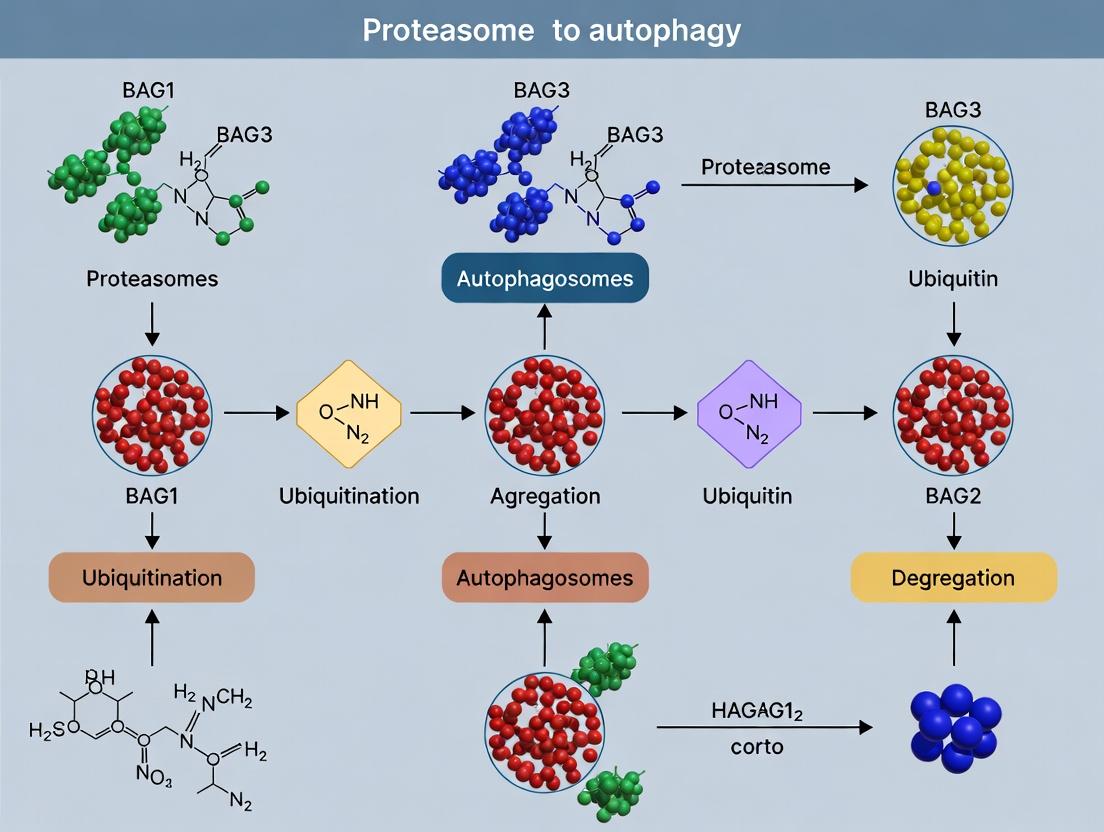
Proteostasis, or protein homeostasis, is the cellular process that ensures the proper synthesis, folding, trafficking, and degradation of proteins. Its precise regulation is fundamental to cellular health, and its dysregulation is implicated in numerous diseases, including neurodegeneration and cancer. The ubiquitin-proteasome system (UPS) and autophagy are the two primary degradation pathways. The UPS rapidly degrades short-lived, soluble, and misfolded proteins, while autophagy, particularly macroautophagy, clears long-lived proteins, aggregates, and damaged organelles. A critical and dynamic balance exists between these systems, coordinated by a network of chaperones, co-chaperones, and stress sensors. Central to this regulatory nexus is the switch between BAG1 and BAG3 co-chaperones, which directs client proteins toward the proteasome or autophagy, respectively, in response to proteostatic stress.
The BAG Domain Family and the BAG1/BAG3 Switch
BAG (Bcl-2-associated athanogene) proteins are a family of co-chaperones that bind to the ATPase domain of Hsp70/Hsc70 via their conserved BAG domain, modulating chaperone activity and client protein fate. BAG1 and BAG3 play opposing yet complementary roles in protein degradation pathways.
- BAG1 channels Hsc70-bound clients to the proteasome via its ubiquitin-like domain, which interacts directly with the proteasome. It is predominant under basal conditions.
- BAG3, under cellular stress (e.g., proteotoxic, heat, oxidative), displaces BAG1. BAG3, via its IPV motif, recruits clients to the autophagy machinery through its interaction with sequestosome-1 (p62/SQSTM1) and LC3 on the autophagosome membrane.
This switch represents a fundamental cellular strategy to adapt degradation capacity to the nature and severity of proteotoxic insult.
Table 1: Functional Characteristics of BAG1 and BAG3
| Feature | BAG1 | BAG3 |
|---|---|---|
| Primary Degradation Pathway | Ubiquitin-Proteasome System (UPS) | Selective Macroautophagy |
| Expression Condition | Basal, Constitutive | Stress-Induced (Heat, Proteotoxicity) |
| Key Binding Domain for Hsc70 | BAG Domain (C-terminal) | BAG Domain (C-terminal) |
| Unique Targeting Domain | Ubiquitin-like (Ubl) Domain | PxxP motif (binds HSPB8), IPV motif (binds p62/LC3) |
| Client Examples | Short-lived nuclear factors (e.g., steroid hormone receptors), misfolded soluble proteins | Aggregation-prone proteins (e.g., mutant Huntingtin, SOD1), damaged organelles |
| Cellular Localization | Nucleus, Cytoplasm | Cytoskeleton, Cytosol, perinuclear quality control (JUNQ) sites |
| Knockout Phenotype (Mouse) | Embryonic lethal, apoptosis defects | Neurodegeneration, myopathy, cardiomyopathy |
Table 2: Experimental Readouts in BAG1/BAG3 Switch Studies
| Experimental Assay | BAG1-Dominant (Proteasomal) Signature | BAG3-Dominant (Autophagic) Signature |
|---|---|---|
| Protein Half-life (CHX chase) | Shortened for specific clients (e.g., CFTRΔF508) | Stabilized or degraded via longer half-life kinetics |
| Aggregate Clearance (Filter Trap/IF) | Inefficient at clearing aggregates | Efficient clearance of protein aggregates |
| Inhibitor Sensitivity | Sensitive to MG132/Bortezomib (proteasome inhibitor) | Sensitive to 3-MA/Bafilomycin A1 (autophagy inhibitor) |
| Key Molecular Interaction | Co-immunoprecipitation with Proteasome subunits | Co-immunoprecipitation with p62, LC3, HSPB8 |
| Marker Expression | Decreased p62 levels, high ubiquitin conjugates | Accumulation of p62, increased LC3-II/I ratio |
Detailed Experimental Protocols
Protocol 1: Co-immunoprecipitation to Assess BAG Complex Formation
Objective: To validate the stress-induced interaction between BAG3, Hsc70, and the autophagic adapter p62.
- Cell Lysis: Treat HEK293 cells expressing FLAG-BAG3 with 10µM MG132 or DMSO (control) for 6 hours. Lyse cells in NP-40 lysis buffer (50mM Tris-HCl pH 8.0, 150mM NaCl, 1% NP-40) supplemented with protease/phosphatase inhibitors.
- Pre-clearance: Incubate lysate with Protein A/G agarose beads for 1h at 4°C. Centrifuge and collect supernatant.
- Immunoprecipitation: Incubate supernatant with anti-FLAG M2 affinity gel for 4h at 4°C with rotation.
- Wash: Pellet beads and wash 5x with cold lysis buffer.
- Elution: Elute bound proteins with 2x Laemmli buffer containing 100mM DTT at 95°C for 5 min.
- Analysis: Resolve proteins by SDS-PAGE and immunoblot for BAG3, Hsc70, and p62.
Protocol 2: Quantitative Analysis of Pathway Switching Using Fluorescence Microscopy
Objective: To visualize the shift of a client protein (e.g., mutant Huntingtin-Q74) from a diffuse/proteasomal localization to an autophagic aggregate upon BAG3 induction.
- Transfection: Co-transfect HeLa cells with mCherry-Htt-Q74 and GFP-BAG3 (or GFP-BAG1) using a lipid-based transfection reagent.
- Stress Induction: At 24h post-transfection, treat cells with 1µM Bortezomib for 12h to induce proteotoxic stress.
- Fixation & Staining: Fix cells with 4% PFA, permeabilize with 0.1% Triton X-100, and immunostain for p62 (autophagy marker) and ubiquitin.
- Imaging & Quantification: Acquire high-resolution confocal images. Quantify the percentage of mCherry-Htt-Q74 puncta that co-localize with p62 (Manders' coefficient) in GFP-BAG1 vs. GFP-BAG3 expressing cells. Analyze ≥50 cells per condition.
Signaling Pathways and Workflow Diagrams
Diagram 1: The BAG1/BAG3 Switch in Proteostasis Pathways.
Diagram 2: Experimental Workflow for BAG3 Complex Analysis.
The Scientist's Toolkit: Research Reagent Solutions
Table 3: Essential Reagents for BAG1/BAG3 and Proteostasis Research
| Reagent | Category/Supplier (Example) | Function in Research |
|---|---|---|
| MG-132 / Bortezomib | Proteasome Inhibitor (Sigma, Selleckchem) | Induces proteotoxic stress to trigger the BAG1-to-BAG3 switch and inhibit UPS function. |
| Bafilomycin A1 / Chloroquine | Autophagy Inhibitor (Cayman Chemical) | Inhibits autophagosome-lysosome fusion; used to monitor autophagic flux (LC3-II accumulation). |
| Cycloheximide (CHX) | Protein Synthesis Inhibitor (Sigma) | Used in chase experiments to measure protein half-life and degradation kinetics. |
| Anti-BAG3 / Anti-BAG1 Antibodies | Primary Antibodies (Cell Signaling, Abcam) | For detection, immunoprecipitation, and localization of key co-chaperones. |
| Anti-p62/SQSTM1 Antibody | Primary Antibody (MBL, Cell Signaling) | Marker for autophagic cargo and BAG3 interaction partner. |
| Anti-LC3B Antibody | Primary Antibody (Novus, Sigma) | Gold standard for monitoring autophagy (LC3-I to LC3-II conversion). |
| Anti-Ubiquitin (FK2) Antibody | Primary Antibody (Enzo) | Detects polyubiquitinated proteins, indicating proteasomal targeting or stress. |
| FLAG-M2 Affinity Gel | Immunoprecipitation Resin (Sigma) | For affinity purification of FLAG-tagged BAG proteins and their complexes. |
| Proteasome Activity Assay Kit | Biochemical Assay (Enzo, Abcam) | Measures chymotrypsin-, trypsin-, and caspase-like activities of the proteasome. |
| siRNA/shRNA for BAG1/BAG3 | Gene Knockdown Tools (Dharmacon, Sigma) | For loss-of-function studies to delineate specific roles of each co-chaperone. |
| HSP70/HSC70 Inhibitor (VER-155008) | Small Molecule Inhibitor (Tocris) | Tests chaperone-dependence of observed degradation phenotypes. |
The BAG (Bcl-2-associated athanogene) protein family comprises a group of co-chaperones that modulate the activity of Hsp70/Hsc70 molecular chaperones. These proteins share a conserved BAG domain (≈110 amino acids) near the C-terminus, which binds directly to the ATPase domain of Hsp70/Hsc70, facilitating nucleotide exchange (the release of ADP to allow ATP binding). This interaction regulates the chaperone cycle, influencing client protein folding, trafficking, and degradation. The broader thesis of contemporary research posits a critical cellular switch between proteasomal degradation and autophagy, orchestrated in part by the differential functions of BAG1 and BAG3. This switch is a pivotal adaptive response to proteotoxic stress, with implications in cancer, neurodegeneration, and aging.
Structural Biology of the BAG Domain
The BAG domain forms a three-helix bundle that interacts with Hsp70/Hsc70's ATPase domain. Despite low sequence homology, the tertiary structure is highly conserved across the family. The binding interface involves specific hydrophobic and electrostatic contacts that displace ADP, promoting the ATP-bound, low-substrate-affinity state of Hsp70.
Table 1: Core Human BAG Family Members
| Protein | Gene | Domains (Besides BAG) | Primary Localization | Key Binding Partners |
|---|---|---|---|---|
| BAG1 | BAG1 | Ubiquitin-like (Ubl) | Nucleus, Cytoplasm | Hsc70/Hsp70, Proteasome, Raf-1 |
| BAG2 | BAG2 | - | Cytoplasm | Hsc70/Hsp70, CHIP |
| BAG3 | BAG3 | WW, PxxP, IPV motifs | Cytoskeleton, Cytosol | Hsc70/Hsp70, Synaptopodin-2, HSPB8 |
| BAG4 | BAG4 | - | Cytoplasm | Hsc70/Hsp70, TNF-R1 |
| BAG5 | BAG5 | - | Cytoplasm | Hsc70/Hsp70, Parkin |
| BAG6 | BAG6 | Ubl, Pro-rich | Cytosol, Nucleus | HSP70, SGTA, GET complex |
Functional Dichotomy: The BAG1 vs. BAG3 Switch
The BAG1 and BAG3 co-chaperones represent two opposing poles in the cellular triage of misfolded proteins.
- BAG1: Directs clients towards the proteasome. Its N-terminal ubiquitin-like (Ubl) domain binds directly to the proteasome's 19S regulatory particle, coupling Hsc70-mediated substrate recognition to degradation. BAG1 is predominant under basal conditions.
- BAG3: Directs clients towards selective autophagy (aggrephagy). Under cellular stress (e.g., heat shock, proteasome inhibition), BAG3 expression is upregulated. It recruits the autophagic machinery via interactions with synaptopodin-2 and LC3. BAG3 also forms a complex with HSPB8, enhancing the recognition of misfolded proteins.
This "switch" from BAG1-mediated proteasomal degradation to BAG3-mediated autophagy is a critical stress adaptation, clearing large, aggregated proteins that cannot be processed by the proteasome.
Table 2: Quantitative Comparison of BAG1 and BAG3 Functions
| Parameter | BAG1 | BAG3 |
|---|---|---|
| Primary Degradation Pathway | Proteasome (26S) | Macroautophagy (Chaperone-Assisted Selective Autophagy - CASA) |
| Stress Induction | Constitutively expressed; often downregulated during severe stress. | Strongly upregulated by heat shock, oxidative stress, proteasome inhibition (HSF1-dependent). |
| Hsp70 Nucleotide Exchange Activity (kcat) | High (~15-20 fold stimulation)* | Moderate (~10-15 fold stimulation)* |
| Key Client Examples | ERα, Raf-1 kinase, glucocorticoid receptor | Mutant Huntingtin, SOD1, Tau, viral proteins |
| Half-life (Protein) | ~4-6 hours | >24 hours |
| Disease Association | Often oncogenic in cancer (e.g., breast, prostate). | Neuroprotection in neurodegeneration; pro-survival in some cancers. |
Note: Representative values based on *in vitro assays; exact rates vary by experimental conditions.*
Detailed Experimental Protocols
Protocol: Co-Immunoprecipitation (Co-IP) to Assess BAG-Hsp70 Complex Formation
Objective: To validate physical interaction between a BAG protein (e.g., BAG3) and Hsp70/Hsc70 in cell lysates under stress conditions.
- Cell Culture & Treatment: Plate HEK293T cells in 10 cm dishes. At 80% confluency, treat one set with 10 μM MG-132 (proteasome inhibitor) or 42°C heat shock for 2 hours to induce BAG3.
- Lysis: Rinse cells with cold PBS. Lyse in 1 mL NP-40 lysis buffer (50 mM Tris-HCl pH 7.4, 150 mM NaCl, 1% NP-40, 1 mM EDTA, plus protease/phosphatase inhibitors) on ice for 30 min. Centrifuge at 16,000 x g for 15 min at 4°C.
- Pre-clearing: Incubate supernatant with 20 μL of Protein A/G agarose beads for 30 min at 4°C. Pellet beads, retain supernatant.
- Immunoprecipitation: Incubate 500 μg of lysate with 2-5 μg of anti-BAG3 antibody (or IgG control) overnight at 4°C with gentle rotation. Add 40 μL of Protein A/G beads and incubate for 2-4 hours.
- Washing: Pellet beads, wash 4x with 1 mL lysis buffer.
- Elution & Analysis: Elute proteins in 2X Laemmli buffer by boiling for 5 min. Analyze by SDS-PAGE and Western blot using anti-Hsp70/Hsc70 and anti-BAG3 antibodies.
Protocol:In VitroNucleotide Exchange Assay
Objective: To measure the stimulation of ADP release from Hsp70 by purified BAG domain.
- Protein Purification: Express and purify recombinant human Hsp70 (ATPase domain) and His-tagged BAG1 domain (e.g., residues 1-150) from E. coli.
- Hsp70 Loading: Incubate 1 μM Hsp70 with 0.5 μM Mant-ADP (a fluorescent ADP analog) in assay buffer (20 mM HEPES pH 7.6, 50 mM KCl, 5 mM MgCl₂) for 15 min at 25°C.
- Nucleotide Exchange: In a fluorescence spectrophotometer (excitation 355 nm, emission 448 nm), add 10 μM of unlabeled ATP alone (control) or ATP plus 5 μM purified BAG1 protein to the Hsp70•Mant-ADP complex. The decrease in Mant-ADP fluorescence indicates its displacement.
- Data Analysis: Plot fluorescence decrease over time. Calculate the initial rate of exchange. The rate constant (kex) can be derived by fitting the curve to a single-exponential decay function.
Protocol: Monitoring the BAG1-BAG3 Switch via Luciferase-Based Aggregation Reporter
Objective: To visualize the shift from proteasomal to autophagic clearance.
- Reporter Construct: Use a luciferase (e.g., firefly luciferase) fused to an aggregation-prone protein (e.g., mutant Huntingtin exon1 with 74Q polyglutamine repeat).
- Cell Transfection: Co-transfect HeLa cells with the aggregation reporter and siRNA targeting BAG1 or BAG3, or with overexpression plasmids for each.
- Treatment & Readout: 24h post-transfection, treat cells with DMSO or 5 μM MG-132 for 12h.
- For Proteasomal Activity: Measure soluble luciferase activity using a standard luciferase assay kit (loss of activity indicates aggregation).
- For Autophagic Flux: Perform Western blot for LC3-II and p62/SQSTM1. Alternatively, use a tandem mRFP-GFP-LC3 reporter; autolysosomes show red-only puncta (GFP quenched in acidic pH), while autophagosomes show yellow (red+green) puncta.
- Analysis: Quantify aggregation (loss of soluble luciferase) and autophagic flux (LC3-II turnover, p62 degradation, RFP/GFP puncta ratio) under different BAG1/BAG3 modulation and stress conditions.
Visualizations
BAG Proteins Catalyze Hsp70 Nucleotide Exchange
The BAG1 to BAG3 Switch Under Proteotoxic Stress
Workflow for In Vitro Nucleotide Exchange Assay
The Scientist's Toolkit: Research Reagent Solutions
Table 3: Essential Reagents for BAG-Hsp70 Research
| Reagent/Catalog Example | Supplier Examples | Function & Application |
|---|---|---|
| Recombinant Proteins | ||
| Human Hsp70/Hsc70 (Pure) | Enzo, StressMarq | In vitro ATPase/nucleotide exchange assays, binding studies. |
| BAG1/BAG3 (BAG Domain) | Abcam, Origene | Purified for in vitro functional assays and interaction studies. |
| Antibodies | ||
| Anti-BAG1 (Monoclonal) | Cell Signaling, Abcam | Immunoprecipitation, Western blot, immunofluorescence to localize BAG1. |
| Anti-BAG3 (Polyclonal) | Proteintech, Novus | Detection of stress-induced BAG3 expression and Co-IP with Hsp70. |
| Anti-Hsp70/Hsc70 | Santa Cruz, CST | Detection of total Hsp70/Hsc70 in Co-IP and expression analysis. |
| Anti-LC3B | CST, MBL | Marker for autophagosome formation (LC3-I to LC3-II conversion). |
| Anti-p62/SQSTM1 | CST, Abnova | Monitor autophagic flux (degraded upon successful autophagy). |
| Chemical Modulators | ||
| MG-132 (Proteasome Inhibitor) | Selleckchem, MedChemExpress | Induces proteotoxic stress and BAG3 upregulation; triggers the switch. |
| VER-155008 (Hsp70 Inhibitor) | Tocris, MedChemExpress | Competitive ATP-site inhibitor; used to probe Hsp70 function in BAG pathways. |
| Bafilomycin A1 (V-ATPase Inhib.) | Sigma, Cayman | Blocks autophagosome-lysosome fusion; used to measure autophagic flux. |
| Cell Lines & Reporters | ||
| HEK293T, HeLa | ATCC | Standard cell models for transfection, Co-IP, and stress experiments. |
| Tandem mRFP-GFP-LC3 | Addgene (ptfLC3) | Live-cell imaging of autophagic flux (differentiates autophagosomes/lysosomes). |
| Luciferase-Aggregation Reporter | Custom construct | Quantify protein aggregation and pathway-specific clearance (proteasome vs. autophagy). |
| siRNA/shRNA Libraries | Dharmacon, Origene | Knockdown of BAG1, BAG3, HSPA8 (Hsc70) to study functional consequences. |
| Assay Kits | ||
| ATPase Colorimetric Assay Kit | Sigma, Innova | Measure Hsp70 ATPase activity with or without BAG protein stimulation. |
| Luciferase Assay System | Promega | Quantify soluble luciferase activity in aggregation-clearance assays. |
The BAG (Bcl-2-associated athanogene) family of co-chaperones integrates stress signaling with protein quality control. A central thesis in this field posits that cells employ a molecular switch between BAG1 and BAG3 to triage substrates between the two primary degradation pathways. BAG1, the focus of this guide, is the archetypal proteasome liaison, steering ubiquitinated clients bound to Hsp70/Hsc70 for degradation via the 26S proteasome. In contrast, BAG3, induced under persistent stress, recruits autophagy adaptors (e.g., p62/SQSTM1) to shuttle aggregates and large clients towards autophagic clearance. This BAG1-BAG3 switch represents a critical regulatory node in cellular proteostasis, with profound implications in aging, neurodegeneration, and cancer.
Core Mechanism & Quantitative Data
BAG1's function hinges on its modular domain architecture and specific interactions. Its ubiquitin-like (UBL) domain binds directly to the 19S regulatory particle of the proteasome, while its BAG domain interacts with the ATPase domain of Hsp70/Hsc70, promoting nucleotide exchange and client release.
Table 1: BAG1 Isoforms and Key Interactions
| Isoform | Length (aa) | Major Domains | Primary Localization | Key Quantitative Affinity (Kd) |
|---|---|---|---|---|
| BAG1S (p36) | 219 | BAG, UBL | Nucleus/Cytoplasm | Hsc70 BAG Domain: ~90 nM |
| BAG1M (p46) | 274 | NLS, BAG, UBL | Nucleus | Proteasome Rpn1/S2: ~0.5 µM |
| BAG1L (p50) | 345 | NLS, RF, BAG, UBL | Nucleus | Androgen Receptor: Data varies |
Table 2: BAG1 vs. BAG3 Functional Comparison
| Parameter | BAG1 | BAG3 |
|---|---|---|
| Primary Pathway | Proteasomal Degradation | Selective Autophagy (e.g., aggrephagy) |
| Stress Induction | Constitutive / Downregulated by stress | Strongly Upregulated by stress (HSF-1) |
| Hsp70 Binding | Promotes ADP release, substrate release | Stabilizes Hsp70-client complex |
| Key Binding Partner | 19S Proteasome (Rpn1, S2) | p62/SQSTM1, LC3, 14-3-3γ |
| Client Preference | Soluble, ubiquitinated proteins | Aggregated, large, ubiquitinated clients |
| Impact on Apoptosis | Pro-apoptotic (binds Bcl-2) | Anti-apoptotic (upregulates Bcl-2) |
Detailed Experimental Protocols
Protocol 1: Co-immunoprecipitation of the BAG1-Hsp70-Proteasome Complex
Objective: To validate the ternary complex formation under proteasomal targeting conditions.
- Cell Lysis: Harvest HEK293T cells overexpressing FLAG-BAG1. Lyse in mild NP-40 lysis buffer (50 mM Tris pH 7.5, 150 mM NaCl, 1% NP-40, 10% glycerol, 1 mM DTT) with protease and proteasome inhibitors (MG-132, 10 µM).
- Pre-clearing: Incubate lysate with control IgG and Protein A/G beads for 1h at 4°C.
- Immunoprecipitation: Incubate supernatant with anti-FLAG M2 affinity gel for 4h at 4°C.
- Washing: Wash beads 5x with lysis buffer.
- Elution & Analysis: Elute proteins with 3xFLAG peptide (150 ng/µL). Resolve by SDS-PAGE and immunoblot for BAG1 (FLAG), Hsp70/Hsc70, and 19S proteasome subunit Rpt5. Key Control: Use a BAG domain mutant (R231A) that cannot bind Hsp70 as a negative control.
Protocol 2: In Vitro Ubiquitinated Client Degradation Assay
Objective: To reconstitute BAG1-mediated targeting using purified components.
- Component Preparation:
- Purified 26S proteasome from bovine red blood cells.
- Recombinant human Hsc70, Hsp40 (DNAJA1), BAG1 (p36 isoform).
- Model Client: ({}^{35})S-methionine-labeled, ubiquitinated dihydrofolate reductase (Ub-DHFR) generated via rabbit reticulocyte lysate.
- Reaction Setup: In degradation buffer (50 mM Tris pH 7.5, 5 mM MgCl2, 2 mM ATP, 0.5 mM DTT), combine:
- Hsc70 (1 µM), Hsp40 (0.5 µM), ATP-regenerating system.
- ({}^{35})S-Ub-DHFR client (nM range).
- Test Conditions: +/- BAG1 (2 µM), +/- proteasome (50 nM).
- Incubation: Incubate at 30°C. Remove aliquots at t=0, 15, 30, 60 min.
- Analysis: Resolve by non-reducing SDS-PAGE (to preserve ubiquitin chains), dry gel, and quantify client loss via phosphorimaging. Degradation is plotted as % initial signal remaining.
Visualization: Signaling Pathways and Workflows
Title: BAG1 Mediated Hsp70 Client Targeting to the Proteasome
Title: The BAG1-BAG3 Switch in Proteostasis During Stress
Title: Co-IP Workflow for BAG1 Complex Analysis
The Scientist's Toolkit: Research Reagent Solutions
Table 3: Essential Reagents for BAG1/Proteasome Targeting Research
| Reagent | Function & Role in Experiment | Example Product/Assay |
|---|---|---|
| Recombinant BAG1 (Isoforms) | Purified protein for in vitro reconstitution assays, binding studies (SPR, ITC), and structural biology. | Human BAG1S (p36), His-tag, from E. coli. |
| Proteasome Inhibitors (MG-132, Bortezomib) | To inhibit proteasomal degradation, stabilizing ubiquitinated clients and complexes for pull-down assays. Validate pathway specificity. | Cell-permeable MG-132 (Z-Leu-Leu-Leu-al). |
| Hsp70/Hsc70 ATPase Assay Kit | Measure the nucleotide exchange activity of BAG1 by quantifying ATP hydrolysis by Hsp70 in presence of co-chaperones. | Colorimetric/Malon green-based kits. |
| Ubiquitinated Model Substrate | Defined client for degradation assays (e.g., Ub-DHFR, Ub-GFP). Can be generated in vitro via E1/E2/E3 enzymes or purchased. | Fluorescent Ubiquitinated GFP (Ub-GFP). |
| Anti-Polyubiquitin (K48-linkage) Antibodies | Specific detection of K48-linked chains, the canonical proteasomal degradation signal, on BAG1-associated clients. | Monoclonal antibody (e.g., Apu2 clone). |
| BAG1-Specific siRNAs/shRNAs | For loss-of-function studies to assess the dependency of client degradation on BAG1 vs. BAG3 or other pathways. | Validated siRNA pools targeting all isoforms. |
| Proteasome Activity Probe | Cell-permeable fluorescent or biotinylated activity-based probe to monitor 20S proteasome activity upon BAG1 manipulation. | MV151 (pan-proteasome) or subunit-specific probes. |
| BAG Domain Mutant (R231A) Plasmid | Critical negative control for Hsp70 binding. Mutation disrupts BAG domain interaction, abolishing function. | Available in FLAG-tagged mammalian vectors. |
Cellular protein homeostasis is maintained by two primary degradation systems: the ubiquitin-proteasome system (UPS) and the autophagy-lysosome pathway. A pivotal regulatory switch in stress response involves the transition from BAG1 to BAG3 co-chaperone function. Under basal conditions, BAG1, through its ubiquitin-like domain, directs Hsp70-bound client proteins to the proteasome. However, under acute stress (e.g., proteotoxic, oxidative, or thermal), BAG1 expression decreases while BAG3 expression is strongly upregulated. BAG3 then displaces BAG1 on the Hsp70 complex, redirecting polyubiquitinated cargo away from the proteasome. Instead, BAG3, via its interaction with the selective autophagy receptor p62/SQSTM1, facilitates the sequestration of clients into autophagosomes for lysosomal degradation. This "co-chaperone switch" represents a critical adaptive mechanism, allowing the cell to handle large, aggregated, or misfolded protein species that are unsuitable for proteasomal degradation. This whitepaper focuses on BAG3's role as a central node in mediating selective macroautophagy.
BAG3 Structure-Function and Interaction with p62/SQSTM1
BAG3 contains several conserved domains essential for its autophagy-regulatory function:
- BAG Domain (C-terminal): Binds to the ATPase domain of Hsp70/Hsc70, regulating its chaperone cycle.
- WW Domain: Mediates interaction with proline-rich motifs in other proteins.
- IPV Motifs (two repeats): Facilitate binding to the small heat shock proteins (HSPBs), crucial for chaperone-assisted selective autophagy (CASA).
- PxxP Motifs: Allow interaction with other SH3 domain-containing proteins.
The interaction with p62/SQSTM1 is primarily mediated through the PxxP motif in BAG3 and the SH2 domain of p62. This physical tether links the BAG3-Hsp70-client complex to the core autophagy machinery. p62, itself an autophagy receptor, oligomerizes and binds both ubiquitin (via its UBA domain) and LC3 on the forming autophagosome (via its LIR motif), ensuring targeted cargo encapsulation.
Quantitative Data on the BAG1/BAG3 Switch and Autophagy Flux
Table 1: Quantitative Changes in the BAG1/BAG3 Switch Under Stress Conditions
| Parameter | Basal Conditions (e.g., 37°C, No Stress) | Proteotoxic Stress (e.g., 42°C, 2h; 10µM MG132, 6h) | Measurement Method | Reference (Example) |
|---|---|---|---|---|
| BAG1 mRNA Level | 1.0 (relative units) | 0.2 - 0.4 | qRT-PCR | Gamerdinger et al., Nat Cell Biol, 2009 |
| BAG3 mRNA Level | 1.0 | 5.0 - 15.0 | qRT-PCR | Ibid. |
| BAG1 Protein Half-life | ~8 hours | Reduced by ~50% | Cycloheximide Chase | Ibid. |
| BAG3 Protein Half-life | ~6 hours | Increased (>10 hours) | Cycloheximide Chase | Ibid. |
| Hsp70-BAG1 Complexes | High | Low | Co-Immunoprecipitation | Behl, Cell Death Diff, 2016 |
| Hsp70-BAG3 Complexes | Low | High | Co-Immunoprecipitation | Ibid. |
| Proteasome Activity | 100% | 40-60% | Fluorogenic Peptide Substrate Assay | Myeku & Figueiredo-Pereira, J Neurosci, 2011 |
| Autophagic Flux | Baseline | 200-300% increase | LC3-II turnover (BafA1 assay) | Klimek et al., Cell Rep, 2017 |
Table 2: Key Binding Affinities in the BAG3-p62 Autophagy Pathway
| Interaction | Affinity (Kd) | Method | Functional Consequence |
|---|---|---|---|
| BAG3 BAG Domain : Hsp70 ATPase Domain | ~0.5 - 2 µM | ITC, SPR | Nucleotide exchange, client release |
| BAG3 IPV Motif : HSPB8 | ~1 - 5 µM | SPR, FP | CASA complex formation |
| BAG3 PxxP Motif : p62 SH2 Domain | ~10 - 20 µM | NMR, ITC | Recruits client complex to autophagosome |
| p62 UBA Domain : K48-Ubiquitin Chain | ~20 - 50 µM | ITC | Substrate recognition |
| p62 LIR Motif : LC3 | ~1 - 10 µM | NMR | Anchoring to phagophore |
Detailed Experimental Protocols
Protocol 1: Assessing the BAG1/BAG3 Switch via Co-Immunoprecipitation and Immunoblot
- Objective: To demonstrate stress-induced replacement of BAG1 with BAG3 on Hsp70 complexes.
- Methodology:
- Cell Treatment: Treat HEK293 or HeLa cells with 10µM MG132 (proteasome inhibitor) or subject to heat shock (42°C) for 2-6 hours.
- Lysis: Harvest cells in Nonidet P-40 lysis buffer (50 mM Tris-HCl pH 8.0, 150 mM NaCl, 1% NP-40, supplemented with protease inhibitors). Clear lysate by centrifugation.
- Immunoprecipitation: Incubate 500 µg lysate with 2 µg anti-Hsp70 antibody overnight at 4°C. Add Protein A/G beads for 2 hours.
- Washing: Wash beads 4x with lysis buffer.
- Elution & Analysis: Elute proteins in 2X Laemmli buffer. Separate by SDS-PAGE. Immunoblot for Hsp70 (loading control), BAG1, and BAG3.
Protocol 2: Measuring BAG3-p62 Dependent Autophagic Flux
- Objective: To quantify autophagy flux mediated by the BAG3-p62 axis using LC3 turnover.
- Methodology:
- Cell Transfection: Co-transfect cells with mCherry-GFP-LC3 tandem reporter and either siRNA against BAG3/p62 or a non-targeting control.
- Stress Induction: Apply stress (e.g., 400 nM Torin1 for 2h, or serum starvation).
- Inhibition: Treat one set with 100 nM Bafilomycin A1 (BafA1) for the last 4 hours to block autolysosome degradation.
- Imaging & Quantification: Image via confocal microscopy. The reporter is pH-sensitive: mCherry signal is stable, but GFP is quenched in acidic lysosomes. Thus:
- Yellow puncta (mCherry+GFP+): Autophagosomes.
- Red puncta (mCherry+GFP-): Autolysosomes.
- Flux Calculation: Autophagic Flux = (Red puncta in stressed cells) - (Red puncta in stressed + BafA1 cells). Compare flux between control and BAG3/p62 knockdown cells.
Protocol 3: In Vitro Reconstitution of BAG3-p62-LC3 Linkage
- Objective: To demonstrate direct, ubiquitin-dependent client targeting via the BAG3-p62-LC3 bridge.
- Methodology:
- Protein Purification: Purify recombinant FLAG-BAG3, His-p62, GST-LC3, and a model ubiquitinated client (e.g., Ub~Tau).
- Pull-down Assay: Immobilize GST-LC3 on glutathione-sepharose beads.
- Binding Reaction: Incubate beads with a mixture containing His-p62, FLAG-BAG3, Hsp70, ATP, and Ub~Tau client in binding buffer for 1 hour at 30°C.
- Washing & Elution: Wash extensively. Elute bound complexes with reduced glutathione.
- Analysis: Analyze eluates by SDS-PAGE and immunoblotting for the client (Tau), BAG3, and p62. Successful reconstitution shows all components in the eluate only when the complete mixture is present.
Signaling Pathways and Mechanisms
Diagram Title: BAG1-to-BAG3 Switch and Selective Autophagy Pathway
Diagram Title: BAG3-p62 Molecular Interaction Map
The Scientist's Toolkit: Key Research Reagent Solutions
Table 3: Essential Reagents for Studying BAG3-p62 Mediated Autophagy
| Reagent | Category | Example Product (Supplier) | Function in Experiment |
|---|---|---|---|
| BAG3 siRNA/shRNA | Genetic Tool | Silencer Select siRNA s17014 (Thermo Fisher); TRCN000006500 (Sigma MISSION) | Knockdown of BAG3 to establish functional necessity in pathway. |
| p62/SQSTM1 Antibody | Detection | Clone D5L7G (CST #39749); Clone 3/P62 (BD Transduction) | Immunoblotting, immunofluorescence, IP to monitor expression, localization, and interactions. |
| LC3B Antibody | Detection | Clone D11 (CST #3868); NanoTools 0231-100/LC3-5F10 | Detection of lipidated LC3-II (autophagosome marker) by WB and IF. |
| Tandem mCherry-GFP-LC3 | Reporter Plasmid | ptfLC3 (Addgene #21074) | Quantifying autophagic flux via fluorescence microscopy; distinguishes autophagosomes from autolysosomes. |
| Bafilomycin A1 | Pharmacological Inhibitor | B1793 (Sigma-Aldrich); 11038 (Cayman Chemical) | V-ATPase inhibitor that blocks autophagosome-lysosome fusion, used in flux assays. |
| Recombinant Human BAG3 Protein | Biochemical Tool | TP723190 (Origene); custom purification from E.coli | For in vitro binding assays, reconstitution experiments, or as a standard. |
| Proteasome Inhibitor | Stress Inducer | MG132 (Sigma C2211); Bortezomib (Selleckchem S1013) | Induces proteotoxic stress and triggers the BAG1/BAG3 switch. |
| Hsp70 Inhibitor | Pathway Modulator | VER-155008 (Tocris 3803) | Inhibits Hsp70 ATPase activity, useful for probing chaperone dependence of the pathway. |
| P62-LIR Competitive Peptide | Mechanistic Probe | TAT-LIR (Peptide International) | Cell-permeable peptide that disrupts p62-LC3 interaction, serving as a negative control. |
Abstract Within the cellular proteostasis network, the BAG (Bcl-2-associated athanogene) family of co-chaperones are critical regulators of Hsp70 function. Under basal conditions, BAG1 dominates, steering Hsp70-client complexes toward the ubiquitin-proteasome system (UPS) for degradation. During proteotoxic stress—induced by heat shock, oxidative stress, or proteasome inhibition—a molecular switch occurs, upregulating BAG3. BAG3 then recruits Hsp70 clients to the autophagy pathway via its interaction with LC3 and p62/SQSTM1. This whitepaper provides an in-depth technical analysis of this switch, its regulatory mechanisms, experimental investigation, and implications for diseases of protein aggregation.
1. Introduction: The BAG1-BAG3 Axis in Proteostasis The BAG domain is a conserved region that binds the ATPase domain of Hsp70, determining the fate of the chaperone complex. BAG1 contains a ubiquitin-like domain (UBL) that directs substrates to the proteasome. In contrast, BAG3 contains an IPV (Ile-Pro-Val) motif that binds to the autophagy receptor p62/SQSTM1 and a WW domain for interaction with other regulators. The competitive displacement of BAG1 by BAG3 on Hsp70 represents a fundamental cellular strategy to manage an overload of misfolded proteins by shifting from the high-capacity, selective UPS to the bulk-degradative autophagy machinery.
2. Molecular Mechanisms of the Switch The switch is orchestrated at transcriptional, post-transcriptional, and competitive binding levels.
- Transcriptional Regulation: The BAG3 gene promoter contains Heat Shock Elements (HSEs) bound by HSF1 upon stress. Conversely, BAG1 expression is often suppressed under prolonged stress.
- Post-translational Modifications: Phosphorylation of BAG3 (e.g., by MAPKAPK-2) enhances its stability and binding affinity for Hsp70 and p62.
- Competitive Binding: Increased BAG3 expression and its modified state allow it to outcompete BAG1 for the shared binding site on Hsp70's nucleotide-binding domain (NBD).
Diagram: BAG1/BAG3 Switch Mechanism
3. Quantitative Data Summary Table 1: Key Characteristics of BAG1 vs. BAG3
| Feature | BAG1 | BAG3 |
|---|---|---|
| Primary Domain | BAG Domain, UBL Domain | BAG Domain, IPV Motif, WW Domain, PxxP |
| Hsp70 Binding Affinity (Kd) | ~30-100 nM (strain-dependent) | ~50-200 nM (increases upon phosphorylation) |
| Degradation Pathway | Ubiquitin-Proteasome System (UPS) | Macroautophagy (Chaperone-Assisted Selective Autophagy, CASA) |
| Key Binding Partners | Proteasome (via UBL), Hsc70/Hsp70 | p62/SQSTM1, LC3, 14-3-3γ, HspB8 |
| Cellular Localization | Cytosol, Nucleus | Cytosol, Perinuclear, Stress Granules |
| Response to Stress | Often Downregulated | Strongly Upregulated (HSF1-mediated) |
| Knockout Phenotype (Mouse) | Embryonic Lethal | Juvenile-onset myopathy, cardiomyopathy |
Table 2: Experimental Stressors and Observed Switch Dynamics
| Stressor | Concentration/Dose | Time to BAG3 Upregulation | Key Readout |
|---|---|---|---|
| Heat Shock | 42-43°C | 2-6 hours | HSF1 translocation, BAG3 mRNA/protein increase |
| Proteasome Inhibitor (MG132) | 10-20 µM | 4-12 hours | p62 accumulation, LC3-II conversion, BAG1 degradation |
| Oxidative Stress (H₂O₂) | 200-500 µM | 1-4 hours | BAG3 phosphorylation, increased BAG3-Hsp70 binding |
| Arsenite (Proteasomal/Autophagic Stress) | 0.5 mM | 2-8 hours | Stress granule formation, BAG3-p62 co-aggregation |
4. Core Experimental Protocols
Protocol 4.1: Monitoring the Switch via Co-Immunoprecipitation (Co-IP) Objective: To assess the competitive displacement of BAG1 by BAG3 on Hsp70 under stress.
- Cell Treatment: Seed HEK293 or HeLa cells. Treat with 20 µM MG132 or 42°C heat shock for 6 hours vs. DMSO/37°C controls.
- Lysis: Harvest cells in mild lysis buffer (e.g., 1% NP-40, 150 mM NaCl, 50 mM Tris pH 8.0) supplemented with protease/phosphatase inhibitors. Avoid harsh detergents to preserve complexes.
- Pre-clearing: Incubate lysate with control IgG and Protein A/G beads for 1h at 4°C.
- Immunoprecipitation: Incubate pre-cleared lysate with 2 µg of anti-Hsp70 antibody overnight at 4°C. Add beads for 2h.
- Washing: Wash beads 3x with lysis buffer.
- Elution & Analysis: Elute with 2X Laemmli buffer. Analyze by Western blot for Hsp70 (load control), BAG1, and BAG3. Quantify band intensity ratios (BAG3:BAG1 bound to Hsp70).
Protocol 4.2: Assessing Functional Autophagic Flux via the BAG3-p62-LC3 Axis Objective: To confirm BAG3-dependent substrate routing to autophagy.
- Cell Manipulation: Transfect cells with siRNA targeting BAG3 or a non-targeting control. 48h post-transfection, induce stress (e.g., 10 µM MG132 for 12h). Include a group treated with 100 nM Bafilomycin A1 (BafA1) for the final 4 hours to inhibit autophagosome-lysosome fusion.
- Lysis and Western Blot: Lyse cells in RIPA buffer.
- Key Blots:
- LC3: Monitor conversion of cytosolic LC3-I to lipidated, autophagosome-associated LC3-II. Increased LC3-II in BafA1-treated vs. untreated indicates active flux.
- p62: p62 levels inversely correlate with autophagic degradation under stress. BAG3 knockdown should increase p62 accumulation.
- Ubiquitinated Proteins: Filter-trap assay or anti-polyubiquitin blot to assess aggregate load.
- Immunofluorescence: Co-stain for BAG3, p62, and LC3. Colocalization (Manders' coefficient) increases upon stress.
Diagram: Experimental Workflow for Switch Analysis
5. The Scientist's Toolkit: Essential Research Reagents
Table 3: Key Reagents for Investigating the BAG1/BAG3 Switch
| Reagent | Function/Application | Example (Vendor) |
|---|---|---|
| Proteasome Inhibitor | Induces proteotoxic stress, triggers the switch. | MG132 (Sigma, Selleckchem) |
| Autophagy Inhibitor | Blocks lysosomal degradation to measure autophagic flux (LC3-II accumulation). | Bafilomycin A1 (Cayman Chemical) |
| HSF1 Inhibitor | Tests HSF1-dependence of BAG3 upregulation. | KRIBB11 (Tocris) |
| siRNA/shRNA Plasmids | For knockdown of BAG1, BAG3, p62, or Hsp70 to establish functional necessity. | Dharmacon, Sigma Mission |
| BAG3 Phospho-specific Antibodies | Detect activating phosphorylation events (e.g., at Ser-377). | PhosphoSolutions, Cell Signaling |
| Hsp70/BAG Co-IP Kits | Optimized buffers and controls for pulling down chaperone complexes. | Thermo Fisher Scientific |
| LC3B Antibody | Gold standard for monitoring autophagy via WB (LC3-I vs II) and IF. | Novus Biologicals, MBL |
| p62/SQSTM1 Knockout Cell Line | Ideal background to study BAG3-mediated autophagy pathways. | ATCC, Horizon Discovery |
| Fluorescent Ubiquitin-Based Aggregation Sensor (FUBA) | Visualize aggregate formation and clearance in live cells. | Addgene plasmids |
6. Implications and Therapeutic Outlook The BAG1-to-BAG3 switch is implicated in cancer (chemoresistance), neurodegeneration (aggregate clearance), and myopathies. In cancer, elevated BAG3 promotes survival under stress, making it a drug target. In neurodegeneration, enhancing the switch may boost clearance of toxic aggregates. Future drug development aims at modulating this switch—either inhibiting BAG3 in oncology or promoting its function in protein aggregation diseases.
Key Client Proteins and Pathways Governed by the BAG1/BAG3 Toggle
1. Introduction: The BAG Domain Toggle Hypothesis
The BAG (Bcl-2-associated athanogene) family of co-chaperones are critical regulators of cellular proteostasis, functioning as nucleotide exchange factors for the heat shock protein 70 (HSP70) family. BAG1 and BAG3 represent two pivotal yet functionally opposing members. BAG1, via its ubiquitin-like domain, shuttles HSP70-bound clients to the proteasome for degradation. Conversely, BAG3, through its interaction with the small heat shock protein HSPB8 and dynein motors, promotes the sequestration of aggregation-prone clients into perinuclear aggressomes and their subsequent clearance via selective macroautophagy (aggrephagy). The "BAG1/BAG3 toggle" describes the competitive, stress-regulated switch in co-chaperone binding that determines the fate of HSP70 client proteins, directing them toward either proteasomal degradation or autophagic clearance. This whitepaper details the key client proteins, governed pathways, and experimental methodologies central to this research axis.
2. Key Client Proteins and Pathways
The fate of specific client proteins is decisively influenced by the prevailing BAG co-chaperone. The table below summarizes quantitatively characterized client proteins and their regulated pathways.
Table 1: Key Client Proteins, Fates, and Associated Pathways Governed by BAG1 vs. BAG3
| Client Protein | Primary BAG Binder | Cellular Fate | Governing Pathway / Process | Key Functional Consequence |
|---|---|---|---|---|
| HSF1 | BAG1 | Stabilization & Proteasomal Turnover | Heat Shock Response | BAG1 binding modulates HSF1 transcriptional activity and its own degradation. |
| Androgen Receptor (AR) | BAG1 | Proteasomal Degradation | Steroid Hormone Signaling | BAG1 promotes degradation of ligand-bound AR, attenuating signaling. |
| RAF-1 Kinase | BAG1 | Proteasomal Degradation | MAPK/ERK Signaling | BAG1-HSP70 complex facilitates RAF-1 turnover, influencing cell proliferation. |
| Mutant p53 | BAG3 | Aggresome/Autophagy Clearance | Tumor Suppressor Misfolding | BAG3 sequesters oncogenic mutant p53 for autophagic degradation. |
| Huntingtin (mHTT) | BAG3 | Aggresome/Autophagy Clearance | Protein Aggregation (PolyQ) | BAG3-HSPB8 complex targets mHTT aggregates for selective autophagy. |
| SOD1 (mutant) | BAG3 | Aggresome/Autophagy Clearance | Protein Aggregation (ALS) | BAG3 facilitates clearance of misfolded, aggregated SOD1. |
| Tau (hyperphosphorylated) | BAG3 | Aggresome/Autophagy Clearance | Neurofibrillary Tangle Pathology | BAG3 promotes clearance of pathological Tau species. |
| α-Synuclein | BAG3 | Aggresome/Autophagy Clearance | Lewy Body Formation | BAG3-HSPB8 complex is crucial for autophagic removal of α-synuclein oligomers. |
The mechanistic interplay is governed by a stress-sensitive switch, depicted in the following pathway diagram.
3. Detailed Experimental Protocols
3.1. Co-Immunoprecipitation (Co-IP) to Assess BAG-Client-HSP70 Complex Formation
Objective: To validate the physical interaction between BAG1/BAG3, HSP70, and a specific client protein under basal and stress conditions.
Protocol:
- Cell Culture & Transfection: Seed HEK293T or relevant cell line. Transfect with plasmids encoding tagged versions of the client protein (e.g., FLAG-tagged mutant p53) and either BAG1-Myc or BAG3-HA.
- Stress Induction (Optional): 24h post-transfection, treat cells with a proteasome inhibitor (e.g., 10µM MG-132 for 6h) to induce BAG3 pathway bias.
- Cell Lysis: Harvest cells in ice-cold IP lysis buffer (e.g., 50mM Tris-HCl pH 7.4, 150mM NaCl, 1% NP-40, 1mM EDTA) supplemented with protease and phosphatase inhibitors. Centrifuge at 16,000×g for 15 min at 4°C.
- Immunoprecipitation: Incubate cleared lysate with anti-FLAG M2 affinity gel for 2h at 4°C with rotation.
- Washing: Wash beads 3-4 times with cold lysis buffer.
- Elution: Elute bound proteins by boiling in 2× Laemmli sample buffer.
- Analysis: Resolve proteins by SDS-PAGE and perform Western blotting. Probe for the client (anti-FLAG), the BAG co-chaperone (anti-Myc or anti-HA), and endogenous HSP70.
3.2. Protein Turnover Assay via Cycloheximide Chase
Objective: To determine the effect of BAG1 or BAG3 overexpression/knockdown on the half-life of a client protein.
Protocol:
- Cell Manipulation: Establish stable cell lines with inducible shRNA targeting BAG1 or BAG3, or transiently overexpress each BAG protein.
- Translation Inhibition: Treat cells with 100µg/mL cycloheximide (CHX) to halt new protein synthesis.
- Time-Course Harvest: Collect cell pellets at defined time points (e.g., 0, 1, 2, 4, 8h) post-CHX addition.
- Lysis and Western Blot: Lyse cells and quantify protein. Analyze equal amounts of protein by Western blot for the client protein and loading control (e.g., GAPDH).
- Quantification: Densitometry analysis of band intensity. Plot client protein remaining (%) vs. time. Calculate half-life; BAG1 overexpression should shorten it (proteasomal), while BAG3 overexpression should extend it (autophagic).
3.3. Immunofluorescence Microscopy for Aggresome/Autophagosome Visualization
Objective: To visually confirm BAG3-mediated targeting of a client protein to aggressomes and autophagosomes.
Protocol:
- Cell Culture & Transfection: Plate cells on glass coverslips. Co-transfect with plasmids for the aggregation-prone client (e.g., mCherry-tagged mutant Huntingtin) and GFP-LC3 (autophagosome marker).
- Stress Induction: Treat with 5µM MG-132 for 12-16h to induce aggregation and BAG3 pathway activation.
- Fixation and Permeabilization: Fix cells with 4% paraformaldehyde for 15 min, permeabilize with 0.1% Triton X-100 for 10 min.
- Immunostaining: Block with 5% BSA, then incubate with primary antibody against BAG3 overnight at 4°C. Incubate with fluorescent secondary antibody (e.g., Alexa Fluor 647).
- Mounting and Imaging: Mount with DAPI-containing medium. Image using a confocal microscope. Co-localization of mCherry-client (red), BAG3 (cyan), and GFP-LC3 (green) puncta in the perinuclear region confirms BAG3-mediated aggrephagy.
4. The Scientist's Toolkit: Key Research Reagent Solutions
Table 2: Essential Reagents for Investigating the BAG1/BAG3 Toggle
| Reagent / Material | Supplier Examples | Function in BAG1/BAG3 Research |
|---|---|---|
| BAG1/BAG3 Specific Antibodies | Cell Signaling, Abcam, Santa Cruz | Detection of endogenous protein expression, localization (IF), and complex formation (IP). |
| Plasmids: BAG1/BAG3 (Tagged) | Addgene, Origene | For overexpression, domain-mutation studies, and co-localization experiments. |
| shRNA/siRNA for BAG1/BAG3 | Dharmacon, Sigma-Aldrich | Knockdown studies to assess loss-of-function phenotypes on client fate. |
| Proteasome Inhibitor (MG-132) | Sigma-Aldrich, Calbiochem | Induces proteotoxic stress to shift the toggle towards the BAG3/autophagy pathway. |
| Autophagy Inhibitor (Bafilomycin A1) | Sigma-Aldrich, Cayman Chemical | Blocks autophagic flux; used with BAG3 modulation to confirm autophagy-dependent client clearance. |
| CHX (Cycloheximide) | Sigma-Aldrich | Used in chase experiments to measure protein half-life and degradation kinetics. |
| HSP70 Inhibitor (VER-155008) | Tocris, Selleckchem | Pharmacologically disrupts HSP70 function to validate chaperone-dependence of observed effects. |
| LC3-GFP/RFP Tandem Reporter | Addgene (ptfLC3) | Monitors autophagic flux; distinguishes autophagosomes (GFP+/RFP+) from autolysosomes (GFP-/RFP+). |
| Aggresome Detection Kit | Cayman Chemical, Millipore | Dye-based (e.g., Proteostat) or antibody-based kit for specific detection of aggressomes. |
Techniques and Models: How to Study the BAG1/BAG3 Switch in Disease Research
This technical guide details the core genetic tools enabling the seminal research into the BAG1 vs BAG3 co-chaperone switch from proteasome-mediated degradation to autophagy. This molecular switch is critical in cellular stress response, cancer, and neurodegenerative diseases. Precise manipulation of BAG1 and BAG3 expression is fundamental to dissecting their distinct and overlapping roles in proteostasis.
Core Technologies: Mechanisms and Applications
siRNA (Small Interfering RNA)
- Mechanism: Synthetic 21-23 bp duplexes that are loaded into the RNA-induced silencing complex (RISC). The guide strand directs RISC to complementary mRNA for endonucleolytic cleavage and degradation.
- Primary Use: Transient knockdown (3-7 days). Ideal for rapid screening of BAG1/BAG3 function in acute stress assays.
shRNA (Short Hairpin RNA)
- Mechanism: DNA-encoded RNA sequences transcribed in vivo as a stem-loop, processed by Dicer into siRNA. Delivered via viral vectors (lentivirus, retrovirus) for stable integration.
- Primary Use: Stable, long-term knockdown. Essential for studying the BAG1/BAG3 switch in prolonged models like senescence or chronic proteotoxic stress.
CRISPR-Cas9 for Knockout & Knock-in
- Mechanism: The Cas9 nuclease, guided by a single guide RNA (sgRNA), creates double-strand breaks (DSBs) at specific genomic loci. Repair via error-prone non-homologous end joining (NHEJ) leads to frameshift knockouts. Precise edits (e.g., tagging) are achieved via homology-directed repair (HDR).
- Primary Use: Complete, permanent gene knockout or precise allele engineering (e.g., creating endogenous GFP-tagged BAG3).
CRISPR-Cas9 for Knockdown (CRISPRi) & Overexpression (CRISPRa)
- Mechanism (CRISPRi): Catalytically dead Cas9 (dCas9) fused to transcriptional repressors (e.g., KRAB) binds to promoter/enhancer regions to block transcription—a "chemical-free" knockdown.
- Mechanism (CRISPRa): dCas9 fused to transcriptional activators (e.g., VP64, p65AD) binds to promoter regions to upregulate gene expression.
- Primary Use: Reversible, tunable transcriptional modulation without altering the genomic DNA sequence. Ideal for studying dose-dependent effects of BAG1/BAG3 levels on the proteasome-autophagy switch.
Quantitative Comparison of Technologies
Table 1: Strategic Comparison of Genetic Manipulation Tools for BAG1/BAG3 Research
| Feature | siRNA | shRNA (Lentiviral) | CRISPR-Cas9 Knockout | CRISPRi/a (dCas9) |
|---|---|---|---|---|
| Target | Cytoplasmic mRNA | Cytoplasmic mRNA (via transcription) | Genomic DNA | Genomic DNA (regulatory regions) |
| Duration | Transient (3-7 days) | Stable, long-term | Permanent | Stable & reversible |
| Primary Outcome | mRNA degradation | mRNA degradation | Frameshift mutations, gene disruption | Transcriptional repression (i) or activation (a) |
| Key Application in BAG1/BAG3 Switch | Acute functional validation; rapid screens | Chronic stress models; in vivo studies | Complete loss-of-function models; studying redundancy | Tunable, dose-dependent studies of the switch |
| Off-Target Risk | Moderate (seed region effects) | Moderate (same as siRNA) | Low (but requires careful sgRNA design) | Very Low (for well-designed sgRNAs) |
| Typical Efficiency | 70-90% knockdown | >80% knockdown (pool), near 100% (clonal) | Variable; requires clonal isolation for 100% | 50-90% repression (i) or 5-50x activation (a) |
| Delivery | Lipid transfection, electroporation | Viral transduction | Plasmid/RNP transfection, viral transduction | Viral transduction (for stable lines) |
Detailed Experimental Protocols
Protocol: Establishing a Stable BAG3 Knockdown Cell Line via Lentiviral shRNA for Autophagy Flux Studies
Objective: To generate a stable cell line with reduced BAG3 expression to assay its necessity for stress-induced autophagy.
- shRNA Design: Select 3-4 validated shRNA sequences targeting human BAG3 mRNA from public databases (e.g., TRC, Sigma).
- Virus Production:
- Co-transfect HEK293T cells with the shRNA plasmid (in pLKO.1 vector), packaging plasmid (psPAX2), and envelope plasmid (pMD2.G) using PEI transfection reagent.
- Harvest virus-containing supernatant at 48 and 72 hours post-transfection. Concentrate via ultracentrifugation.
- Cell Transduction:
- Incubate target cells (e.g., HeLa or U2OS) with viral supernatant + 8 µg/mL polybrene for 24h.
- Selection & Validation:
- At 48h post-transduction, add 2 µg/mL puromycin for 7-10 days to select transduced cells.
- Harvest polyclonal population and validate knockdown via Western blot (anti-BAG3 antibody) and qRT-PCR.
Protocol: CRISPR-Cas9-Mediated Knock-in of an Endogenous Tag on BAG1
Objective: To insert a fluorescent tag (e.g., mNeonGreen) at the C-terminus of the endogenous BAG1 gene for localization studies.
- sgRNA Design: Design a sgRNA targeting the sequence just before the BAG1 stop codon using an online tool (e.g., Benchling).
- Donor Template Construction: Synthesize an ssODN or dsDNA donor template containing: a 5’ homology arm (~80 bp), the mNeonGreen sequence (no start codon), a P2A self-cleaving peptide sequence (optional), and a 3’ homology arm (~80 bp).
- Delivery & Editing:
- Transfect cells with a ribonucleoprotein (RNP) complex: 3 µg recombinant Cas9 protein + 1 µg in vitro transcribed sgRNA, along with 2 µM ssODN donor, using nucleofection.
- Clonal Isolation & Screening:
- At 48h post-nucleofection, single-cell sort into 96-well plates.
- Expand clones for 3-4 weeks. Screen via PCR (junction amplification) and confirm by Western blot (size shift) and fluorescence microscopy.
Signaling Pathways & Experimental Workflows
Title: BAG1 vs. BAG3 Regulation of Proteostasis Under Stress
Title: Decision Workflow for Selecting Genetic Tool
The Scientist's Toolkit: Research Reagent Solutions
Table 2: Essential Reagents for Genetic Manipulation Studies
| Item | Function & Relevance to BAG1/BAG3 Research | Example Supplier/Product |
|---|---|---|
| Validated siRNA/shRNA Pools | Pre-designed, sequence-verified RNAs for consistent knockdown of BAG1 or BAG3; reduces initial screening time. | Dharmacon ON-TARGETplus, Sigma MISSION shRNA |
| Lentiviral Packaging Plasmids | Essential for producing safe, high-titer lentivirus to deliver shRNA or CRISPR components; enables stable integration. | Addgene: psPAX2 (packaging), pMD2.G (envelope) |
| Recombinant Cas9 Protein | For RNP complex delivery; offers high editing efficiency, rapid turnover, and reduced off-target effects compared to plasmid DNA. | IDT Alt-R S.p. Cas9 Nuclease V3 |
| Chemically Modified sgRNA | Increased stability and reduced immunogenicity; crucial for high-efficiency RNP delivery in sensitive cell types. | Synthego (sgRNA EZ Kit) |
| Single-Stranded ODNs (ssODNs) | Serve as donor templates for precise HDR-mediated knock-in (e.g., tagging BAG1). | IDT Ultramer DNA Oligos |
| Puromycin/Drug Selection | Antibiotics for selecting cells successfully transduced with shRNA (puromycin) or CRISPR (e.g., blasticidin) vectors. | Thermo Fisher Scientific |
| Anti-BAG1 & Anti-BAG3 Antibodies | Critical validation tools for confirming knockdown/overexpression at the protein level via Western blot or immunofluorescence. | Cell Signaling Tech (#8682 BAG3), Abcam (ab79424 BAG1) |
| Autophagy Flux Reporter | Tandem fluorescent LC3 (mRFP-GFP-LC3) to monitor autophagic flux, a key readout of BAG3 function. | PtfLC3 (Addgene #21074) |
The BAG (Bcl-2-associated athanogene) family of co-chaperones are critical regulators of cellular proteostasis, linking molecular chaperones like Hsp70 to downstream degradation pathways. A pivotal concept in stress biology and disease (e.g., cancer, neurodegeneration) is the stress-induced switch from BAG1 to BAG3. Under basal conditions, BAG1, with its ubiquitin-like domain, directs Hsp70-client complexes to the proteasome for degradation. During cellular stress (e.g., proteotoxic, oxidative), BAG3 expression is upregulated. BAG3, containing an LC3-interacting region (LIR) and binding to the autophagy adapter p62/SQSTM1, reroutes misfolded clients towards selective autophagy (aggrephagy). Detecting this switch and its functional consequences is essential for understanding disease mechanisms and therapeutic targeting. This guide details the core assays for investigating this pathway.
Table 1: Characteristic Signatures of BAG1 vs. BAG3 in Proteostasis
| Parameter | BAG1 (Proteasome Route) | BAG3 (Autophagy Route) | Key Detection Assay |
|---|---|---|---|
| Primary Degradation Pathway | Ubiquitin-Proteasome System (UPS) | Macroautophagy / Aggrephagy | WB, IF (LC3/p62 markers) |
| Hsp70 Binding Affinity (Kd) | ~0.5 - 2.0 nM (high) | ~5.0 - 20 nM (lower, regulated) | Co-IP, SPR (cited data) |
| Stress-Induced Expression | Downregulated or stable | Strongly upregulated (>10-fold in some stresses) | qPCR, WB |
| Half-life of Client Proteins | Shortened (e.g., Raf-1: <30 min) | Prolonged (stabilized in aggregates) | Cycloheximide Chase + WB |
| Key Domain for Degradation | Ubiquitin-like (Ubl) domain | LC3-Interacting Region (LIR), PXXP motif | Co-IP (mutant constructs) |
| Colocalization with Markers | 26S Proteasome (Rpt subunits) | p62/SQSTM1, LC3-positive puncta | Immunofluorescence |
| Inhibition Effect | MG-132/Lactacystin blocks degradation | Bafilomycin A1/Chloroquine blocks degradation | Degradation Assay + WB |
Table 2: Expected Experimental Outcomes in a Model of Proteotoxic Stress (e.g., 10μM MG-132, 4h)
| Assayed Component | BAG1-KO/Condition | BAG3-KO/Condition | Wild-type (Stressed) | Assay |
|---|---|---|---|---|
| Hsp70-BAG1 Complex | Absent | Increased (~150%) | Decreased (~50%) | Co-IP |
| Hsp70-BAG3 Complex | Increased (~200%) | Absent | Increased (~300%) | Co-IP |
| Polyubiquitinated Proteins | Drastic Increase | Moderate Increase | Increase | WB (FK2 antibody) |
| LC3-II/LC3-I Ratio | No change or decrease | No conversion | Increased (~4-fold) | WB |
| p62/SQSTM1 Level | Accumulation | Strong Accumulation | Initial Increase then Clearance | WB, IF |
| BAG1 Protein Level | N/A | Stable | Downregulated (~40%) | WB |
| BAG3 Protein Level | Upregulated (~5-fold) | N/A | Upregulated (~8-fold) | WB |
Experimental Protocols
Co-immunoprecipitation (Co-IP) to Monitor Hsp70 Co-chaperone Complex Dynamics
Purpose: To physically demonstrate the stress-induced dissociation of Hsp70-BAG1 complexes and formation of Hsp70-BAG3 complexes. Protocol:
- Cell Lysis: Harvest HEK293 or stressed (e.g., 37°C, 10μM MG-132, 6h) cells in non-denaturing lysis buffer (e.g., 50mM Tris-HCl pH7.4, 150mM NaCl, 1% NP-40, 1mM EDTA, plus protease/phosphatase inhibitors). Centrifuge at 16,000×g, 20 min, 4°C.
- Pre-clearing: Incubate 500-1000μg lysate with 20μL Protein A/G agarose beads for 1h at 4°C. Pellet beads, keep supernatant.
- Immunoprecipitation: Incubate supernatant with 2-5μg of anti-Hsp70 antibody (or anti-BAG1/BAG3 for reciprocal IP) overnight at 4°C with gentle rotation.
- Bead Capture: Add 30μL equilibrated Protein A/G beads for 2h at 4°C.
- Washing: Pellet beads, wash 3x with cold lysis buffer.
- Elution: Resuspend beads in 2X Laemmli sample buffer, boil for 5 min.
- Analysis: Resolve by SDS-PAGE and perform Western blotting for Hsp70, BAG1, BAG3, and potential client proteins (e.g., HSF1, Raf-1).
Western Blotting to Quantify the Switch and Autophagic Flux
Purpose: To measure expression changes of BAG1/BAG3 and key autophagy markers. Protocol:
- Sample Preparation: Lyse cells in RIPA buffer. Determine protein concentration via BCA assay.
- Electrophoresis: Load 20-30μg protein per lane on 4-20% gradient SDS-PAGE gels.
- Transfer: Transfer to PVDF membrane using standard wet or semi-dry transfer.
- Blocking: Block with 5% non-fat milk in TBST for 1h.
- Primary Antibody Incubation: Incubate overnight at 4°C with specific antibodies:
- BAG1 (1:1000), BAG3 (1:1000), LC3B (1:2000), p62/SQSTM1 (1:2000), GAPDH/β-actin (loading control, 1:5000).
- Secondary Antibody Incubation: Incubate with HRP-conjugated anti-rabbit or anti-mouse IgG (1:5000) for 1h at RT.
- Detection: Use enhanced chemiluminescence (ECL) substrate and image.
- Autophagic Flux Note: Include samples treated with lysosomal inhibitors (e.g., 100nM Bafilomycin A1 for 4h) to distinguish increased LC3-II accumulation due to induction vs. blocked degradation.
Immunofluorescence to Visualize Subcellular Re-localization
Purpose: To visualize the colocalization of BAG3 with autophagic machinery upon stress. Protocol:
- Cell Seeding: Seed cells on poly-L-lysine-coated glass coverslips in 24-well plates.
- Stress Induction: Treat cells with stressor (e.g., 10μM MG-132, 17h).
- Fixation: Fix with 4% paraformaldehyde in PBS for 15 min at RT. Permeabilize with 0.1% Triton X-100 for 10 min.
- Blocking: Block with 3% BSA in PBS for 1h.
- Antibody Staining: Incubate with primary antibodies (e.g., anti-BAG3, anti-p62, anti-LC3) diluted in blocking buffer overnight at 4°C. Wash 3x with PBS.
- Secondary Staining: Incubate with fluorophore-conjugated secondary antibodies (e.g., Alexa Fluor 488, 568) and DAPI (for nuclei) for 1h at RT in the dark.
- Mounting: Mount coverslips with anti-fade mounting medium.
- Imaging: Acquire high-resolution images using a confocal microscope. Analyze colocalization using Manders' or Pearson's coefficient (e.g., for BAG3 and p62 puncta).
Pathway and Workflow Diagrams
BAG1-BAG3 Molecular Switch in Proteostasis Pathways
Integrated Workflow to Detect the BAG1-BAG3 Switch
The Scientist's Toolkit: Research Reagent Solutions
Table 3: Essential Reagents for BAG1/BAG3 Switch Research
| Reagent/Category | Specific Example/Product | Function in Experimental Context |
|---|---|---|
| Cell Stress Inducers | MG-132 (Proteasome Inhibitor), Bafilomycin A1 (Autophagy Inhibitor), HSP90 inhibitors (e.g., 17-AAG) | Induce proteotoxic stress to trigger the switch; inhibit autophagy flux for marker accumulation. |
| Validated Antibodies | Anti-BAG1 (CST #8689), Anti-BAG3 (CST #8550), Anti-LC3B (CST #3868), Anti-p62/SQSTM1 (CST #5114), Anti-Hsp70 (CST #4872) | Specific detection and immunoprecipitation of key pathway components. |
| Co-IP Grade Beads | Protein A/G Plus Agarose beads | Efficient capture of antibody-protein complexes for interaction studies. |
| LysoTracker & Dyes | LysoTracker Red DND-99, DAPI | Label acidic compartments (e.g., lysosomes, autolysosomes) and nuclei for live-cell or fixed-cell imaging. |
| siRNA/CRISPR Tools | BAG1-specific siRNA, BAG3 CRISPR Knockout Kit | Genetically perturb the system to establish causality in the switch phenotype. |
| Autophagy Reporter | mCherry-EGFP-LC3B tandem reporter (tfLC3) | Distinguish autophagosomes (yellow mCherry+EGFP+) from autolysosomes (red mCherry+ only) via fluorescence microscopy. |
| Lysis Buffers | Non-denaturing IP Lysis Buffer, RIPA Buffer | Extract proteins while preserving native interactions (Co-IP) or efficiently solubilizing all components (WB). |
| Fluorescent Secondaries | Alexa Fluor 488/568/647 conjugated anti-IgG | High-sensitivity, multi-color detection for immunofluorescence colocalization studies. |
Thesis Context: The BAG1/BAG3 Molecular Switch
This guide is framed within the broader thesis that the BAG (Bcl-2-associated athanogene) co-chaperones, specifically BAG1 and BAG3, orchestrate a critical switch in cellular protein quality control. BAG1, via its ubiquitin-like domain, typically directs Hsp70-bound client proteins to the proteasome for degradation. Under conditions of proteotoxic stress, a molecular switch occurs: BAG3 is upregulated, displacing BAG1 from Hsp70. BAG3, through its interaction with macroautophagy (hereafter autophagy) adaptors like p62/SQSTM1, redirects polyubiquitinated cargo and aggregation-prone proteins to the autophagic pathway via LC3 binding. This functional switch from proteasomal to autophagic flux is a crucial adaptive mechanism, and its dysregulation is implicated in cancer, neurodegeneration, and aging.
Table 1: Key Quantitative Readouts for Proteasomal vs. Autophagic Flux
| Pathway | Primary Measurement | Common Assay/Reagent | Typical Data Output | Interpretation |
|---|---|---|---|---|
| Proteasomal Flux | Chymotrypsin-like activity | Fluorogenic substrate (e.g., Suc-LLVY-AMC) | Fluorescence (RFU) over time | Increased RFU = increased proteasomal activity. |
| Protein ubiquitination | Western Blot (anti-Ubiquitin) | Ubiquitin-conjugate accumulation | Accumulation upon proteasome inhibition indicates flux. | |
| Reporter degradation | Ubiquitin-Fusion Degradation (UFD) reporters (e.g., UbG76V-GFP) | Fluorescence loss / Western Blot | Faster GFP loss = higher proteasomal flux. | |
| Autophagic Flux | LC3-II turnover | Western Blot (anti-LC3) with/without lysosome inhibitors (BafA1, CQ) | LC3-II ratio (+inhibitor/-inhibitor) | Ratio >1 confirms active autophagic flux. |
| p62/SQSTM1 degradation | Western Blot (anti-p62) with/without inhibitors | p62 level decrease | Decrease indicates autophagic degradation; blocked by inhibitors. | |
| Autophagosome accumulation | Fluorescent reporter (e.g., GFP-LC3, mRFP-GFP-LC3 tandem) | Puncta count & colocalization | Increased puncta; GFP quenching in lysosomes (mRFP signal only) indicates flux. | |
| BAG Modulation | BAG1/BAG3 Expression | qPCR, Western Blot | Fold-change mRNA, protein level | BAG3↑/BAG1↓ correlates with autophagy switch. |
| Client Protein Partitioning | Co-immunoprecipitation (Hsp70, BAG1, BAG3, p62) | Interaction strength | Stress shifts Hsp70 binding from BAG1 to BAG3; BAG3-p62 interaction increases. |
Experimental Protocols
Protocol 1: Concurrent Measurement of Proteasomal and Autophagic Flux Objective: To assess the functional shift in degradation pathways upon siRNA-mediated BAG1/BAG3 modulation under basal and stressed (e.g., 10µM MG132, 2h) conditions.
- Cell Treatment: Seed HEK293 or U2OS cells in 6-well plates. Transfect with siBAG1, siBAG3, or non-targeting siRNA.
- Flux Inhibition: 48h post-transfection, treat cells with DMSO (control), 100nM Bafilomycin A1 (BafA1; inhibits autophagic flux), or 10µM MG132 (inhibits proteasomal flux) for 4-6 hours.
- Sample Harvest: Lyse cells in RIPA buffer + protease inhibitors.
- Western Blot Analysis:
- Load equal protein amounts for SDS-PAGE.
- Probe with antibodies: LC3, p62, Ubiquitin, BAG1, BAG3, Hsp70, and loading control (β-Actin/GAPDH).
- Quantify: LC3-II (normalized to actin) with/without BafA1 to calculate autophagic flux. Measure p62 degradation. Assess ubiquitin conjugate accumulation with/without MG132.
Protocol 2: Live-Cell Kinetic Analysis with Tandem Reporter Objective: Visualize and quantify autophagic flux dynamics in real-time.
- Transfection: Transduce cells with adenovirus encoding mRFP-GFP-LC3 tandem reporter.
- BAG Modulation & Imaging: 24h later, transfect with siBAG1/BAG3 or treat with a pharmacological inducer of the switch (e.g., 5µM Ver-155008, an Hsp70 inhibitor). Include controls.
- Confocal Microscopy: Image live cells at 37°C, 5% CO₂ at 0, 12, 24h post-modulation.
- Analysis: Count GFP+/mRFP+ (yellow) puncta (autophagosomes) and GFP-/mRFP+ (red-only) puncta (autolysosomes). The red-only puncta count is a direct measure of autophagic flux completion.
Protocol 3: In Vitro Proteasomal Activity Assay Objective: Measure direct proteasome function from cell lysates after BAG modulation.
- Lysate Prep: Prepare cytosolic fractions from control and BAG-modulated cells in assay buffer.
- Reaction Setup: In a black 96-well plate, mix 20µg lysate with 100µM fluorogenic substrate Suc-LLVY-AMC in buffer. Include control wells with 20µM MG132 to confirm specificity.
- Kinetic Reading: Measure fluorescence (Ex/Em: 380/460 nm) every 5 minutes for 1-2 hours at 37°C using a plate reader.
- Analysis: Calculate the slope of the linear increase in RFU (Relative Fluorescence Units) as proteasomal activity.
Signaling Pathway & Workflow Diagrams
Diagram 1: BAG1/BAG3 Switch in Protein Degradation Pathways
Diagram 2: Integrated Experimental Workflow for Flux Analysis
The Scientist's Toolkit: Key Research Reagents
Table 2: Essential Reagents for BAG Modulation & Flux Studies
| Reagent / Material | Function & Application | Example Product/Catalog # |
|---|---|---|
| siRNA/shRNA for BAG1 & BAG3 | Specific knockdown to modulate the co-chaperone switch and observe pathway effects. Validated sequences are critical. | Origene, Dharmacon SMARTpool |
| Bafilomycin A1 (BafA1) | V-ATPase inhibitor. Blocks autophagosome-lysosome fusion, used to measure autophagic flux (LC3-II accumulation). | Sigma, B1793 |
| Chloroquine (CQ) | Lysosomotropic agent. Raises lysosomal pH, inhibiting degradation; used as an alternative flux inhibitor. | Sigma, C6628 |
| MG-132 / Epoxomicin | Potent proteasome inhibitors. Induce ubiquitin-conjugate accumulation and used to measure proteasome-dependent clearance. | Selleckchem, S2619 / S7038 |
| Anti-LC3B Antibody | Detects both cytosolic LC3-I and lipidated, autophagosome-associated LC3-II by Western Blot/IF. Essential for flux assays. | Novus Biologicals, NB100-2220 |
| Anti-p62/SQSTM1 Antibody | Monitors autophagy adaptor degradation. Decrease indicates active autophagic flux. | Abcam, ab109012 |
| mRFP-GFP-LC3 Tandem Reporter | Live-cell sensor. Differential pH sensitivity (GFP quenched in lysosomes, mRFP stable) allows quantification of autophagic flux stages. | ptfLC3 (Addgene, 21074) |
| Fluorogenic Proteasome Substrate (Suc-LLVY-AMC) | Sensitive, kinetic measurement of chymotrypsin-like proteasome activity in lysates or live cells. | Sigma, I-1395 |
| Hsp70 Inhibitor (Ver-155008) | Pharmacologically mimics stress by binding Hsp70, can induce the BAG3-dependent autophagy switch experimentally. | Tocris, 3803 |
| Ubiquitin Fusion Degradation (UFD) Reporter (UbG76V-GFP) | Constitutively targeted to proteasome. GFP signal loss correlates with proteasomal activity. | (Custom construct) |
Within the broader thesis on the BAG1 vs. BAG3 co-chaperones switch from proteasome to autophagy, modeling neurodegenerative diseases provides a critical experimental framework. This chaperone switch represents a fundamental cellular stress response, shifting degradation from the proteasome to macroautophagy when the proteasome is overwhelmed. This paper details technical modeling approaches for Alzheimer's disease (AD), Huntington's disease (HD), and Amyotrophic Lateral Sclerosis (ALS) to investigate this pivotal switch and its failure in neurodegeneration.
The BAG1/BAG3 Switch in Proteostasis
BAG1 and BAG3 are nucleotide exchange factors for Hsc70/Hsp70 with opposing degradation targeting. BAG1, through its ubiquitin-like domain, directs client proteins to the proteasome. BAG3, containing an IPV motif, recruits clients to the autophagy machinery via HSPB8 and p62/SQSTM1. Under acute stress, BAG1-mediated proteasomal degradation predominates. Chronic stress induces a switch to BAG3-mediated selective autophagy (chaperone-assisted selective autophagy, CASA). Neurodegeneration is characterized by the accumulation of aggregation-prone proteins (Aβ, tau, huntingtin, TDP-43, SOD1), suggesting a failure in this adaptive switch, leading to proteostatic collapse.
Disease-Specific Modeling & Technical Protocols
Alzheimer's Disease Modeling
Thesis Context: Modeling Aβ and tau pathology to test if BAG3 upregulation can ameliorate proteotoxic stress by enhancing autophagic clearance.
In Vitro Models:
- Cell Lines: SH-SY5Y, HEK293T stably expressing APP Swedish mutant (APPswe), or tau (e.g., tau P301L).
- Primary Neurons: Cortical/hippocampal neurons from transgenic mice (e.g., 3xTg-AD) or wild-type treated with oligomeric Aβ42.
Key Experimental Protocol: Inducing and Measuring Tau Aggregation & Clearance
- Transfection: Transfect HEK293T cells with plasmids for tau P301L-GFP and either BAG1-mCherry or BAG3-mCherry.
- Aggregation Induction: Treat cells with 10 μM proteasome inhibitor (MG132) for 12 hours to simulate proteasomal impairment and induce tau aggregation.
- Autophagy Modulation: Co-treat with 100 nM rapamycin (inducer) or 10 mM 3-Methyladenine (3-MA, inhibitor) for 12 hours.
- Analysis:
- Quantification: Image cells using high-content microscopy. Quantify tau-GFP puncta (aggregates) per cell.
- Biochemical: Perform filter trap assay for insoluble tau or Sarkosyl-insoluble fractionation followed by tau immunoblot.
- Pathway Activation: Immunoblot for LC3-I/II conversion, p62 degradation, and BAG1/BAG3 expression.
Table 1: Quantitative Metrics in AD Models
| Metric | Control (WT tau) | Tau P301L + MG132 | + BAG1 Overexpression | + BAG3 Overexpression | Measurement Technique |
|---|---|---|---|---|---|
| Tau Aggregates/Cell | 2.1 ± 0.5 | 25.3 ± 4.7 | 31.2 ± 5.1 | 8.4 ± 2.3 | High-content imaging |
| Insoluble Tau (A.U.) | 1.0 ± 0.2 | 15.7 ± 3.1 | 18.9 ± 3.8 | 5.2 ± 1.4 | Filter trap assay |
| LC3-II/LC3-I Ratio | 1.0 ± 0.3 | 2.1 ± 0.5 | 1.5 ± 0.4 | 4.7 ± 1.1 | Western Blot |
| p62 Level (A.U.) | 1.0 ± 0.2 | 3.5 ± 0.7 | 4.1 ± 0.8 | 0.6 ± 0.2 | Western Blot |
BAG1/BAG3 Switch in Alzheimer's Disease Pathology
Huntington's Disease Modeling
Thesis Context: Modeling polyQ-expanded huntingtin (HTT) aggregation to investigate BAG3's role in sequestering HTT into p62-positive aggresomes/autophagosomes.
In Vitro Models:
- Striatal Neuron Cell Lines: ST14A or STHdhQ111/Q111 knock-in cells.
- Primary Neurons: Striatal neurons from R6/2 or zQ175 knock-in mice.
Key Experimental Protocol: Monitoring HTT Aggresome Formation & Autophagic Flux
- Cell Modeling: Use STHdhQ111/Q111 cells or transfect cells with HTT-exon1-Q74-GFP.
- BAG Modulation: Knockdown BAG3 using siRNA or overexpress BAG3-mCherry.
- Live-Cell Imaging: Treat cells with 50 nM Bafilomycin A1 (inhibits autophagosome-lysosome fusion) for 6 hours. Image HTT-GFP and mCherry-p62 (transfected) or LysoTracker Red.
- Analysis:
- Co-localization: Calculate Manders' coefficients for HTT-GFP with mCherry-p62 (aggresome) or LysoTracker (lysosomal delivery).
- FRAP: Perform Fluorescence Recovery After Photobleaching on HTT-GFP aggregates to measure protein mobility, indicating sequestration strength.
- Biochemical: Sequential extraction (Triton X-100, Sarkosyl) to separate soluble, oligomeric, and insoluble HTT.
Table 2: Quantitative Metrics in HD Models
| Metric | STHdhQ7/Q7 (Control) | STHdhQ111/Q111 (HD) | HD + BAG3 siRNA | HD + BAG3 OE | Measurement Technique |
|---|---|---|---|---|---|
| HTT-p62 Coloc. (M1) | 0.08 ± 0.03 | 0.45 ± 0.09 | 0.15 ± 0.05 | 0.72 ± 0.11 | Confocal Microscopy |
| Insoluble HTT (A.U.) | 1.0 ± 0.3 | 22.5 ± 5.2 | 35.1 ± 6.8 | 9.8 ± 2.4 | Sarkosyl-insoluble blot |
| Autophagic Flux (LC3-II Accum.) | 1.0 ± 0.2 | 2.8 ± 0.6 | 1.5 ± 0.3 | 5.2 ± 1.3 | WB: BafA1-treated/untreated |
| Cell Viability (%) | 100 ± 5 | 62 ± 8 | 45 ± 7 | 85 ± 6 MTT assay |
Amyotrophic Lateral Sclerosis Modeling
Thesis Context: Modeling TDP-43 or mutant SOD1 aggregation to assess the specificity of the BAG switch for different pathogenic clients.
In Vitro Models:
- Motor Neuron-like Cells: NSC-34 or induced pluripotent stem cell (iPSC)-derived motor neurons (iMNs).
- Cell Lines: HEK293T for TDP-43 aggregation assays.
Key Experimental Protocol: TDP-43 Cytoplasmic Mislocalization & Clearance Assay
- Model Generation: Differentiate iMNs from ALS-patient iPSCs (TDP-43 mutation) or transfect NSC-34 cells with TDP-43-GFP wild-type or ΔNLS mutant.
- Stress Induction: Apply oxidative stress (200 μM sodium arsenite, 1h) or inhibit nuclear export (10 μM Leptomycin B, 6h).
- Intervention: Transduce with lentivirus expressing BAG3 or a BAG3 mutant lacking the IPV motif (ΔIPV).
- Analysis:
- Subcellular Fractionation: Separate nuclear/cytoplasmic fractions. Immunoblot for TDP-43, Lamin B1 (nuclear), GAPDH (cytoplasmic).
- Immunofluorescence: Score percentage of cells with predominant cytoplasmic TDP-43 inclusion.
- Co-IP: Immunoprecipitate BAG3 and blot for TDP-43, Hsp70, and p62 to confirm complex formation.
Table 3: Quantitative Metrics in ALS Models
| Metric | Control iMNs | TDP-43 Mut iMNs | + BAG3 OE | + BAG3 ΔIPV | Measurement Technique |
|---|---|---|---|---|---|
| Cytoplasmic TDP-43 (%) | 12 ± 4 | 68 ± 10 | 30 ± 7 | 65 ± 9 | Immunofluorescence scoring |
| Cyt/Nuc TDP-43 Ratio | 0.3 ± 0.1 | 2.8 ± 0.6 | 1.1 ± 0.3 | 2.5 ± 0.5 | Subcellular fractionation |
| BAG3-TDP-43 Co-IP (A.U.) | 1.0 ± 0.3 | 5.5 ± 1.2 | 8.9 ± 1.8 | 1.5 ± 0.4 | Co-Immunoprecipitation |
| Motor Neuron Survival | 100 ± 6 | 55 ± 9 | 80 ± 8 | 58 ± 10 | Viability assay |
BAG3-Mediated CASA Pathway & Failure Points
The Scientist's Toolkit: Research Reagent Solutions
Table 4: Essential Reagents for BAG1/BAG3 Switch Research
| Item | Function & Application in Research | Example Product/Catalog # |
|---|---|---|
| Anti-BAG1 Antibody | Immunoblot, immunofluorescence to monitor BAG1 expression and localization. | Cell Signaling Tech #3251 |
| Anti-BAG3 Antibody | Critical for detecting BAG3 upregulation upon stress and its co-localization with aggregates. | Proteintech 10599-1-AP |
| p62/SQSTM1 Antibody | Marker for protein aggregates/aggresomes targeted for autophagy. | Abcam ab109012 |
| LC3B Antibody | Detects LC3-I to LC3-II conversion, standard for monitoring autophagic activity. | Novus Biologicals NB100-2220 |
| Proteasome Inhibitor (MG132) | Induces proteotoxic stress and triggers the BAG1/BAG3 switch in vitro. | Sigma-Aldrich C2211 |
| Autophagy Inhibitor (Bafilomycin A1) | Inhibits V-ATPase, blocks autophagosome-lysosome fusion; used in flux assays. | Cayman Chemical 11038 |
| Lentiviral BAG3 shRNA | For stable knockdown of BAG3 in cultured neurons to study loss-of-function. | Sigma TRCN0000295863 |
| Recombinant BAG3 Protein | For in vitro binding assays with Hsp70 and client proteins (e.g., tau). | Origene TP722002 |
| Hsp70/Hsc70 Inhibitor (VER-155008) | To dissect the dependency of BAG1/BAG3 functions on Hsp70 activity. | Tocris 3803 |
| IPV Motif Peptide (Competitor) | Peptide mimicking BAG3's HSPB8 binding site; disrupts CASA complex. | Custom synthesis |
| Filter Trap Assay Kit | Quantifies insoluble protein aggregates from cell/lysate samples. | DotBlot Apparatus |
| Live-Cell LysoTracker Dye | Stains acidic lysosomes to assess autophagic cargo delivery. | Thermo Fisher L12492 |
| iPSC-derived Motor Neurons (ALS mutant) | Physiologically relevant model for studying TDP-43/SOD1 pathology and BAG3 function. | Fujifilm Cellular Dynamics |
Experimental modeling of AD, HD, and ALS provides distinct yet convergent platforms to interrogate the BAG1/BAG3 switch. The protocols and quantitative frameworks detailed here allow for rigorous testing of the hypothesis that reinforcing the BAG3-mediated autophagic pathway can compensate for proteasomal insufficiency, a common theme in neurodegeneration. This research directly informs therapeutic strategies aimed at modulating co-chaperone networks to restore proteostasis.
Within the cellular stress response, the BAG (Bcl-2-associated athanogene) family of co-chaperones regulates critical protein homeostasis decisions. A pivotal concept in modern cancer biology is the BAG1 vs. BAG3 switch, which governs a strategic shift from proteasomal degradation to autophagic clearance. Under basal conditions, BAG1, through its ubiquitin-like domain, directs Hsp70-client complexes to the proteasome. Under acute stress (e.g., chemotherapy, hypoxia, nutrient deprivation), BAG3 expression is upregulated. BAG3, via its IPV (Ile-Pro-Val) motif, recruits the autophagic machinery, while simultaneously inhibiting proteasomal activity. This switch allows cancer cells to survive by disposing of large, aggregated toxic proteins and damaged organelles via macroautophagy, conferring therapeutic resistance. This whitepaper details the role of BAG3 in this adaptive mechanism.
Quantitative Data on BAG3 in Cancer
Table 1: Correlation of BAG3 Expression with Clinical and Experimental Parameters
| Cancer Type | High BAG3 Association | Quantitative Measure (Example) | Reported Hazard Ratio (HR) / p-value |
|---|---|---|---|
| Pancreatic Ductal Adenocarcinoma | Poor Overall Survival | mRNA & IHC Score | HR: 2.45 (95% CI: 1.67–3.58), p<0.001 |
| Glioblastoma | Temozolomide Resistance | Protein Level (Western) | IC50 increase >3-fold in BAG3-high cells |
| Triple-Negative Breast Cancer | Metastasis & Recurrence | IHC Score in patient tissue | p=0.003 for recurrence-free survival |
| Ovarian Cancer | Platinum Resistance | mRNA fold-change | 4.8-fold increase in resistant cell lines |
| Hepatocellular Carcinoma | Proliferation Index | Ki-67 correlation coefficient | r=0.72, p<0.01 |
Table 2: Key Functional Consequences of BAG3 Upregulation
| Cellular Process | Experimental Readout | Typical Change with BAG3 Overexpression |
|---|---|---|
| Apoptosis Resistance | Caspase-3/7 activity after stress | Reduction of 60-80% |
| Autophagic Flux | LC3-II turnover (blot) / GFP-LC3 puncta | Increase of 2-5 fold |
| Proteasomal Activity | GFPu degradation assay / Proteasome peptidase activity | Inhibition of 40-60% |
| Senescence Bypass | SA-β-Gal staining | Reduction of 70% in positive cells |
| Migration/Invasion | Transwell Matrigel assay | Increase of 2-3 fold |
Core Experimental Protocols
Protocol 1: Validating the BAG1-to-BAG3 Switch In Vitro
- Objective: To demonstrate the stress-induced switch at the protein level.
- Methodology:
- Cell Treatment: Subject cancer cell lines (e.g., MIA PaCa-2, U87MG) to relevant stress (e.g., 10µM Sorafenib, 2µM Paclitaxel, or Serum Starvation).
- Time-Course Harvest: Lyse cells at 0, 6, 12, 24, and 48h post-treatment.
- Western Blot Analysis: Probe membranes with anti-BAG1, anti-BAG3, anti-LC3, anti-p62/SQSTM1, anti-polyubiquitin (K48-linked), and loading control (e.g., GAPDH) antibodies.
- Expected Result: BAG1 levels decrease over time, while BAG3, LC3-II, and ubiquitinated proteins increase, with p62 degradation.
Protocol 2: Assessing BAG3-Dependent Autophagic Flux
- Objective: To confirm functional autophagy reliance on BAG3.
- Methodology:
- Genetic Manipulation: Establish stable cell lines with BAG3 shRNA knockdown or CRISPR-Cas9 knockout alongside scramble controls.
- Tandem Fluorescence Reporter (mRFP-GFP-LC3): Transfect the tandem sensor. Autophagosomes (yellow puncta: mRFP+GFP+) and autolysosomes (red-only puncta: mRFP+GFP-) are quantified via confocal microscopy.
- Pharmacological Inhibition: Treat cells with 100nM Bafilomycin A1 (V-ATPase inhibitor) for 4h to block autolysosomal acidification and LC3-II degradation.
- Quantification: Calculate autophagic flux as the difference in LC3-II levels or red-only puncta with and without Bafilomycin A1 in control vs. BAG3-deficient cells.
Protocol 3: BAG3-Protein Interaction Co-Immunoprecipitation (Co-IP)
- Objective: To map BAG3 interactions with Hsp70/Hsc70 and autophagy receptors.
- Methodology:
- Cell Lysis: Use mild lysis buffer (e.g., 1% CHAPS or 0.5% NP-40) with protease/phosphatase inhibitors.
- Immunoprecipitation: Incubate lysates with anti-BAG3 antibody-conjugated beads or anti-FLAG beads for FLAG-tagged BAG3. Use IgG as control.
- Wash & Elution: Wash beads stringently, elute proteins with 2X Laemmli buffer.
- Analysis: Subject eluates to Western blotting for Hsp70/Hsc70, p62, SYNPO2 (an IPV motif-binding partner), and ubiquitin.
Signaling Pathways and Workflow Visualizations
Diagram 1: The BAG1-to-BAG3 Switch Pathway
Diagram 2: Experimental Workflow for Autophagic Flux
The Scientist's Toolkit: Key Research Reagent Solutions
Table 3: Essential Reagents for BAG3/Autophagy Switch Research
| Reagent / Material | Supplier Examples | Primary Function in Context |
|---|---|---|
| Anti-BAG3 Antibody | Cell Signaling Tech (CAT#: 12594), Abcam | Detection of BAG3 protein via Western blot, IHC, Co-IP. Validated for human samples is critical. |
| Anti-LC3B Antibody | Novus Biologicals (NB100-2220), CST | Marker for autophagosomes (LC3-II form). Essential for flux assays. |
| mRFP-GFP-LC3 Tandem Reporter | Addgene (ptfLC3, #21074) | Gold-standard live-cell sensor for tracking autophagic flux stages. |
| Bafilomycin A1 | Sigma-Aldrich (B1793), Cayman Chemical | Inhibits autolysosome acidification; used to block degradation and measure flux. |
| Proteasome Activity Assay Kit | Cayman Chemical (ab107921), Biomol | Fluorogenic substrate-based kit to measure chymotrypsin-/caspase-like proteasome activity. |
| Hsp70/Hsc70 Inhibitor (VER-155008) | Tocris Bioscience, Selleckchem | Pharmacological inhibitor to dissect Hsp70-BAG3 complex dependence. |
| BAG3-specific siRNA/shRNA Pools | Horizon Discovery (M-012267), Santa Cruz Biotech | For targeted gene knockdown studies to establish functional necessity. |
| Live-Cell Apoptosis Assay (Annexin V/ PI) | Thermo Fisher Scientific, BioLegend | Quantify apoptosis resistance conferred by BAG3 upregulation. |
High-Throughput Screening (HTS) for Modulators of BAG1/BAG3 Expression or Interaction
Within the molecular chaperone network, the BAG family proteins function as nucleotide exchange factors for Hsp70. The switch between BAG1 and BAG3 as co-chaperone partners for Hsp70 determines the fate of client proteins, directing them either to the proteasome (via BAG1) or to selective autophagy (via BAG3). Dysregulation of this switch is implicated in cancer, neurodegeneration, and aging. High-throughput screening (HTS) for modulators of BAG1/BAG3 expression or their interaction with Hsp70 represents a strategic approach to identify pharmacological agents capable of reprogramming cellular proteostasis, offering novel therapeutic avenues.
Biological Context: The BAG1/BAG3 Proteostasis Switch
Core Pathways and Interactions
The following diagram illustrates the competing pathways governing client protein fate via the BAG1/BAG3-Hsp70 axis.
Title: BAG1 vs BAG3 Client Fate Decision Pathway
Quantitative Data on BAG1 and BAG3 Properties
Table 1: Comparative Molecular and Functional Properties of BAG1 and BAG3
| Property | BAG1 (Isoform p50) | BAG3 |
|---|---|---|
| Primary Domain | Ubiquitin-like (UBL) Domain, BAG Domain | WW Domain, PxxP Motifs, IPV Motif, BAG Domain |
| Key Binding Partner | Hsp70/Hsc70, Proteasome | Hsp70/Hsc70, HspB8, Synaptopodin-2, LC3 |
| Client Fate | Proteasomal Degradation | Autophagic (Macroautophagy/Chaperone-Assisted Selective Autophagy) |
| Cellular Response | Rapid Turnover, Apoptosis Promotion | Stress Adaptation, Apoptosis Inhibition, Cytoprotection |
| Reported Kd for Hsp70 | ~0.5 - 2 µM (BAG domain) | ~0.1 - 1 µM (BAG domain) |
| Disease Link | Often downregulated in some cancers; can be pro-apoptotic | Overexpressed in many cancers (e.g., glioblastoma, pancreatic); associated with resistance to therapy |
HTS Strategy and Assay Design
Strategic Workflow for HTS Campaign
The overall HTS process from assay selection to hit validation is outlined below.
Title: HTS Campaign Workflow for BAG1/BAG3 Modulators
Primary Assay Methodologies
Reporter Gene Assays for Expression Modulation
- Principle: Utilize promoters for BAG1 or BAG3 genes driving expression of a luciferase (e.g., NanoLuc, Firefly) or fluorescent protein (e.g., GFP) reporter.
- Detailed Protocol:
- Reporter Construct Cloning: Clone a ~1-2 kb genomic region upstream of the BAG1 or BAG3 transcription start site (TSS) into a promoterless vector upstream of the reporter gene (e.g., pGL4.10[luc2]).
- Cell Line Generation: Stably transfect the reporter construct into a relevant cell line (e.g., U-2 OS, HEK293, or a cancer cell line with imbalanced BAG1/BAG3). Use a selection marker (e.g., puromycin) to generate a polyclonal or monoclonal stable cell line.
- Assay Plate Preparation: Seed cells in white, solid-bottom 384-well plates at an optimized density (e.g., 5,000 cells/well in 40 µL medium). Incubate for 24 hours.
- Compound Addition: Using a liquid handler, transfer 100 nL of compounds from a DMSO library stock (typically 10 mM) to achieve a final concentration of ~10-20 µM. Include controls: DMSO-only (negative), known stress inducers (e.g., 10 µM MG-132 for BAG3 positive control).
- Incubation: Incubate plates for 16-24 hours at 37°C, 5% CO₂.
- Signal Detection: For luciferase, add 20 µL of ONE-Glo or Nano-Glo Luciferase Assay System reagent, incubate for 5-10 minutes, and read luminescence on a plate reader (e.g., EnVision).
- Data Analysis: Calculate Z' factor using controls. Normalize luminescence to DMSO controls. Hits are defined as compounds causing a significant change (e.g., >3 SD from mean) in reporter activity.
Protein-Protein Interaction (PPI) Assays for BAG-Hsp70 Disruption/Stabilization
- Principle: Use technologies like Bioluminescence Resonance Energy Transfer (BRET) or Fluorescence Polarization (FP) to monitor the BAG domain-Hsp70 interaction in cells or solution.
- Detailed Protocol for NanoBRET:
- Construct Design: Fuse Hsp70 (HSPA1A or HSPA8) to a NanoLuc luciferase (Hsp70-NL). Fuse the BAG domain of BAG1 or BAG3 to a suitable fluorescent acceptor (e.g., HaloTag, HT).
- Cell Transfection: Co-transfect HEK293T cells with constant amounts of Hsp70-NL and varying amounts of BAG-HT constructs in a 96-well format to establish an optimal donor:acceptor ratio for a robust BRET signal.
- Assay Optimization: In a 384-well plate, seed transiently or stably expressing cells. Prior to reading, add the cell-permeable HaloTag ligand conjugated to the BRET acceptor dye (e.g., NanoBRET 618 Ligand) according to manufacturer's protocol.
- Compound Screening: Add library compounds and incubate (2-6 hours). Add the furimazine substrate to activate NanoLuc.
- Dual Detection: Read luminescence at 450 nm (donor) and 618 nm (acceptor) using a compatible plate reader (e.g., GloMax Discover).
- BRET Ratio Calculation: Calculate the BRET ratio as (Em618 / Em450). Normalize to vehicle control (0% inhibition) and a control with excess unlabeled BAG peptide (100% inhibition). Hits shift the BRET ratio.
Key Quantitative Parameters for Assay Validation
Table 2: HTS Assay Performance Metrics and Targets
| Assay Parameter | Reporter Gene Assay | NanoBRET PPI Assay | Acceptance Criterion |
|---|---|---|---|
| Z' Factor | Calculated from (Positive Ctrl - Negative Ctrl) | Calculated from (No Competitor - High Competitor) | > 0.5 |
| Signal-to-Background (S/B) | Luminescence (Inducer/Uninduced) | BRET ratio (No Comp/High Comp) | > 3 |
| Coefficient of Variation (CV) | Across replicate negative control wells | Across replicate no-competitor wells | < 10% |
| Assay Volume | 20 - 50 µL | 20 - 50 µL | Minimized for cost |
| Library Concentration | 10 - 20 µM final | 10 - 20 µM final | Standard for primary screen |
The Scientist's Toolkit: Research Reagent Solutions
Table 3: Essential Reagents and Materials for HTS on BAG1/BAG3
| Reagent / Material | Supplier Examples | Function in HTS Context |
|---|---|---|
| BAG1 (full-length & BAG domain) Recombinant Protein | Origene, Abcam, BPS Bioscience | Target protein for biochemical PPI assays (FP, TR-FRET). |
| BAG3 (full-length & BAG domain) Recombinant Protein | Novus Biologicals, R&D Systems, Proteintech | Target protein for biochemical PPI assays and selectivity testing. |
| Hsp70/Hsc70 (HSPA1A/HSPA8) Recombinant Protein | Enzo Life Sciences, StressMarq | Binding partner for BAG proteins in in vitro assays. |
| Anti-BAG1 Antibody (ChIP-grade) | Cell Signaling Technology (CST #8682) | Validation of expression changes via Western blot or ICC. |
| Anti-BAG3 Antibody (ChIP-grade) | CST #12599, Proteintech 10599-1-AP | Validation of expression changes and monitoring autophagy flux. |
| NanoBRET PPI System | Promega | For intracellular, live-cell monitoring of BAG-Hsp70 interactions. |
| HaloTag Technology | Promega | Protein tagging for cellular localization and interaction studies. |
| ONE-Glo / Nano-Glo Luciferase Assay Systems | Promega | Detection for reporter gene assays; high sensitivity, low background. |
| Proteasome Inhibitor (MG-132) | Selleckchem, MilliporeSigma | Positive control for BAG3 induction and proteostasis stress. |
| Autophagy Inhibitor (Bafilomycin A1) | Cayman Chemical, Tocris | Control for validating autophagy-mediated effects of BAG3 modulators. |
| 384-well, White, Solid-Bottom Assay Plates | Corning, Greiner Bio-One | Optimal plate format for luminescence and BRET assays. |
| Automated Liquid Handler (e.g., Bravo, Echo) | Agilent, Labcyte | For precise, high-throughput compound and reagent dispensing. |
Secondary Assays and Hit Validation
Primary HTS hits require stringent triage to exclude artifacts and identify true mechanistic modulators.
Title: Hit Validation Cascade for BAG1/BAG3 Modulators
- Key Validation Experiments:
- Quantitative RT-PCR: Measure endogenous BAG1 and BAG3 mRNA levels in treated vs. untreated cells to confirm transcriptional regulation.
- Immunoblotting: Quantify BAG1, BAG3, Hsp70, LC3-II (autophagy marker), and polyubiquitinated proteins (proteasome substrate load) to assess functional consequences.
- Cellular Protein Turnover Assay: Utilize a fluorescently tagged reporter protein (e.g., GFPu, a destabilized GFP) to monitor whether hits shift degradation from proteasomal to autophagic pathways.
- Selectivity Profiling: Test hits against BAG family members (BAG2, BAG4, BAG5) and other Hsp70 co-chaperones (e.g., Hsp40) to ensure specificity.
Implementing a robust HTS campaign for modulators of the BAG1/BAG3 switch requires careful integration of appropriate assay technologies, rigorous validation, and a deep understanding of the underlying proteostasis biology. The identified chemical probes or lead compounds will not only serve as potential therapeutics but also as essential tools to further dissect the critical decision-making process at the proteasome-autophagy interface, advancing the core thesis of BAG co-chaperone switching in health and disease.
Challenges and Solutions in BAG1/BAG3 Experimental Design and Data Interpretation
The BAG (Bcl-2-associated athanogene) family of co-chaperones are critical regulators of cellular proteostasis, with BAG1 and BAG3 playing opposing yet pivotal roles in the switch from proteasomal degradation to autophagy. Accurate detection and differentiation of these isoforms via Western blotting (WB) and immunohistochemistry (IHC) is foundational to research in neurodegeneration, cancer, and aging. However, antibody cross-reactivity, inappropriate validation, and a lack of isoform-specific reagents are pervasive issues that confound data interpretation. This guide details the technical pitfalls and provides validated protocols to ensure specificity in the context of the BAG1-BAG3 proteostasis switch.
The Specificity Challenge: BAG Protein Isoforms and Epitope Homology
BAG1 exists as multiple isoforms (p50, p46, p33, p29) generated from alternative translation start sites, while BAG3 is a single, larger protein. Significant sequence homology, particularly in the conserved BAG domain (approximately 45% identity between BAG1 and BAG3), is a primary source of antibody cross-reactivity. Commercially available antibodies often target this domain, leading to false-positive signals.
Table 1: Key Homology Regions and Common Cross-Reactive Epitopes
| Protein | Isoforms | Molecular Weight (kDa) | Conserved BAG Domain (AA) | Common Cross-Reactive Region |
|---|---|---|---|---|
| BAG1 | p50, p46, p33, p29 | 29-50 | 218-345 | C-terminal BAG domain (≈45% identity to BAG3) |
| BAG3 | - | ~74 | 420-575 | C-terminal BAG domain (≈45% identity to BAG1) |
Validated Experimental Protocols for Specific Detection
Knockdown/Knockout Validation (Mandatory Control)
- Purpose: To confirm antibody specificity by eliminating the target protein.
- Protocol: Transfect target cells (e.g., HEK293, HeLa) with siRNA targeting BAG1 or BAG3 mRNA. A non-targeting siRNA serves as control. After 48-72 hours, perform lysis and WB.
- Lysis Buffer: RIPA buffer with protease/phosphatase inhibitors.
- Gel: 12% SDS-PAGE (to separate BAG1 isoforms from BAG3).
- Transfer: PVDF membrane, 100V for 90 min.
- Antibodies: Test anti-BAG1 (e.g., C-term) and anti-BAG3 (e.g., N-term) antibodies.
- Expected Result: A specific antibody shows signal loss only in its respective knockdown lane, not in the other.
Isoform Discrimination via High-Resolution Gel Electrophoresis
- Purpose: To separate and identify specific BAG1 isoforms, preventing misidentification.
- Protocol: Use extended electrophoresis on 12-15% gels. Include a full-range protein ladder.
- Running Conditions: 80V through stacking gel, 120V through resolving gel for ~2 hours or until the 25 kDa marker is near the bottom.
- Loading Control: GAPDH (37 kDa) helps orient the blot. BAG3 runs at ~74 kDa, distinct from BAG1 isoforms.
- Key: Overexpression of individual FLAG-tagged BAG1 isoforms can serve as migration controls.
Peptide Blocking Competiton Assay
- Purpose: To confirm signal is derived from binding to the intended epitope.
- Protocol: Incubate the primary antibody (at working dilution) with a 10-fold molar excess of the immunizing peptide (or a recombinant protein fragment) for 1 hour at room temperature prior to applying to the membrane. Compare signal intensity to antibody incubated with a non-relevant peptide.
- Outcome: Specific signal should be abolished or dramatically reduced in the peptide-blocked sample.
Visualization of the BAG1-BAG3 Functional Switch in Proteostasis
Diagram Title: BAG1 vs. BAG3 Proteostasis Pathway Switch
Research Reagent Solutions: Essential Toolkit
Table 2: Critical Reagents for Specific BAG Protein Research
| Reagent | Example Catalog # / Type | Function & Critical Note |
|---|---|---|
| BAG1 siRNA Pool | siRNA targeting all human BAG1 isoforms | Essential negative control for antibody validation. |
| BAG3 siRNA | siRNA targeting human BAG3 | Essential negative control for antibody validation. |
| BAG1 Isoform Expression Vectors | Plasmids encoding p50, p46, p33 FLAG-tagged | Positive controls for WB migration and antibody specificity. |
| BAG3 Expression Vector | Plasmid encoding full-length BAG3 FLAG-tagged | Positive control to distinguish from BAG1 signals. |
| Validated Primary Antibodies | Anti-BAG1 (C-term, isoform-specific); Anti-BAG3 (N-term specific) | Use antibodies validated in knockout systems. Avoid "BAG domain" antibodies. |
| Blocking Peptides | Recombinant BAG1/BAG3 protein fragments | For competition assays to confirm antibody-epitope binding. |
| High-Range Protein Ladder | Precision Plus Protein Dual Color standards | Crucial for accurate identification of BAG1 isoform sizes. |
| Proteasome Inhibitor | MG-132 (10 µM) | Induces stress, upregulates BAG3, useful for functional assays. |
| Autophagy Flux Inhibitor | Bafilomycin A1 (100 nM) | Used to monitor autophagic turnover of BAG3-client complexes. |
Data Interpretation and Quantification Guidelines
Table 3: Troubleshooting Common BAG Detection Results
| Observed Result | Potential Cause | Recommended Validation Experiment |
|---|---|---|
| Single band at ~74 kDa with anti-BAG1 | Likely cross-reactivity with BAG3 | Perform BAG3 siRNA knockdown. |
| Multiple bands (30-50 kDa) with anti-BAG1 | Expected for BAG1 isoforms. | Confirm with isoform-specific overexpression. |
| High background in IHC | Non-specific binding of primary or secondary antibody. | Optimize blocking (use 5% normal serum from secondary host); include no-primary control. |
| Band in siRNA knockout sample | Non-specific antibody or incomplete knockout. | Use CRISPR/Cas9 KO cell line as definitive control; try alternative antibody. |
Rigorous validation of antibody specificity is non-negotiable for meaningful research into the BAG1-BAG3 functional switch. Researchers must employ a combinatorial strategy of genetic controls (siRNA/KO), careful gel optimization, and peptide competition to generate reliable data. Investing in these validation steps upfront is essential to avoid the pervasive pitfalls of cross-reactivity and to accurately elucidate the complex roles of these co-chaperones in proteostasis and disease.
1. Introduction In the proteostasis network, the BAG family co-chaperones act as nucleotide exchange factors for Hsp70/Hsc70, directing client protein fate. A critical mechanistic switch occurs between BAG1, which shuttles clients to the proteasome, and BAG3, which couples Hsc70 to the autophagy machinery. Deciphering disease states and therapeutic interventions requires precise quantification of both the total cellular pools of BAG1/BAG3 and, crucially, the fraction actively engaged with client proteins. This guide details optimized assays to distinguish these pools within the research context of the BAG1-to-BAG3 functional switch from proteasomal degradation to autophagic clearance.
2. Quantitative Overview of BAG1 vs. BAG3 Table 1: Core Functional & Quantitative Distinctions Between BAG1 and BAG3
| Parameter | BAG1 (Isoforms) | BAG3 |
|---|---|---|
| Primary Fate Destination | Proteasome (via ubiquitin-like domain) | Macroautophagy (via LC3 interaction) |
| Key Binding Partners | Hsc70/Hsp70, Proteasome, CHIP | Hsc70/Hsp70, HSPB8, LC3, p62/SQSTM1 |
| Stress Response | Often downregulated | Robustly upregulated (Heat Shock, Proteotoxic) |
| Reported Half-life | ~4-6 hours (variable by isoform) | >12 hours (stabilized by HSPB8 interaction) |
| Basal Expression (HeLa) | ~5-20 ng/mg total protein | ~2-10 ng/mg total protein |
| Induced Expression (Stress) | ≤ 2-fold increase | 5- to 50-fold increase |
| Critical Domain for Client Binding | C-terminal BAG Domain (Hsc70 binding) | BAG Domain & IPV motifs (client interaction) |
3. Experimental Protocols for Pool Discrimination
3.1. Sequential Extraction for Total vs. Insoluble-Bound Pool Analysis Principle: BAG3, particularly under stress, sequesters clients into insoluble aggressomes or protein aggregates. A differential detergent extraction isolates soluble (free/loosely bound) and insoluble (tightly client-bound) fractions.
Protocol:
- Lyse cells in Buffer A (50 mM Tris-HCl pH 7.5, 150 mM NaCl, 1% NP-40, 0.5% sodium deoxycholate, protease/phosphatase inhibitors) on ice for 30 min.
- Centrifuge at 16,000 x g, 4°C, 15 min. Collect supernatant as the Soluble Fraction.
- Wash the pellet twice gently with cold Buffer A.
- Solubilize the pellet in Buffer B (Buffer A + 2% SDS) by sonication and heating at 95°C for 10 min. Centrifuge at 16,000 x g, RT, 10 min. Collect supernatant as the Insoluble Fraction.
- Analyze both fractions by immunoblotting for BAG1, BAG3, clients (e.g., Tau, mutant SOD1), and loading controls (GAPDH for soluble; Vinculin/Coomassie for insoluble).
3.2. Co-Immunoprecipitation (Co-IP) for Client-Complexed Pool Principle: Capturing BAG1 or BAG3 in native conditions preserves interactions with Hsc70 and client proteins, allowing quantification of the actively chaperone-bound pool.
Optimized Protocol:
- Prepare lysate in Non-Denaturing Lysis Buffer (50 mM HEPES pH 7.4, 150 mM NaCl, 1% Triton X-100, 1.5 mM MgCl2, 1 mM EGTA, 10% glycerol, fresh inhibitors).
- Pre-clear lysate with species-matched control IgG and protein A/G beads for 1h at 4°C.
- Incubate 500-1000 µg of pre-cleared lysate with 2-4 µg of anti-BAG1 (C-terminal specific) or anti-BAG3 antibody overnight at 4°C with gentle rotation. Use isotype control for background.
- Add 30 µL of equilibrated protein A/G magnetic beads for 2h at 4°C.
- Wash beads 4x with lysis buffer. Elute proteins in 2X Laemmli buffer at 95°C for 5 min.
- Immunoblot for Hsc70 (confirms co-chaperone function), target client (e.g., TDP-43), and the BAG protein itself.
3.3. Proximity Ligation Assay (PLA) for In Situ Visualization of Client Engagement Principle: PLA detects endogenous protein-protein complexes (<40 nm apart) as discrete fluorescent foci, allowing single-cell visualization of BAG-client interactions.
Protocol Outline:
- Culture cells on chamber slides. Fix with 4% PFA, permeabilize with 0.1% Triton X-100.
- Block and incubate with primary antibodies from two different host species (e.g., mouse α-BAG3, rabbit α-Hsc70 or α-client).
- Apply PLA probes (anti-mouse PLUS, anti-rabbit MINUS). Perform ligation and amplification steps per manufacturer's instructions (Duolink kit).
- Mount with DAPI-containing medium. Image with fluorescence microscopy.
- Quantify foci per cell using image analysis software (e.g., ImageJ). Foci count corresponds to abundance of client-bound BAG complexes.
4. Visualizing the BAG1/BAG3 Switch and Assay Workflow
Title: BAG1 vs. BAG3 Client Fate Decision Pathway
Title: Multiplex Assay Workflow for BAG Pool Analysis
5. The Scientist's Toolkit: Key Research Reagent Solutions
Table 2: Essential Reagents for BAG1/BAG3 Pool Analysis
| Reagent / Material | Function & Application | Critical Consideration |
|---|---|---|
| Isoform-Specific BAG Antibodies | WB, IP, IF. Must distinguish isoforms (e.g., BAG1 p50 vs p36) and avoid cross-reactivity with other BAGs. | Validate knockdown/knockout cells for specificity. |
| Hsc70/Hsp70 Antibodies | Confirm functional co-chaperone complexes in Co-IP; loading control. | Monoclonal for consistency in quantification. |
| Validated Client Proteins | Disease-relevant substrates (e.g., Tau, α-synuclein, mutant p53). | Use models with pathogenic aggregation propensity. |
| Non-Denaturing Detergents | Maintain native protein complexes for Co-IP (Triton X-100, Digitonin). | Avoid SDS in lysis for interaction studies. |
| Proteasome Inhibitor (MG132) | Blocks BAG1-mediated degradation, enriches client-bound pools for detection. | Use pulsed treatment to avoid compensatory autophagy. |
| Autophagy Inducer (Rapamycin) / Inhibitor (Bafilomycin A1) | Modulate BAG3 pathway flux; test pool redistribution. | Bafilomycin blocks lysosomal degradation, accumulates autophagic cargo. |
| Duolink PLA Kit | In situ detection of BAG-client/Hsc70 proximity. | Optimize antibody pairs with high specificity. |
| Differential Centrifugation Columns | Rapid separation of soluble/insoluble fractions. | Prevents cross-contamination of pools. |
| Magnetic Protein A/G Beads | Efficient, low-background immunoprecipitation. | Reduce non-specific binding vs. agarose beads. |
Within the molecular chaperone network, the switch between BAG1 and BAG3 represents a critical regulatory node determining the fate of misfolded proteins. The BAG1 co-chaperone directs client substrates to the proteasome for degradation, while BAG3, often in conjunction with HSPB8, facilitates the targeting of aggregation-prone proteins to the autophagic-lysosomal pathway, specifically via selective autophagy (e.g., aggrephagy). This BAG1/BAG3 switch is a cellular adaptation to proteotoxic stress, such as that induced by heat shock or proteasome inhibition. Conflicting flux data often arise when interpreting activity readouts of these two degradation systems, as they are dynamically interconnected and compensatory. This guide provides a technical framework for dissecting these complexities.
The Core Challenge: Interconnected and Compensatory Pathways
A primary source of conflicting data is the compensatory upregulation of autophagy when the proteasome is inhibited, and vice-versa. Isolating the flux through each system requires specific pharmacological and genetic tools alongside carefully timed assays. Simply measuring substrate accumulation can be misleading without determining the rate of flux through the pathway.
Essential Methodologies for Isolating Flux
Proteasomal Activity and Flux Assays
Protocol: Fluorogenic Peptide Substrate Cleavage Assay (Chymotrypsin-like Activity)
- Principle: Use of suc-LLVY-AMC substrate. Upon proteasomal cleavage, free AMC fluoresces.
- Procedure:
- Prepare cell lysates in assay buffer (e.g., 50 mM Tris-HCl, pH 7.5, 5 mM MgCl2, 1 mM DTT).
- Distribute lysate (10-20 µg protein) into a black 96-well plate.
- Add suc-LLVY-AMC to a final concentration of 40 µM.
- Immediately measure fluorescence (Ex/Em 350/440 nm) kinetically every 5 minutes for 60-90 minutes at 37°C.
- Critical Control: Include replicate wells pre-treated for 30 minutes with 20 µM MG-132 to establish background signal.
- Interpretation: The slope of the MG-132-inhibitable fluorescence increase represents proteasomal activity.
Protocol: Monitoring Ubiquitinated Protein Turnover (Pulse-Chase)
- Principle: Track the degradation of a pulse-labeled protein under different conditions.
- Procedure:
- Starve cells for 30 min in methionine/cysteine-free medium.
- "Pulse" with medium containing L-azidohomoalanine (AHA) or 35S-labeled Met/Cys for 20 min.
- "Chase" with complete medium for various time points (0, 30, 60, 120 min).
- At each time point, perform click-chemistry to biotinylate AHA-labeled proteins or perform immunoprecipitation of the protein of interest.
- Analyze by Western blot. Co-treatment with proteasome (MG-132, 10 µM) or autophagy inhibitors (Bafilomycin A1, 100 nM) during the chase clarifies the responsible pathway.
Autophagic Flux Assays
Protocol: LC3-II Turnover Assay via Immunoblotting
- Principle: LC3-II levels correlate with autophagosome number but must be interpreted with lysosomal inhibition to measure flux.
- Procedure:
- Seed cells in 6-well plates. Set up two sets: DMSO control and Bafilomycin A1 (BafA1, 100 nM) or chloroquine (CQ, 50 µM).
- Treat cells with experimental conditions (e.g., stress inducer) for desired time.
- Treat one set with BafA1/CQ for the final 4-6 hours to block autophagosome-lysosome fusion/degradation.
- Harvest cells, perform SDS-PAGE and Western blot for LC3.
- Also blot for p62/SQSTM1, an autophagic substrate.
- Interpretation: A true increase in autophagic flux is indicated by an increase in LC3-II and decrease in p62 in the absence of BafA1, with a further accumulation of LC3-II in the presence of BafA1.
Protocol: Tandem Fluorescent LC3 (mRFP-GFP-LC3) Reporter Assay
- Principle: GFP is quenched in the acidic lysosome, while mRFP is more stable. This differentiates autophagosomes (yellow puncta, RFP+GFP+) from autolysosomes (red puncta, RFP+GFP-).
- Procedure:
- Transfect cells with an mRFP-GFP-LC3 plasmid.
- 24-48h post-transfection, treat cells as required.
- Fix cells and image via confocal microscopy.
- Quantify the number of yellow and red puncta per cell using image analysis software.
- Interpretation: An increase in autophagic flux is shown by an increase in red puncta. An increase only in yellow puncta suggests a block in fusion or degradation.
Simultaneous Monitoring in the Context of BAG1/BAG3
Protocol: Co-immunoprecipitation and Degradation Tracking
- Objective: Determine the binding preference of a misfolded protein client to BAG1/HSC70 or BAG3/HSC70 complexes under stress, and correlate with degradation route.
- Procedure:
- Treat cells (e.g., with proteasome inhibitor or heat shock) to induce the BAG switch.
- Lyse cells in mild lysis buffer.
- Perform immunoprecipitation (IP) for the client protein, BAG1, or BAG3.
- Immunoblot co-precipitated partners (HSC70, BAG1/BAG3, ubiquitin).
- In parallel, perform cycloheximide chase (CHX, 50 µg/mL) to monitor client degradation rate, with and without pathway-specific inhibitors.
Table 1: Key Pharmacological and Genetic Tools for Pathway Dissection
| Target/Manipulation | Tool (Example) | Mechanism of Action | Primary Use in Flux Assays |
|---|---|---|---|
| Proteasome Inhibition | MG-132 (10-20 µM) | Reversible peptide aldehyde inhibitor of chymotrypsin-like site. | Control for proteasome-specific activity; induces BAG3 and autophagy. |
| Lysosome Inhibition | Bafilomycin A1 (50-100 nM) | V-ATPase inhibitor; blocks autophagosome-lysosome fusion and acidification. | Essential for LC3-II turnover assay to measure autophagic flux. |
| Global Protein Synthesis Inhibition | Cycloheximide (50 µg/mL) | Inhibits eukaryotic translation elongation. | Used in chase experiments to monitor pre-existing protein degradation. |
| BAG3 Knockdown | siRNA/shRNA targeting BAG3 | Reduces BAG3 protein expression. | To test necessity of BAG3 for autophagic clearance of aggregates under stress. |
| BAG1 Overexpression | BAG1 expression plasmid | Increases BAG1-HSC70 complex formation. | To test sufficiency for proteasomal targeting and inhibition of the autophagy switch. |
Table 2: Interpretation of Conflicting Readout Scenarios
| Observed Data | Potential Confound | Resolution Experiment | Conclusion if Resolved |
|---|---|---|---|
| High ubiquitinated proteins + High LC3-II | Compensatory autophagy activation due to proteasome impairment. | Perform LC3-II turnover assay ± BafA1. If BafA1 causes further LC3-II rise, flux is high. | Proteasome is compromised; autophagy is upregulated as compensatory response (BAG3-mediated). |
| Rapid client degradation despite proteasome inhibitor | Client is switched to BAG3-mediated autophagy. | Co-IP client with BAG3 after inhibitor treatment; test if degradation is blocked by BafA1 or BAG3 KD. | Client degradation pathway has switched from proteasomal (BAG1) to autophagic (BAG3). |
| Accumulation of p62 with increased LC3-II | Block in autophagic degradation, not induction. | Perform mRFP-GFP-LC3 assay. Prevalence of yellow puncta indicates a block. | Autophagosome formation is induced, but flux is impaired downstream (fusion/lysis defect). |
Signaling Pathways and Workflows
Title: BAG1 vs BAG3 Pathway Switching Under Stress
Title: Decision Workflow for Resolving Conflicting Degradation Data
The Scientist's Toolkit: Research Reagent Solutions
| Reagent/Tool | Supplier Examples | Function in BAG1/BAG3 & Flux Research |
|---|---|---|
| Fluorogenic Proteasome Substrates (e.g., Suc-LLVY-AMC) | Cayman Chemical, Enzo Life Sciences | Directly measures chymotrypsin-like proteasome activity in lysates or live cells. |
| LC3B Antibody | Cell Signaling Tech (#3868), Novus Biologicals | Gold-standard for monitoring autophagosome association (LC3-I to LC3-II shift) by immunoblot. |
| p62/SQSTM1 Antibody | Abcam (ab109012), Cell Signaling Tech (#5114) | Marks autophagic substrates; degradation inversely correlates with autophagic flux. |
| Bafilomycin A1 | Sigma-Aldrich (B1793), InvivoGen | Lysosomal inhibitor essential for conclusive autophagic flux measurement via LC3-II blots or reporters. |
| Tandem mRFP-GFP-LC3 Plasmid | Addgene (#21074) | Enables quantitative distinction between autophagosomes and autolysosomes via microscopy. |
| BAG1 & BAG3 Specific Antibodies | Santa Cruz Biotechnology, Proteintech | For monitoring expression shifts and performing co-immunoprecipitation experiments. |
| HSC70/HSPA8 Antibody | Enzo Life Sciences (ADI-SPA-815) | Identifies the core chaperone complex interacting with both BAG1 and BAG3. |
| Recombinant BAG1/BAG3 Proteins | Origene, Abnova | Used for in vitro binding assays or as standards for quantitative analysis. |
| Live-Cell Proteasome Sensor (e.g., ZsProSensor-1) | Clontech/Takara | Reports real-time proteasome function in live cells via fluorescence. |
| Cycloheximide | Sigma-Aldrich (C7698) | Protein synthesis inhibitor used in chase experiments to track degradation kinetics of existing proteins. |
The BAG (Bcl-2-associated athanogene) family of co-chaperones are critical regulators of cellular proteostasis, bridging Hsp70/Hsc70 chaperone activity to downstream degradation pathways. A central paradigm in modern proteostasis research is the "BAG switch"—the context-dependent shift from BAG1-mediated proteasomal degradation to BAG3-mediated selective macroautophagy (hereafter autophagy). This whitepaper provides a technical guide for researchers on selecting appropriate cellular and animal models, and designing experiments to dissect this switch, which is pivotal in stress response, aging, cancer, and neurodegenerative diseases.
The BAG1-BAG3 Switch: Molecular Mechanisms
BAG1, through its ubiquitin-like domain, recruits the proteasome to Hsc70, directing client proteins for degradation. BAG3, containing a WW domain and IPV motifs, competes with BAG1 for Hsc70 binding and couples clients to the autophagy machinery via its interaction with p62/SQSTM1 and LC3. The switch is governed by:
- Cellular Stress: Proteotoxic, oxidative, and thermal stress upregulate BAG3.
- Signaling Pathways: The HSF1-driven heat shock response upregulates BAG3, while the FOXO and NRF2 pathways also contribute.
- Oncogenic Signaling: In many cancers, constitutive BAG3 expression supports survival.
- Post-translational Modifications: Phosphorylation of BAG3 by MAPKAPK2 enhances its pro-autophagic activity.
BAG1 vs. BAG3 Functional Domains and Interactions
Table 1: Core Functional Domains of BAG1 and BAG3
| Protein | Key Domains/Motifs | Binding Partner | Functional Consequence |
|---|---|---|---|
| BAG1 | BAG Domain (N-terminal) | Hsc70/Hsp70 ATPase domain | Regulates chaperone cycle, promotes substrate release. |
| Ubiquitin-Like (Ubl) Domain | 26S Proteasome | Targets Hsc70-client complex for proteasomal degradation. | |
| (Isoform-specific) | |||
| BAG3 | BAG Domain | Hsc70/Hsp70 ATPase domain | Competes with BAG1, redirects clients. |
| WW Domain | PPXY motif proteins (e.g., SYNPO2) | Links to cytoskeleton and signaling. | |
| IPV Motifs (2x) | HSPB8 (Small heat shock protein) | Forms complex for targeting misfolded clients. | |
| PxxP Motifs | SH3 domain proteins | Regulatory signaling interactions. |
Key Signaling Pathways Regulating the Switch
Title: Signaling Pathways Upregulating BAG3 Expression and Activity
Model System Selection: A Practical Guide
The choice of model is critical and depends on the research context (e.g., basal physiology vs. acute stress vs. chronic disease).
Table 2: Model Systems for Studying the BAG1-BAG3 Switch
| Model Type | Specific Model | Context/Advantage | Limitation | Key Readout for "Switch" |
|---|---|---|---|---|
| Immortalized Cell Lines | HeLa, HEK293, U2OS | High reproducibility, easy genetic manipulation. | May have altered basal proteostasis. | BAG3/BAG1 mRNA/protein ratio; LC3-II flux vs. proteasome activity. |
| Primary Cells | Cardiac Myocytes, Neurons | Physiologically relevant cell types for disease. | Finite lifespan, donor variability. | Accumulation of aggregate-prone proteins (e.g., mutant huntingtin). |
| Cancer Cell Lines | MDA-MB-231 (Breast), SK-MEL-28 (Melanoma) | Study constitutive BAG3 in oncogenesis & chemo-resistance. | Highly adapted to culture. | Apoptosis resistance upon BAG3 knockdown + proteotoxic drug. |
| Differentiation Models | C2C12 (Myoblast→Myotube), iPSC-derived Neurons | Study switch during development or tissue specialization. | Time-intensive protocol. | BAG3 upregulation during differentiation; autophagy dependency. |
| Animal Models | Bag3 KO mice (lethal), Bag3 heterozygous mice, Tissue-specific KO | In vivo systemic physiology, chronic adaptation. | Costly, complex analysis. | Premature aging, myopathy, aggregated protein clearance in vivo. |
| Inducible Systems | Tet-On BAG3/BAG1 overexpression, Cre-ERT2 | Temporal control of expression, study acute effects. | Potential overexpression artifacts. | Clearance kinetics of reporter clients (e.g., ΔF508-CFTR, mutant SOD1). |
Experimental Protocol: Inducing and Quantifying the Switch in Cultured Cells
Protocol Title: Stress-Induced BAG1-to-BAG3 Switch Assay
- Objective: To measure the shift from proteasomal to autophagic degradation in response to thermal stress.
- Materials: HeLa cells, DMEM + 10% FBS, 42°C CO2 incubator, DMSO, MG132 (proteasome inhibitor), Bafilomycin A1 (autophagy inhibitor), RIPA buffer, antibodies (BAG1, BAG3, LC3, p62, GAPDH).
- Procedure:
- Seed cells in 6-well plates (3x10^5 cells/well). Grow overnight.
- Stress Induction: Place experimental plates in a 42°C incubator for 2 hours. Maintain control plates at 37°C.
- Inhibitor Treatment (Optional Parallel): For the last 4 hours of recovery (at 37°C), treat cells with:
- DMSO (vehicle control).
- MG132 (10 µM) to block proteasomes.
- Bafilomycin A1 (100 nM) to block autophagic flux.
- Harvest: Lyse cells in RIPA buffer with protease/phosphatase inhibitors at 0, 2, 4, 8, and 24 hours post-stress.
- Analysis: Perform Western blotting. Quantify band intensity.
- Key Metrics:
- BAG3/BAG1 Ratio: Increases post-heat shock.
- Autophagic Flux: Calculate LC3-II levels (Baf A1-treated minus untreated) – should increase post-stress.
- p62 Dynamics: Clearance indicates functional autophagy; accumulation with Baf A1 confirms flux.
Title: Workflow for Stress-Induced BAG Switch Assay
The Scientist's Toolkit: Essential Research Reagents
Table 3: Key Reagents for BAG Co-chaperone Research
| Reagent Category | Specific Item / Assay | Function & Application |
|---|---|---|
| Antibodies | Anti-BAG1 (monoclonal, C-terminal), Anti-BAG3 (polyclonal, recognizes IPV region), Anti-LC3B (for autophagy flux), Anti-p62/SQSTM1, Anti-polyubiquitin (FK2). | Detection of protein expression, localization (IF), and complex formation (Co-IP). |
| Chemical Inhibitors | MG132 / Bortezomib (Proteasome inhibitor), Bafilomycin A1 / Chloroquine (Lysosome/V-ATPase inhibitor), 3-Methyladenine (Class III PI3K inhibitor). | Pharmacological dissection of degradation pathway contribution. |
| Activators | HSF1 Activators (e.g., Celastrol), NRF2 Activators (e.g., Sulforaphane), Rapamycin (mTOR inhibitor, induces autophagy). | Induce pathways that modulate BAG3 expression. |
| siRNA/shRNA | Validated siRNA pools targeting human/mouse BAG1, BAG3, HSPB8, Hsc70. | Loss-of-function studies to establish necessity. |
| Expression Vectors | Mammalian expression plasmids for FLAG/GFP-tagged BAG1, BAG3, BAG3 mutants (ΔWW, ΔIPV), Hsp70. | Gain-of-function and structure-function studies. |
| Reporters | Proteasome Activity Reporter (e.g., UbG76V-GFP), Autophagy Reporter (mRFP-GFP-LC3, tfLC3). | Live-cell imaging and flow cytometry to monitor pathway activity. |
| Assay Kits | Proteasome Activity Assay (Chymotrypsin-like, Caspase-like, Trypsin-like), ATPase Activity Assay (for Hsp70/BAG interaction studies). | Quantitative biochemical analysis. |
Critical Data Interpretation and Pitfalls
Table 4: Quantitative Changes Indicative of the BAG Switch
| Parameter | Basal State (BAG1-Dominant) | Stressed/Adapted State (BAG3-Dominant) | Assay |
|---|---|---|---|
| BAG3/BAG1 Protein Ratio | Low (e.g., 0.2 - 0.5) | High (e.g., 2.0 - 10.0) | Western Blot Quantification |
| Proteasomal Degradation Rate | High | Suppressed | UbG76V-GFP decay; Proteasome activity kit. |
| Autophagic Flux | Low | High | LC3-II turnover (with/without Baf A1); p62 clearance. |
| Client Protein Solubility | Client-specific | Aggregation-prone proteins shift to insoluble fraction. | Sequential detergent extraction. |
| Cell Viability upon Proteasome Inhibition | Sensitive | Resistant (Autophagy compensates) | Viability assay + MG132. |
- Pitfall 1: Antibody Specificity. BAG1 has multiple isoforms; ensure antibody recognizes the correct one. BAG3 antibodies may cross-react with other proteins; use knockout cell lysates as a control.
- Pitfall 2: Autophagic Flux vs. Marker Accumulation. Increased LC3-II or p62 can mean increased induction OR blocked degradation. Always measure flux with lysosomal inhibitors.
- Pitfall 3: Compensatory Mechanisms. Chronic BAG1 knockdown may upregulate BAG3, and vice versa. Use acute/inducible systems for clean mechanistic studies.
The contextual shift from BAG1-proteasome to BAG3-autophagy is a fundamental adaptive response. Rigorous investigation requires careful model selection (matching the biological question), precise pharmacological and genetic tools, and assays that dynamically measure both pathways. Understanding this switch offers high therapeutic potential for diseases of proteostasis, guiding strategies to modulate specific degradation pathways for clinical benefit.
In the study of cellular proteostasis, the regulated switch between the proteasome and autophagy pathways, mediated by the co-chaperones BAG1 and BAG3, represents a critical adaptive mechanism. This switch allows cells to prioritize degradation pathways in response to proteotoxic stress. BAG1, through its ubiquitin-like domain, directs Hsc70/Hsp70-bound clients to the proteasome. In contrast, BAG3, containing a WW domain and IPV motifs, recruits clients into autophagy via its interaction with LC3 and dynein motors, facilitating their sequestration into aggressomes and subsequent autophagic degradation. A precise and standardized set of stress induction protocols is essential to reliably trigger this switch for reproducible research and therapeutic exploration. This guide details these protocols within the context of BAG1/BAG3 co-chaperone research.
Core Stressors and Their Molecular Targets
The BAG1-to-BAG3 switch is triggered by specific proteotoxic insults that overwhelm the proteasomal capacity. The following table summarizes the primary inductors, their targets, and key quantitative outcomes from recent studies (2023-2024).
Table 1: Standardized Stress Induction Protocols for Triggering the BAG1/BAG3 Switch
| Stressor & Concentration | Primary Molecular Target | Key Quantitative Readout (Switch Induction) | Typical Exposure Time | Key Reference (2023-2024) |
|---|---|---|---|---|
| Proteasome Inhibitor: Bortezomib (100 nM) | 26S proteasome catalytic activity | ≥5-fold increase in BAG3 mRNA; BAG1 protein decrease by ~60-70% | 6-24 hours | Leidal et al., Cell Rep, 2023 |
| Hsp90 Inhibitor: 17-AAG (1 µM) | Hsp90 ATPase activity; misfolded client protein accumulation | BAG3 protein upregulation 3-4 fold; Increased BAG3::LC3 co-localization (Pearson's coeff. >0.7) | 12-18 hours | Behnke et al., JBC, 2024 |
| Oxidative Stress: Sodium Arsenite (500 µM) | Protein sulfhydryl groups; Induces widespread misfolding | BAG1 dissociation from Hsc70 complex; 4-fold increase in BAG3-bound ubiquitinated proteins | 1-3 hours | Minoia et al., Redox Biol, 2023 |
| Thermal Stress: 42°C | Global protein unfolding/aggregation | BAG3 promoter activation (Luciferase assay: 8-fold); Proteasomal activity decrease by ~40% | 30-120 min | Rosati et al., FEBS J, 2024 |
| Protein Aggregation Inducer: MG132 (10 µM) | Proteasome (reversible); Aggresome formation | Aggresome formation in >80% of cells co-localizing with BAG3 & p62/SQSTM1 | 8-16 hours | Chaplot et al., Autophagy, 2023 |
Detailed Experimental Protocol: Bortezomib-Induced Switch
This protocol serves as a benchmark for reliably inducing the BAG1/BAG3 switch in adherent mammalian cell lines (e.g., HEK293, HeLa, U2OS).
A. Materials and Reagents
- Cell Line: U2OS (osteosarcoma) cells, recommended for clear aggressome visualization.
- Stressor: Bortezomib (MedChemExpress, HY-10227). Prepare a 10 mM stock in DMSO. Store at -20°C.
- Controls: Vehicle control (0.1% DMSO in culture medium).
- Growth Medium: DMEM, high glucose, supplemented with 10% FBS and 1% penicillin/streptomycin.
- Fixation Solution: 4% paraformaldehyde (PFA) in PBS.
- Permeabilization/Blocking Solution: PBS containing 0.3% Triton X-100 and 5% normal goat serum.
- Primary Antibodies: Anti-BAG3 (Proteintech, 10599-1-AP), Anti-BAG1 (Abcam, ab32109), Anti-Ubiquitin (FK2, MilliporeSigma, 04-263), Anti-LC3B (Cell Signaling, 3868).
- Secondary Antibodies: Alexa Fluor 488, 555, or 647 conjugates.
- Nuclear Stain: DAPI (300 nM).
- Lysis Buffer: RIPA buffer supplemented with protease and phosphatase inhibitors.
B. Procedure
- Cell Seeding: Seed U2OS cells at 60-70% confluence on sterile glass coverslips in 12-well plates. Culture for 24 hours.
- Stress Induction: Replace medium with fresh medium containing 100 nM Bortezomib or vehicle control (0.1% DMSO). Incubate cells at 37°C, 5% CO₂ for 16 hours (optimal time point for switch manifestation).
- Cell Fixation and Staining: Aspirate medium. Wash cells once with PBS. Fix with 4% PFA for 15 min at RT. Permeabilize and block with blocking solution for 1 hour. Incubate with primary antibody cocktails (e.g., BAG3/LC3B or BAG1/Ubiquitin) diluted in blocking solution overnight at 4°C. Wash 3x with PBS, then incubate with appropriate secondary antibodies for 1 hour at RT in the dark. Wash and mount with DAPI-containing mounting medium.
- Image Acquisition & Analysis: Acquire images using a confocal microscope with a 63x oil objective. Quantify BAG3-positive aggressomes per cell (>30 cells per condition). Measure co-localization coefficients (e.g., Manders' or Pearson's) between BAG3 and LC3 or ubiquitin using ImageJ/Fiji with JACoP plugin.
- Biochemical Validation: Harvest parallel cell samples in RIPA lysis buffer. Perform Western blotting for BAG3, BAG1, LC3-I/II, and ubiquitin. Use GAPDH or β-actin as loading control. Densitometric analysis should show increased BAG3: BAG1 protein ratio (typically >3-fold).
Pathway and Workflow Visualization
BAG1 to BAG3 Switch Triggered by Proteotoxic Stress
Experimental Workflow for Validating the Co-chaperone Switch
The Scientist's Toolkit: Key Research Reagent Solutions
Table 2: Essential Reagents for BAG1/BAG3 Switch Research
| Reagent/Catalog Number | Supplier | Function in Protocol | Critical Note |
|---|---|---|---|
| Bortezomib (HY-10227) | MedChemExpress | Gold-standard proteasome inhibitor to induce the switch. | Reconstitute in DMSO; use fresh aliquots to avoid activity loss. |
| Anti-BAG3 Antibody (10599-1-AP) | Proteintech | Primary antibody for detecting BAG3 upregulation and localization in IF/WB. | Validated for IF, WB, IP; rabbit polyclonal. |
| Anti-BAG1 Antibody (ab32109) | Abcam | Primary antibody for monitoring BAG1 downregulation/displacement. | Mouse monoclonal; suitable for WB and IF. |
| pCMV-FLAG-BAG3 Plasmid (24818) | Addgene | Expression vector for BAG3 overexpression/gain-of-function studies. | Allows study of forced autophagic routing. |
| siRNA Human BAG3 (L-004873-00) | Horizon Discovery | siRNA for BAG3 knockdown/loss-of-function validation. | Use with appropriate non-targeting control. |
| CQ (Chloroquine, HY-17589A) | MedChemExpress | Lysosomotropic agent; blocks autophagic flux to measure LC3-II turnover. | Essential control for autophagy assays (use 50-100 µM). |
| Proteasome-Glo Assay (G8531) | Promega | Luminescent assay to quantitatively measure proteasome chymotrypsin-like activity. | Confirm functional proteasome inhibition post-stress. |
| mCherry-EGFP-LC3B Tandem Sensor (22418) | Addgene | Plasmid for tandem fluorescence autophagy flux assay. | EGFP quenched in acidic lysosome; mCherry stable. Red-only puncta indicate autolysosomes. |
Data Normalization Strategies for Quantitative Comparative Studies
Within the investigation of the BAG1 to BAG3 co-chaperone switch—a pivotal regulatory node shifting cellular protein degradation from the proteasome to autophagy—robust quantitative data normalization is paramount. Comparative studies measuring mRNA, protein levels, or ubiquitinated substrate flux demand stringent analytical frameworks to ensure biological differences are distinguishable from technical artifacts. This guide details core normalization strategies, framed within this specific molecular context.
Core Normalization Strategies: A Comparative Framework
The selection of a normalization strategy depends on the experimental design, technology, and biological question. Below is a structured comparison of primary methods.
Table 1: Core Normalization Strategies for Quantitative Studies
| Strategy | Primary Use Case | Key Assumption | Advantage in BAG1/B3 Studies | Potential Limitation |
|---|---|---|---|---|
| Housekeeping Gene (HKG) | qRT-PCR, Western Blot | HKG expression is constant across conditions. | Simple; validates transcriptional shifts. | HKG stability (e.g., GAPDH, β-actin) can be altered during proteotoxic stress. |
| Total Signal (e.g., Total Protein) | Proteomics, Western Blot, Imaging | Total cellular protein content is consistent. | Controls for loading; useful for global proteostasis assays. | Fails if autophagic/proteasomal inhibition alters total protein. |
| Spike-in Controls | RNA-seq, Mass Spectrometry | Exogenous control is added in equal amounts pre-processing. | Accounts for technical losses; ideal for degraded samples. | Requires early addition; not suitable for fixed samples. |
| Quantile / Global Scaling | High-throughput 'omics (RNA-seq, LC-MS) | The overall distribution of signal intensities is similar. | Non-parametric; robust for large feature sets. | May mask global expression changes. |
| Reference Sample / Pool | Multi-batch experiments | A reference standardizes all batches. | Essential for longitudinal or drug-response studies. | Requires careful reference creation and aliquoting. |
Experimental Protocols for Key Assays
Protocol 1: Normalizing BAG Co-chaperone mRNA Levels via qRT-PCR with Geometric Mean of HKGs
- Cell Treatment: Induce proteotoxic stress (e.g., 10µM MG-132 for 4h to inhibit proteasome; 200nM Bafilomycin A1 for 4h to inhibit autophagic flux) in relevant cell lines.
- RNA Isolation: Use TRIzol reagent with DNase I treatment. Verify integrity (RIN > 9.0 via Bioanalyzer).
- cDNA Synthesis: Use 1µg total RNA with a High-Capacity cDNA Reverse Transcription Kit, including RNase inhibitor.
- qPCR Setup: Perform in triplicate with SYBR Green master mix. Primer sequences must span exon-exon junctions.
- Targets: BAG1, BAG3, MAP1LC3B, PSMB5.
- Candidate HKGs: HPRT1, PPIA, RPLP0.
- Normalization: Calculate ∆Ct = Ct(target) - Ct(HKG). Use the geometric mean of at least 3 validated HKGs as the normalizer. Validate HKG stability with algorithms like geNorm or NormFinder.
- Analysis: Perform comparative ∆∆Ct analysis relative to the untreated control.
Protocol 2: Protein-Level Analysis via Western Blot with Total Protein Normalization (TPN)
- Sample Lysis: Lyse cells in RIPA buffer with protease/phosphatase inhibitors. Determine protein concentration via RC DC Assay (compatible with detergents).
- Gel Loading: Load equal mass (e.g., 20µg) per lane on a 4-12% Bis-Tris gel.
- Transfer & Stain: Transfer to PVDF membrane. Stain membrane with REVERT 700 Total Protein Stain for 5 minutes. Image on the 700nm channel (Licor Odyssey or equivalent).
- Immunoblotting: Destain, block, and probe with primary antibodies overnight at 4°C.
- Primary Antibodies: Anti-BAG1, Anti-BAG3, Anti-p62/SQSTM1, Anti-K48-linkage Specific Polyubiquitin.
- Detection: Use fluorophore-conjugated secondary antibodies (e.g., IRDye 800CW). Image on appropriate channels.
- Normalization: For each lane, quantify target band intensity. Normalize target signal to the total protein signal from the entire lane of the REVERT image for that sample.
Protocol 3: Spike-in Controlled RNA-seq for Autophagy/Proteasome Gene Expression
- Spike-in Addition: Immediately after cell lysis, add a defined quantity of External RNA Controls Consortium (ERCC) Spike-in Mix to each lysate.
- Library Prep: Proceed with poly-A selection and standard library construction (e.g., Illumina TruSeq). The spike-in sequences will be co-amplified and sequenced.
- Sequencing & Alignment: Sequence to appropriate depth (e.g., 30M reads/sample). Align reads to a combined reference genome (host + ERCC sequences).
- Normalization: Use spike-in counts to calculate scaling factors (e.g., via DESeq2's
estimateSizeFactorsfunction). This corrects for differences in RNA recovery and library preparation efficiency. - Differential Expression: Analyze normalized counts for endogenous genes to identify pathways altered in the BAG1/BAG3 switch.
Visualizing the Experimental and Conceptual Workflow
Diagram 1: BAG Switch Study & Normalization Workflow (93 chars)
Diagram 2: BAG1 vs BAG3 Degradation Pathway Logic (78 chars)
The Scientist's Toolkit: Key Research Reagent Solutions
Table 2: Essential Reagents for BAG1/BAG3 Switch Studies
| Reagent / Kit Name | Provider (Example) | Primary Function in Context |
|---|---|---|
| MG-132 (Proteasome Inhibitor) | Sigma-Aldrich, Selleckchem | Induces proteotoxic stress, triggering the BAG1-to-BAG3 switch. |
| Bafilomycin A1 | Cayman Chemical, Tocris | Inhibits autophagosome-lysosome fusion; essential for measuring autophagic flux. |
| ERCC RNA Spike-In Mix | Thermo Fisher Scientific | Exogenous RNA controls for normalizing RNA-seq data across varying sample integrity. |
| REVERT 700 Total Protein Stain | LI-COR Biosciences | Fluorescent total protein stain for Western blot normalization, superior to single HKGs. |
| K48-linkage Specific Ubiquitin Antibody | Cell Signaling Technology, Millipore | Detects proteasome-targeting polyubiquitin chains; a key readout of pathway choice. |
| LC3B Antibody Kit | Novus Biologicals | Monitors autophagosome formation and turnover via LC3-I to LC3-II conversion. |
| TurboFect or Lipofectamine 3000 | Thermo Fisher Scientific | For transfection of BAG1/BAG3 siRNA or expression plasmids to manipulate the switch. |
| CellTiter-Glo Luminescent Viability Assay | Promega | Measures ATP levels as a viability proxy during proteostasis stress experiments. |
| RIPA Lysis Buffer | Thermo Fisher Scientific | Effective lysis buffer for co-chaperone complexes and ubiquitinated proteins. |
Therapeutic Validation: Comparing BAG1 and BAG3 as Drug Targets Across Diseases
This analysis is situated within a broader thesis investigating the functional switch between the co-chaperones BAG1 and BAG3, which orchestrates a critical shift in cellular protein quality control from proteasome-mediated degradation to autophagy. BAG1, by tethering Hsp70 to the proteasome, promotes the degradation of ubiquitinated clients. Conversely, BAG3 recruits Hsp70 clients to the autophagy machinery via its interaction with LC3 and p62/SQSTM1. This "co-chaperone switch" is implicated in cellular adaptation to proteotoxic stress, aging, and disease. Profiling the expression of BAG1 and BAG3 across tissue states is fundamental to understanding the initiation and consequences of this switch in pathogenesis.
Quantitative Expression Profiles in Tissue States
Recent data (last 24 months) from transcriptomic (RNA-Seq) and proteomic studies reveal distinct and often reciprocal expression patterns for BAG1 and BAG3 in various disease contexts compared to healthy tissues.
Table 1: BAG1 vs. BAG3 Expression in Selected Cancers
| Tissue/Cancer Type | BAG1 Expression (vs. Normal) | BAG3 Expression (vs. Normal) | Proposed Functional Implication |
|---|---|---|---|
| Glioblastoma | ↓ Downregulated (2-5 fold) | ↑↑ Upregulated (5-10 fold) | Strong switch to pro-survival autophagy, chemoresistance. |
| Pancreatic Ductal Adenocarcinoma | ↓ Downregulated | ↑ Upregulated | Enhanced removal of aggregated proteins, tumor cell resilience. |
| Triple-Negative Breast Cancer | Variable | ↑↑ Upregulated (3-8 fold) | BAG3-mediated selective autophagy supports metastasis. |
| Colorectal Cancer | ↑ Upregulated in some subtypes | ↑ Upregulated | Possible co-regulation under high stress; context-dependent roles. |
Table 2: BAG1 vs. BAG3 Expression in Neurodegenerative & Cardiac Disease
| Disease/Model | BAG1 Expression | BAG3 Expression | Proposed Functional Implication |
|---|---|---|---|
| Alzheimer's Disease (Post-mortem brain) | ↓ Decreased protein levels | ↑ Increased protein levels | Failed proteostasis; BAG3 upregulation may attempt clearance of aggregates via autophagy. |
| Huntington's Disease Model | No significant change | ↑↑ Induced upon polyQ expression | Compensatory autophagy induction for mutant huntingtin clearance. |
| Diabetic Cardiomyopathy (Mouse) | ↓ Downregulated | ↑ Upregulated | Shift from proteasome to autophagy correlates with cardiac dysfunction. |
| Pressure-Overload Heart Failure | ↓ | ↑↑ | BAG3 essential for Z-disc maintenance via autophagy; loss leads to cardiomyopathy. |
Key Experimental Protocols for Expression & Functional Analysis
3.1. Quantitative PCR (qPCR) for Transcriptional Profiling
- Objective: Quantify BAG1 and BAG3 mRNA levels in paired healthy/diseased tissue samples.
- Protocol Summary:
- RNA Extraction: Homogenize tissue in TRIzol. Isolve total RNA using chloroform phase separation and isopropanol precipitation. Treat with DNase I.
- Reverse Transcription: Use 1 µg RNA with oligo(dT) or random primers and reverse transcriptase (e.g., M-MLV) to generate cDNA.
- qPCR Reaction: Prepare SYBR Green or TaqMan master mix. Use validated primer pairs:
- BAG1 (Human): F: 5'-CAGCAGATCCAGGACCTCAAG-3', R: 5'-GTAGATGCCATCACCAGGGTC-3'
- BAG3 (Human): F: 5'-AAGACCCAGATGGACAAGCC-3', R: 5'-TGTTGCTGGGTTGAAGGAGT-3' Include housekeeping genes (e.g., GAPDH, ACTB).
- Data Analysis: Calculate ∆Ct [Ct(target) - Ct(housekeeping)], then ∆∆Ct relative to healthy control. Express as fold-change (2^(-∆∆Ct)).
3.2. Immunoblotting (Western Blot) for Protein-Level Validation
- Objective: Assess BAG1 and BAG3 protein abundance and potential post-translational modifications.
- Protocol Summary:
- Protein Extraction: Lyse tissues in RIPA buffer supplemented with protease and phosphatase inhibitors. Centrifuge at 14,000g, 15 min, 4°C. Quantify supernatant.
- Electrophoresis: Load 20-40 µg protein per lane on 4-12% Bis-Tris polyacrylamide gels. Transfer to PVDF membrane.
- Immunodetection: Block in 5% non-fat milk/TBST. Incubate with primary antibodies overnight at 4°C:
- Anti-BAG1 (mouse monoclonal, e.g., Clone 2.12B6)
- Anti-BAG3 (rabbit polyclonal, e.g., ab47124)
- Anti-β-Actin (loading control).
- Visualization: Incubate with HRP-conjugated secondary antibodies. Develop using enhanced chemiluminescence (ECL) and image. Perform densitometric analysis.
3.3. Immunofluorescence & Co-localization Analysis
- Objective: Determine subcellular localization and co-localization with proteasome (e.g., Rpt1) or autophagy (e.g., LC3) markers.
- Protocol Summary:
- Tissue Sectioning: Fix tissue in 4% PFA, embed in paraffin, section (5 µm), or use frozen sections.
- Staining: Perform antigen retrieval (citrate buffer, 95°C). Permeabilize with 0.1% Triton X-100. Block with 10% normal goat serum.
- Incubation: Incubate with primary antibodies against BAG1/BAG3 and target marker (e.g., LC3) overnight. Use species-specific fluorophore-conjugated secondary antibodies (e.g., Alexa Fluor 488, 568).
- Imaging & Analysis: Image with confocal microscopy. Quantify co-localization using Manders' or Pearson's coefficient via ImageJ/Fiji software.
Signaling Pathways & Experimental Workflow Diagrams
Title: BAG1 to BAG3 Functional Switch in Proteostasis
Title: Experimental Workflow for BAG1/BAG3 Profiling
The Scientist's Toolkit: Key Research Reagents
Table 3: Essential Reagents for BAG1/BAG3 Research
| Reagent/Solution | Function & Application | Key Considerations |
|---|---|---|
| BAG1 siRNA/shRNA Pool | Knockdown of BAG1 expression to study loss-of-function and validate antibody specificity. | Off-target effects must be controlled via rescue experiments. |
| BAG3 CRISPR/Cas9 Knockout Cell Line | Generate isogenic cell lines to delineate BAG3-specific phenotypes in autophagy flux. | Use validated clones; monitor compensatory BAG1 expression. |
| Recombinant BAG3 Protein (Full-length) | For in vitro binding assays (e.g., with Hsp70, LC3, p62) or to stimulate autophagy in permeabilized cells. | Ensure proper folding and post-translational modification status. |
| pCMV-HA-BAG1 Plasmid | Ectopic overexpression to study BAG1 function in proteasomal targeting and cell survival assays. | Promoter strength can lead to non-physiological aggregation. |
| LC3B-GFP/RFP Reporter Construct | Monitor autophagosome formation and flux; co-transfect with BAG1/BAG3 constructs to assess impact. | Use tandem mRFP-GFP-LC3 to distinguish autophagosomes from lysosomes. |
| Proteasome Inhibitor (MG-132) | Induce proteotoxic stress to trigger the BAG1/BAG3 switch in vitro; positive control for ubiquitinated protein accumulation. | Cytotoxic; titrate carefully for time-course experiments. |
| Autophagy Inhibitor (Bafilomycin A1) | Inhibit lysosomal degradation to measure autophagic flux (accumulation of LC3-II and BAG3 clients). | Distinguishes between induction and blockade of autophagy. |
| Anti-p62/SQSTM1 Antibody | Key marker for protein aggregates targeted for autophagy; co-immunoprecipitation with BAG3. | Monitor migration shift as indicator of phosphorylation state. |
| Hsp70/Hsc70 ATPase Assay Kit | Measure the effect of BAG1 or BAG3 on Hsp70 ATPase activity, fundamental to their co-chaperone function. | Requires purified proteins; BAG3 typically stimulates ATPase more potently. |
This whitepaper details an in-depth technical guide for validating the functional consequences of the BAG co-chaperone switch—specifically the antagonistic balance between BAG1 and BAG3—in animal models of proteinopathy. The BAG1 vs. BAG3 switch represents a critical cellular decision point, shifting protein triage from proteasomal degradation (BAG1-proteasome axis) to macroautophagy (BAG3-Selective Autophagy axis). In pathological states such as neurodegenerative diseases (e.g., Alzheimer's, ALS) and certain myopathies, this switch is often dysregulated. Phenotypic rescue via targeted modulation of this switch in vivo constitutes a cornerstone validation step for transitioning from mechanistic discovery to therapeutic development.
The BAG Switch: Core Mechanism and Therapeutic Rationale
BAG1 and BAG3 are nucleotide exchange factors for Hsc70/Hsp70. Their distinct domain structures dictate substrate fate:
- BAG1: Contains a ubiquitin-like domain, facilitating delivery of Hsp70-bound clients to the proteasome. Predominantly nuclear/cytosolic.
- BAG3: Contains an IPV motif, recruiting Hsp70 clients to the autophagy machinery via its interaction with LC3 and p62/SQSTM1. Forms a complex with HSPB8 to facilitate the clearance of aggregation-prone proteins.
The "switch" is context-dependent. Under acute stress or in basal conditions, BAG1-mediated proteasomal degradation predominates. Under chronic stress, during aging, or upon proteasome impairment, BAG3 expression is upregulated, rerouting misfolded proteins to autophagosomes. Disease-associated accumulation of aggregation-prone proteins (e.g., mutant SOD1, Tau, polyQ-expanded Huntingtin) is often linked to an insufficient BAG3-mediated autophagic response. Therefore, therapeutic strategies aim to either potentiate BAG3 function or inhibit BAG1 to redirect substrates toward autophagy-dependent clearance.
Animal Model Selection & Characterization
Validation requires models that recapitulate both protein aggregation and a discernable phenotypic readout.
Table 1: Representative Animal Models for BAG Switch Validation
| Disease Context | Model (Species) | Genetic/Induced Lesion | Key Aggregated Substrate | Primary Phenotypic Readouts |
|---|---|---|---|---|
| Amyotrophic Lateral Sclerosis (ALS) | SOD1-G93A (Mouse) | Transgenic expression of mutant human SOD1 | Misfolded SOD1 | Motor neuron loss, grip strength decline, rotarod performance, survival time. |
| Tauopathy | PS19 (Mouse) | Transgenic expression of human P301S mutant Tau | Hyperphosphorylated Tau | Cognitive deficits (Morris water maze), motor impairment, brain atrophy (histology). |
| Polyglutamine Disease | R6/2 (Mouse) | Transgenic expression of exon 1 Huntingtin with ~120 CAG repeats | mutant Huntingtin (mHTT) aggregates | Motor coordination (rotarod, clasping), body weight loss, reduced lifespan. |
| Cardiomyopathy | Desmin-related cardiomyopathy (Mouse) | Overexpression of mutant αB-Crystallin (CryAB-R120G) | Desmin, CryAB | Cardiac hypertrophy, systolic dysfunction (echocardiography), fibrosis, survival. |
| Chemotherapy-Induced Neuropathy | Paclitaxel-treated (Rat) | Intraperitoneal injection of paclitaxel | Misfolded tubulin? | Mechanical allodynia (von Frey test), thermal hyperalgesia. |
Experimental Protocols for Modulation and Validation
Protocol A: Genetic Modulation via Viral Vectors
Aim: To overexpress BAG3 or knock down BAG1 in a target tissue (e.g., spinal cord, brain, heart).
- Vector Construction: Clone full-length BAG3 cDNA or shRNA targeting BAG1 (e.g.,
5'-CCGGGCCTCTGACTACTTCGAGAATCTCGAGATTCTCGAAGTAGTCAGAGGCTTTTTG-3') into an AAV serotype (e.g., AAV9 for widespread CNS or cardiac tropism). - Stereotaxic or Systemic Delivery: For CNS, inject 2-3 µL of AAV (titer ≥ 1x10^13 vg/mL) into the relevant brain region (e.g., hippocampus, cortex) or intrathecally. For heart or systemic effect, administer via tail vein (dose: 1x10^14 vg/kg).
- Timeline: Administer at pre-symptomatic or early symptomatic stage. Assess phenotype 4-12 weeks post-injection.
- Validation of Modulation:
- Western Blot: Quantify BAG3 upregulation or BAG1 knockdown in target tissue lysates.
- Immunohistochemistry: Co-stain for BAG3/LC3/p62 or BAG1/proteasome subunits.
Protocol B: Pharmacological Modulation
Aim: To evaluate small molecule modulators of the BAG switch.
- Candidate Compounds: (Search for most current candidates) HSF1A (HSF1 activator, upregulates BAG3), Colivelin (neuroprotective peptide, may affect BAG3), YM-1 (Hsp70 inhibitor, disrupts both arms).
- Dosing Regimen: Adminstrate via oral gavage or i.p. injection. Begin at symptom onset. Example: HSF1A at 10 mg/kg/day, i.p., for 30 days in SOD1-G93A mice.
- Endpoint Analysis: Behavioral testing 24h after last dose, followed by tissue collection.
Protocol C: Phenotypic & Biochemical Rescue Assessment
Aim: To quantify rescue following modulation.
- Behavioral/Cognitive Testing: Rotarod (motor coordination), grip strength, Morris water maze (spatial memory), von Frey filaments (pain).
- Histopathological Analysis:
- Immunofluorescence: Stain tissue sections for disease substrate (pTau, SOD1), autophagy marker (LC3-II puncta), and ubiquitin/p62. Quantify aggregate load and colocalization.
- Electron Microscopy: Assess autophagosome abundance and morphology.
- Biochemical Assays:
- Filter Trap Assay: Quantify insoluble aggregates from tissue homogenates.
- Western Blot for Autophagy Flux: Compare LC3-II levels in presence/absence of lysosomal inhibitors (chloroquine 50 mg/kg, 6h prior to sacrifice).
- Proteasome Activity Assay: Use fluorogenic substrates (Suc-LLVY-AMC) in tissue lysates to assess 20S proteasome activity.
Data Presentation: Expected Outcomes of Phenotypic Rescue
Table 2: Quantitative Rescue Metrics Following Pro-Autophagy BAG Switch Modulation
| Parameter | Disease Model (Untreated) | After BAG3 OE / BAG1 KD | Measurement Technique | Significance Threshold |
|---|---|---|---|---|
| Aggregate Load | 100% (Baseline) | 40-60% reduction | Filter trap densitometry | p < 0.01 |
| LC3-II/I Ratio | 1.0 (Baseline) | 2.5 - 4.0 fold increase | Western Blot (+/- chloroquine) | p < 0.05 |
| Rotarod Latency (s) | 120 ± 15 | 180 ± 20 | Accelerating rotarod | p < 0.01 |
| Grip Strength (g) | 80 ± 10 | 115 ± 12 | Grip strength meter | p < 0.01 |
| Survival Extension | Median 125 days | Median 145 days | Kaplan-Meier curve | p < 0.001 (Log-rank) |
The Scientist's Toolkit: Research Reagent Solutions
Table 3: Essential Reagents for BAG Switch Validation Studies
| Reagent / Material | Supplier Examples | Function in Experiment |
|---|---|---|
| AAV-hBAG3 & AAV-shBAG1 | Vector Biolabs, Vigene Biosciences | Genetic modulation of the BAG switch in vivo. |
| Anti-BAG3 Antibody | Proteintech (10599-1-AP), Abcam (ab47124) | Detection of BAG3 expression via WB/IHC. |
| Anti-BAG1 Antibody | Cell Signaling (70648), Santa Cruz (sc-33705) | Detection of BAG1 expression. |
| Anti-LC3B Antibody | Novus Biologicals (NB100-2220) | Key marker for autophagosome formation. |
| Anti-p62/SQSTM1 Antibody | Abcam (ab109012) | Marker for autophagic flux and protein aggregates. |
| Fluorogenic Proteasome Substrate (Suc-LLVY-AMC) | Sigma-Aldrich, Enzo Life Sciences | Measuring chymotrypsin-like proteasome activity. |
| Chloroquine Diphosphate | Sigma-Aldrich (C6628) | Lysosomal inhibitor for assessing autophagic flux in vivo. |
| HSF1A Compound | Tocris Bioscience ( assay-dependent) | Small molecule activator of HSF1, upregulating BAG3. |
| Tissue Protein Extraction Reagent (with Protease Inhibitors) | Thermo Fisher (78510) | Preparation of tissue lysates for biochemical assays. |
Pathway and Workflow Visualizations
Diagram 1: BAG Switch Mechanism and Therapeutic Modulation
Diagram 2: In Vivo Validation Workflow
The molecular chaperone system is a critical regulator of cellular proteostasis. A pivotal concept in oncology is the BAG1 vs. BAG3 co-chaperone switch, where cancer cells shift their survival dependence from BAG1-mediated proteasomal degradation to BAG3-mediated selective macroautophagy (hereafter autophagy). This switch, often induced by stressors like chemotherapy, promotes tumor cell survival, drug resistance, and aggressive phenotypes. Consequently, targeted inhibition of BAG3 presents a promising therapeutic strategy to disrupt this adaptive mechanism and sensitize tumors to treatment.
The BAG1/BAG3 Switch: A Mechanistic Foundation
BAG1 and BAG3 are nucleotide exchange factors for Hsp70, yet they direct client proteins to distinct fates. BAG1, via its ubiquitin-like domain, shuttles Hsp70-bound clients to the proteasome for degradation. In contrast, BAG3, through its interaction with HSPB8 and the dynein motor complex, facilitates the retrograde transport of polyubiquitinated clients to the perinuclear aggresome, followed by their clearance via LC3-mediated autophagy. Under stress (e.g., proteotoxic, oxidative, chemotherapeutic), BAG3 expression is upregulated, often via HSF1 activation, while BAG1 levels may decrease, executing the functional switch that prioritizes autophagy-dependent survival.
Diagram 1: BAG3-Mediated Autophagy Pathway Under Stress
Preclinical Evidence for BAG3 Inhibition in Oncology
Accumulating preclinical data validate BAG3 as a high-value target across multiple cancer types. The evidence primarily revolves around genetic knockdown (si/shRNA) and pharmacological inhibition.
Table 1: Key Preclinical Findings of BAG3 Modulation in Cancer Models
| Cancer Type | Model System | Intervention | Key Phenotypic Outcomes | Proposed Mechanism | Ref. (Example) |
|---|---|---|---|---|---|
| Pancreatic Ductal Adenocarcinoma (PDAC) | MIA PaCa-2, PANC-1 cell lines; Xenograft | shRNA knockdown | Reduced cell proliferation, increased apoptosis, sensitization to gemcitabine. | Disruption of autophagy-dependent mutant p53 clearance. | [1] |
| Glioblastoma Multiforme (GBM) | U87, U251 cell lines; Orthotopic mouse model | siRNA knockdown | Impaired cell migration/invasion, reduced tumor growth, enhanced temozolomide efficacy. | Inhibition of BAG3-HSPB8-mediated autophagy of oncogenic kinases. | [2] |
| Triple-Negative Breast Cancer (TNBC) | MDA-MB-231, HCC1937 cell lines | shRNA & Small Molecule Inhibitor | Blocked tumor sphere formation, reduced metastasis in vivo, induced apoptosis. | Attenuation of pro-survival autophagy and NF-κB signaling. | [3] |
| Colorectal Cancer (CRC) | HCT116, SW620 cell lines; PDX model | CRISPR/Cas9 KO | Potentiated oxaliplatin and 5-FU cytotoxicity, reduced tumor volume in PDX. | Loss of autophagy-mediated chemoprotection and ER stress induction. | [4] |
| Ovarian Cancer | HeyA8, SKOV3ip1 cell lines; Metastatic mouse model | BAG3-targeting peptide | Inhibited peritoneal metastasis, reduced ascites formation. | Interference with BAG3-mediated focal adhesion turnover. | [5] |
Compound Development and Pharmacological Strategies
The development of BAG3-targeted therapeutics is an emerging field, with strategies focusing on disrupting specific protein-protein interactions (PPIs) within the BAG3 autophagy complex.
Strategy A: BAG3-HSPB8 PPI Inhibitors. This interface is critical for client recognition and loading. Novel compounds designed to block this interaction have shown promise in preclinical models.
- Example Protocol: Fluorescence Polarization (FP) Assay for PPI Inhibition Screening.
- Reagent Prep: Purify recombinant BAG3 (BAG domain) and label it with a fluorescent dye (e.g., TAMRA). Prepare purified HSPB8.
- Binding Reaction: In a 96-well plate, mix fixed concentration of TAMRA-BAG3 with increasing concentrations of HSPB8 in assay buffer (PBS, 0.01% Triton X-100) to establish binding curve and calculate Kd.
- Inhibition Test: Co-incubate TAMRA-BAG3 and HSPB8 (at ~Kd concentration) with test compounds (typically 10 µM initial dose).
- Measurement & Analysis: Read FP (mP units) using a plate reader. A decrease in mP signal indicates compound disruption of the BAG3-HSPB8 complex. Calculate % inhibition and IC50.
Strategy B: BAG3 Homodimerization Inhibitors. BAG3 dimerization is functionally important. A cell-penetrating peptide mimicking the dimerization interface has been explored.
- Example Protocol: Co-Immunoprecipitation (Co-IP) for Dimerization Disruption.
- Transfection: Co-transfect HEK293T cells with plasmids for FLAG-tagged BAG3 and MYC-tagged BAG3.
- Treatment & Lysis: Treat cells with candidate inhibitor or vehicle control for 24h. Lyse cells in non-denaturing IP lysis buffer supplemented with protease inhibitors.
- Immunoprecipitation: Incubate lysates with anti-FLAG M2 affinity gel for 4h at 4°C.
- Wash & Elution: Wash beads thoroughly with lysis buffer. Elute bound proteins with 3xFLAG peptide or SDS sample buffer.
- Detection: Analyze eluates and input lysates by Western blot using anti-MYC and anti-FLAG antibodies. Reduced MYC-BAG3 in the FLAG-IP lane indicates disrupted dimerization.
Strategy C: BAG3 Expression Suppressors. Some natural compounds (e.g., certain phytochemicals) have been reported to downregulate BAG3 transcriptionally.
- Example Protocol: Quantitative PCR (qPCR) for BAG3 mRNA Analysis.
- Treatment & RNA Extraction: Treat cancer cell lines with compound. After 24-48h, extract total RNA using a silica-membrane column kit.
- cDNA Synthesis: Perform reverse transcription with 1 µg RNA using random hexamers and M-MLV reverse transcriptase.
- qPCR Setup: Prepare reactions with SYBR Green master mix, gene-specific primers for BAG3 and a housekeeping gene (e.g., GAPDH, ACTB). Run in triplicate on a real-time PCR instrument.
- Data Analysis: Calculate ΔΔCt values to determine relative BAG3 mRNA expression in treated vs. control samples.
Table 2: Representative BAG3-Targeting Compounds in Development
| Compound/Agent | Type | Primary Target/MOA | Development Stage | Reported Efficacy (Model) |
|---|---|---|---|---|
| YM-1 | Small Molecule | Disrupts BAG3-HSPB8 interaction | Early Preclinical | Synergizes with gemcitabine in PDAC cells. |
| BAG3-derived Peptide (Pep-3) | Cell-penetrating peptide | Inhibits BAG3 homodimerization | Preclinical in vivo | Reduces metastasis in ovarian cancer models. |
| Withanolide D (Natural Product) | Steroidal lactone | Downregulates BAG3 expression (transcriptional) | Preclinical in vitro | Induces apoptosis in GBM and TNBC cells. |
| siRNA/shRNA (Polymeric NP) | RNAi | Degrades BAG3 mRNA | Preclinical in vivo | Inhibits tumor growth in CRC and breast cancer xenografts. |
Diagram 2: BAG3 Inhibitor Development Workflow
The Scientist's Toolkit: Essential Research Reagents
Table 3: Key Reagent Solutions for BAG3 Research
| Reagent / Material | Supplier Examples | Function & Application |
|---|---|---|
| Anti-BAG3 Antibody (for WB/IHC) | Cell Signaling Tech, Abcam, Santa Cruz | Detection of BAG3 protein expression and localization in cells/tissues. |
| Recombinant Human BAG3 & HSPB8 Proteins | Abcam, Proteintech, R&D Systems | In vitro binding assays (FP, SPR), crystallography, screening. |
| BAG3 siRNA/shRNA Libraries | Horizon Discovery, Sigma-Aldrich, Origene | Genetic knockdown studies to elucidate BAG3 function. |
| LC3B Antibody (for Autophagy Flux) | Novus Biologicals, MBL International | Monitoring autophagy induction (LC3-I to LC3-II conversion) via WB. |
| Bafilomycin A1 | Cayman Chemical, Sigma-Aldrich | Lysosomal V-ATPase inhibitor used in autophagy flux assays (blocks LC3-II degradation). |
| HSP70/HSC70 Activity Assay Kit | Assay Biotech, Enzo Life Sciences | Measures Hsp70 ATPase activity to assess impact of BAG3 inhibition on co-chaperone function. |
| BAG3 Reporter Plasmid (Luciferase) | Custom builds (e.g., GenScript) | Screening for compounds that modulate BAG3 promoter activity. |
| Polymeric Nanoparticles for RNA delivery | Polysciences, Creative PEGWorks | In vivo delivery of BAG3-targeting siRNA/shRNA. |
Targeting the BAG3-mediated autophagy pathway represents a rational strategy to counteract the adaptive survival mechanism defined by the BAG1-to-BAG3 switch. Robust preclinical evidence across solid tumors supports the therapeutic potential of BAG3 inhibition, especially as a combination therapy to overcome chemoresistance. Future work must focus on optimizing the pharmacokinetics and selectivity of emerging small-molecule PPI inhibitors, developing robust biomarkers for patient stratification (e.g., high BAG3: BAG1 ratio), and exploring the therapeutic window to minimize potential on-target toxicity in normal tissues reliant on basal autophagy. The integration of BAG3 inhibitors into conventional oncology regimens holds significant promise for improving therapeutic outcomes.
The functional antagonism between BAG1 and BAG3 represents a pivotal regulatory switch in cellular proteostasis, determining the route for damaged protein clearance. BAG1 directs substrates to the ubiquitin-proteasome system (UPS), while BAG3, in concert with HSP70 and the co-chaperone HSPB8, promotes selective macroautophagy, known as BAG3-mediated selective autophagy. In neurodegeneration, particularly in proteinopathies like Alzheimer's disease (AD), Amyotrophic Lateral Sclerosis (ALS), and Huntington's disease (HD), the proteasome becomes overwhelmed. The therapeutic hypothesis posits that enhancing the BAG3-mediated autophagic pathway can compensate for impaired UPS function, facilitating the clearance of toxic aggregates and improving neuronal survival. This whitepaper details proof-of-concept studies validating this strategy.
Core Signaling Pathways and Mechanisms
Diagram 1: BAG1/BAG3 Proteostasis Switch Pathway
Key Proof-of-Concept Study Data
Table 1: Summary of Key In Vitro Proof-of-Concept Studies
| Disease Model | Intervention | Key Outcome Measures | Quantitative Result (vs. Control) | Reference (Year) |
|---|---|---|---|---|
| ALS (SOD1-G93A NSC-34 cells) | BAG3 overexpression | SOD1-G93A aggregation | ~60% reduction in aggregates | Crippa et al., 2016 |
| Cell viability (MTT assay) | Increased by ~40% | |||
| AD (HEK293-Tau P301L) | BAG3 siRNA vs. OE | Tau P301L insoluble levels | siRNA: +150%; OE: -70% | Lei et al., 2020 |
| HD (STHdh-Q111 cells) | Pharmacological BAG3 induction (HSF1A) | mHTT aggregates (filter trap) | ~50% reduction | Grison et al., 2021 |
| Caspase-3/7 activity | Decreased by ~35% | |||
| PD (SH-SY5Y α-synuclein A53T) | BAG3 overexpression | α-synuclein oligomers | ~55% reduction | Xu et al., 2023 |
| Lysosomal activity (LysoTracker) | Increased by ~65% |
Table 2: Summary of Key In Vivo Proof-of-Concept Studies
| Animal Model | Intervention Method | Key Findings | Behavioral Improvement | Pathological Improvement |
|---|---|---|---|---|
| Tau P301S transgenic mice | AAV-BAG3 hippocampal injection | Reduced insoluble tau. Enhanced autophagic flux. | Preserved spatial memory in Morris water maze. | ~40% fewer tau-positive neurons in CA1. |
| SOD1-G93A mouse model (ALS) | CNS-targeted BAG3 gene therapy | Delayed disease onset. Reduced motor neuron loss. | Rotarod performance: onset delayed by ~12 days. | Motor neuron survival: +25% at end-stage. |
| α-synuclein A53T transgenic mice | Pharmacological BAG3 enhancer (BAG3-M) | Reduced α-synuclein burden in substantia nigra. | Improved motor coordination on beam walk. | ~30% reduction in phosphorylated α-synuclein. |
Experimental Protocols
Protocol 4.1: Assessing BAG3-Mediated Clearance of Aggregates (In Vitro)
Aim: To quantify the reduction of specific pathogenic protein aggregates following BAG3 overexpression.
- Cell Model: Transfect disease-relevant cell line (e.g., NSC-34 for ALS) with pathogenic protein plasmid (e.g., SOD1-G93A) and a BAG3 expression plasmid (or empty vector control) using a lipid-based transfection reagent (e.g., Lipofectamine 3000).
- Harvesting: 48h post-transfection, lyse cells in Triton X-100 buffer (1% Triton X-100, 150 mM NaCl, 50 mM Tris pH 7.5) with protease/phosphatase inhibitors.
- Fractionation: Centrifuge lysate at 100,000 x g for 30 min at 4°C. Collect supernatant (soluble fraction). Solubilize the pellet (insoluble aggregate fraction) in 2% SDS buffer.
- Analysis: Perform SDS-PAGE and Western blot on both fractions. Probe for target protein (e.g., anti-SOD1) and BAG3 (to confirm overexpression). Use β-actin for soluble fraction loading control.
- Quantification: Measure aggregate burden as the ratio of insoluble signal to total (soluble + insoluble) signal using densitometry software (e.g., ImageJ).
Protocol 4.2: Validating Autophagic Flux Enhancement by BAG3
Aim: To confirm that BAG3 reduction of aggregates is autophagy-dependent.
- Cell Treatment: Treat BAG3-overexpressing and control cells (as in 4.1) with either vehicle, an autophagy inhibitor (e.g., 10 mM Bafilomycin A1 for 6h), or an inducer (e.g., 100 nM Rapamycin for 24h) as controls.
- Lysate Preparation: Harvest cells in RIPA buffer.
- Western Blot Analysis: Probe for:
- LC3-I/II: Calculate LC3-II/LC3-I ratio; Bafilomycin A1 should increase this ratio in cells with active flux.
- p62/SQSTM1: BAG3 enhancement should decrease p62 levels, blocked by Bafilomycin A1.
- Use GAPDH as a loading control.
- Immunofluorescence: Fix cells and stain for LC3 (puncta formation) and the aggregate protein. Colocalization analysis confirms targeted autophagy.
Diagram 2: BAG3 Flux Validation Workflow
The Scientist's Toolkit: Research Reagent Solutions
Table 3: Essential Research Reagents for BAG3 Neurodegeneration Studies
| Reagent/Material | Function/Application in BAG3 Research | Example Product/Catalog # (Representative) |
|---|---|---|
| Anti-BAG3 Antibody | Detection of endogenous and overexpressed BAG3 in WB, IF, IHC. Crucial for validation. | Proteintech 10599-1-AP; Abcam ab47124 |
| BAG3 Expression Plasmid | For gain-of-function studies via transient or stable transfection. | Addgene plasmid # 86812 (pCMV-BAG3) |
| BAG3-targeting siRNA/sgRNA | For loss-of-function/CRISPR-Cas9 knockout studies to establish necessity. | Dharmacon ON-TARGETplus; Santa Cruz Biotechnology sc-61839 |
| Pathogenic Protein Plasmids | To model neurodegenerative disease (e.g., mutant Tau, α-synuclein, mHTT, SOD1). | Addgene resources (e.g., # 46904 for Tau P301L) |
| Autophagy Modulators | Pharmacological control of autophagic flux (BafA1, Chloroquine, Rapamycin). | Sigma-Aldrich B1793 (BafA1), SML2230 (Rapamycin) |
| LC3B Antibody | Gold-standard marker for autophagosomes (LC3-II form). | Cell Signaling Technology #3868 |
| p62/SQSTM1 Antibody | Marker for autophagic cargo and flux (accumulates when autophagy is inhibited). | Abcam ab91526 |
| Lysotracker Dyes | Live-cell imaging of lysosomal mass and acidity to infer activity. | Thermo Fisher Scientific L7528 |
| Proteasome Inhibitor (MG132) | To induce proteostatic stress and shift balance towards BAG3/autophagy. | Sigma-Aldrich C2211 |
| Cell Death Assay Kit | To correlate BAG3 effects with cytotoxicity (e.g., Caspase-3/7, LDH, MTT). | Promega Caspase-Glo 3/7; Sigma TOX1 (LDH) |
This whitepaper examines the critical challenge of off-target effects and therapeutic window in the pharmacological modulation of cellular stress pathways. The analysis is framed within a specific research thesis investigating the functional switch between the co-chaperones BAG1 and BAG3, which governs a pivotal cellular decision point: shunting client proteins towards the ubiquitin-proteasome system (UPS) or the macroautophagy (hereafter autophagy) pathway. The BAG1-to-BAG3 switch, often induced by proteotoxic stress, represents a paradigm of pathway modulation where precise targeting is essential. Off-target engagement in this tightly regulated network can disrupt protein homeostasis, leading to either insufficient efficacy or excessive toxicity, thereby narrowing the therapeutic window. This guide details the risks, assessment methodologies, and experimental strategies for developing targeted therapies within this domain.
Core Concepts: BAG1/BAG3 Switch, Off-Targets, and Therapeutic Index
BAG1 vs. BAG3 Molecular Functions:
- BAG1: Predominantly nuclear and cytosolic. Its ubiquitin-like domain (ULD) binds the 26S proteasome, actively directing Hsp70/Hsc70 client proteins for degradation via the UPS. It is associated with rapid clearance of soluble, misfolded proteins.
- BAG3: Forms a complex with Hsp70 and the chaperone-associated ubiquitin ligase STUB1/CHIP. BAG3's IPV (Ile-Pro-Val) motif recruits the autophagic adapter p62/SQSTM1, coupling clients to LC3-positive autophagosomes. It is upregulated during stress (e.g., heat shock, proteasome inhibition) to handle aggregated and bulk cytotoxic material.
The "switch" from BAG1-mediated proteasomal degradation to BAG3-mediated autophagic clearance is a fundamental adaptive response. Pharmacologically modulating this switch—either by inhibiting one arm or promoting the other—aims to treat diseases like neurodegenerative disorders (enhance autophagy) or cancer (inhibit autophagy/proteasome). However, off-target effects pose significant risks:
- Pathway Crosstalk: The UPS and autophagy are compensatory. Inhibiting the proteasome potently upregulates autophagy via BAG3 induction. An agent intended to modulate BAG3 may inadvertently affect proteasomal activity or vice versa.
- Shared Components: Both pathways utilize Hsp70/Hsc70. Off-target binding to this chaperone or other BAG domain-containing proteins (BAG2, BAG5, BAG6) can have pleiotropic effects.
- Therapeutic Window Consequence: Off-target effects can cause toxic protein aggregation (if autophagy is inhibited) or uncontrolled proteolysis (if the proteasome is overactivated), severely compromising the dose range between efficacy and toxicity.
Table 1: Key Quantitative Parameters in BAG1/BAG3 Pathway Modulation
| Parameter | BAG1-Mediated UPS | BAG3-Mediated Autophagy | Assay Method | Implications for Therapeutic Window |
|---|---|---|---|---|
| Typical Degradation Half-Life | Minutes to hours (fast, specific) | Hours to days (slow, bulk) | Cycloheximide chase + immunoblot | UPS inhibitors show rapid cytotoxicity; autophagy modulators may have delayed efficacy/toxicity. |
| Expression Shift During Stress | Downregulated (∼50-70% decrease) | Upregulated (∼5-20 fold increase) | qPCR, Proteomics | Targeting BAG3 in stress conditions (e.g., tumors) is more feasible but essential for cell survival. |
| Reported IC50 for Tool Compounds | BAG1-ULD:Proteasome inhibitors: 10 nM - 1 µM | BAG3-Hsp70 inhibitors: 0.5 - 5 µM | FP, SPR, Cell Viability | Wider range suggests differential binding site accessibility; lower potency can require higher doses, increasing off-target risk. |
| Common Off-Target Engagement (Ki) | 26S Proteasome (>90% activity loss at 10x IC50) | Hsp70 ATPase (2-10 µM), BAG2 (∼5-15 µM) | Biochemical selectivity panels | Narrow selectivity margin directly shrinks therapeutic window. |
| Therapeutic Index (In Vitro, Cancer Models) | 2-10 (e.g., Proteasome inhibitors) | 1.5-8 (e.g., Autophagy inhibitors) | LD50(healthy cell) / LD50(cancer cell) | Generally narrow indices underscore the risk of pathway modulation. |
Table 2: Experimental Readouts for Assessing Off-Target Effects
| System Level | Primary Readout | Off-Target Indicator |
|---|---|---|
| Biochemical | Target protein binding affinity (KD, IC50) | >50% activity change in a panel of 50+ related kinases, proteases, chaperones at 10 µM. |
| Cellular | LC3-II flux (autophagy), Ubiquitin conjugates (UPS) | Unexpected activation of the compensatory pathway; e.g., UPS inhibitor causing rapid LC3 puncta formation. |
| Transcriptomic | Gene signature of intended pathway modulation (e.g., SQSTM1, GABARAP up) | Signature enrichment for unrelated pathways (e.g., DNA damage, unfolded protein response). |
| Phenotypic | Clearance of specific aggregation-prone protein (e.g., mutant huntingtin) | General cytotoxicity in primary cells lacking the target protein (via CRISPR knockout). |
Experimental Protocols for Risk Assessment
Protocol 1: Quantifying the BAG1/BAG3 Switch and Compensatory Response Aim: To determine if a candidate modulator specifically affects one degradation arm or triggers an off-target compensatory response. Methodology:
- Cell Treatment: Seed HEK293 or U2OS cells in 6-well plates. Treat with candidate compound (at IC50, 5xIC50) and controls (DMSO, 5 µM MG132 (proteasome inhibitor), 100 nM Bafilomycin A1 (autophagy inhibitor)) for 3, 6, 12h.
- Immunoblot Analysis: Lyse cells in RIPA buffer. Resolve 30 µg protein by SDS-PAGE. Probe with antibodies:
- Primary: BAG1, BAG3, LC3B, p62/SQSTM1, K48-linked ubiquitin, Hsp70, GAPDH (loading control).
- Secondary: HRP-conjugated anti-mouse/rabbit IgG.
- Quantification: Normalize band intensity to GAPDH. Calculate:
- Autophagy Flux: LC3-II ratio (+Baf A1 / -Baf A1).
- UPS Activity: Levels of high-MW ubiquitin conjugates.
- Switch Index: (BAG3/BAG1) ratio post-treatment. Interpretation: An ideal BAG3-specific inducer should increase BAG3, LC3 flux, and p62 degradation while not accumulating ubiquitin conjugates. Concurrent ubiquitin accumulation indicates off-target proteasome inhibition.
Protocol 2: CRISPRi Transcriptomic Off-Target Profiling Aim: To identify genome-wide transcriptional changes beyond the intended pathway. Methodology:
- Engineered Cell Line: Use a dCas9-KRAB HEK293 cell line with stably expressed sgRNAs targeting BAG1 (knockdown) or a non-targeting control.
- Compound Treatment: Treat isogenic BAG1-KD and control cells with the candidate modulator for 24h.
- RNA-seq: Extract total RNA, prepare libraries, and perform 150bp paired-end sequencing.
- Bioinformatic Analysis: Align reads to human genome (GRCh38). Perform differential expression (DE) analysis (DESeq2). Compare DE genes in:
- Compound-treated vs. untreated CONTROL cells.
- Compound-treated vs. untreated BAG1-KD cells. Interpretation: Genes differentially expressed in control cells but not in BAG1-KD cells are likely on-target effects. Genes altered in both conditions suggest off-target effects independent of the primary BAG1 mechanism.
Protocol 3: Thermal Proteome Profiling (TPP) for Direct Target Engagement Aim: To experimentally identify all cellular proteins bound by the candidate compound, revealing off-targets. Methodology:
- Cell Lysate Preparation: Lyse cells in physiological buffer. Divide lysate into aliquots.
- Compound Incubation: Incubate aliquots with candidate compound (10 µM) or DMSO vehicle.
- Heat Denaturation: Subject each aliquot to a range of temperatures (e.g., 37°C - 67°C in 10 steps).
- Soluble Protein Recovery: Centrifuge to remove aggregated proteins. Recover soluble fraction.
- Mass Spectrometry: Digest soluble proteins with trypsin, label with TMT, and analyze by LC-MS/MS.
- Data Analysis: Calculate melting curves for all proteins. A rightward shift in melting temperature (ΔTm) in the drug-treated sample indicates direct binding. Interpretation: Proteins showing significant ΔTm (e.g., >2°C) are candidate binding targets. Identification of proteins beyond BAG1/BAG3/Hsp70 reveals direct off-targets.
Visualization of Pathways and Workflows
Diagram 1 Title: BAG1 to BAG3 Switch Under Cellular Stress
Diagram 2 Title: Off-Target Risk Assessment Workflow
The Scientist's Toolkit: Research Reagent Solutions
Table 3: Essential Reagents for BAG1/BAG3 Pathway & Off-Target Research
| Reagent | Category | Function in Research | Example Supplier/Product Code |
|---|---|---|---|
| Recombinant BAG1/BAG3 Proteins | Protein | In vitro binding assays (SPR, FP), structural studies, antibody validation. | Sino Biological (BAG1: HG10001-M; BAG3: HG10306-M) |
| Validated BAG1/BAG3 Antibodies | Antibody | Immunoblot, immunofluorescence, IP to monitor expression, localization, and interactions. | Cell Signaling Tech (BAG1: #3254; BAG3: #8550) |
| LC3B Antibody Kit | Antibody | Gold-standard for monitoring autophagy flux via immunoblot and immunofluorescence. | MBL International (PM036) |
| Ubiquitinylation Assay Kit | Biochemical Assay | Measures global K48-linked ubiquitin conjugates to assess UPS activity. | Enzo Life Sciences (BML-UW8955) |
| Hsp70/Hsc70 Inhibitor (VER-155008) | Small Molecule Tool | Positive control for Hsp70 ATPase inhibition, a common off-target. | Tocris Bioscience (3803) |
| CRISPRi sgRNA Library (kinase/chaperone-focused) | Molecular Biology | For targeted knockout/knockdown screens to identify off-target mediators. | Addgene (Kit #1000000069) |
| Tandem Fluorescent LC3 (mRFP-GFP-LC3) Reporter | Cell Line | Distinguishes autophagosomes (yellow) from autolysosomes (red) via fluorescence microscopy. | AMSBIO (GGT-101) |
| Proteasome Activity Probe | Chemical Probe | Live-cell imaging and biochemical assessment of 20S proteasome activity. | Bio-Techne (UbiQ-088) |
| Thermal Proteome Profiling Kit | Proteomics | Streamlined workflow for TPP experiments, including buffers and TMT labels. | Bio-Rad (TPP Starter Kit) |
| BAG3-Hsp70 Inhibitor (YC-1) | Small Molecule Tool | Research compound for modulating the BAG3-autophagy pathway. | Sigma-Aldrich (SML0417) |
This whitepaper provides an in-depth technical comparison of key proteostasis switches, extending beyond the well-characterized BAG1/BAG3 system. Operating within the established framework of the BAG1 vs BAG3 chaperone-mediated switch from proteasomal degradation to autophagy, we detail the structure, function, and experimental interrogation of two other critical nodal switches: CHIP (Carboxyl terminus of Hsc70-Interacting Protein) and UBQLN2 (Ubiquilin-2). The content is designed to equip researchers and drug development professionals with a consolidated resource of mechanistic insights, quantitative data, and validated methodologies for probing these proteostatic networks.
Cellular protein homeostasis (proteostasis) relies on a network of chaperones and degradation pathways. The BAG family proteins exemplify critical decision points: BAG1, via its ubiquitin-like domain, directs Hsc70/Hsp70-bound clients to the proteasome, while BAG3, via its interaction with dynein and synaptonemal complex proteins, promotes autophagic clearance, especially of aggregated proteins. This switch is crucial under stress conditions. Other proteins, like CHIP and UBQLN2, perform analogous gating functions, integrating ubiquitination status, chaperone binding, and receptor engagement to route substrates to either the 26S proteasome or selective autophagy (e.g., aggrephagy, mitophagy).
Core Molecular Switches: A Comparative Analysis
The CHIP (STUB1) Switch
CHIP is an E3 ubiquitin ligase and co-chaperone. It binds Hsc70/Hsp70 via its TPR domain and catalyzes substrate ubiquitination. The switch outcome—proteasomal degradation vs. autophagy—depends on ubiquitin chain topology (K48 vs. K63 linkages) and collaborative interactions with other E3/E4 ligases like PARKIN.
The UBQLN2 Switch
UBQLN2 contains a ubiquitin-associated (UBA) domain for binding ubiquitinated substrates and a ubiquitin-like (UBL) domain for proteasomal interaction. It also possesses LC3-interacting regions (LIRs) for autophagy. It functions as a shuttle, but its role as a switch is dictated by post-translational modifications (e.g., phosphorylation) and conformational changes that bias UBL (proteasome) vs. LIR (autophagy) engagement.
Table 1: Quantitative Comparison of Proteostasis Switches
| Feature | BAG1 | BAG3 | CHIP (STUB1) | UBQLN2 |
|---|---|---|---|---|
| Primary Domains | BAG, UBL | BAG, WW, PxxP | TPR, U-box | UBL, UBA, STI1-like, LIR |
| Chaperone Binding | Hsc70/Hsp70 (BAG domain) | Hsc70/Hsp70 (BAG domain) | Hsc70/Hsp70, Hsp90 (TPR domain) | Indirect via UBA/UBL |
| Degradation Signal | UBL (Direct to 26S) | HSPB8, dynein, LC3 interaction | E3 Ubiquitin Ligase (U-box) | UBL (26S), LIR (Autophagy) |
| Preferred Pathway | Proteasome | Macroautophagy | Switch: K48-Ub → Proteasome; K63-Ub → Autophagy | Switch: UBL-active → Proteasome; LIR-active → Autophagy |
| Key Regulatory Input | BAG3 competition, substrate ubiquitination | Cellular stress, HSPB8 levels | Co-chaperones (e.g., BAG3), E4 ligases (e.g., PARKIN) | Phosphorylation (e.g., at S405), Oligomerization |
| Disease Links | Cancer (altered ratios) | Neurodegeneration, Myopathy | Neurodegeneration (ALS, Ataxia), Cancer | ALS/FTD, UBQLN2 mutations |
Table 2: Experimental Readouts for Switch Activity
| Assay Type | BAG1/BAG3 Switch | CHIP Switch | UBQLN2 Switch |
|---|---|---|---|
| Pathway Bias | LC3-II/p62 turnover vs. proteasomal activity | Ubiquitin chain linkage analysis (K48 vs. K63) | Co-localization: Proteasomes (Ubqln2-UBL) vs. Autophagosomes (Ubqln2-LIR) |
| Quantitative Metric | BAG3:BAG1 mRNA/protein ratio | In vitro ubiquitination assay product profiling | Phospho-specific antibody signal (e.g., pS405) |
| Functional Output | Aggregate clearance (microscopy), cell viability under stress | Degradation kinetics of model substrates (e.g., Tau, α-synuclein) | Soluble vs. insoluble protein fraction analysis |
Detailed Experimental Protocols
Protocol: Assessing the CHIP-Mediated Switch via Ubiquitin Chain Typing
Objective: Determine if CHIP activity favors K48- or K63-linked ubiquitin chain formation on a substrate. Materials: See "The Scientist's Toolkit" (Section 5). Method:
- In Vitro Ubiquitination Reaction: Incubate purified CHIP (E3), UBE1 (E1), UbcH5a (E2), ATP, FLAG-tagged substrate (e.g., mutant HSPB8), and ubiquitin (wild-type or mutants: K48-only, K63-only) in reaction buffer (25 mM Tris-HCl pH 7.5, 50 mM NaCl, 5 mM MgCl2, 2 mM ATP) for 90 min at 30°C.
- Chain Restriction: Terminate reaction with SDS sample buffer +/- 1µM USP2 (broad deubiquitinase) control.
- Immunoblotting: Resolve proteins by SDS-PAGE. Probe with anti-FLAG to detect poly-ubiquitinated substrate laddering. Use linkage-specific anti-Ub antibodies (K48-linkage, K63-linkage) for definitive typing.
- Quantification: Densitometry of smeared high-MW regions normalized to unmodified substrate band.
Protocol: Imaging the UBQLN2 Switch via Co-localization
Objective: Visualize UBQLN2's association with proteasomes versus autophagosomes under stress. Materials: See "The Scientist's Toolkit" (Section 5). Method:
- Cell Transfection & Treatment: Co-transfect HEK293T cells with mCherry-UBQLN2 (WT and phospho-mutant S405A/S405E) and GFP-LC3 (autophagosome marker) or GFP-Rpn10 (proteasome marker). At 24h post-transfection, treat cells with 10µM MG132 (proteasome inhibition) or 100nM Bafilomycin A1 (autophagy inhibition) for 6h.
- Fixation & Imaging: Fix cells with 4% PFA, permeabilize with 0.1% Triton X-100, and mount. Acquire z-stacks using a confocal microscope with 63x oil objective.
- Image Analysis: Calculate Manders' overlap coefficients (M1, M2) for UBQLN2 with LC3 or Rpn10 using Fiji/ImageJ with JACoP plugin. Statistically compare coefficients across conditions (n>30 cells).
Pathway & Workflow Visualizations
Diagram Title: BAG1 vs BAG3 Proteostasis Switch Mechanism
Diagram Title: CHIP as a Ubiquitin Chain-Type Switch
Diagram Title: Phospho-Switching of UBQLN2 Degradation Pathway
Diagram Title: CHIP Ubiquitin Chain Typing Workflow
The Scientist's Toolkit: Essential Research Reagents
Table 3: Key Reagents for Proteostasis Switch Research
| Reagent | Function & Purpose in Experiments | Example (Vendor/ Cat#) |
|---|---|---|
| Recombinant CHIP (STUB1) Protein | E3 ligase for in vitro ubiquitination assays; assess direct activity. | Active Motif, 81157 |
| Linkage-Specific Anti-Ubiquitin Antibodies | Discriminate K48 vs K63 polyUb chains on blots or in IF. | MilliporeSigma, 05-1307 (K48), 05-1308 (K63) |
| UBQLN2 Phospho-Specific Antibody (pS405) | Detect switch-relevant phosphorylation state in cell lysates or tissue. | Proteintech, 28769-1-AP (needs validation for phospho) |
| FLAG-tagged HSPB8 (K141E mutant) | Model CHIP substrate for ubiquitination assays. | Generate via transfection of plasmid (e.g., Addgene #133450) |
| mCherry-UBQLN2 WT & S405 Mutants | Visualize subcellular localization and pathway bias via live/IF imaging. | Generate via site-directed mutagenesis of WT (Addgene #86699) |
| Proteasome Inhibitor (MG132) | Block proteasomal degradation to study pathway compensation/block. | Cayman Chemical, 10012628 |
| Autophagy Inhibitor (Bafilomycin A1) | V-ATPase inhibitor; blocks autophagosome-lysosome fusion. | Cell Signaling Technology, 54645S |
| USP2 Catalytic Domain (Recombinant) | Broad-spectrum deubiquitinase; control for confirming Ub ladders. | R&D Systems, E-508 |
| Hsp70/Hsc70 Inhibitor (VER-155008) | Inhibit chaperone function to disrupt switch complex formation. | Tocris, 3803 |
Conclusion
The BAG1-to-BAG3 switch represents a fundamental, stress-responsive decision point in cellular proteostasis, directing misfolded proteins toward distinct clearance fates. Understanding this toggle provides profound insight into disease mechanisms where proteostasis is compromised, notably in neurodegeneration and cancer. While methodological advances allow detailed study, careful experimental design is required to avoid artifacts. Therapeutically, promoting the BAG3-autophagy axis may benefit aggregate-prone diseases, whereas inhibiting BAG3 could sensitize cancer cells. Future research must focus on developing precise small-molecule or gene therapy modulators of this switch, validate their efficacy in complex human disease models, and define the spatiotemporal control of this pathway in vivo. Successfully targeting the BAG node offers a promising avenue for restoring proteostatic balance in a wide spectrum of clinical disorders.