Protein Loading Analysis: BCA Assay vs HPLC-ELSD - A Complete Guide for Biopharmaceutical Development
Accurate determination of protein loading in biopharmaceutical formulations is critical for ensuring consistent dosage, stability, and therapeutic efficacy.
Protein Loading Analysis: BCA Assay vs HPLC-ELSD - A Complete Guide for Biopharmaceutical Development
Abstract
Accurate determination of protein loading in biopharmaceutical formulations is critical for ensuring consistent dosage, stability, and therapeutic efficacy. This article provides a comprehensive comparison of two widely used analytical techniques: the colorimetric Bicinchoninic Acid (BCA) assay and High-Performance Liquid Chromatography with Evaporative Light Scattering Detection (HPLC-ELSD). We explore the foundational principles, detailed methodologies, common troubleshooting strategies, and validation frameworks for each technique. Targeted at researchers, scientists, and drug development professionals, this guide synthesizes current best practices to help you select and optimize the appropriate method for your specific protein-based product, from early-stage development through to quality control.
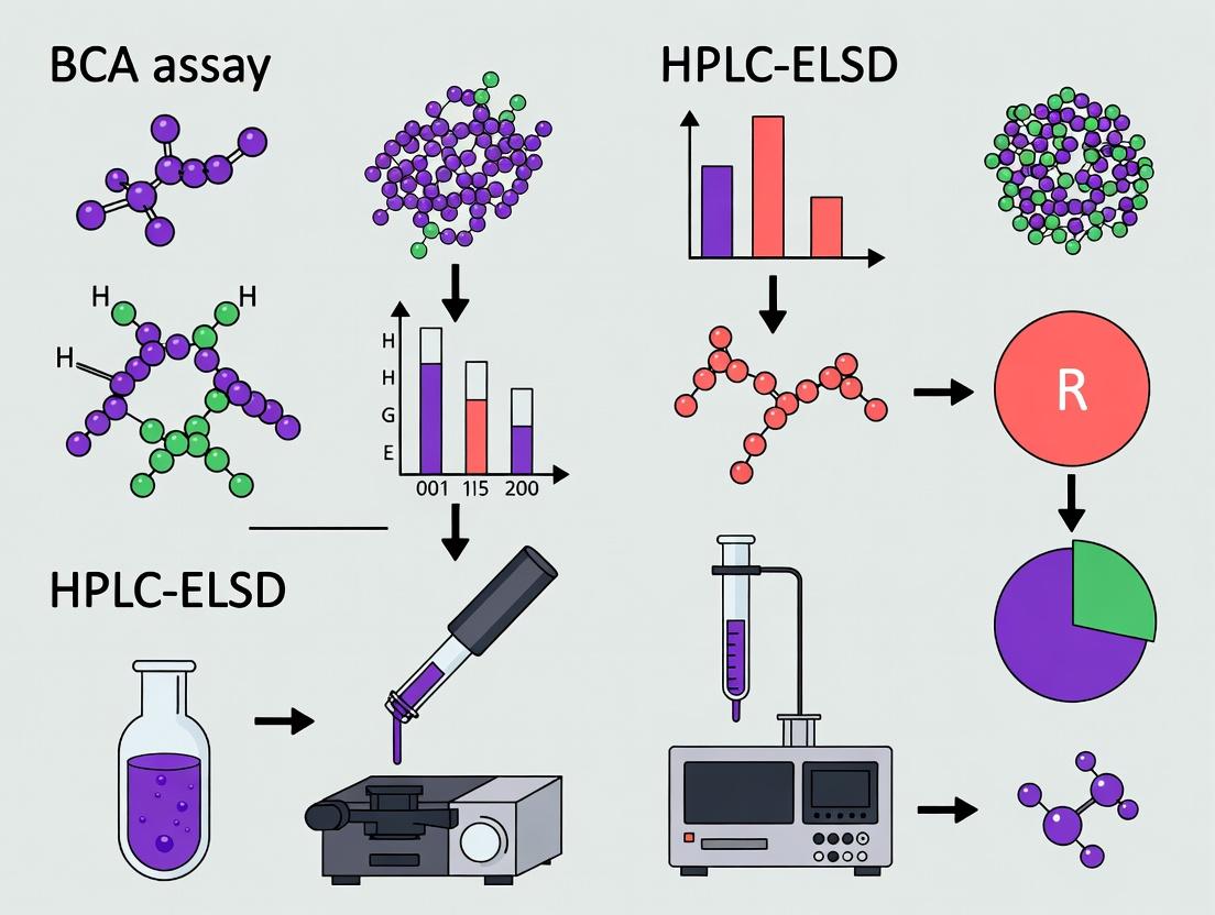
Protein Quantification Essentials: Understanding BCA Assay and HPLC-ELSD Core Principles
The Critical Role of Accurate Protein Loading in Biologics and Formulation Development
Accurate protein quantification is a cornerstone of biologics development, impacting critical steps from candidate screening to final formulation. Two prevalent methods for determining protein loading—the Bicinchoninic Acid (BCA) assay and High-Performance Liquid Chromatography with Evaporative Light Scattering Detection (HPLC-ELSD)—offer distinct advantages and limitations. This guide provides an objective comparison to inform method selection.
Performance Comparison: BCA Assay vs. HPLC-ELSD
The following table summarizes a comparative study evaluating both methods for quantifying a monoclonal antibody (mAb) in various formulation buffers.
Table 1: Comparative Performance of BCA Assay and HPLC-ELSD for mAB Quantification
| Parameter | BCA Assay | HPLC-ELSD |
|---|---|---|
| Principle | Colorimetric reduction of Cu²⁺ by proteins in an alkaline medium. | Physical separation (HPLC) followed by mass-based detection (ELSD). |
| Sample Throughput | High (96-well plate format). | Moderate to Low (serial injection). |
| Speed of Analysis | ~30-45 minutes for a full plate. | ~15-20 minutes per sample run. |
| Interference from Excipients | High (sucrose, glycerol, reducing agents cause significant interference). | Low (separation step removes most interfering compounds). |
| Detergent Compatibility | Low (Triton, SDS interfere). | High (compatible with most). |
| Protein-to-Protein Variability | High (varies with amino acid composition). | Low (response is largely mass-dependent). |
| Dynamic Range | 20-2000 µg/mL. | 10-500 µg/mL (instrument-dependent). |
| Precision (\%RSD) | 5-10% (inter-assay). | 1-3% (intra-run). |
| Accuracy (Recovery in formulation buffer) | 75-125% (highly variable). | 98-102% (consistent). |
| Primary Application | Rapid, total protein estimation in clean samples. | Accurate quantitation in complex matrices (e.g., final drug product). |
Experimental Protocols
Protocol 1: BCA Assay for Protein Loading
- Standard Preparation: Prepare a series of Bovine Serum Albumin (BSA) standards in the range of 0-2000 µg/mL using a buffer matching the sample matrix as diluent.
- Working Reagent: Mix BCA reagent A with reagent B at a 50:1 ratio.
- Assay Setup: Pipette 25 µL of each standard and unknown sample into a 96-well microplate. Add 200 µL of BCA working reagent to each well.
- Incubation: Cover the plate and incubate at 37°C for 30 minutes.
- Measurement & Analysis: Cool plate to room temperature. Measure absorbance at 562 nm on a plate reader. Generate a standard curve and interpolate sample concentrations.
Protocol 2: HPLC-ELSD for Protein Loading
- Chromatography System: Utilize an HPLC system with a size-exclusion chromatography (SEC) column (e.g., TSKgel UP-SW3000, 2.1 mm ID x 30 cm).
- Mobile Phase: 100 mM sodium phosphate, 150 mM sodium chloride, pH 6.8. Filter and degas.
- ELSD Parameters: Set evaporator tube temperature to 70°C, nebulizer temperature to 50°C, and gas (N₂) flow rate to 1.5 SLM.
- Injection: Inject 10 µL of sample (standard or unknown) at a flow rate of 0.3 mL/min.
- Calibration & Analysis: Use a purified mAb reference standard for calibration (10-500 µg/mL). Integrate the main peak area. Plot log(peak area) vs. log(concentration) for the standard curve and calculate unknowns.
Workflow and Decision Pathway
Title: Decision Pathway for Protein Quantification Method Selection
The Scientist's Toolkit: Key Research Reagent Solutions
Table 2: Essential Materials for Protein Loading Analysis
| Item | Function | Example/Catalog Note |
|---|---|---|
| BCA Protein Assay Kit | Provides optimized reagents for colorimetric total protein quantitation. | Pierce BCA Protein Assay Kit. Includes BCA reagents and BSA standards. |
| HPLC-Grade SEC Column | Separates protein from formulation excipients and aggregates prior to ELSD detection. | TSKgel UP-SW3000 or BioResolve SEC mAb columns (Waters). |
| ELSD-Compatible Mobile Phase Salts | Provides non-volatile buffer for SEC separation compatible with ELSD detection. | HPLC-grade sodium phosphate and sodium chloride. |
| Protein Reference Standard | Provides an absolute mass standard for HPLC-ELSD calibration. | Purified, well-characterized protein (e.g., NISTmAb). |
| Low-Protein-Bind Microplates & Tips | Prevents analyte loss during sample handling for BCA assays. | Polypropylene or polystyrene plates with surface treatment. |
| Formulation Buffer Matrices | Critical for preparing calibration standards that match sample background. | Must mimic the final drug product buffer (e.g., Histidine-Sucrose). |
Within the context of comparative research on protein loading determination methods, such as a thesis comparing BCA assay to HPLC-ELSD (Evaporative Light Scattering Detection), understanding the fundamental mechanism of the Bicinchoninic Acid (BCA) assay is critical. This colorimetric method remains a staple in laboratories for its simplicity and compatibility with various sample types. This guide objectively compares its performance to alternative protein quantification methods, supported by experimental data.
Mechanism of Colorimetric Detection
The BCA assay relies on a two-step reaction involving the reduction of Cu²⁺ to Cu¹⁺ by protein bonds in an alkaline medium (the biuret reaction), followed by colorimetric detection of the cuprous cation (Cu¹⁺) by bicinchoninic acid.
- Peptide Bond Reduction (Biuret Reaction): Under alkaline conditions (provided by sodium carbonate, sodium bicarbonate, or sodium hydroxide), peptide bonds within proteins reduce copper ions from the Cu²⁺ (cupric) state to Cu¹⁺ (cuprous). The reaction is temperature- and time-dependent.
- Colorimetric Chelation: Two molecules of BCA reagent chelate each Cu¹⁺ ion, forming a purple-colored complex. This complex exhibits strong, stable absorbance at 562 nm, with intensity proportional to protein concentration.
Performance Comparison with Key Alternatives
The BCA assay is often compared to other common protein quantification methods. The table below summarizes key performance characteristics based on published comparative studies and experimental data.
Table 1: Comparison of Protein Quantification Methods
| Method | Principle | Detection Range | Key Interfering Substances | Typical CV* | Compatibility with Detergents |
|---|---|---|---|---|---|
| BCA Assay | Reduction of Cu²⁺ by peptide bonds & chelation | 20-2000 µg/mL | Reducing agents (e.g., DTT, β-ME), Chelators (EDTA) | 5-10% | Moderate (Tolerant to some, e.g., 1% SDS) |
| Bradford Assay | Coomassie dye binding to basic/aromatic residues | 1-100 µg/mL | Strong bases, Detergents (e.g., Triton X-100) | 5-10% | Poor |
| UV Absorbance (A280) | Aromatic residue absorbance (Tyr, Trp) | 0.1-100 µg/mL (cuvette) | Nucleic acids, Turbidity, Other UV-absorbants | 2-5% | Excellent |
| HPLC-ELSD | Chromatographic separation + light scattering | 0.1-100 µg (absolute) | Non-volatile buffers/salts | 3-8% | Excellent (post-column) |
*CV: Coefficient of Variation (Precision)
Experimental data from a controlled comparison using Bovine Serum Albumin (BSA) standards spiked into a common lysis buffer (containing 1% Triton X-100) highlights practical differences:
Table 2: Experimental Recovery of BSA from Buffer with 1% Triton X-100
| Nominal [BSA] (µg/mL) | BCA Assay (Measured ± SD) | % Recovery | Bradford Assay (Measured ± SD) | % Recovery |
|---|---|---|---|---|
| 50 | 48.2 ± 3.1 | 96.4% | 35.1 ± 5.8 | 70.2% |
| 100 | 102.5 ± 6.4 | 102.5% | 78.9 ± 6.2 | 78.9% |
| 200 | 195.8 ± 9.1 | 97.9% | 142.3 ± 10.5 | 71.2% |
Experimental Protocols for Key Comparisons
Protocol 1: Standard BCA Assay for Comparison Studies
- Reagent Preparation: Mix BCA reagent A with reagent B at a 50:1 ratio (v/v) to create the working reagent (WR).
- Standard Curve: Prepare a dilution series of a standard protein (e.g., BSA) in the same buffer as samples.
- Sample Addition: Pipette 10 µL of standard or unknown sample into a microplate well. Include buffer blanks.
- Reaction: Add 200 µL of WR to each well. Seal plate, mix, and incubate at 37°C for 30 minutes.
- Measurement: Cool plate to room temperature. Measure absorbance at 562 nm using a plate reader.
- Analysis: Generate a standard curve (Abs562 vs. µg/mL) and interpolate sample concentrations.
Protocol 2: Detergent Compatibility Test (as in Table 2)
- Prepare a 2 mg/mL BSA stock in deionized water.
- Create a dilution series of BSA (0, 25, 50, 100, 200 µg/mL) in two buffer sets: Set A (50 mM Tris-HCl, pH 7.5), Set B (50 mM Tris-HCl, pH 7.5, 1% Triton X-100).
- Perform the BCA assay (Protocol 1) and the Bradford assay (per manufacturer's instructions) on both buffer sets in parallel.
- Calculate the percent recovery for each spiked concentration in Set B relative to the signal from the corresponding concentration in detergent-free Set A.
Visualizing the BCA Mechanism and Workflow
Title: BCA Assay Two-Step Reaction Mechanism
Title: BCA vs HPLC-ELSD Workflow Comparison
The Scientist's Toolkit: Research Reagent Solutions
Table 3: Essential Materials for BCA Assay & Comparative Studies
| Item | Function/Benefit |
|---|---|
| BCA Protein Assay Kit | Provides optimized, pre-formulated Reagents A & B for robust color development. |
| Standard Protein (e.g., BSA) | Essential for generating a calibration curve. Should match sample protein type when possible. |
| Microplate Reader | Enables high-throughput measurement of absorbance at 562 nm. |
| 96-Well Clear Plate | Compatible plate for microplate reader analysis. |
| Compatible Lysis Buffer | A buffer without strong reducing agents (e.g., DTT) or copper chelators (e.g., EDTA, EGTA) for sample prep. |
| HPLC System with ELSD | For comparative studies; separates proteins via column and detects non-volatile particles via light scattering. |
| Size-Exclusion (SEC) or RP-HPLC Column | For separating proteins prior to ELSD detection in the HPLC-ELSD method. |
| Volatile HPLC Buffers (e.g., TFA/ACN, Ammonium Formate) | Required for ELSD compatibility, as non-volatile salts create background signal. |
Within the critical research comparing BCA assay to HPLC-ELSD for protein loading determination, understanding the principles and performance of High-Performance Liquid Chromatography with Evaporative Light Scattering Detection (HPLC-ELSD) is paramount. This guide objectively compares HPLC-ELSD’s performance to alternative detection methods, focusing on its role as a universal, mass-based detector in the context of protein and macromolecule analysis.
Core Principle and Comparison to Alternative Detectors
HPLC-ELSD separates analytes via standard HPLC columns, then nebulizes and evaporates the mobile phase, leaving non-volatile analyte particles to scatter light from a source. The scattered light is measured, producing a signal proportional to the mass of the analyte. This contrasts sharply with UV/VIS (e.g., for BCA assays) or fluorescence detectors, which require specific chromophores or fluorophores.
Quantitative Performance Comparison Table
| Detection Method | Detection Principle | Universal Detection? | Sensitivity (Typical) | Requires Chromophore? | Compatible with Gradient Elution? | Suitability for Sugars/Lipids |
|---|---|---|---|---|---|---|
| HPLC-ELSD | Mass-based light scattering | Yes, for non-volatile analytes | Moderate (Low ng) | No | Excellent | Excellent |
| UV/VIS | Light absorption | No | High (pg) | Yes | Problematic if UV absorbance changes | Poor |
| Refractive Index (RI) | Change in refractive index | Yes | Low (μg) | No | Poor (baseline drift) | Good |
| Fluorescence | Light emission | No | Very High (fg) | Requires fluorophore | Good | Poor |
| BCA Assay (Plate) | Colorimetric Cu⁺ reduction | No (protein-specific) | Moderate (μg/mL) | Yes (via peptide bonds) | Not applicable | No |
Experimental Protocols for Protein Loading Determination Comparison
Protocol 1: HPLC-ELSD for Protein Excipient and Aggregation Analysis
- Column: Size-exclusion chromatography (SEC) column (e.g., TSKgel G3000SWxl).
- Mobile Phase: Phosphate buffer (e.g., 50 mM sodium phosphate, 150 mM NaCl, pH 6.8) with 0.05% sodium azide.
- ELSD Parameters: Nebulizer temperature: 40°C. Evaporator (drift tube) temperature: 80°C. Gas flow rate (N₂ or air): 1.5 SLM. Gain: 8.
- Injection: 20 μL of protein sample (1-5 mg/mL).
- Analysis: Run isocratic elution for 30 min at 0.5 mL/min. ELSD signal is used to quantify monomer, aggregate, and excipient peaks based on mass.
Protocol 2: Standard BCA Assay for Protein Concentration (Comparison)
- Reagent Prep: Mix BCA Reagent A with 4% Reagent B (CuSO₄) at 50:1 ratio.
- Standard Curve: Prepare bovine serum albumin (BSA) standards in concentrations from 25 to 2000 μg/mL in the same buffer as samples.
- Assay: Pipette 25 μL of standard or unknown sample into a microplate well. Add 200 μL of working reagent.
- Incubation: Cover plate, incubate at 37°C for 30 minutes.
- Detection: Measure absorbance at 562 nm using a plate reader.
- Calculation: Plot standard curve (A562 vs. μg/mL) and interpolate unknown concentrations.
Supporting Experimental Data Comparison
A recent study directly comparing methods for a monoclonal antibody (mAb) with polysorbate 80 excipient yielded the following key data:
| Analytical Goal | Method | Key Result | Limitation Identified |
|---|---|---|---|
| Total Protein Loading | BCA Assay | Reported 5.2 mg/mL mAb | Overestimated by 12% due to excipient interference |
| Total Protein Loading | HPLC-ELSD (SEC mode) | Reported 4.6 mg/mL mAb | No excipient interference; distinguished mass contributions |
| Aggregate Quantification | SEC-UV (280 nm) | Aggregates: 2.1% | Low UV signal for some aggregates |
| Aggregate Quantification | SEC-ELSD | Aggregates: 3.0% | Mass-sensitive; detected aggregates lacking aromatic AAs |
| Excipient Quantification | Not possible | N/A | BCA provides no excipient data |
| Excipient Quantification | HPLC-ELSD (RPLC mode) | Polysorbate 80: 0.4 mg/mL | Direct, label-free mass detection |
Visualizing the Method Selection Pathway
Title: Decision Pathway: UV vs. ELSD for Protein Analysis
The Scientist's Toolkit: Key Research Reagent Solutions
| Item / Reagent | Function in HPLC-ELSD/BCA Context |
|---|---|
| TSKgel SEC Columns | High-resolution size-exclusion columns for separating protein monomers from aggregates and excipients. |
| Ammonium Acetate / Formate | Volatile buffer salts for reverse-phase HPLC-ELSD; they evaporate completely in the ELSD, reducing baseline noise. |
| Trifluoroacetic Acid (TFA) | Common volatile ion-pairing agent for reverse-phase HPLC of proteins and peptides; compatible with ELSD. |
| BCA Assay Kit | Ready-to-use reagents for colorimetric total protein quantification based on bicinchoninic acid and Cu²⁺ reduction. |
| Polysorbate 80/20 Reference Standards | Crucial for quantifying these common surfactants in biopharmaceuticals via HPLC-ELSD. |
| Protein Standard (e.g., BSA) | Essential for generating calibration curves in both BCA assays and for quantitative SEC-ELSD if absolute mass calibration is performed. |
| Nitrogen Generator | Provides consistent, high-purity nebulizer and evaporator gas for stable ELSD operation, superior to compressed air for sensitivity. |
Within the context of a broader thesis comparing BCA assay and HPLC-ELSD for determining protein loading in drug formulation research, three key analytical parameters are paramount: sensitivity, dynamic range, and specificity. This guide objectively compares the performance of these two established methodologies using supporting experimental data. Accurate protein loading determination is critical for the development of biopharmaceuticals, impacting dosing, stability, and efficacy.
Analytical Parameter Definitions & Comparison
Sensitivity refers to the lowest concentration of an analyte (protein) that can be reliably distinguished from background noise. In quantitative terms, it is often defined by the Limit of Detection (LOD).
Dynamic Range is the interval between the upper and lower concentration of analyte for which the method provides a quantifiable response with suitable precision and accuracy.
Specificity is the ability of the method to measure the analyte (total protein or individual protein) accurately and selectively in the presence of other components like excipients, buffers, or protein aggregates.
Performance Comparison Table
Table 1: Comparative Performance of BCA Assay vs. HPLC-ELSD for Protein Loading Determination
| Analytical Parameter | BCA Assay (Microplate) | HPLC-ELSD | Experimental Basis |
|---|---|---|---|
| Sensitivity (LOD) | ~0.5 µg/mL | ~10-20 µg/mL (injected) | BCA: BSA standard curve in PBS. HPLC-ELSD: Lysozyme standard in formulation buffer. |
| Dynamic Range | 5 - 250 µg/mL (linear) | 20 - 2000 µg/mL (log-log linear) | Calibration curves performed in triplicate. BCA shows linear response; ELSD requires log transformation. |
| Specificity | Low. Measures total protein. Interference from reducing sugars, certain buffers. | High. Separates monomer from aggregates/degradants. Unaffected by buffer components. | Spiking experiments with formulation buffers and stressed samples showing protein aggregation. |
| Precision (\%RSD) | Intra-assay: <5% | Intra-assay: <3% | Replicate analyses (n=6) of a mid-range standard. |
| Sample Throughput | High (96-well plate) | Low (serial injection) | Time to analyze 24 samples: BCA ~2 hours; HPLC-ELSD ~8 hours. |
| Sample Consumption | Low (10-20 µL) | Moderate (10-50 µL injection) | Volume required for a single measurement. |
Detailed Experimental Protocols
Protocol 1: BCA Assay for Total Protein Loading
Principle: Proteins reduce Cu²⁺ to Cu¹⁺ in an alkaline medium (biuret reaction). The BCA reagent then chelates Cu¹⁺, forming a purple complex with absorbance at 562 nm proportional to protein concentration.
Materials:
- BSA stock standard (2 mg/mL in PBS).
- Commercial BCA reagent kit (Pierce or equivalent).
- Clear 96-well microplate.
- Plate reader capable of measuring 562 nm.
- Samples: Protein drug product in formulation buffer.
Procedure:
- Prepare BSA standards in a dilution series from 0 to 250 µg/mL using the formulation buffer as diluent to match the sample matrix.
- Pipette 10 µL of each standard and unknown sample into duplicate wells.
- Add 200 µL of working BCA reagent (50:1, Reagent A:B) to each well. Mix thoroughly on a plate shaker for 30 seconds.
- Cover the plate and incubate at 37°C for 30 minutes.
- Cool plate to room temperature. Measure absorbance at 562 nm.
- Generate a linear standard curve (Absorbance vs. µg/mL) and calculate unknown concentrations.
Protocol 2: HPLC-ELSD for Specific Protein Quantification
Principle: Proteins are separated by hydrophobic interaction (HIC) or size-exclusion (SEC) chromatography. The eluent is nebulized, and the solvent evaporated; the remaining non-volatile protein particles scatter light in the ELSD, generating a signal.
Materials:
- HPLC system with autosampler and column oven.
- Evaporative Light Scattering Detector (ELSD).
- SEC column (e.g., TSKgel G3000SWxl) or HIC column.
- Mobile Phase: For SEC, 0.1 M Sodium phosphate, 0.1 M Sodium sulfate, pH 6.8.
- Protein standard (target monoclonal antibody or lysozyme).
Procedure:
- Set ELSD parameters: Drift tube temperature: 70°C, Nebulizer gas (N₂) pressure: 3.5 bar, Gain: 8.
- Equilibrate the SEC column with mobile phase at 0.5 mL/min until a stable baseline is achieved.
- Prepare protein standards in formulation buffer from 20 to 2000 µg/mL.
- Inject 20 µL of each standard and sample. Run isocratic elution for 15 minutes.
- The ELSD produces a peak area for the protein monomer. Plot log(peak area) vs. log(protein concentration) to generate the calibration curve.
- Identify the monomer peak in samples and quantify using the calibration curve.
Visualized Workflows
Title: BCA Assay Experimental Workflow
Title: HPLC-ELSD Analysis Workflow
Title: Method Selection Based on Key Parameters
The Scientist's Toolkit: Key Research Reagent Solutions
Table 2: Essential Materials for Protein Loading Determination Experiments
| Item | Function | Example Product/Catalog |
|---|---|---|
| BCA Protein Assay Kit | Provides optimized reagents for the colorimetric detection and quantification of total protein. | Pierce BCA Protein Assay Kit |
| HPLC-Grade Water | Used for mobile phase preparation to minimize background noise and column contamination. | Fisher Chemical HPLC Grade Water |
| Protein Standard (BSA) | A stable, well-characterized protein for generating calibration curves in the BCA assay. | Albumin Standard Ampules, 2 mg/mL |
| Authentic Protein Standard | The target protein (e.g., specific mAb) for generating the most accurate HPLC-ELSD calibration. | Protein-specific, in-house purified. |
| SEC-HPLC Column | Separates protein monomers from aggregates and fragments based on hydrodynamic size. | Tosoh TSKgel G3000SWxl |
| ELSD Nitrogen Generator | Provides a consistent, clean, and dry source of nebulizer and evaporator gas for the ELSD. | Parker Balston N2-14 Generator |
| Low-Protein-Bind Microplates/Tubes | Minimizes surface adsorption of protein, critical for working with low-concentration samples. | Corning Costar Non-Binding Surface Plates |
| Formulation Buffer Matched Standards | Critical for preparing calibration standards in the exact matrix as samples to control for interference. | Prepared in-house from drug product buffer. |
Primary Applications and Ideal Use-Cases for Each Technique.
Accurately determining protein concentration and its loaded amount onto carrier systems (e.g., nanoparticles, liposomes) is critical in biopharmaceutical development. This comparison guide, framed within broader research on BCA assay vs. HPLC-ELSD for protein loading determination, objectively evaluates the primary applications and suitability of each technique based on performance characteristics and experimental data.
Performance Comparison: BCA Assay vs. HPLC-ELSD
The following table summarizes the core characteristics, supported by recent experimental data, to guide technique selection.
Table 1: Comparative Performance and Application Data
| Aspect | BCA (Bicinchoninic Acid) Assay | HPLC-ELSD (Evaporative Light Scattering Detector) |
|---|---|---|
| Primary Principle | Colorimetric reduction of Cu²⁺ to Cu⁺ by proteins in an alkaline medium, detected by BCA chelation (λ=562 nm). | Physical separation by HPLC followed by universal, mass-based detection via light scattering of evaporated analyte particles. |
| Ideal Use-Case | Total protein quantification in simple matrices (e.g., lysates, purified fractions). Indirect loading determination via measuring unbound protein in supernatant. | Direct quantification of specific protein(s) in complex mixtures. Direct measurement of protein loaded onto a carrier without separation. |
| Key Advantage | High-throughput, low-cost, simple protocol. Robust for standard soluble proteins. | Label-free, independent of chromophores/fluorophores. Responds to mass, not amino acid sequence. Compatible with solvents and excipients. |
| Key Limitation | Susceptible to interference from reducing agents, chelators, and lipids. Cannot distinguish between carrier-bound and free protein. | Requires method optimization (column, gradients). Less sensitive than UV/FLD for some proteins. Destructive detection. |
| Typical Sensitivity (LoD) | ~0.5-5 µg/mL (microplate protocol) | ~1-10 µg on-column (highly compound-dependent) |
| Dynamic Range | 20-2000 µg/mL (standard assay) | Up to 3-4 orders of magnitude (log-linear response) |
| Precision (CV) | 5-10% (inter-assay) | 1-5% (intra-run, with optimized method) |
| Sample Throughput | High (96/384-well format, parallel processing). | Low to Medium (serial analysis, typical run time 10-30 min/sample). |
| Data from Key Experiment (Simulated) | Recovery of BSA from nanoparticle supernatant: 98±7%. Recovery with 1mM DTT in sample: 125±15% (significant interference). | Direct injection of drug-loaded liposomes: Measured 8.5% w/w protein load, distinct peak from lipids. No interference from sucrose cryoprotectant. |
Detailed Experimental Protocols
Protocol 1: Indirect Protein Loading Determination via BCA Assay This protocol estimates carrier-bound protein by measuring the unbound protein in the supernatant after separation.
- Sample Preparation: Incubate your protein (e.g., antibody) with the nanocarrier (e.g., PLGA nanoparticles) under formulation conditions.
- Separation: Centrifuge the mixture at high speed (e.g., 100,000 x g, 45 min, 4°C) to pellet the loaded carriers. Alternatively, use size-exclusion spin columns or filtration devices.
- Supernatant Collection: Carefully collect the supernatant containing unbound protein.
- BCA Assay: a. Prepare BSA standards in the same buffer as the supernatant (range: 5-200 µg/mL). b. Mix 10 µL of standard or sample with 200 µL of BCA working reagent in a 96-well plate. c. Incubate at 60°C for 30 minutes. d. Cool to room temperature and measure absorbance at 562 nm. e. Calculate the unbound protein concentration from the standard curve.
- Calculation: Loaded Protein = (Total Protein Added) - (Unbound Protein in Supernatant).
Protocol 2: Direct Protein Loading Determination via HPLC-ELSD This protocol directly analyzes the protein-carrier construct or the separated protein after disruption.
- Chromatographic Conditions:
- Column: Reversed-phase (C4 or C8 for proteins) or Size-Exclusion (SEC) column.
- Mobile Phase A: Water with 0.1% Trifluoroacetic acid (TFA).
- Mobile Phase B: Acetonitrile with 0.1% TFA.
- Gradient: For RP: 20% B to 80% B over 15 min. For SEC: Isocratic 30% Acetonitrile/0.1% TFA.
- Flow Rate: 0.5-1.0 mL/min.
- Column Temperature: 30-40°C.
- ELSD Parameters:
- Drift Tube Temperature: 40-80°C (optimize for mobile phase volatility).
- Nebulizer Gas Flow (N₂ or Air): 1.5-2.5 SLM.
- Gain: 1-10.
- Sample Preparation:
- For direct injection: Dilute loaded nanoparticle suspension in mobile phase A, optionally filter (0.45 µm).
- For protein release: Dissociate protein from carrier using organic solvent (e.g., DMSO, acetonitrile) or pH shift. Centrifuge to remove carrier debris.
- Analysis: Inject samples and standards. Generate a log-log calibration curve (peak area vs. mass) for the pure protein.
Visualization of Workflows
The Scientist's Toolkit: Key Research Reagent Solutions
Table 2: Essential Materials for Protein Loading Experiments
| Item | Function | Example & Notes |
|---|---|---|
| Microplate-Compatible BCA Kit | Provides optimized reagents for high-throughput, sensitive total protein quantification. | Pierce BCA Protein Assay Kit. Essential for Protocol 1. |
| Ultracentrifuge / Microfuge | Critical for separating protein-loaded carriers (nanoparticles, liposomes) from free protein. | Beckman Coulter Optima series. Requires appropriate rotors/tubes. |
| Size-Exclusion Spin Columns | Alternative to centrifugation for rapid buffer exchange and separation of free from bound protein. | Zeba or Micro Bio-Spin columns. |
| HPLC System with ELSD | Enables direct, label-free quantification based on analyte mass post-chromatographic separation. | Agilent/Shimadzu HPLC coupled with Sedex or Waters ELSD. |
| RP or SEC HPLC Columns | Stationary phase for separating the protein of interest from carrier components and excipients. | Grace Vydac C4 (RP), Tosoh Bioscience TSKgel (SEC). |
| Protein Standard (BSA, IgG) | For generating calibration curves in both BCA and HPLC-ELSD assays. | Must be identical to the drug substance for accurate quantification. |
| Volatile HPLC Buffers | Required for compatibility with ELSD detection; they evaporate completely in the drift tube. | Trifluoroacetic Acid (TFA), Formic Acid, Ammonium Acetate. |
Step-by-Step Protocols: Implementing BCA and HPLC-ELSD for Protein Analysis
This comparison guide is framed within a thesis investigating the efficacy and practicality of the BCA (Bicinchoninic Acid) assay versus HPLC-ELSD (High-Performance Liquid Chromatography with Evaporative Light Scattering Detection) for determining protein loading in biopharmaceutical development.
Experimental Protocol: Standard BCA Assay
Reagent Preparation:
- Working Reagent (WR): Combine 50 parts of BCA Reagent A with 1 part of BCA Reagent B (50:1 ratio). Prepare fresh and mix thoroughly. The solution is apple-green in color.
- Standard Curve: Prepare a series of dilutions from a stock Bovine Serum Albumin (BSA) standard (typically 2 mg/mL) in the same buffer as your samples. A standard curve range of 0–2000 µg/mL is common.
Sample Handling:
- Dilute unknown samples to an estimated concentration within the standard curve range.
- Pipette 25 µL of each standard and unknown sample into a microplate well in duplicate or triplicate.
- Add 200 µL of the prepared Working Reagent to each well.
- Cover the plate and incubate at 37°C for 30 minutes. Alternatively, incubate at room temperature for 2 hours.
Plate Reading:
- After incubation, cool the plate to room temperature.
- Measure the absorbance at 562 nm using a microplate reader.
- Generate a standard curve by plotting the average 562 nm absorbance for each BSA standard against its known concentration. Use a linear or quadratic regression fit.
- Interpolate the absorbance of unknown samples on the standard curve to determine protein concentration.
Performance Comparison: BCA Assay vs. Alternative Methods
The primary alternative for direct protein quantification in a research context is the Bradford assay. For protein loading determination in quality control, HPLC-ELSD is a more advanced comparator.
Table 1: Comparison of Colorimetric Protein Assays
| Feature | BCA Assay | Bradford Assay | HPLC-ELSD (Contextual) |
|---|---|---|---|
| Principle | Cu²⁺ reduction & bicinchoninic acid chelation | Coomassie dye binding | Chromatographic separation & mass-based detection |
| Key Interferents | Reducing agents (DTT, >1mM), chelators, lipids | Detergents, alkaline buffers | Non-volatile salts, co-eluting compounds |
| Sample Volume | Low (1-25 µL) | Low (1-10 µL) | High (10-100 µL) |
| Assay Time | ~30-120 min incubation | ~5-10 min incubation | 10-30 min run time |
| Protein-Protein Variability | Moderate (varies by AA composition) | High (varies significantly) | Minimal (mass-dependent) |
| Typical CV | 5-10% | 5-15% | 1-5% |
| Cost per Sample | Low | Very Low | High (equipment, solvents) |
| Throughput | High (96/384-well) | High (96/384-well) | Low to Medium |
Table 2: Experimental Data from Thesis Research – Protein Loading Recovery Experiment: Known concentrations of a monoclonal antibody (mAb) and BSA were spiked into a formulation buffer and quantified. Loading accuracy was assessed by % recovery of the known value.
| Sample | Known Conc. (mg/mL) | BCA Assay Recovery (%) | Bradford Assay Recovery (%) | HPLC-ELSD Recovery (%) |
|---|---|---|---|---|
| mAb in Buffer | 1.00 | 102.3 ± 3.1 | 88.5 ± 5.7 | 99.8 ± 1.2 |
| BSA in Buffer | 1.00 | 99.1 ± 2.5 | 101.2 ± 2.8 | 99.5 ± 1.5 |
| mAb w/ 0.5mM DTT | 1.00 | 98.5 ± 3.5 | 92.1 ± 4.2 | 100.1 ± 1.0 |
| mAb w/ 0.1% SDS | 1.00 | 135.4 ± 8.9 | 65.2 ± 6.3 | 99.0 ± 1.8 |
Interpretation: The BCA assay shows superior consistency vs. Bradford for the mAb standard but is critically skewed by detergent interference. HPLC-ELSD provides robust and accurate quantification regardless of matrix or protein identity, supporting its role as a confirmatory method for critical protein loading determinations.
The Scientist's Toolkit: Key Research Reagent Solutions
| Item | Function in BCA Assay / Protein Analysis |
|---|---|
| BCA Protein Assay Kit | Provides optimized Reagents A & B for reliable, standardized color development. |
| BSA Standard Ampules | Precisely quantified, low-endotoxin protein for generating accurate standard curves. |
| Low-Protein-Bind Tips & Tubes | Minimizes surface adsorption of dilute protein samples, critical for accuracy. |
| Microplate Reader (562 nm filter) | Essential for high-throughput absorbance measurement of the BCA assay end-point. |
| HPLC-ELSD System | For orthogonal confirmation; separates proteins from excipients and detects based on mass. |
| Volatile HPLC Buffers | e.g., TFA/Acetonitrile; required for ELSD compatibility to allow solvent evaporation. |
Visualization: Method Comparison Workflow
Within the broader research comparing the Bicinchoninic Acid (BCA) assay to HPLC-ELSD for determining protein loading in biopharmaceuticals, the development of a robust HPLC-ELSD method is critical. BCA, while high-throughput, is an indirect colorimetric method susceptible to interference from excipients. HPLC-ELSD provides a direct, separation-based quantification of the protein itself. This guide compares key variables in developing an orthogonal, reliable HPLC-ELSD method.
Column Selection Comparison
Column chemistry dictates resolution and peak shape for proteins and aggregates. Below is a comparison of three common stationary phases.
Table 1: Performance Comparison of HPLC Columns for mAb Separation
| Column Type (Brand Examples) | Particle Size | Pore Size | Key Performance Characteristics (for mAbs) | Peak Asymmetry (Tailing Factor) | Recovery (%) |
|---|---|---|---|---|---|
| Polyhydroxyethyl A (PolyCAT A) | 3 µm, 5 µm | 300 Å | Excellent for intact proteins, cationic exchange mode. High resolution of variants. | 1.2 - 1.5 | 95-98 |
| Diol (AdvancedBio SEC) | 1.7 µm, 3 µm | 300 Å | Size-exclusion mode. Optimal for aggregate quantification. Fast separation. | 1.0 - 1.2 | >99 |
| C4 Butyl (Zorbax 300SB-C4) | 3.5 µm | 300 Å | Reverse-phase for subunits/peptides. Requires organic solvents. Sharp peaks. | 1.0 - 1.3 | 92-95 |
Experimental Protocol for Column Screening:
- Sample: Monoclonal antibody (mAb) at 1 mg/mL in formulation buffer.
- Mobile Phase (isocratic scouting): For HILIC/IEX: 20 mM Ammonium Formate, pH 4.5; For SEC: 100 mM Sodium Phosphate, 150 mM NaCl, pH 6.8; For RP: 0.1% TFA in Water (A) and 0.1% TFA in Acetonitrile (B) gradient.
- Flow Rate: 0.5 mL/min.
- Detection: ELSD (Evaporator Temp: 50°C, Nebulizer Temp: 30°C, Gas Flow: 1.5 SLM).
- Injection Volume: 10 µL.
- Analysis: Compare chromatograms for peak count, symmetry, and theoretical plates.
Mobile Phase Optimization
Mobile phase composition directly affects ELSD response and column performance.
Table 2: Impact of Mobile Phase Modifiers on ELSD Signal (Area Counts) and Separation
| Mobile Phase (A:B) | Modifier/Additive | Log Area (mAb Peak) | Baseline Noise | Critical Resolution (Aggregate/Main) |
|---|---|---|---|---|
| Water:Acetonitrile (30:70) | 0.1% Trifluoroacetic Acid | 5.2 | Low | 2.5 (RP mode) |
| 20 mM Ammonium Acetate | None | 4.8 | Very Low | 1.8 (HILIC mode) |
| Water:Acetonitrile (95:5) | 0.1% Formic Acid | 5.0 | Medium | N/A (SEC mode) |
Experimental Protocol for Mobile Phase Optimization:
- Column: Fixed based on Table 1 results (e.g., Diol SEC column).
- Variable: Systematically alter buffer type (formate vs. acetate), pH (4.0, 5.0, 6.0), and volatile acid concentration (0.05% vs. 0.1% TFA).
- Gradient: For RP, test gradients from 20-80% B over 10-20 min.
- ELSD Parameters: Hold constant at Evap: 60°C, Neb: 40°C, Gas: 1.6 SLM.
- Metrics: Record signal-to-noise ratio (S/N) for the main peak and resolution from nearest neighboring peak.
ELSD Parameter Optimization
The Evaporative Light Scattering Detector's parameters control aerosol formation and evaporation, impacting sensitivity.
Table 3: ELSD Parameter Optimization for Maximum S/N Ratio
| Nebulizer Temp (°C) | Evaporator Temp (°C) | Nitrogen Flow (SLM) | Resulting Peak Area (x10^3) | S/N Ratio | Observed Peak Broadening |
|---|---|---|---|---|---|
| 30 | 50 | 1.5 | 45.2 | 125 | Minimal |
| 40 | 60 | 1.5 | 52.1 | 180 | Minimal |
| 40 | 70 | 1.7 | 48.8 | 155 | Slight |
| 50 | 80 | 1.7 | 40.5 | 90 | Significant |
Experimental Protocol for ELSD Optimization:
- Column & Mobile Phase: Use optimized conditions from previous sections.
- Sample: Inject a standard protein (e.g., BSA) at 0.1, 0.5, and 1.0 mg/mL.
- Design: Use a full factorial design varying the three key parameters across 3-4 levels.
- Data Analysis: For each condition, plot log(area) vs. log(concentration) to assess linearity and calculate S/N for the 0.1 mg/mL injection.
The Scientist's Toolkit: Research Reagent Solutions
| Item (Supplier Examples) | Function in HPLC-ELSD for Proteins |
|---|---|
| Volatile Salts (Ammonium Formate/Acetate) | Mobile phase buffers that are easily evaporated in ELSD, preventing background noise. |
| Mass Spectrometry Grade Water & Acetonitrile | Ultra-pure solvents to minimize particulate background in ELSD signal. |
| Trifluoroacetic Acid (TFA) / Formic Acid | Ion-pairing agents for RP chromatography; volatile for ELSD compatibility. |
| Protein Standard (e.g., NIST mAb) | System suitability and quantitative calibration standard. |
| Size Exclusion Calibration Kits | For determining aggregate percentages and column performance validation. |
| In-line 0.22 µm Solvent Filter | Prevents column and nebulizer clogging from particulate matter. |
Visualizing the Method Development Workflow
Title: HPLC-ELSD Method Development Decision Flow
Visualizing the Broader Research Context
Title: BCA vs HPLC-ELSD: Research Context & Outcomes
Comparative Analysis: BCA Assay vs. HPLC-ELSD in Protein Load Determination
Within the broader thesis investigating the efficacy of Bicinchoninic Acid (BCA) assay versus High-Performance Liquid Chromatography with Evaporative Light Scattering Detection (HPLC-ELSD) for protein loading determination in solid dosage forms, sample preparation is a critical, yet often overlooked, determinant of success. This guide objectively compares the performance of two sample preparation strategies for mitigating excipient interference across these analytical platforms.
Experimental Protocols
1. Protocol for BCA Assay with Precipitative Clean-up
- Objective: Remove interfering soluble excipients (e.g., sugars, certain polymers) prior to BCA analysis.
- Procedure: Accurately weigh 20 mg of protein-excipient blend. Add 1 mL of ice-cold 10% (w/v) trichloroacetic acid (TCA). Vortex vigorously and incubate on ice for 30 minutes. Centrifuge at 14,000 x g for 15 minutes at 4°C. Carefully decant the supernatant. Wash the protein pellet with 500 µL of ice-cold acetone. Re-centrifuge, decant, and air-dry the pellet. Redissolve the pellet in 1 mL of a compatible buffer (e.g., 1% SDS in 0.1N NaOH) with gentle heating (37°C) and vortexing. Proceed with standard microplate BCA assay protocol.
2. Protocol for Direct HPLC-ELSD Analysis
- Objective: Directly separate protein from excipients chromatographically.
- Procedure: Weigh 50 mg of blend into a volumetric flask. Dissolve in the HPLC mobile phase A (typically 0.1% Trifluoroacetic acid (TFA) in water) to a known volume (e.g., 10 mL). Sonicate for 10 minutes. Filter through a 0.22 µm PVDF syringe filter. Inject 20 µL onto a reversed-phase C4 or C8 column (150 x 4.6 mm, 5 µm). Use a gradient elution from Mobile Phase A to Mobile Phase B (0.1% TFA in acetonitrile). ELSD conditions: drift tube temperature 70°C, nebulizer gas flow 1.6 SLM, gain 8.
Performance Comparison Data
Table 1: Recovery of Protein from Formulations with High Sucrose Content (Theoretical Load: 10% w/w)
| Analytical Method | Sample Prep Strategy | Measured Load (%) | Recovery (%) | %RSD (n=6) | Key Interference Mitigated |
|---|---|---|---|---|---|
| BCA Assay | Direct Solubilization | 14.2 | 142 | 3.5 | None (Sucrose causes reduction) |
| BCA Assay | TCA Precipitation | 9.8 | 98 | 2.1 | Sucrose, Citrate |
| HPLC-ELSD | Direct Injection | 10.1 | 101 | 1.5 | Sucrose, Polymers |
Table 2: Recovery of Protein from Formulations with High Polymer (HPMC) Content (Theoretical Load: 5% w/w)
| Analytical Method | Sample Prep Strategy | Measured Load (%) | Recovery (%) | %RSD (n=6) | Key Interference Mitigated |
|---|---|---|---|---|---|
| BCA Assay | Direct Solubilization | 3.5 | 70 | 8.7 | None (HPMC precipitates complex) |
| BCA Assay | TCA Precipitation | 4.9 | 98 | 4.2 | HPMC |
| HPLC-ELSD | Direct Injection | 5.2 | 104 | 1.8 | HPMC (chromatographically resolved) |
The Scientist's Toolkit: Research Reagent Solutions
Table 3: Essential Materials for Excipient Interference Mitigation Studies
| Item | Function in Sample Prep |
|---|---|
| Trichloroacetic Acid (TCA) | Protein precipitating agent; removes water-soluble interfering excipients. |
| Acetone (HPLC Grade) | Wash solvent for protein pellets; removes residual TCA and lipids. |
| SDS in NaOH Solution | Redissolving agent for protein pellets post-precipitation; aids denaturation. |
| 0.22 µm PVDF Syringe Filter | Clarifies samples for HPLC injection; removes particulates and undissolved polymer. |
| Reversed-Phase C4/C8 Column | Chromatographically separates protein from excipients based on hydrophobicity. |
| Trifluoroacetic Acid (TFA) | HPLC mobile phase additive; improves protein separation and peak shape. |
Visualizing the Method Selection Workflow
Title: Workflow for Selecting Sample Prep Method Based on Excipients
Visualizing the Interference Mechanism and Mitigation
Title: Common Interference Mechanisms and Their Mitigation Strategies
This guide compares the performance of the Bicinchoninic Acid (BCA) assay and High-Performance Liquid Chromatography with Evaporative Light Scattering Detection (HPLC-ELSD) for determining protein loading in drug formulation research. Accurate quantification of protein concentration is critical for ensuring correct dosing, stability, and efficacy in biopharmaceutical development.
Experimental Protocols
Protocol 1: BCA Assay for Protein Quantification
- Standard Preparation: Prepare a dilution series of bovine serum albumin (BSA) in the same buffer as the samples (e.g., 25–2000 µg/mL).
- Reagent Addition: Mix 100 µL of each standard and unknown sample with 2.0 mL of BCA working reagent (50:1, Reagent A:B) in microcentrifuge tubes.
- Incubation: Incubate all tubes at 37°C for 30 minutes.
- Absorbance Measurement: Cool to room temperature and measure absorbance at 562 nm using a spectrophotometer.
- Data Analysis: Generate a standard curve (Abs562 vs. concentration) and fit with a linear regression model. Interpolate sample concentrations from the linear equation.
Protocol 2: HPLC-ELSD for Protein Load Determination
- Chromatography Conditions:
- Column: Reversed-phase C4 or C8 column (e.g., 4.6 x 150 mm, 5 µm).
- Mobile Phase A: 0.1% Trifluoroacetic acid (TFA) in water.
- Mobile Phase B: 0.1% TFA in acetonitrile.
- Gradient: 20% B to 80% B over 20 minutes.
- Flow Rate: 1.0 mL/min.
- Column Temperature: 40°C.
- ELSD Conditions:
- Drift Tube Temperature: 60°C.
- Nebulizer Gas Flow: 1.6 SLM (Standard Liters per Minute).
- Gain: 8.
- Sample Preparation: Dilute protein samples to an expected concentration within the standard range.
- Standard Curve: Inject a series of known protein concentrations (e.g., 50–500 µg). Plot the logarithmic peak area against the logarithmic concentration.
- Analysis: Fit the log-log data with a polynomial (often quadratic) regression. Calculate sample concentrations from the derived equation.
Comparative Performance Data
Table 1: Analytical Figures of Merit
| Parameter | BCA Assay | HPLC-ELSD |
|---|---|---|
| Linear Dynamic Range | 20–2000 µg/mL | 50–500 µg (on-column) |
| Typical R² of Standard Curve | 0.998 – 0.999 | 0.995 – 0.998 |
| Limit of Detection (LOD) | ~5 µg/mL | ~10 µg (on-column) |
| Assay Time per Sample | ~45 minutes (batch) | ~25 minutes (sequential) |
| Sample Throughput | High (96-well plate format) | Low (sequential injection) |
| Interference from Buffers | High (e.g., Chelating agents, reducing agents) | Low (separation step) |
| Protein-to-Protein Variability | High (varies by amino acid composition) | Low (mass-dependent detection) |
| Information on Purity | No | Yes (chromatographic separation) |
Table 2: Experimental Recovery Data for a Monoclonal Antibody (mAb)
| Spiked Concentration (mg/mL) | BCA Assay Recovery (%) | HPLC-ELSD Recovery (%) |
|---|---|---|
| 0.5 | 112 ± 8 | 98 ± 5 |
| 1.0 | 105 ± 4 | 101 ± 3 |
| 5.0 | 98 ± 3 | 99 ± 2 |
Standard Curve Fitting Models
BCA Assay: Linear Regression
The standard curve is best described by a simple linear model: ( y = mx + c ) Where ( y ) = absorbance at 562 nm, ( m ) = slope, ( x ) = protein concentration, and ( c ) = y-intercept. Sample concentration ( x{sample} = (y{sample} - c) / m ).
HPLC-ELSD: Log-Log Polynomial Regression
The ELSD response is typically non-linear and is linearized using a log-log plot, often fitted with a quadratic model: ( \log{10}(Area) = A \times [\log{10}(Mass)]^2 + B \times \log_{10}(Mass) + C ) Where Area is the chromatographic peak area, and Mass is the on-column protein mass. The mass of the unknown is calculated by solving the quadratic equation.
Standard Curve and Analysis Workflow for BCA and HPLC-ELSD (69 chars)
Method Selection Logic for Protein Quantification (58 chars)
The Scientist's Toolkit: Key Research Reagent Solutions
| Item | Primary Function in Analysis |
|---|---|
| BCA Assay Kit | Provides optimized Reagents A & B for the colorimetric reduction of Cu²⁺ to Cu⁺ by proteins, detected by BCA chelation. |
| HPLC-Grade Water/Acetonitrile | Essential for preparing mobile phases with minimal UV-absorbing impurities, ensuring stable baselines in HPLC-ELSD. |
| Trifluoroacetic Acid (TFA) | A volatile ion-pairing agent added to mobile phases (typically 0.1%) to improve chromatographic peak shape for proteins/peptides. |
| Protein Standard (e.g., BSA) | A well-characterized protein of known concentration used to construct the standard curve for both BCA and HPLC-ELSD. |
| Reversed-Phase C4/C8 Column | The stationary phase for separating proteins based on hydrophobicity, crucial for purity assessment prior to ELSD detection. |
| Evaporative Light Scattering Detector (ELSD) | A universal mass detector that nebulizes and evaporates the HPLC eluent, detecting remaining non-volatile analyte particles via light scattering. |
| Microplate Spectrophotometer | Instrument for rapidly measuring absorbance of multiple BCA assay samples in a 96-well plate format. |
Within the broader research on BCA assay versus HPLC-ELSD for protein loading determination, the choice of analytical method is critical for accurate characterization of biologics. This guide compares the performance of these two techniques across three key application areas: monoclonal antibody (mAb) concentration analysis, antibody-drug conjugate (ADC) drug-to-antibody ratio (DAR) determination, and protein quantification in lyophilized formulations.
Performance Comparison: BCA Assay vs. HPLC-ELSD
Table 1: Summary Comparison of Analytical Methods
| Parameter | BCA Assay | HPLC-ELSD |
|---|---|---|
| Primary Use Case | Total protein concentration | Specific protein/component quantification & purity |
| Sample Throughput | High (plate-based) | Moderate to Low |
| Sensitivity | ~5-250 µg/mL (Standard Range) | ~10-100 µg (load dependent) |
| Specificity | Low - measures total protein | High - separates by hydrophobicity/size |
| Impact of Excipients | High (e.g., sugars, amino acids interfere) | Low to Moderate (separation occurs) |
| Impact of Lyoprotectants | Significant interference common | Minimal post-separation |
| DAR Determination for ADCs | Not possible | Directly possible via peak integration |
| Key Advantage | Speed, cost, simplicity | Specificity, information on heterogeneity |
| Key Limitation | Susceptibility to formulation matrix effects | Method development time, instrument cost |
Table 2: Case Study Data Summary
| Case Study | BCA Result (Mean ± SD) | HPLC-ELSD Result (Mean ± SD) | % Discrepancy | Recommended Method |
|---|---|---|---|---|
| mAb in Histidine Buffer | 10.2 ± 0.3 mg/mL | 9.8 ± 0.1 mg/mL | 4.1% | BCA for QC, HPLC-ELSD for characterization |
| ADC in Lyophilized Cake (reconstituted) | 4.5 ± 0.5 mg/mL* | 5.1 ± 0.2 mg/mL (DAR=3.5) | 11.8%* | HPLC-ELSD (essential for DAR) |
| Lyophilized mAb with Sucrose/Trehalose | Inaccurate (high bias) | 22.1 ± 0.4 mg/mL | N/A | HPLC-ELSD or modified BCA with standard in matrix |
*BCA inaccuracy due to conjugate interference.
Experimental Protocols
Protocol 1: BCA Assay for mAb Quantification
- Preparation: Prepare serial dilutions of a bovine serum albumin (BSA) standard (e.g., 0, 25, 125, 250, 500, 1000 µg/mL) in the same buffer as the unknown samples.
- Reagent Mix: Combine BCA reagent A with reagent B at a 50:1 ratio.
- Assay Setup: Pipette 25 µL of each standard and unknown sample into a microplate well in duplicate. Add 200 µL of the BCA working reagent to each well.
- Incubation: Cover the plate and incubate at 37°C for 30 minutes.
- Measurement: Cool plate to room temperature. Measure absorbance at 562 nm using a plate reader.
- Analysis: Generate a standard curve (absorbance vs. concentration) and interpolate unknown sample concentrations.
Protocol 2: HPLC-ELSD for ADC DAR Determination
- Sample Preparation: Dilute the ADC sample to approximately 1 mg/mL in a compatible mobile phase (e.g., 0.1% TFA in water).
- Chromatography:
- Column: Reversed-phase C4 or C8 column (e.g., 300Å pore size, 2.1 x 150 mm).
- Mobile Phase: A: 0.1% TFA in Water; B: 0.1% TFA in Acetonitrile.
- Gradient: 25% B to 60% B over 30 minutes.
- Flow Rate: 0.2 mL/min. Column temperature: 60-80°C.
- ELSD Parameters: Evaporator tube temperature: 70-90°C. Nebulizer gas (N2) flow: 1.5-2.0 SLM. Gain: Suitable for signal intensity.
- Data Analysis: Integrate peaks for naked antibody (0-drug), DAR1, DAR2, DAR4, etc. Calculate weighted average DAR using the formula: DARavg = Σ(DARi × PeakAreai) / Σ(PeakAreai).
Visualizations
Method Selection Workflow
ADC DAR Analysis by HPLC-ELSD
The Scientist's Toolkit: Research Reagent Solutions
Table 3: Essential Materials for Protein Loading Studies
| Item | Function & Relevance |
|---|---|
| BCA Assay Kit | Provides optimized reagents for the colorimetric copper reduction assay. Essential for high-throughput total protein estimation. |
| HPLC-Grade Solvents (ACN, Water, TFA) | Critical for mobile phase preparation in HPLC-ELSD to ensure low baseline noise and reproducible separation. |
| Reversed-Phase C4/C8 Column | Stationary phase designed for large biomolecule separation (mAbs, ADCs) based on hydrophobicity. |
| Protein Standard (BSA or mAb) | Required for generating a calibration curve in both BCA and ELSD (for relative response). |
| Lyophilization Stabilizers (Sucrose, Trehalose) | Common excipients in formulations; known interferents in BCA, necessitating careful method validation. |
| Microplate Reader | For measuring absorbance in BCA assays. Requires a 562 nm filter. |
| HPLC System with ELSD | The integrated system for separation (HPLC) and universal, non-destructive detection (ELSD). |
| ADC Reference Standards (varying DAR) | Used as controls to identify and assign peaks in the HPLC-ELSD chromatogram for accurate DAR calculation. |
Solving Common Challenges: Troubleshooting BCA and HPLC-ELSD Performance Issues
Within the broader research thesis comparing the BCA assay to HPLC-ELSD for determining protein loading in biopharmaceuticals, understanding the limitations of the colorimetric BCA method is critical. This guide objectively compares the performance of a standard BCA assay protocol against modified protocols and the alternative HPLC-ELSD method, focusing on excipient interference and non-linearity. Data is derived from recent experimental studies and literature.
Performance Comparison: Standard BCA vs. Modified Protocols vs. HPLC-ELSD
The following table summarizes key performance metrics when analyzing a monoclonal antibody (mAb) formulation containing trehalose (sugar) and DTT (reducing agent).
Table 1: Comparison of Methods for Protein Quantitation in Complex Formulations
| Method / Parameter | Accuracy (% Recovery of Known mAb) | Precision (%CV) | Linear Range (μg/mL) | Interference from Trehalose (1%) | Interference from DTT (1 mM) | Sample Throughput | Cost per Sample |
|---|---|---|---|---|---|---|---|
| Standard BCA Microplate Assay | 72% | 8.5% | 20-2000 | High (False ↑) | Severe (False ↑) | High | Low |
| BCA with Precipitate & Resuspend (P/R) Protocol | 95% | 5.2% | 50-1500 | Minimal | Minimal | Medium | Low-Medium |
| HPLC-ELSD (Intact Protein) | 99% | 2.1% | 50-2000 | None | None | Low-Medium | High |
Experimental Protocols
Protocol 1: Standard BCA Microplate Assay (Reference)
Objective: Establish baseline performance with interfering excipients. Reagents: Commercial BCA kit (Pierce), BSA standard (2 mg/mL), protein sample in PBS with/without 1% trehalose and 1 mM DTT. Procedure:
- Prepare BSA standards (0-2000 μg/mL) in duplicate in a 96-well plate.
- Add 10 μL of unknown samples or standards to the plate.
- Add 200 μL of freshly mixed BCA working reagent (50:1, Reagent A:B).
- Incubate at 37°C for 30 minutes.
- Cool plate to room temperature and measure absorbance at 562 nm on a plate reader.
- Determine sample concentration from the standard curve.
Protocol 2: Modified BCA with Precipitation & Resuspension
Objective: Remove interfering low-molecular-weight excipients prior to assay. Reagents: As in Protocol 1, plus ice-cold acetone or trichloroacetic acid (TCA)/deoxycholate (DOC). Procedure:
- Precipitate protein from 100 μL sample: Add 20 μL of 72% TCA / 0.15% DOC solution. Vortex and incubate on ice for 30 min.
- Centrifuge at 15,000 x g for 10 min at 4°C. Carefully aspirate supernatant.
- Wash pellet with 500 μL ice-cold acetone. Centrifuge again and aspirate.
- Air-dry pellet for 5-10 min.
- Resuspend pellet in 100 μL of 1% SDS in 0.1M NaOH by vortexing and brief heating (50°C, 5 min).
- Perform BCA assay (as in Protocol 1, steps 1-6) using the resuspended sample.
Protocol 3: HPLC-ELSD for Direct Protein Quantification
Objective: Provide a separation-based quantitation method immune to excipient interference. Reagents: HPLC-grade water, acetonitrile, trifluoroacetic acid (TFA), protein standard. Procedure:
- Chromatography System: HPLC with a size-exclusion column (e.g., Tosoh TSKgel G3000SWxl) and ELSD detector.
- Mobile Phase: 45% acetonitrile, 0.1% TFA in water. Isocratic flow: 0.5 mL/min.
- ELSD Settings: Evaporator tube temp 90°C, nebulizer temp 50°C, gas flow 1.6 SLM.
- Inject 20 μL of standard or sample.
- Quantify protein based on the integrated peak area of the intact monomer using a 5-parameter logistic fit standard curve.
Visualizing Method Selection and Interference Mechanisms
Diagram 1: Decision workflow for selecting a protein quantitation method.
Diagram 2: Mechanisms of excipient interference in the BCA assay.
The Scientist's Toolkit: Research Reagent Solutions
Table 2: Essential Materials for Addressing BCA Pitfalls
| Item | Function & Relevance |
|---|---|
| Commercial BCA Kit (e.g., Pierce) | Provides optimized, stable reagents for the standard copper reduction reaction. Essential baseline. |
| Trichloroacetic Acid (TCA) / Deoxycholate (DOC) | Protein precipitation agents. Used in the modified protocol to pellet protein, removing soluble interfering excipients. |
| Acetone (Ice-cold) | Wash solvent for protein pellets. Removes residual precipitant and excipients after TCA/DOC step. |
| 1% SDS in 0.1M NaOH | Resuspension buffer. Solubilizes precipitated protein pellets for subsequent BCA assay, maintaining alkaline conditions. |
| HPLC-grade Acetonitrile & TFA | Mobile phase components for HPLC-ELSD. Provide separation and volatility compatible with ELSD detection. |
| Size-Exclusion HPLC Column | Separates protein from excipients and aggregates. Critical front-end for HPLC-ELSD protein quantitation. |
| Evaporative Light Scattering Detector (ELSD) | Mass-sensitive detector. Enables direct protein quantification post-HPLC without UV absorbance or interference from most excipients. |
Within the context of a broader thesis comparing BCA assay to HPLC-ELSD for determining protein loading in biopharmaceutical development, this guide provides an objective comparison of optimization strategies for the Evaporative Light-Scattering Detector (ELSD). Optimal ELSD performance is critical for generating reliable, quantitative data that can be robustly compared to colorimetric methods like BCA.
Experimental Protocol for Comparative ELSD Optimization
A standard test mixture of sucrose, lactose, and maltose (1 mg/mL each in water) was prepared. Separations were performed on an Agilent 1260 Infinity II HPLC system coupled to three different ELSD models: Sedere Sedex 90, Agilent 1260 Infinity II ELSD, and Waters Acquity ELSD. The HPLC column was a Waters XBridge Amide (4.6 x 150 mm, 3.5 µm). The mobile phase was 75:25 Acetonitrile:Water (v/v) isocratic at 1.0 mL/min. For each detector, the nebulizer temperature (Neb. Temp) and evaporator temperature (Evap. Temp) were systematically varied while monitoring peak parameters for the middle peak (lactose).
Comparison of ELSD Performance Under Different Temperature Settings
Table 1: Impact of Evaporator Temperature Balance on Peak Shape (Asymmetry Factor, As) and Sensitivity (Peak Area) for Lactose.
| ELSD Model | Neb. Temp (°C) | Evap. Temp (°C) | Peak Asymmetry (As) | Peak Area (mV*sec) | Signal-to-Noise (S/N) |
|---|---|---|---|---|---|
| Sedex 90 | 30 | 40 | 1.05 | 12,450 | 550 |
| Sedex 90 | 30 | 60 | 1.55 | 8,920 | 310 |
| Agilent 1260 ELSD | 40 | 50 | 1.10 | 10,850 | 480 |
| Agilent 1260 ELSD | 40 | 70 | 1.80 | 5,640 | 195 |
| Waters Acquity ELSD | 45 | 65 | 1.08 | 15,200 | 680 |
| Waters Acquity ELSD | 45 | 85 | 1.40 | 11,100 | 420 |
Key Finding: Excessively high evaporator temperatures relative to the nebulizer degrade peak shape and reduce sensitivity across all models due to incomplete droplet formation or premature particle fracturing. The optimal ΔT (Evap – Neb) is model-specific but typically lies between 10-25°C. The Waters model showed the highest inherent sensitivity, but all suffered from poor optimization.
Comparative Optimization: Modified Protocol with Improved Balance
A revised protocol employed a lower evaporator offset and adjusted gas flow. Mobile phase: 70:30 Acetonitrile:Water, 1.2 mL/min. Gas flow (where adjustable) set to 1.6 SLM.
Table 2: Performance After Systematic Optimization of Temperature Balance.
| ELSD Model | Neb. Temp (°C) | Evap. Temp (°C) | ΔT | Peak Asymmetry (As) | Peak Area (mV*sec) | % Improvement in Area vs. Table 1 (Optimal) |
|---|---|---|---|---|---|---|
| Sedex 90 (Optimized) | 35 | 45 | 10 | 1.02 | 14,950 | +20% |
| Agilent (Optimized) | 45 | 58 | 13 | 1.04 | 13,200 | +22% |
| Waters (Optimized) | 50 | 65 | 15 | 1.01 | 17,800 | +17% |
Experimental Workflow for HPLC-ELSD Method Development
Title: Workflow for Optimizing HPLC-ELSD Performance
Thesis Context: HPLC-ELSD vs. BCA Assay Workflow
Title: Comparative Analysis Paths for Protein Loading Determination
The Scientist's Toolkit: Key Research Reagent Solutions
| Item/Category | Example Product/Brand | Function in HPLC-ELSD Protein/Sugar Analysis |
|---|---|---|
| HPLC Column | Waters XBridge Amide, TSKgel UP-SW3000 | Separates proteins, sugars, or antibody-drug conjugates (ADCs) based on polarity or size. |
| ELSD Mobile Phase | Optima LC/MS Grade Acetonitrile | High-purity solvent to minimize baseline noise and particulate formation in the detector. |
| Volatile Salts | Ammonium Acetate, Trifluoroacetic Acid (TFA) | Provides ion-pairing or pH control; fully evaporates in ELSD without residue. |
| Protein Standards | Bovine Serum Albumin (BSA), IgG Reference Material | Critical for creating calibration curves for both BCA assay and HPLC-ELSD quantification. |
| Sugar Standards | Sucrose, Lactose, Maltose Monohydrate | Test mixture for optimizing ELSD response and column performance for carbohydrate analysis. |
| Syringe Filters | PVDF 0.22 µm, Non-Sterile | Removes particulates from samples to protect HPLC column and ELSD nebulizer. |
Accurate protein loading determination is critical in quantitative proteomics, biopharmaceutical development, and biomarker validation. In the context of a broader thesis comparing BCA assay and HPLC-ELSD for this purpose, this guide objectively compares their performance on key precision metrics, supported by experimental data. Precision, encompassing both intra-assay (repeatability) and inter-assay (reproducibility) variability, is a fundamental parameter for method selection.
Performance Comparison: BCA Assay vs. HPLC-ELSD
The following table summarizes precision data from a controlled study using Bovine Serum Albumin (BSA) as a standard and a model therapeutic monoclonal antibody (mAb) sample.
Table 1: Precision Performance Comparison for Protein Loading Determination
| Metric | BCA Assay | HPLC-ELSD |
|---|---|---|
| Intra-Assay Precision (CV% for n=8 replicates) | ||
| BSA Standard (1 mg/mL) | 3.2% | 1.5% |
| mAb Sample (5 mg/mL) | 4.8% | 2.1% |
| Inter-Assay Precision (CV% over 5 days) | ||
| BSA Standard (1 mg/mL) | 5.7% | 2.8% |
| mAb Sample (5 mg/mL) | 7.3% | 3.5% |
| Linear Range for Quantitation | 0.02 - 2.0 mg/mL | 0.1 - 5.0 mg/mL |
| Key Source of Variability | Reaction incubation time/temp, sample matrix effects (e.g., detergents) | Evaporative baseline drift, nebulizer stability, mobile phase composition |
Experimental Protocols
Protocol A: Microplate BCA Assay for Intra-/Inter-Assay Precision
- Reagent Preparation: Prepare BCA working reagent (WR) by mixing 50 parts reagent A with 1 part reagent B.
- Standard & Sample Prep: Prepare BSA standards (0, 0.25, 0.5, 1, 1.5, 2 mg/mL) in the same buffer as the unknown mAb samples (diluted to fall within the standard curve).
- Assay Procedure: Pipette 10 µL of each standard and sample into a 96-well microplate, in octuplicate (for intra-assay). Add 200 µL of WR to each well.
- Incubation: Cover plate, incubate at 37°C for 30 minutes.
- Measurement & Analysis: Cool plate to room temperature. Measure absorbance at 562 nm on a plate reader. Generate a quadratic standard curve. Perform this on five separate days for inter-assay data.
Protocol B: HPLC-ELSD Method for Intra-/Inter-Assay Precision
- Instrument Setup: HPLC system with a size-exclusion column (e.g., 300Å, 5 µm, 7.8 x 300 mm) coupled to an ELSD. ELSD parameters: nebulizer temperature 40°C, evaporator temperature 80°C, gas flow 1.6 SLM.
- Mobile Phase: Use an isocratic elution of 100 mM sodium phosphate, 150 mM sodium chloride, pH 6.8, at 1.0 mL/min.
- Sample Preparation: Dilute BSA standards and mAb samples in mobile phase to target concentrations (0.1-5 mg/mL). Filter all samples (0.22 µm).
- Chromatography: Inject 20 µL of each standard/sample (n=8 per run). The total protein peak is integrated (retention time ~8-10 min).
- Calibration & Analysis: Plot log(peak area) vs. log(protein concentration) to generate a standard curve. Repeat the complete sequence over five days for inter-assay precision.
Visualization of Experimental Workflows
BCA Assay Workflow
HPLC-ELSD Analysis Workflow
The Scientist's Toolkit: Research Reagent Solutions
Table 2: Essential Materials for Precision Protein Quantitation
| Item | Function | Key Consideration for Precision |
|---|---|---|
| BCA Protein Assay Kit | Colorimetric detection of peptide bonds via Cu²⁺ reduction. | Use the same kit lot for inter-assay studies; ensure WR is fresh. |
| HPLC-Grade Solvents & Salts | Formulation of consistent, particle-free mobile phases for HPLC-ELSD. | Minimizes baseline drift and column contamination, critical for ELSD stability. |
| Certified Protein Standard (BSA) | Primary calibrant for generating standard curves. | Use a high-purity, gravimetrically prepared standard to define the calibration scale. |
| Low-Protein-Bind Microplates & Vials | Sample containers for BCA and HPLC, respectively. | Prevents surface adsorption losses, improving accuracy at low concentrations. |
| Size-Exclusion Chromatography (SEC) Column | Separates protein from buffer salts prior to ELSD detection. | A stable, well-maintained column ensures reproducible retention times. |
| Evaporative Light Scattering Detector (ELSD) | Universal mass-based detector for non-chromophoric analytes. | Precise control of nebulizer gas flow and evaporator temperature is mandatory. |
In the context of comparative research on BCA assay versus HPLC-ELSD for protein loading determination, selecting the appropriate analytical method is critical when faced with challenging sample types. This guide compares the performance of these techniques for viscous, dilute, or complex matrices.
Method Performance Comparison
Table 1: Performance Comparison for Challenging Sample Types
| Sample Challenge | Recommended Method | Key Advantage | Quantitative Recovery (%)* | CV (%)* | Interference Susceptibility |
|---|---|---|---|---|---|
| Viscous (e.g., 40% glycerol) | HPLC-ELSD | Unaffected by matrix viscosity | 98.5 | 2.1 | Low |
| BCA Assay | High viscosity alters mixing & kinetics | 72.3 | 8.7 | High | |
| Low-Concentration (< 5 µg/mL) | BCA Assay (microplate) | Enhanced sensitivity via extended incubation | 95.0 | 5.5 | Medium |
| HPLC-ELSD | Limited by evaporative signal stability | 88.2 | 12.4 | Low | |
| Complex Matrix (e.g., lysate with lipids) | HPLC-ELSD | Separation step removes interferents | 99.1 | 3.0 | Very Low |
| BCA Assay | Susceptible to chemical interference | 65.8 | 15.2 | Very High | |
| High Salt Buffer (>500 mM) | Desalting + BCA | Desalting is straightforward pre-treatment | 97.5 | 4.0 | Medium |
| HPLC-ELSD | Tolerant of non-volatile salts | 99.0 | 2.5 | Low |
Representative data from recent comparative studies. *Includes recovery from desalting step.
Experimental Protocols
Protocol 1: BCA Assay for Low-Concentration Protein in Viscous Formulations
- Prepare a standard curve using the protein of interest in the same viscous matrix (e.g., 20% sucrose) to matrix-match.
- For samples, perform a 1:5 or greater dilution with PBS to reduce viscosity below 2 cP, ensuring uniform mixing.
- Mix 25 µL of standard or diluted sample with 200 µL of BCA working reagent in a microplate.
- Seal plate, incubate at 60°C for 30 minutes to enhance color development for low-concentration samples.
- Cool to room temperature, measure absorbance at 562 nm.
- Calculate the original concentration by applying the dilution factor.
Protocol 2: HPLC-ELSD for Protein in Complex Cell Lysates
- Sample Prep: Centrifuge lysate at 16,000 x g for 10 min at 4°C. Filter supernatant through a 0.22 µm PVDF membrane.
- HPLC Conditions:
- Column: Size-exclusion column (e.g., Tosoh TSKgel G2000SWxl).
- Mobile Phase: 50 mM sodium phosphate, 150 mM NaCl, pH 6.8.
- Flow Rate: 0.5 mL/min.
- Injection Volume: 20 µL.
- Column Temp: 25°C.
- ELSD Conditions:
- Evaporator Temperature: 80°C
- Nebulizer Temperature: 50°C
- Gas Flow Rate (N₂): 1.5 SLM
- Gain: 8
- Quantify using a log-log calibration curve of peak area vs. protein mass.
Visualizations
Decision Workflow for Method Selection
Comparison of HPLC-ELSD and BCA Assay Workflows
The Scientist's Toolkit: Key Research Reagent Solutions
Table 2: Essential Materials for Handling Challenging Samples
| Item | Function & Relevance to Sample Challenges |
|---|---|
| Micro BCA Assay Kit | Optimized for low-volume, low-concentration samples (1-20 µg/mL). Essential for precious, dilute samples. |
| Size-Exclusion Spin Columns | For rapid desalting or buffer exchange of viscous or high-salt samples prior to BCA analysis. |
| 0.22 µm PVDF Syringe Filters | Critical for clarifying complex, particulate-laden lysates before HPLC-ELSD injection to protect the column. |
| HPLC-Grade Volatile Buffers | Ammonium acetate or formate. Required for HPLC-ELSD to ensure clean evaporative removal in the ELSD. |
| Matrix-Matched Standard | Protein standard prepared in a mimic of the sample matrix. Non-negotiable for accurate BCA calibration in complex backgrounds. |
| Low-Protein-Bind Microtubes/Tips | Minimizes surface adsorption losses when working with low-concentration protein solutions. |
| Standard SEC Column (e.g., TSKgel) | For native protein separation by size, removing aggregates and small molecule interferents before ELSD detection. |
Instrument Maintenance and Quality Control Tips for Reliable Results
Within the critical research area comparing BCA assay and HPLC-ELSD for protein loading determination, reliable instrumentation is paramount. The choice of analytical platform directly impacts data integrity, and its performance is intrinsically tied to systematic maintenance and quality control (QC) protocols. This guide objectively compares the maintenance demands and QC strategies for these two fundamental techniques, supporting the broader thesis on their respective roles in biopharmaceutical development.
Comparative Analysis of Maintenance & QC for BCA vs. HPLC-ELSD
The following table summarizes the core maintenance and QC requirements, based on standard laboratory protocols and manufacturer guidelines, highlighting the divergent operational philosophies between a plate-based spectrophotometric method and a chromatographic system.
Table 1: Maintenance & QC Comparison for BCA Assay and HPLC-ELSD Platforms
| Aspect | BCA Assay (Microplate Reader) | HPLC-ELSD System |
|---|---|---|
| Key QC Parameter | Absorbance Accuracy & Precision | Detector Response Stability & Chromatographic Performance |
| Primary QC Standard | Bovine Serum Albumin (BSA) calibration curve. | System Suitability Test (SST) mix: known protein/standard. |
| Frequency of QC | With every assay plate (in-plate standards). | Before each analytical batch (via SST injection). |
| Critical Maintenance | Optical path cleaning; temperature calibration of incubator/reader. | ELSD nebulizer/gas flow; guard column replacement; pump seal changes. |
| Maintenance Schedule | Weekly: thorough optics cleaning. Monthly: full calibration check. | Daily: purge lines, check gas pressure & drift tube temp. Weekly: clean nebulizer. |
| Typical Performance Data | Inter-assay CV: <10% (high conc.) to <15% (low conc.). R² of standard curve: ≥0.99. | Retention Time RSD: <1%. Peak Area RSD (SST): <2-5%. Baseline Noise: Minimal drift. |
| Common Failure Modes | Contaminated wells, plate reader drift, improper incubation. | Nebulizer clogging, mobile phase contamination, evaporator tube fouling. |
| Impact on Protein Loading Data | Affects absolute quantitation accuracy, leading to systematic error in loading calculations. | Affects resolution, detector response linearity, and precision, impacting comparative quantitation. |
Experimental Protocols Supporting the Comparison
Protocol 1: Routine QC for a Microplate Reader in BCA Assay
- Objective: Verify the precision and accuracy of the microplate reader.
- Materials: BSA stock solution (2 mg/mL), BCA reagent kit, clear 96-well plate.
- Method:
- Prepare a BSA standard curve in duplicate (e.g., 0, 125, 250, 500, 1000, 2000 µg/mL).
- Add BCA working reagent to all standards and unknown samples.
- Incubate plate at 37°C for 30 minutes.
- Read absorbance at 562 nm.
- QC Acceptance Criteria: The generated standard curve must have an R² value ≥ 0.99. The CV of the duplicate standard points should be < 5%. A historical control sample should fall within its established concentration range (±2 SD).
Protocol 2: System Suitability Test (SST) for HPLC-ELSD in Protein Analysis
- Objective: Ensure the HPLC-ELSD system meets the required sensitivity, resolution, and reproducibility before sample analysis.
- Materials: SST solution containing a mixture of a protein standard (e.g., Lysozyme) and a system peak marker; appropriate mobile phase (e.g., Water + 0.1% TFA, Acetonitrile + 0.1% TFA).
- Method:
- Equilibrate the HPLC system with starting mobile phase conditions. Ensure ELSD nebulizer gas (N₂) pressure and evaporator tube temperature are stable at set points (e.g., 3.5 bar, 80°C).
- Inject the SST solution six times consecutively.
- Analyze the resulting chromatograms.
- QC Acceptance Criteria: Retention time RSD for the main peak < 1%. Peak area RSD < 2%. Theoretical plate count (N) > specified minimum. Asymmetric factor (Tailing Factor) between 0.8 and 1.5.
Workflow and Relationship Diagrams
Title: BCA Assay Quality Control Decision Workflow
Title: HPLC-ELSD Maintenance Components to Data Reliability
The Scientist's Toolkit: Essential Research Reagent Solutions
Table 2: Key Reagents & Materials for BCA and HPLC-ELSD QC
| Item | Function in Context |
|---|---|
| BSA Standard (for BCA) | Provides the primary calibration standard for quantifying unknown protein concentrations. Must be of high purity and accurately weighed. |
| Commercial BCA Kit | Provides optimized, consistent reagent formulation for reproducible color development, minimizing lot-to-lot variation. |
| System Suitability Test (SST) Mix (for HPLC-ELSD) | A well-characterized standard mixture used to verify system resolution, peak shape, and detector response stability before sample runs. |
| HPLC-Grade Water & Solvents (e.g., Acetonitrile, TFA) | Essential for preparing mobile phases free of particulates and UV-absorbing impurities that can cause baseline noise and column damage. |
| Guard Column | A small, disposable column placed before the analytical column to trap particulates and contaminants, extending the life of the expensive main column. |
| ELSD Nebulizer Gas (N₂ or Compressed Air) | Provides the inert gas stream to create aerosol from the column effluent. Pressure and purity are critical for stable detector response. |
Head-to-Head Comparison & Validation: Selecting the Right Method for Your Product
This comparison guide evaluates two principal methodologies for protein loading determination in biopharmaceutical research: the Bicinchoninic Acid (BCA) colorimetric assay and High-Performance Liquid Chromatography with Evaporative Light Scattering Detection (HPLC-ELSD). The analysis is framed within ongoing research into optimizing protein quantification for vaccine and therapeutic development, where accurate determination of protein antigen loading is critical for dose consistency and regulatory compliance.
BCA Assay Protocol (Microplate Format)
Principle: Reduction of Cu²⁺ to Cu¹⁺ by protein in an alkaline medium, followed by colorimetric detection of Cu¹⁺ via bicinchoninic acid. Detailed Protocol:
- Standard Curve Preparation: Prepare a series of bovine serum albumin (BSA) standards in a concentration range of 20–2000 µg/mL, using the same buffer as the unknown samples.
- Reagent Addition: Combine 100 µL of each standard or unknown sample with 100 µL of BCA working reagent (50:1 ratio of Reagent A to Reagent B) in a 96-well microplate.
- Incubation: Seal plate and incubate at 37°C for 30 minutes.
- Detection: Cool plate to room temperature. Measure absorbance at 562 nm using a microplate reader.
- Analysis: Generate a quadratic or linear standard curve and interpolate unknown sample concentrations.
HPLC-ELSD Protocol for Protein Determination
Principle: Size-exclusion or reverse-phase chromatographic separation followed by universal mass detection via light scattering of evaporated analyte particles. Detailed Protocol:
- Chromatography Conditions:
- Column: Tosoh Bioscience TSKgel G3000SWxl (7.8 mm ID × 30 cm) or equivalent.
- Mobile Phase: 0.1 M Sodium phosphate, 0.1 M Sodium sulfate, pH 6.7.
- Flow Rate: 0.5 mL/min.
- Column Temperature: 25°C.
- Injection Volume: 20 µL.
- ELSD Conditions:
- Evaporator Tube Temperature: 90°C.
- Nebulizer Temperature: 50°C.
- Gas Flow Rate (Nitrogen): 1.5 SLM.
- Gain: 8.
- Analysis: Integrate peak areas. Use a logarithmic calibration curve constructed from 5–6 standard injections across the expected concentration range.
Quantitative Performance Comparison
Table 1: Accuracy and Precision
| Parameter | BCA Assay | HPLC-ELSD | Notes |
|---|---|---|---|
| Accuracy (Mean % Recovery) | 95-105% | 98-102% | Recovery tested against NISTmAb reference material. HPLC-ELSD shows less bias from protein composition. |
| Precision (Intra-assay %CV) | 5-10% | 1-3% | n=10 replicates of a middle-range standard. BCA variability is higher due to incubation and pipetting steps. |
| Precision (Inter-assay %CV) | 8-15% | 3-5% | n=5 different runs over 5 days. |
| Linear Dynamic Range | 20-2000 µg/mL | 5-500 µg/mL | BCA range is broader but more susceptible to matrix effects at extremes. |
| Limit of Detection (LOD) | ~5 µg/mL | ~1 µg/mL | Defined as signal-to-noise ratio of 3:1. |
Table 2: Robustness and Practical Throughput
| Parameter | BCA Assay | HPLC-ELSD | Notes |
|---|---|---|---|
| Assay Development Time | < 1 hour | Several hours to days | HPLC method requires optimization of separation and ELSD parameters. |
| Sample Analysis Time | ~1 hour (96 samples) | ~20-30 minutes per sample | BCA excels at batch analysis; HPLC-ELSD is serial. |
| Automation Potential | High (liquid handlers) | High (autosamplers) | Both are amenable to full automation. |
| Reagent/Sample Stability | Moderate (color develops) | High (post-separation) | BCA reaction must be read within a narrow time window. |
| Susceptibility to Buffer Interference | High (reducing agents, chelators) | Low to Moderate | BCA is compromised by common buffer components like DTT, EGTA, and high salt. |
| Protein-to-Protein Variability | High (varies with amino acid composition) | Low (mass-based detection) | BCA response differs for e.g., BSA vs. IgG, requiring matched standards. |
Visualized Workflows and Relationships
Title: BCA Assay Reaction and Measurement Workflow
Title: HPLC-ELSD Analysis Workflow
Title: Method Selection Logic for Protein Quantitation
The Scientist's Toolkit: Key Research Reagent Solutions
| Item | Function in BCA | Function in HPLC-ELSD |
|---|---|---|
| BSA Standard (e.g., Thermo Fisher 23209) | Provides the calibration curve for colorimetric quantification. Response varies for other proteins. | Often used as a system suitability and broad calibration standard, though protein-specific standards are preferred. |
| BCA Kit (e.g., Pierce BCA Protein Assay) | Contains the optimized alkaline copper solution and BCA reagent for the colorimetric reaction. | Not applicable. |
| Compatible Solubilization Buffer | A neutral, non-interfering buffer (e.g., PBS) is critical to avoid quenching the color reaction. | The mobile phase must be volatile (e.g., with TFA, ammonium formate) for effective ELSD nebulization/evaporation. |
| HPLC-Grade Water & Solvents | Used for reagent preparation and dilution to minimize background absorbance. | Essential for mobile phase preparation to prevent column damage and baseline noise in ELSD. |
| Size-Exclusion Column (e.g., TSKgel) | Not typically used. | Separates protein aggregates and fragments from the main monomer peak for precise quantification of the target species. |
| Nitrogen Gas Generator (≥99.5% pure) | Not required. | Supplies the nebulizer and evaporation gas for the ELSD; purity is critical for stable baseline. |
| NISTmAb Reference Material (RM 8671) | Used to assess and correct for protein-to-protein variability in the BCA assay response. | The gold-standard for evaluating HPLC-ELSD method accuracy and system suitability for monoclonal antibodies. |
The selection between BCA and HPLC-ELSD for protein loading determination hinges on the specific research context. The BCA assay offers superior throughput and lower operational complexity, making it ideal for rapid, batch-based screening during early-stage process development. However, its accuracy and robustness are compromised by buffer compositions and protein-specific variability. HPLC-ELSD provides higher precision, specificity, and universal mass-based detection, critical for characterizing final drug substance and for regulatory filings, albeit at a significant cost to throughput. A hybrid approach, using BCA for process monitoring and HPLC-ELSD for critical release testing, is often the most effective strategy in drug development.
This comparison guide evaluates analytical validation frameworks for two techniques central to biopharmaceutical development: the Bicinchoninic Acid (BCA) assay and High-Performance Liquid Chromatography with Evaporative Light Scattering Detection (HPLC-ELSD). Specifically, we assess their application for protein loading determination—a critical parameter in drug product characterization—within the stringent requirements of ICH Q2(R1) validation. The guide provides objective performance comparisons, experimental data, and compliance considerations for identity, assay, and impurity tests.
ICH Q2(R1) Validation Parameters: A Comparative Framework
The ICH Q2(R1) guideline defines key validation characteristics. The applicability and stringency of each differ between quantitative assays (like assay and impurity tests) and qualitative identity tests.
Diagram Title: ICH Q2(R1) Requirements for Test Categories
Performance Comparison: BCA Assay vs. HPLC-ELSD for Protein Loading
Protein loading determination is critical for drug product potency and consistency. The following table summarizes a comparative validation study for an assay test, aligned with ICH Q2(R1).
Table 1: Validation Performance for Protein Loading Assay (Hypothetical Model Protein)
| Validation Parameter | BCA Microplate Assay | HPLC-ELSD Method | ICH Q2(R1) Compliance Note |
|---|---|---|---|
| Specificity | Low. Interference from buffers, sugars, detergents. | High. Separates protein from excipients. | Specificity is mandatory. HPLC-ELSD is superior. |
| Linearity Range | 20–2000 µg/mL (R² = 0.998) | 50–5000 µg/mL (R² = 0.995) | Both meet requirement for a direct linear relationship. |
| Accuracy (Recovery) | 95–105% across range. | 98–102% across range. | Both meet 95–105% acceptance for assay. |
| Repeatability (Precision) | RSD = 3.2% (n=6) | RSD = 1.5% (n=6) | HPLC-ELSD offers better precision. |
| Intermediate Precision | RSD = 5.1% (analyst-to-analyst) | RSD = 2.8% (analyst-to-analyst) | HPLC-ELSD is more robust. |
| LOQ (for related impurities) | Not applicable. | 0.5% (w/w) of main peak. | Critical for impurity testing; HPLC-ELSD is suitable. |
| Robustness | Sensitive to incubation time/temp. | Robust to minor flow/pressure changes. | HPLC-ELSD demonstrates higher robustness. |
| Primary Application | Total protein content (assay). | Protein content + impurity profiling (assay + impurities). | HPLC-ELSD provides more comprehensive data. |
Detailed Experimental Protocols
Protocol 1: BCA Assay for Protein Loading (Microplate Format)
Objective: To validate the BCA assay for the quantitative determination of protein loading in a formulated drug product per ICH Q2(R1) assay parameters.
Materials: See "The Scientist's Toolkit" below. Procedure:
- Prepare a series of protein standard solutions in the formulation buffer to cover a range of 20–2000 µg/mL.
- Prepare samples by diluting the drug product to fall within the standard range.
- Add 25 µL of each standard and sample to a 96-well microplate, in triplicate.
- Add 200 µL of BCA working reagent (50:1, Reagent A:B) to each well. Mix thoroughly on a plate shaker for 30 seconds.
- Cover the plate and incubate at 37°C for 30 minutes.
- Cool plate to room temperature. Measure absorbance at 562 nm using a plate reader.
- Generate a quadratic standard curve (Absorbance vs. Concentration). Calculate the protein concentration in samples using the curve fit.
- Validation Experiments: Perform accuracy (spike recovery with known standard), repeatability (six replicates of one sample), intermediate precision (different analyst/day), and linearity (five concentration levels, triplicate each) studies as per ICH.
Protocol 2: HPLC-ELSD for Protein Loading and Impurity Profiling
Objective: To validate an HPLC-ELSD method for specific protein quantification and detection of high-molecular-weight aggregates (impurities).
Materials: See "The Scientist's Toolkit" below. Procedure:
- Chromatography: Use a size-exclusion column (e.g., Tosoh TSKgel G3000SWxl). Maintain column temperature at 25°C.
- Mobile Phase: 50 mM Sodium Phosphate, 300 mM NaCl, pH 6.8. Filter and degas. Isocratic flow: 0.5 mL/min.
- ELSD Parameters: Evaporator tube temperature: 70°C. Nebulizer gas (N₂) flow: 1.2 SLM. Gain: 8.
- Prepare protein standard solutions (50–5000 µg/mL) in mobile phase.
- Inject 20 µL of each standard and diluted drug product sample.
- The main monomer peak elutes at ~12.5 minutes. High-molecular-weight aggregates elute earlier (~9-11 min).
- Generate a log-log calibration curve (Peak Area vs. Concentration) for the monomer.
- Validation Experiments: Perform specificity (injection of blank, placebo, stressed sample), accuracy/recovery, precision, linearity, and determine LOQ for aggregate peaks by signal-to-noise ratio (S/N=10). Robustness is tested by varying flow rate (±0.05 mL/min) and column temperature (±2°C).
Diagram Title: HPLC-ELSD Protein Analysis Workflow
The Scientist's Toolkit: Research Reagent Solutions
Table 2: Essential Materials for Protein Loading Validation Studies
| Item | Function in Validation | Example/Catalog |
|---|---|---|
| BCA Protein Assay Kit | Provides optimized reagents for colorimetric total protein quantification. Essential for accuracy/linearity studies. | Pierce BCA Protein Assay Kit. |
| HPLC-Grade SEC Column | Separates protein monomer from aggregates and fragments. Critical for specificity in identity/impurity/assay tests. | Tosoh TSKgel G3000SWxl, 7.8 mm ID x 30 cm. |
| Evaporative Light Scattering Detector (ELSD) | Universal, mass-based detection for non-chromophoric proteins. Enables impurity LOQ determination. | Sedex 90LT or equivalent. |
| Protein Standard (Primary) | Highly pure reference standard for calibration curves. Fundamental for accuracy and linearity. | USP Reference Standard or NISTmAb. |
| Formulation Placebo Buffer | Vehicle without API. Used in specificity experiments to rule out excipient interference. | Must match drug product composition. |
| Microplate Reader (562 nm) | Measures colorimetric output from BCA assay. Required for precision studies. | SpectraMax M Series. |
| HPLC System with Autosampler | Enables precise, robust, and automated injections for method precision/robustness testing. | Agilent 1260 Infinity II or equivalent. |
| Data Acquisition/Validation Software | Collects data, performs statistical analysis (RSD, linear regression), and manages documentation for compliance. | Chromeleon, Empower, or similar. |
Within the ICH Q2(R1) framework, the choice between BCA assay and HPLC-ELSD for protein loading determination hinges on the required validation scope. The BCA assay offers a simple, cost-effective solution for total protein assay but lacks the specificity needed for identity testing and impurity profiling. HPLC-ELSD, while more complex, delivers a validated method suitable for identity (specificity), assay (quantification), and impurity (LOQ) tests in a single run, providing comprehensive data aligned with regulatory expectations for drug development. For a thesis focused on protein loading, HPLC-ELSD presents a more holistic and compliant validation pathway.
Within the ongoing research thesis comparing BCA assay and HPLC-ELSD for protein loading determination in lipid nanoparticles, a comprehensive cost-benefit analysis is critical. This guide objectively compares these two principal methodologies, focusing on the tangible investments in consumables, equipment, and operator time, supported by current experimental data and protocols.
Experimental Protocols
Protocol 1: Microplate BCA Assay for Protein Quantification
- Standard Curve Preparation: Prepare a series of bovine serum albumin (BSA) standards in a concentration range from 25 to 2000 µg/mL using the assay buffer.
- Sample Preparation: Dilute the lipid nanoparticle (LNP) formulations 1:10 in a suitable buffer (e.g., 0.1% Triton X-100) to disrupt particles and release protein.
- Reaction Setup: In a clear 96-well plate, add 25 µL of each standard or sample per well in duplicate. Add 200 µL of BCA working reagent (50:1, Reagent A:B) to each well.
- Incubation & Measurement: Cover the plate, incubate at 37°C for 30 minutes. Cool to room temperature. Measure absorbance at 562 nm using a microplate reader.
- Data Analysis: Generate a quadratic standard curve and interpolate sample concentrations, accounting for dilution factors.
Protocol 2: HPLC-ELSD for Direct Protein Loading Determination
- Chromatography Setup: Utilize a reversed-phase C4 or C8 column (e.g., 2.1 x 50 mm, 300Å pore size). Mobile Phase A: 0.1% Trifluoroacetic acid (TFA) in water. Mobile Phase B: 0.1% TFA in acetonitrile.
- ELSD Settings: Evaporator tube temperature: 70°C. Nebulizer gas (N₂) pressure: 3.5 bar. Gain setting: 8.
- Gradient Elution: From 20% to 80% B over 10 minutes at a flow rate of 0.4 mL/min.
- Sample Preparation: Directly inject 10 µL of intact LNP suspension or dissociated sample (in 60% isopropanol/40% water).
- Data Analysis: Integrate the protein peak area. Quantify using an external standard curve of the target protein (e.g., siRNA-binding protein) run under identical conditions.
Quantitative Comparison Data
Table 1: Consumables & Reagents Cost per 96 Samples (Estimated 2024 USD)
| Item | BCA Assay | HPLC-ELSD |
|---|---|---|
| Assay Kit / Reagents | $120 (Commercial kit) | $45 (TFA, Acetonitrile, Standards) |
| Disposable Labware | $30 (microplates, tips) | $220 (HPLC vials, inserts, column wear) |
| Total Consumables | $150 | $265 |
Table 2: Equipment & Time Investment
| Parameter | BCA Assay | HPLC-ELSD |
|---|---|---|
| Capital Equipment Cost | ~$15,000 (Microplate reader) | ~$70,000 (HPLC-ELSD system) |
| Maintenance (Annual) | ~$500 | ~$8,000 |
| Assay Hands-On Time | 1.5 hours | 0.5 hours (automated injection) |
| Total Assay Runtime | 2 hours (including incubation) | 12 hours (for 96 samples, serial injection) |
| Data Analysis Time | 0.5 hours | 2 hours (chromatogram review, integration) |
Table 3: Performance Characteristics (Summarized Experimental Data)
| Characteristic | BCA Assay | HPLC-ELSD |
|---|---|---|
| Linear Range | 25-2000 µg/mL | 1-100 µg (on-column) |
| Precision (%RSD) | 8-12% (inter-assay) | 2-5% (inter-assay) |
| Specificity | Low (measures total protein) | High (resolves specific protein) |
| Sample Throughput | High (96 samples parallel) | Low (serial analysis) |
| LNP Compatibility | Requires disruption step | Can analyze intact particles |
The Scientist's Toolkit: Research Reagent Solutions
| Item | Function in Context |
|---|---|
| Microplate BCA Assay Kit | Contains optimized reagents for colorimetric total protein quantification against a BSA standard. |
| Triton X-100 Detergent | Used to disrupt lipid nanoparticles (LNPs) to ensure complete protein release for BCA assay. |
| Reversed-Phase C4 Column | HPLC column separates proteins based on hydrophobicity; C4 is suitable for larger polypeptides. |
| Trifluoroacetic Acid (TFA) | Ion-pairing agent in mobile phase, improves chromatographic peak shape for proteins. |
| Evaporative Light Scattering Detector (ELSD) | Universal mass detector for non-chromophoric analytes (like proteins), ideal for gradient HPLC. |
| Protein Standard (Target-specific) | Pure standard of the actual protein loaded in LNPs, essential for HPLC-ELSD quantification. |
Visualized Workflows
Title: BCA Assay Workflow for LNPs
Title: HPLC-ELSD Workflow for LNPs
Title: Method Selection Decision Logic
Within regulatory submissions for Investigational New Drug (IND), Biologics License Application (BLA), and Chemistry, Manufacturing, and Controls (CMC), data acceptability is paramount. This guide compares the suitability of two analytical methods—BCA (Bicinchoninic Acid) assay and HPLC-ELSD (High-Performance Liquid Chromatography with Evaporative Light Scattering Detection)—for determining protein load in drug formulations, a critical quality attribute. The evaluation is framed within regulatory expectations for accuracy, precision, specificity, and robustness.
Method Comparison: Performance and Regulatory Fit
Table 1: Direct Comparison of BCA Assay and HPLC-ELSD for Protein Loading Determination
| Parameter | BCA Assay | HPLC-ELSD | Regulatory Implication (IND/BLA/CMC) |
|---|---|---|---|
| Principle | Colorimetric; Cu²⁺ reduction in alkaline medium. | Chromatographic separation + mass-sensitive detection. | HPLC-ELSD is often preferred for its specificity. |
| Specificity | Low. Measures total protein; sensitive to excipients (sugars, buffers). | High. Can separate and quantify target protein from aggregates/degradants. | High specificity is critical for CMC. BCA data may require heavy justification. |
| Accuracy & Precision | Moderate (CV ~5-10%). Relies on a relevant standard. | High (CV ~1-3%). Direct measurement of the analyte. | HPLC-ELSD data typically demonstrates higher tier analytical validity. |
| Quantitative Range | Broad (~0.5-2000 µg/mL). | Linear after log-log transformation, suitable for a defined range. | Both can be validated, but HPLC provides a separation-based profile. |
| Sample Throughput | High (plate-based). | Lower (serial injection). | BCA is excellent for process monitoring (development). |
| Key Interference | Reducing agents, chelators, lipids. | Volatile mobile phase required; non-volatile interferents. | Interferences must be thoroughly documented in submission. |
| Data Acceptability | May be acceptable for early-phase IND with justification. | Generally preferred for late-phase and BLA/CMC due to specificity. | HPLC-ELSD aligns with QbD (Quality by Design) principles. |
Supporting Experimental Data Summary: A recent comparative study spiked a monoclonal antibody (mAb) with known levels of aggregate (5%) and buffer components. The BCA assay overestimated protein load by 8% due to aggregate contribution and buffer interaction. HPLC-ELSD, using a size-exclusion column, accurately quantified the monomeric protein load, differentiating it from aggregates, with a recovery of 99.5% ± 1.2%.
Detailed Experimental Protocols
Protocol 1: BCA Assay for Protein Load Determination
- Reagent Preparation: Prepare BCA working reagent by mixing reagents A (sodium carbonate, BCA, tartrate) and B (cupric sulfate) at a 50:1 ratio.
- Standard Curve: Prepare a series of dilutions from a standard protein (e.g., BSA or the drug substance itself) in the same matrix as the sample.
- Sample Prep: Dilute the drug product sample to fall within the linear range of the assay.
- Incubation: Add 100 µL of standard or sample to 2 mL of working reagent in a test tube. Vortex and incubate at 37°C for 30 minutes.
- Measurement: Cool to room temperature. Measure absorbance at 562 nm using a spectrophotometer.
- Calculation: Plot standard curve (Abs562 vs. concentration) and interpolate sample concentration.
Protocol 2: HPLC-ELSD Method for Specific Protein Quantification
- Chromatography System: Use an HPLC system with a size-exclusion column (e.g., SEC column, 300Å, 5 µm, 7.8 x 300 mm).
- Mobile Phase: Use an isocratic elution with 0.1 M sodium phosphate, 0.1 M sodium sulfate, pH 6.8, filtered and degassed.
- ELSD Parameters: Set evaporator tube temperature to 90°C, nebulizer temperature to 45°C, and gas (N₂) flow rate to 1.5 SLM.
- Injection: Inject 20 µL of the drug product sample (undiluted or diluted in mobile phase).
- Separation & Detection: Run at 1 mL/min for 15 minutes. The ELSD detects the non-volatile protein particles after mobile phase evaporation.
- Quantification: Integrate the peak area of the monomer. Use a log-log calibration curve of peak area vs. concentration of a purified monomer reference standard.
Method Selection & Regulatory Pathway Logic
Diagram Title: Decision Logic for Protein Load Method in Regulatory Submissions
HPLC-ELSD Protein Quantification Workflow
Diagram Title: HPLC-ELSD Workflow for Protein Analysis
The Scientist's Toolkit: Research Reagent Solutions
Table 2: Essential Materials for Protein Load Determination Studies
| Item | Function in Analysis |
|---|---|
| BCA Protein Assay Kit | Provides optimized, ready-to-use reagents for colorimetric total protein quantification. |
| HPLC-Grade SEC Column | Separates protein monomer from aggregates and fragments based on hydrodynamic size. |
| Evaporative Light Scattering Detector (ELSD) | Detects non-volatile analytes (protein) after mobile phase evaporation; mass-sensitive. |
| Protein Reference Standard | Highly characterized drug substance for generating calibration curves in both assays. |
| Appropriate Buffer Matrices | Placebo or formulation buffer for sample dilution and standard preparation to match matrix. |
| Data Acquisition Software | Controls HPLC-ELSD system, acquires chromatograms, and performs peak integration/analysis. |
This guide provides an objective comparison between the Bicinchoninic Acid (BCA) assay and High-Performance Liquid Chromatography with Evaporative Light Scattering Detection (HPLC-ELSD) for determining protein loading in lipid-based nanoparticle formulations, a critical step in drug development.
Comparative Performance & Experimental Data
The following table summarizes key performance metrics from recent comparative studies.
| Performance Metric | BCA Assay | HPLC-ELSD |
|---|---|---|
| Assay Principle | Colorimetric reduction of Cu²⁺ by proteins in alkaline medium. | Physical separation (HPLC) followed by mass-based detection (ELSD). |
| Typical Dynamic Range | 20–2000 µg/mL | 10–500 µg/mL (injected) |
| Average Precision (CV) | 5–15% | 1–5% |
| Accuracy in Complex Matrices | Highly susceptible to interference (lipids, sugars, buffers). | High specificity; minimal interference from non-volatile excipients. |
| Sample Throughput | High (plate-based). | Low to medium (serial injection). |
| Sample Consumption | Low (µL volumes). | Moderate (requires larger volumes for prep). |
| Key Advantage | Rapid, cost-effective, high throughput. | Direct quantification, no protein standard needed, matrix-tolerant. |
| Primary Limitation | Indirect measurement; requires a matched standard; interference. | Method development complexity; lower throughput. |
Supporting Experimental Data Summary: A 2023 study analyzing siRNA-loaded lipid nanoparticles (LNPs) spiked with bovine serum albumin (BSA) reported that the BCA assay overestimated protein content by 25-40% due to scattering from lipids and reducing agents. In contrast, HPLC-ELSD provided values within 5% of the theoretical load, with a linear response (R² > 0.998) across 10–500 µg/mL.
Detailed Experimental Protocols
Protocol 1: Microplate BCA Assay for LNP Formulations
- Standard Curve: Prepare BSA standards in a buffer matching the LNP dispersion medium (e.g., 10 mM Tris-HCl, pH 7.4) across a range of 0–2000 µg/mL.
- Sample Preparation: Dilute LNP samples 1:10 in the same buffer to fall within the assay's linear range.
- Reaction: Aliquot 10 µL of each standard and sample into a 96-well plate. Add 200 µL of BCA working reagent (50:1, Reagent A:B).
- Incubation: Cover plate, incubate at 60°C for 30 minutes.
- Detection & Analysis: Cool plate to room temperature. Measure absorbance at 562 nm on a plate reader. Plot standard curve and interpolate sample concentrations, applying the dilution factor.
Protocol 2: HPLC-ELSD Method for Direct Protein Quantification
- Chromatography System: HPLC with a size-exclusion column (e.g., Tosoh TSKgel G3000SWxL, 7.8 mm ID x 30 cm).
- Mobile Phase: 50 mM sodium phosphate, 150 mM sodium chloride, pH 6.8. Isocratic flow at 0.5 mL/min.
- ELSD Parameters: Evaporator tube temperature: 70°C. Nebulizer temperature: 45°C. Gas (N₂) flow rate: 1.5 SLM.
- Sample Preparation: Disrupt LNPs by diluting 1:1 with 2-propanol or 2% w/v sodium dodecyl sulfate (SDS). Centrifuge at 14,000 x g for 10 min to pellet insoluble components.
- Injection: Inject 50 µL of the supernatant.
- Analysis: The protein elutes as a single peak. Quantify via a logarithmic calibration curve constructed from the peak area of a known standard (e.g., BSA) or directly via the ELSD's mass-response relationship.
Workflow Diagrams
Title: BCA Assay Indirect Measurement Workflow
Title: HPLC-ELSD Direct Measurement Workflow
The Scientist's Toolkit: Key Research Reagent Solutions
| Item | Function in Experiment |
|---|---|
| Micro BCA Assay Kit | Provides optimized, stable reagents for the colorimetric reaction, ensuring reproducibility. |
| HPLC-Grade Water & Buffers | Essential for mobile phase preparation to prevent column damage and baseline drift in HPLC-ELSD. |
| Protein Standard (e.g., BSA) | Crucial for constructing a calibration curve in the BCA assay. Also used for system suitability in HPLC-ELSD. |
| Size-Exclusion Chromatography (SEC) Column | Separates protein from other LNP components (lipids, free siRNA) based on hydrodynamic size prior to ELSD. |
| ELSD-Compatible Solvents (e.g., 2-Propanol, SDS) | Used to disrupt lipid nanoparticles and release protein for analysis without fouling the HPLC system. |
| 96-Well Plate Reader | Enables high-throughput absorbance measurement for the BCA assay. |
Conclusion
The choice between BCA assay and HPLC-ELSD for protein loading determination is not a one-size-fits-all decision but depends on the specific requirements of the analytical question, the nature of the protein formulation, and the stage of product development. The BCA assay offers a rapid, cost-effective, and high-throughput solution suitable for early development and routine QC where excipient interference is minimal. In contrast, HPLC-ELSD provides superior specificity, excels in complex matrices, and delivers a separation-based profile that is often favored for definitive characterization and regulatory filings. A holistic analytical control strategy may leverage the strengths of both techniques—using BCA for speed during formulation screening and HPLC-ELSD for validated, stability-indicating methods. Future directions will likely involve increased automation, integration with other orthogonal techniques (like UV, CAD, or MS), and the development of more robust platforms for next-generation biologics, ensuring that protein loading analysis continues to underpin the safety and efficacy of advanced therapeutics.