Protein Integrity Verification by Mass Spectrometry: A Comprehensive Guide for Robust Biomolecular Analysis
This article provides a comprehensive guide to mass spectrometry (MS) methods for verifying protein integrity, a critical step in biomedical research and drug development.
Protein Integrity Verification by Mass Spectrometry: A Comprehensive Guide for Robust Biomolecular Analysis
Abstract
This article provides a comprehensive guide to mass spectrometry (MS) methods for verifying protein integrity, a critical step in biomedical research and drug development. It explores the foundational principles of protein integrity and the challenges posed by degradation and dynamic range. The scope extends to detailed methodological workflows, from sample preparation to advanced LC-MS/MS and data-independent acquisition, highlighting applications in biopharmaceuticals and interactome studies. It further addresses key troubleshooting strategies for common issues like batch effects and missing data, and offers a comparative analysis of software platforms and validation techniques. Designed for researchers and scientists, this resource aims to equip professionals with the knowledge to implement robust, reproducible proteomic verification in their workflows.
The Pillars of Protein Integrity: Defining, Challenging, and Quantifying Biomolecular Stability
In the development of biopharmaceuticals, protein integrity is a critical quality attribute that extends far beyond simple purity. It encompasses the structural, conformational, and functional state of a protein, ensuring it maintains its native conformation and biological activity throughout manufacturing, formulation, and storage. While purity analysis confirms the absence of contaminants, integrity verification confirms the protein itself remains correctly folded, non-aggregated, and functionally competent. Mass spectrometry (MS) has emerged as a powerful analytical platform that provides comprehensive insights into all aspects of protein integrity, from primary structure to higher-order conformations, enabling researchers to ensure product safety, efficacy, and stability [1] [2].
This Application Note details integrated MS-based protocols for the multi-level assessment of protein integrity, providing researchers in drug development with robust methodologies for characterizing therapeutic proteins.
The Multi-Faceted Nature of Protein Integrity
Protein integrity is a multi-dimensional attribute. Conformational stability refers to the maintenance of the secondary, tertiary, and quaternary structure under various environmental stresses. Functional integrity is the retention of biological activity, which is directly dependent on the native three-dimensional structure [3]. Finally, compositional integrity includes the accurate primary sequence and appropriate post-translational modifications (PTMs).
The relationship between structure, stability, and function was clearly demonstrated in a study of lysozyme, DNase I, and lactate dehydrogenase (LDH). Using High Sensitivity Differential Scanning Calorimetry (HSDSC) and FT-Raman spectroscopy, researchers showed that the ability of lysozyme to refold after thermal denaturation was directly linked to the retention of its native structure and enzymatic activity. In contrast, the irreversible denaturation of DNase I and LDH led to a complete loss of function, underscoring the critical link between structural preservation and activity [3].
Table 1: Key Aspects of Protein Integrity and Their Impact
| Aspect of Integrity | Description | Consequence of Loss |
|---|---|---|
| Conformational Integrity | Preservation of secondary, tertiary, and quaternary structure. | Loss of biological function; potential for increased immunogenicity. |
| Functional Integrity | Retention of specific biological or enzymatic activity. | Reduced drug efficacy. |
| Compositional Integrity | Correct primary amino acid sequence and desired PTMs. | Altered pharmacokinetics, efficacy, and stability. |
| Aggregation State | Absence of undesirable higher-order aggregates or fragments. | Product instability, reduced efficacy, and potential safety issues. |
Mass Spectrometry Methods for Structural Integrity Assessment
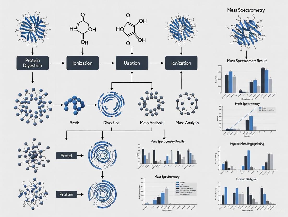
Mass spectrometry techniques provide unparalleled detail on protein structure and dynamics. The following workflow illustrates the pathway for MS-based integrity verification, from sample analysis to data interpretation.
Top-Down and Bottom-Up MS for Primary Structure Analysis
Bottom-up proteomics, the most established approach, involves enzymatic digestion of proteins into peptides followed by LC-MS/MS analysis. It excels at identifying proteins, sequencing peptides, quantifying abundance, and locating PTMs [4] [5]. Top-down proteomics, an advancing methodology, analyzes intact proteins, providing a complete picture of proteoforms, including combinations of PTMs present on a single molecule [4].
Table 2: Mass Spectrometry Techniques for Protein Integrity Analysis
| Technique | Primary Application | Key Strengths | Common Instrumentation |
|---|---|---|---|
| Bottom-Up LC-MS/MS | Sequence confirmation, PTM mapping, quantification. | High sensitivity and robust identification. | Orbitrap platforms (e.g., Exploris, Astral), timsTOF [5]. |
| Top-Down MS | Intact mass analysis, proteoform characterization. | Preserves labile PTM information and protein stoichiometry. | High-resolution systems like Orbitrap Excedion Pro, timsTOF [4]. |
| Targeted (PRM/MRM) | High-precision quantification of specific targets (e.g., impurities). | High sensitivity, accuracy, and multiplexing capability. | Triple quadrupole, Q Exactive HF-X, Orbitrap platforms [6] [5]. |
| Hydrogen-Deuterium Exchange (HDX-MS) | Conformational dynamics, epitope mapping, stability. | Probes solvent accessibility and protein folding. | Coupled with high-resolution MS systems [7]. |
| Size Exclusion Chromatography MS (SEC-MS) | Analysis of size variants and aggregates. | Simultaneously separates and identifies oligomeric states. | LC systems coupled to MS detectors [1]. |
Protocol: Primary Structure and PTM Analysis via Bottom-Up LC-MS/MS
This protocol is adapted from an integrated workflow for plasma proteome analysis [6] and can be applied to most recombinant protein samples.
Sample Preparation (Timing: ~2 hours)
- Denaturation and Reduction: Dilute the protein sample in a buffer containing 6 M urea. Add tris(2-carboxyethyl)phosphine (TCEP) to a final concentration of 20 mM and incubate at 37°C for 60 minutes to reduce disulfide bonds [6].
- Alkylation: Add iodoacetamide (IAA) to a final concentration of 40 mM. Incubate at room temperature in the dark for 30 minutes to alkylate cysteine residues [6].
Enzymatic Digestion (Timing: 6-8 hours)
- Dilute the urea concentration to below 1 M using 50 mM ammonium bicarbonate.
- Add trypsin at an enzyme-to-substrate ratio of 1:50 (w/w). Incubate at 37°C with shaking (500 rpm) for 6-8 hours.
- Stop the digestion by adding formic acid to a final concentration of 1% [6].
Desalting (Timing: ~1 hour)
- Prepare a C18 desalting column (e.g., using StageTips).
- Activate the column with methanol and acetonitrile, then equilibrate with 0.1% formic acid (Solvent A).
- Load the acidified peptide mixture onto the column. Wash with Solvent A to remove salts.
- Elute peptides with 40-60% acetonitrile in 0.1% formic acid. Concentrate the eluate in a vacuum centrifuge [6].
LC-MS/MS Analysis and Data Processing
- Reconstitute desalted peptides in Solvent A and separate using a nano-flow LC system with a C18 column and a gradient of increasing acetonitrile.
- Acquire data on a high-resolution mass spectrometer (e.g., Orbitrap Astral, timsTOF) using data-dependent acquisition (DDA) or data-independent acquisition (DIA).
- Process raw data using software (e.g., MaxQuant, Spectronaut) to search against a protein sequence database for identification and quantification [6] [5].
Assessing Conformational and Functional Integrity
Biophysical Techniques Correlated with MS
While MS is powerful, a full integrity profile requires orthogonal techniques. Fourier-Transform Infrared (FTIR) and Raman Spectroscopy are highly sensitive to changes in protein secondary structure by monitoring the amide I band (~1650 cm⁻¹) [7]. Differential Scanning Calorimetry (DSC) directly measures thermal stability by determining the melting temperature (Tm) and enthalpy (ΔH) of protein unfolding [1] [3]. Dynamic Light Scattering (DLS) analyzes hydrodynamic radius and is used to sensitively detect small quantities of protein aggregates [1].
Protocol: Conformational Stability Analysis via HDX-MS
Hydrogen-Deuterium Exchange coupled to MS (HDX-MS) is a powerful label-free technique for probing protein conformation and dynamics.
Deuterium Labeling (Timing: Variable)
- Dilute the purified protein into a deuterated buffer (e.g., D2-based PBS, pD 7.0). The dilution factor and protein concentration must be optimized to minimize back-exchange.
- Incubate for various time points (e.g., 10 seconds, 1 minute, 10 minutes, 1 hour) at a controlled temperature (e.g., 25°C) to allow deuterium incorporation.
Quenching and Digestion (Timing: < 2 minutes)
- At each time point, withdraw an aliquot and mix with a quench solution (e.g., low pH, low temperature) to reduce the pH to ~2.5 and the temperature to ~0°C. This drastically slows down the exchange reaction.
- Immediately inject the quenched sample onto a cooled, immobilized pepsin column for online digestion (typically < 1 minute).
LC-MS Analysis and Data Processing
- Separate the resulting peptides using a short, steep UPLC gradient under quench conditions to minimize back-exchange.
- Acquire mass spectra on a high-resolution mass spectrometer.
- Process data using dedicated HDX-MS software to identify peptides and calculate deuterium uptake for each peptide at each time point. A change in deuterium uptake under different conditions (e.g., with ligand, after stress) indicates a conformational change.
Application: Monitoring Host Cell Proteins as Integrity Indicators
Residual Host Cell Proteins are process-related impurities that can co-purify with biopharmaceuticals, posing a risk to drug stability and patient safety. MS is uniquely capable of identifying and quantifying individual HCPs, complementing traditional immunoassays [2].
MS Workflow for HCP Monitoring:
- Sample Preparation: Digest the drug substance or process intermediate. Depletion of the therapeutic protein can be performed to enrich for low-abundance HCPs.
- Data Acquisition: Use high-sensitivity DIA or label-free DDA on instruments like the Orbitrap Astral to achieve the depth of coverage needed to detect low-level HCPs [2].
- Data Analysis: Search data against a database of the host organism's proteome. Software tools and artificial intelligence are increasingly used to improve the reliability of HCP identification and to prioritize HCPs based on risk (e.g., enzymatic activity, immunogenicity) [2].
The Scientist's Toolkit: Essential Research Reagents and Materials
Table 3: Key Research Reagent Solutions for Protein Integrity Analysis
| Item | Function/Application | Example/Critical Feature |
|---|---|---|
| Trypsin, MS Grade | Proteolytic digestion for bottom-up proteomics. | High purity to minimize autolysis; modified trypsin to prevent self-cleavage. |
| Urea & IAA | Protein denaturation and cysteine alkylation. | High-purity, fresh urea solutions to avoid cyanate formation that causes artifactual modifications. |
| TCEP | Reduction of disulfide bonds. | Odorless alternative to DTT; stable at room temperature. |
| C18 StageTips | Desalting and cleanup of peptide mixtures. | In-house packed tips for low-cost, high-recovery sample preparation [6]. |
| HDX Buffers | Deuterated buffers for hydrogen-deuterium exchange. | High D-content; precise pD adjustment. |
| Somalogic SomaScan | Affinity-based proteomics platform. | Used for large-scale studies of the circulating proteome; useful for biomarker discovery [8]. |
| Olink Explore HT | Multiplexed, proximity extension assay platform. | Used in large-scale proteomics projects like the UK Biobank [8]. |
| Standard BioTools | Provider of the SomaScan platform. | Enables analysis of thousands of proteins simultaneously [8]. |
A multi-parametric approach is essential for defining protein integrity in modern biopharmaceutical development. By integrating mass spectrometry—spanning top-down, bottom-up, and targeted strategies—with orthogonal biophysical techniques, researchers can build a comprehensive integrity profile that directly links structural conformation to biological function. The protocols and applications detailed herein provide a framework for implementing these powerful MS-based methods to ensure the quality, safety, and efficacy of protein therapeutics.
In mass spectrometry (MS)-based proteomics, the "dynamic range" refers to the span between the most abundant and least abundant proteins in a sample. This presents a central challenge for protein integrity verification research and drug development: high-abundance proteins can suppress the detection and quantification of low-abundance species, many of which are biologically significant targets or potential biomarkers [9] [10] [11]. In complex biological samples like plasma, this dynamic range can exceed 10 orders of magnitude, with 22 proteins constituting about 99% of the protein mass, while the remaining 1% comprises thousands of distinct lower-abundance proteins [12]. This imbalance means that without specialized techniques, the ion signals of low-abundance peptides are often drowned out during ionization, making them invisible to the mass spectrometer [10]. Overcoming this limitation is critical for obtaining a comprehensive view of the proteome, enabling the discovery of novel biomarkers, and advancing precision medicine.
Technological and Methodological Solutions
Researchers have developed a suite of strategies to compress the dynamic range of protein abundances, making low-abundance proteins accessible to MS analysis. The following table summarizes the core approaches.
Table 1: Core Methodologies for Overcoming Dynamic Range Challenges in Proteomics
| Method Category | Key Principle | Key Advantage | Quantitative Performance |
|---|---|---|---|
| Sample Pre-fractionation & Enrichment | Depletes highly abundant proteins or enriches low-abundance targets prior to digestion [10] [11]. | Directly reduces sample complexity and compresses dynamic range. | Improves sensitivity but requires careful validation to avoid co-depletion of targets [13]. |
| Bead-Based Enrichment | Uses paramagnetic beads with specific binders to isolate and concentrate low-abundance proteins from complex samples [11]. | Highly specific; can be automated for high-throughput applications. | The ENRICH-iST kit reports low coefficients of variation (CVs) and high reproducibility [11]. |
| Nanoparticle Protein Corona (Proteograph) | Uses engineered nanoparticles to bind proteins, compressing dynamic range via competitive binding at the nanoparticle surface [12]. | Unbiased, scalable, and enables deep plasma proteome coverage. | Identified >7,000 plasma proteins; maintains fold change accuracy and precision across batches [12]. |
| Advanced MS Acquisition Modes | Multiplexes precursor ions into different m/z range packets during instrument transients [9]. | Maximizes instrument usage without increasing measurement time or cost. | Increases protein identifications by 9% (DDA) and 4% (DIA), while reducing quantitative CV by >50% [9]. |
| Computational Protein Inference | Leverages peptides shared across multiple proteins for quantification using combinatorial optimization [14]. | Allows quantification of proteins that lack unique peptides. | Enables relative abundance calculations for proteins previously discarded from analysis [14]. |
Detailed Experimental Protocols
Protocol: Bead-Based Enrichment for Low-Abundance Proteins
This protocol, based on commercially available kits like the ENRICH-iST, is designed for processing plasma or serum samples to enhance the detection of low-abundance proteins [11].
1. Binding: Incubate the plasma sample with coated paramagnetic beads. The beads are functionalized with specific binders that selectively capture target proteins or a broad range of low-abundance proteins via affinity interactions. 2. Washing: Apply a magnetic field to separate the beads from the solution. Wash the beads thoroughly to remove non-specifically bound contaminants and highly abundant proteins. 3. Lysis and Denaturation: Resuspend the beads in a LYSE reagent to denature the captured proteins. Incubate in a thermal shaker (typically at ~95°C for 10 minutes) to irreversibly break disulfide bonds and fully linearize the proteins. 4. Digestion: Digest the proteins into peptides directly on the beads. Add a proteolytic enzyme (typically trypsin) and incubate under optimized conditions (e.g., 37°C for several hours) for complete digestion. 5. Peptide Purification: Clean up the digested peptides using solid-phase extraction (SPE) to remove salts, detergents, and other impurities that could interfere with downstream LC-MS analysis. 6. MS Analysis: Reconstitute the purified peptides in an appropriate solvent (e.g., 0.1% formic acid, 3% acetonitrile) for injection into the LC-MS/MS system [11].
This entire workflow can be completed in approximately 5 hours and is amenable to automation for processing large sample cohorts [11].
Protocol: Nanoparticle Protein Corona Workflow (Proteograph)
The Proteograph Product Suite employs a multiplexed nanoparticle workflow to compress the dynamic range of plasma proteomes, enabling deep coverage [12].
1. Sample Incubation: Incubate the plasma sample with the proprietary engineered nanoparticles. During incubation, a protein "corona" forms on the nanoparticle surface through competitive binding, where low-abundance proteins with high affinity can displace high-abundance proteins with lower affinity. 2. Corona Isolation and Washing: Separate the nanoparticles with their bound protein corona from the bulk solution, followed by washing steps to remove unbound or weakly associated proteins. 3. Protein Elution and Digestion: Elute the proteins from the nanoparticle corona. Subsequently, denature, reduce, and alkylate the proteins following standard protocols (e.g., using TCEP or DTT for reduction and iodoacetamide for alkylation). Digest the protein mixture into peptides using trypsin. 4. Peptide Clean-up: Desalt and concentrate the resulting peptides using StageTips or SPE plates to ensure compatibility with LC-MS. 5. LC-MS Analysis: Analyze the peptides using a high-performance LC-MS system, typically with a Data-Independent Acquisition (DIA) method like on an Orbitrap Astral mass spectrometer. The data is processed using specialized software (e.g., DIA-NN in library-free mode) for protein identification and quantification [12].
Protocol: Multiple Accumulation Precursor Mass Spectrometry (MAP-MS)
MAP-MS is an instrumental method that enhances dynamic range by using otherwise "wasted" instrument time [9].
1. Instrument Setup: Implement the method on a trapping instrument like an Orbitrap Exploris 480 coupled to an EASY-nLC 1200 and a UHPLC column (e.g., Aurora Ultimate XT 25×75 C18). 2. Accumulation and Multiplexing: During the long transient recording times of the Orbitrap, multiplex precursor ions by accumulating them into several distinct m/z range packets simultaneously, rather than scanning a single broad range. 3. Data Acquisition: Perform this in either Data-Dependent Acquisition (DDA) or Data-Independent Acquisition (DIA) mode. The approach efficiently utilizes the instrument's dynamic range capacity by preventing the detector from being saturated by a few high-abundance ions. 4. Data Analysis: Process the resulting spectra with standard proteomics software suites. The output demonstrates increased protein identifications and improved quantitative precision compared to standard methods [9].
Workflow Visualization
The following diagram illustrates the logical progression of decisions and methodologies for tackling the dynamic range challenge, from sample preparation to data analysis.
The Scientist's Toolkit: Essential Research Reagents & Materials
Successful navigation of the dynamic range challenge relies on a suite of specialized reagents and materials. The following table details key solutions for robust and reproducible results.
Table 2: Key Research Reagent Solutions for Dynamic Range Challenges
| Item | Function & Application | Specific Examples |
|---|---|---|
| Paramagnetic Bead Kits | Selective isolation and concentration of low-abundance proteins from complex samples like plasma/serum; ideal for targeted studies [11]. | ENRICH-iST Kit [11] |
| Nanoparticle Kits | Unbiased dynamic range compression for deep, discovery-phase profiling of biofluids; suited for large-scale cohort studies [12]. | Proteograph XT Assay Kit [12] |
| Lysis Buffers & Inhibitors | Reagent-based cell lysis and solubilization of proteins while inhibiting endogenous proteases/phosphatases to preserve sample integrity [10]. | Custom buffers with protease/phosphatase inhibitors [10] |
| Protein Assays | Accurate quantification of protein concentration after lysis to ensure consistent loading across experiments and control for yield [10]. | Pierce Protein Assays (commercial examples) [10] |
| Digestion Enzymes | High-purity, specific proteases (e.g., trypsin) for complete and reproducible digestion of proteins into peptides for bottom-up MS [10]. | Trypsin, Lys-C [10] |
| Desalting & Clean-up Kits | Removal of salts, detergents, and other interfering substances from peptide digests prior to LC-MS to prevent ion suppression [10] [11]. | Solid-Phase Extraction (SPE) tips, StageTips [10] |
| Stable Isotope Labels | Metabolic (SILAC) or chemical (iTRAQ, TMT) incorporation of tags for precise multiplexed relative quantification across samples [13]. | SILAC, iTRAQ, TMT reagents [13] |
In mass spectrometry imaging (MSI) and quantitative proteomics, data integrity is paramount. The journey from sample preparation to final data visualization is fraught with potential sources of degradation that can compromise analytical results. Protein modifications and data fragmentation introduce significant artifacts that distort biological interpretations, particularly in protein integrity verification research crucial for drug development.
The selection of analytical color schemes represents a frequently overlooked yet critical point of potential data degradation. While rainbow-based colormaps like "jet" remain popular for their visual appeal, they introduce well-documented artifacts that can actively mislead data interpretation [15] [16]. These colormaps are not perceptually uniform, meaning equal changes in data value do not produce equal changes in perceived color intensity. Furthermore, their use of multiple hues makes accurate interpretation challenging for the approximately 8% of males of European descent with color vision deficiencies (CVDs) [16]. This degradation in data representation directly impacts the reliability of protein verification studies, potentially leading to false conclusions in therapeutic protein characterization.
Mechanisms of Data Degradation
Perceptual Distortion from Non-Linear Colormaps
The use of non-perceptually uniform colormaps creates a form of systematic data degradation by distorting the visual representation of quantitative information. In the jet colormap, the perceptual distance for the same quantitative change in signal (e.g., 1.39) can vary significantly (e.g., from 47.5 to 57.0) depending on where it occurs in the data range [16]. This non-linear relationship means that data gradients appear artificially steep in some regions and flattened in others, misleading researchers about the true distribution of protein abundances in MSI heatmaps.
The human visual system is naturally drawn to areas of high luminance and specific hues, particularly yellow. Rainbow colormaps exploit this by placing bright yellow in the middle of their data range, arbitrarily drawing the viewer's attention to medium-intensity values rather than the highest data values [16]. This attentional bias can cause researchers to overlook genuinely high-abundance regions in protein distribution maps, potentially missing critical biomarkers or localization patterns in drug target verification.
Exclusionary Data Visualization Through Color Inaccessibility
The degradation extends beyond mere misrepresentation to active exclusion of researchers with color vision deficiencies (CVDs). Approximately 8% of males of European descent and <1% of females have some form of CVD [16]. The red-green confusion characteristic of the most common forms of CVD (protanopia and deuteranopia) renders many rainbow-based visualizations quantitatively useless for these individuals. This represents not just an accessibility oversight but a fundamental degradation of data communicability across the scientific community.
When MSI data is visualized using problematic colormaps, the resulting heatmaps become scientifically ambiguous for a significant portion of researchers. For example, a protein distribution that appears as a clear gradient to those with normal color vision may show virtually no perceptible variation to someone with deuteranopia [16]. This fragmentation in data interpretation compromises collaborative research efforts and reduces the reproducible value of published findings in protein verification studies.
Analytical Fragmentation in Quantitative Workflows
The degradation pathway extends to methodological fragmentation in quantitative proteomics. In meat authentication studies, traditional peptide targeting strategies require labor-intensive, database-dependent identification of species-specific peptides followed by recovery rate validation [17]. This approach creates workflow inefficiencies where researchers must perform uniqueness queries peptide-by-peptide against entire databases, a process described as "labor-intensive and inefficient" [17].
This fragmentation in analytical methodology introduces validation bottlenecks that delay verification of protein integrity. Without streamlined processes for marker validation, the critical path from raw data to quantitative conclusion becomes fragmented, increasing the risk of analytical errors propagating through to final results. The absence of standardized, efficient workflows represents a systemic vulnerability in protein verification methodologies used throughout drug development pipelines.
Quantitative Assessment of Visualization Artifacts
Table 1: Comparative Performance of Colormaps in MSI Data Representation
| Colormap Type | Perceptual Uniformity | CVD Accessibility | Quantitative Accuracy | Recommended Use |
|---|---|---|---|---|
| Jet (Rainbow) | Poor - introduces artificial boundaries | Problematic for 8% of population | Low - misleading intensity perception | Not recommended |
| Hot | Moderate - linear RGB but not perceptually uniform | Moderate - some differentiation issues | Moderate - better than rainbow | Acceptable alternative |
| Greyscale | High - perceptually linear gradient | High - no hue dependency | High - intuitive intensity mapping | Recommended for accuracy |
| Cividis | High - scientifically derived | High - optimized for CVDs | High - uniform perceptual distance | Recommended for publication |
Table 2: Impact of Colormap Selection on Data Interpretation Accuracy
| Interpretation Parameter | Rainbow Colormaps | Perceptually Uniform Colormaps |
|---|---|---|
| Identification of maximum values | Arbitrarily drawn to yellow, not highest values | Correctly identifies highest intensity regions |
| Perception of data gradients | Inconsistent - varies by data range | Consistent across full data range |
| Accessibility for CVD researchers | Severely compromised | Fully accessible |
| Quantitative comparison between regions | Difficult due to hue variation | Intuitive due to luminance scaling |
| Reproducibility across publications | Low - subjective interpretation | High - objective interpretation |
The quantitative superiority of perceptually uniform colormaps is demonstrated through perceptual distance measurements. For the same quantitative difference in normalized glutathione abundance (approximately 1.3-1.4), the cividis colormap provides nearly identical perceptual distances (24.8 and 25.9), while the jet colormap shows widely varying perceptual distances (47.5 and 57.0) for the same actual data differences [16]. This perceptual inconsistency directly compromises the quantitative integrity of MSI data visualization.
Experimental Protocols for Optimal Data Visualization
Protocol: Validating Perceptual Uniformity in MSI Heatmaps
Principle: Ensure colormap selection accurately represents quantitative protein abundance data without perceptual distortion.
Materials:
- MSI data set (e.g., protein distribution heatmap)
- Multiple colormaps (jet, hot, greyscale, cividis)
- Kovesi test image transformation algorithm
- Perceptual distance calculation utility (e.g., cmaputil module)
Procedure:
- Generate protein distribution heatmap using default laboratory colormap
- Apply Kovesi test by transforming colormap color values with sine function
- Identify regions where sine wave is indistinguishable, indicating nonlinear color gradients
- Calculate perceptual distances between data points with known quantitative differences
- Compare perceptual distance consistency across data range
- Select colormap with most uniform perceptual gradient
- Verify accessibility using CVD simulation software (e.g., Dalton Lens)
Validation: A colormap passes validation when perceptual distance between points with equal quantitative differences varies by less than 10% across the data range [16].
Protocol: Hierarchical Clustering-Driven Peptide Screening for Quantitative Analysis
Principle: Streamline identification of species-specific peptide markers while excluding non-informative signals to prevent analytical fragmentation.
Materials:
- Meat samples (pork, beef)
- Extraction solution (Tris-HCl 0.05 M, urea 7 M, thiourea 2 M, pH 8.0)
- Trypsin, dithiothreitol (DTT), iodoacetamide (IAA)
- C18 solid-phase extraction columns
- UPLC system with Hypersil GOLD C18 column (2.1 mm × 150 mm, 1.9 µm)
- Q Exactive HF-X mass spectrometer
Procedure:
- Sample Preparation: Homogenize 2g meat sample with 20mL pre-cooled extraction solution in ice-water bath
- Centrifugation: Spin at 12,000 rpm for 20 minutes at 4°C
- Digestion:
- Aliquot 200μL supernatant
- Reduce with 30μL 0.1M DTT at 56°C for 60 minutes
- Alkylate with 30μL 0.1M IAA in dark at room temperature for 30 minutes
- Dilute with 1.8mL Tris-HCl buffer (25 mM, pH 8.0)
- Digest with 60μL trypsin (1.0 mg/mL) at 37°C overnight
- Terminate with 15μL formic acid
- Purification:
- Activate C18 SPE column with methanol
- Equilibrate with 0.5% acetic acid
- Load sample, wash with 0.5% acetic acid
- Elute with 2mL ACN/0.5% acetic acid (60/40, v/v)
- Filter through 0.22μm membrane
- HRMS Analysis:
- Employ Full Scan-ddMS2 mode on Q Exactive HF-X
- Use gradient elution: 0.0-0.2min (97-90% A), 0.2-16.0min (90-60% A), 16.0-17.0min (60-20% A), 17.0-17.5min (20% A), 17.5-18.5min (20-97% A), 18.5-25.0min (97% A)
- Mobile phase A: 0.1% FA in water; B: 0.1% FA in ACN
- Flow rate: 0.2mL/min, column temperature: 40°C
- Data Processing:
- Apply hierarchical clustering analysis (HCA) to peptide signals
- Implement positive correlation-based pre-screening
- Verify species-specificity of candidate peptides
- Validate quantitative suitability through recovery rate testing
Validation: The protocol achieves 80% elimination of non-informative peptide signals while maintaining accurate quantification with recoveries of 78-128% and RSD <12% [17].
Pathway Diagram: Data Degradation in Mass Spectrometry
Data Degradation Pathway in MS: This pathway illustrates how multiple degradation sources compromise data integrity throughout the mass spectrometry workflow, leading to erroneous scientific conclusions.
Pathway Diagram: Solution Framework for Data Integrity
Solution Framework for Data Integrity: This framework identifies key mitigation strategies that collectively preserve data integrity throughout mass spectrometry-based protein verification workflows.
Research Reagent Solutions for Protein Integrity Studies
Table 3: Essential Research Reagents for Protein Degradation and Quantification Studies
| Reagent/Category | Specific Examples | Function in Research | Application Context |
|---|---|---|---|
| Digestion Enzymes | Trypsin | Protein cleavage at specific sites for MS analysis | Sample preparation for bottom-up proteomics |
| Reducing Agents | Dithiothreitol (DTT) | Reduction of disulfide bonds | Protein denaturation before digestion |
| Alkylating Agents | Iodoacetamide (IAA) | Cysteine alkylation to prevent reformation | Sample preparation stabilization |
| Solid-Phase Extraction | C18 columns | Peptide purification and concentration | Sample clean-up before LC-MS/MS |
| Chromatography Columns | Hypersil GOLD C18 (2.1 mm × 150 mm, 1.9 µm) | Peptide separation | UPLC separation prior to MS detection |
| Mobile Phases | 0.1% Formic acid in water/acetonitrile | LC solvent system | Liquid chromatography gradient elution |
| Isobaric Labeling | TMT (Tandem Mass Tag) reagents | Multiplexed quantitative proteomics | Simultaneous quantification of multiple samples |
| Fluorescent Reporters | eGFP, GS-eGFP | Protein degradation tracking | Live-cell degradation kinetics measurement |
| Microinjection Markers | Fluorescently labeled dextran (10 kDa) | Injection volume quantification | Normalization in single-cell degradation assays |
The research reagents listed in Table 3 form the foundation of robust protein integrity verification protocols. Specifically, TMT labeling enables high-sensitivity parallel analysis of multiple samples [18], while fluorescent proteins like GS-eGFP serve as critical tools for quantifying degradation kinetics at single-cell resolution [19]. The integration of hierarchical clustering with high-resolution mass spectrometry creates a streamlined workflow that eliminates 80% of non-quantitative peptides while maintaining accurate quantification with recovery rates of 78-128% and RSD under 12% [17].
The integrity of mass spectrometry data in protein verification research faces multiple degradation pathways that require systematic mitigation. Non-perceptual colormaps introduce quantitative distortions that misrepresent protein distribution data, while methodological fragmentation creates analytical bottlenecks that compromise efficiency and reproducibility. Through implementation of perceptually uniform visualization schemes, accessibility-focused design principles, and streamlined analytical workflows, researchers can significantly enhance the reliability of protein integrity verification. These practices establish a foundation for robust, reproducible mass spectrometry methods that maintain data integrity throughout the drug development pipeline, ensuring that critical decisions regarding therapeutic protein characterization rest upon uncompromised analytical results.
In mass spectrometry-based protein integrity verification, two metrics stand as critical indicators of data quality and reliability: the Coefficient of Variation (CV) and Protein Sequence Coverage. For researchers and drug development professionals, these metrics provide the foundational evidence required to confirm protein therapeutic identity, purity, and stability. CV quantifies the precision of quantitative measurements across replicates, offering confidence in reproducibility for pharmacokinetic and biomarker studies. Sequence coverage comprehensively assesses protein identity and integrity by determining the percentage of amino acid sequences verified by detected peptides, thereby confirming the correct sequence of recombinant proteins and detecting potential modifications, degradations, or mutations. Within biopharmaceutical development, these parameters are indispensable for lot-release testing, biosimilar characterization, and stability studies, providing objective criteria for decision-making throughout the drug development pipeline.
Understanding and Calculating the Coefficient of Variation (CV)
Conceptual Foundation of CV
The coefficient of variation serves as a normalized measure of dispersion, enabling comparison of variability across datasets with different units or widely different means. In proteomics, CV calculates the ratio of the standard deviation to the mean of protein expression levels, expressed as a percentage. A lower CV indicates higher reproducibility and precision, which is paramount for reliable quantification in regulated bioanalysis [20]. This metric has gained renewed importance with technological advancements in high-throughput proteomics, where it frequently serves to benchmark the quantitative performance of new instruments, sample preparation workflows, and software tools [21].
Calculation Methods and Statistical Considerations
The standard formula for calculating CV is:
CV = (σ / μ) × 100%
where σ represents the standard deviation and μ denotes the mean of the measurements [20]. However, proteomics data presents specific statistical challenges due to its non-normal distribution. Raw intensity data is right-skewed, while log-transformed data approximates a normal distribution. This characteristic necessitates careful formula selection [21].
Table 1: CV Calculation Formulas for Proteomics Data
| Data Type | Appropriate Formula | Key Characteristics |
|---|---|---|
| Non-log-transformed Intensity | Base formula: CV = (σ / μ) × 100% | Applied directly to raw intensity values; preserves original data dispersion. |
| Log-transformed Intensity | Geometric formula: CV = √(e^(σ²_log) - 1) × 100% | σ_log is standard deviation of log-transformed data; provides comparable results to base formula on raw data. |
A critical error to avoid is applying the base CV formula to log-transformed data, which artificially compresses dispersion and can yield median CV values more than 14 times lower than the true variability, severely misrepresenting data quality [21].
Experimental Factors Influencing CV
Multiple experimental factors significantly impact calculated CV values, making transparency in methods reporting essential:
- Data Normalization: Systematic normalization procedures dramatically reduce CVs by removing technical bias. For example, median normalization can produce a "considerably lower" median CV compared to non-normalized data [21].
- Software Tools and Parameters: Default settings in common data processing tools like DIA-NN and Spectronaut often apply transformations that minimize data dispersion. For instance, selecting "High Precision" versus "High Accuracy" modes in DIA-NN can result in a 45% difference in median CV. Similarly, Spectronaut's default Top 3 filter and global normalization reduce CV by approximately 35% compared to disabled settings [21].
- Acquisition Methodology: Data-Independent Acquisition (DIA) generally provides lower CVs and higher reproducibility than Data-Dependent Acquisition (DDA) due to more consistent peptide sampling, making it particularly suitable for large-scale biomarker studies [22].
Achieving and Interpreting Protein Sequence Coverage
The Role of Sequence Coverage in Protein Verification
Protein sequence coverage represents the percentage of the total protein amino acid sequence detected and confirmed by identified peptides in a mass spectrometry experiment. It provides direct evidence of protein identity, completeness, and authenticity. In biopharmaceutical contexts, sequence coverage analysis is indispensable for confirming the intact expression of recombinant proteins, identifying sequence breakages or mutations, and providing critical data for biomarker discovery, disease diagnosis, and drug development [23]. High sequence coverage builds confidence that the target protein has been correctly synthesized and processed without unexpected alterations.
Methodologies for Maximizing Sequence Coverage
Achieving comprehensive sequence coverage, particularly 100% coverage, requires strategic method design beyond standard tryptic digestion:
- Multi-Enzyme Digestion Strategies: Employing various proteases with different cleavage specificities generates complementary peptide arrays that cover regions inaccessible to a single enzyme. Commonly used enzymes include Trypsin, Chymotrypsin, Asp-N, Pepsin, and Glu-C [23]. Combined digestions (e.g., Trypsin+Asp-N, Trypsin+Glu-C) further enhance coverage [23].
- Advanced Mass Spectrometry Platforms: High-resolution mass spectrometers, such as the Q Exactive Hybrid Quadrupole-Orbitrap and Orbitrap Fusion Lumos Tribrid systems, provide the precise mass measurements and fragmentation data required for confident peptide identification [23].
- Specialized Approaches for Challenging Proteins: Small membrane proteins and hydrophobic segments often require alternative approaches. Top-down MALDI-MS/MS techniques and solvent extraction-based purifications can sequence proteins inaccessible to conventional bottom-up methods [24]. Alternative proteases like chymotrypsin, elastase, or pepsin can generate peptides from transmembrane segments lacking basic residues [24].
Troubleshooting Incomplete Coverage
When sequence coverage falls below 100%, systematic investigation is necessary. Researchers should review protein data and sequences to identify undetected theoretical peptides, then determine whether alternative enzymatic treatments or methodological adjustments could recover missing regions [23]. For proteins with small molecular weights, high-concentration SDS-PAGE separation followed by in-gel digestion can improve detection [23]. In quantification experiments using surrogate peptides, selecting multiple signature peptides for each target protein enables cross-validation and improves quantification accuracy, as demonstrated in the quantification of Cry1Ab protein in genetically modified plants [25].
Integrated Experimental Protocols
Protocol for Determining Protein Sequence Coverage
This protocol outlines the steps for achieving comprehensive protein sequence coverage using multi-enzyme digestion.
Materials:
- Purified target protein (50-100 μg)
- Specific proteases (Trypsin, Chymotrypsin, Asp-N, Glu-C, etc.)
- High-resolution mass spectrometer (e.g., Q Exactive HF-X or Orbitrap Fusion Lumos)
- Liquid chromatography system (e.g., Easy-nLC 1200)
- Professional analysis software (e.g., MaxQuant, DIA-NN, Spectronaut)
Procedure:
- Sample Preparation: Divide the target protein sample into aliquots for separate enzymatic digestions.
- Multi-Enzyme Digestion: Digest each aliquot with different specific proteases (e.g., Trypsin, Chymotrypsin, Asp-N) under optimal conditions for each enzyme.
- LC-MS/MS Analysis: Analyze digested peptides using high-resolution LC-MS/MS with data-dependent or data-independent acquisition.
- Database Search: Identify peptides against the target protein sequence using professional analysis software with appropriate false discovery rate controls (typically ≤1%).
- Sequence Assembly: Combine identification results from multiple enzymatic digestions to reconstruct complete protein sequence information.
- Coverage Calculation: Calculate sequence coverage as (number of amino acids in detected peptides / total number of amino acids in protein) × 100%.
Troubleshooting Tips:
- If coverage remains incomplete after multi-enzyme digestion, consider combining enzymes in simultaneous digestion.
- For membrane proteins, incorporate organic solvent extraction and alternative proteases.
- For low-abundance proteins, implement affinity enrichment prior to digestion.
Protocol for Precision Assessment Using CV
This protocol describes the systematic evaluation of quantitative precision in proteomic experiments using coefficient of variation.
Materials:
- Biological or technical replicates (minimum n=3, recommended n=5-6)
- Stable isotope-labeled internal standards (when applicable)
- Statistical computing environment (e.g., R with proteomicsCV package)
Procedure:
- Experimental Design: Prepare and process sufficient replicates to support robust variability assessment (minimum 3 for validation, 5-6 for discovery studies).
- Data Acquisition: Analyze all replicates using identical LC-MS/MS conditions within a randomized sequence to minimize batch effects.
- Data Preprocessing: Apply consistent normalization procedures while retaining non-log-transformed intensity data for CV calculation.
- CV Calculation:
- For non-log-transformed intensity data: Use base formula CV = (σ / μ) × 100%
- For log-transformed intensity data: Use geometric formula CV = √(e^(σ²_log) - 1) × 100%
- Precision Assessment: Calculate CVs for each protein across replicates, then determine median CV across all quantified proteins as an overall quality metric.
- Data Reporting: Document all normalization procedures, transformation steps, and the specific CV formula used in methods sections.
Troubleshooting Tips:
- If CVs are higher than expected, examine raw data for outliers and check normalization procedures.
- For experiments comparing multiple conditions, ensure consistent CV calculation methodology across all groups.
- Use the R package "proteomicsCV" for standardized calculations [21].
Applications in Protein Therapeutic Development
Regulatory Considerations and Validation Requirements
For protein therapeutics development, regulatory guidelines establish specific acceptance criteria for quantitative bioanalytical methods. The AAPS Bioanalytical Focus Group recommends validation parameters that blend considerations from both small molecule and protein ligand-binding assays [26].
Table 2: Validation Acceptance Criteria for Protein LC-MS/MS Bioanalytical Methods
| Validation Parameter | Small Molecule LC-MS/MS | Protein LBA | Protein LC-MS/MS (Recommended) |
|---|---|---|---|
| Lower Limit of Quantification | Within ±20% | Within ±25% | Within ±25% |
| Calibration Standards | Within ±15% (except LLOQ) | Within ±20% (except LLOQ/ULOQ) | Within ±20% (except LLOQ) |
| Accuracy and Precision | Within ±15% (LLOQ ±20%)Minimum 3 runs | Within ±20% (LLOQ/ULOQ ±25%)Minimum 6 runs | Within ±20% (LLOQ ±25%)Minimum 3 runs |
| Selectivity/Specificity | 6 matrix lots;Blanks <20% of LLOQ | 10 matrix lots;LLOQ accuracy within ±25% for 80% of lots | 6-10 matrix lots;Blanks <20% of LLOQLLOQ accuracy within ±25% for 80% of lots |
| Matrix Effect | IS-normalized CV ≤15% across 6 lots | Not Applicable | IS-normalized CV ≤20% across 6-10 lots |
These validation parameters provide a framework for establishing assays that support non-clinical toxicokinetic and clinical pharmacokinetic studies, with the understanding that method requirements should be tailored to the specific protein therapeutic, intended study population, and analytical challenges [26].
Case Studies in Biopharmaceutical Analysis
Biosimilarity Assessment: Comprehensive sequence coverage analysis provides critical evidence for biosimilar development by verifying identical primary structure to the reference product. Multi-enzyme digestion approaches achieving 100% sequence coverage can confirm amino acid sequence identity, while high-precision quantification (CV < 15%) ensures consistent expression levels across manufacturing batches [23].
Antibody-Drug Conjugate Characterization: For ADCs, sequence coverage verifies the integrity of the antibody scaffold, while CV measurements ensure precise quantification of drug-to-antibody ratio (DAR) and payload distribution. Specialized digestion protocols may be required to characterize conjugation sites and confirm the absence of sequence variants that could impact binding or efficacy.
Biomarker Verification: In clinical proteomics, CV values help identify reliable biomarkers from discovery datasets. Proteins with low CVs across technical and biological replicates demonstrate consistent quantification, increasing confidence in their validity. Sequence coverage provides additional confirmation of biomarker identity, which is particularly important for distinguishing between protein isoforms with high sequence homology [27].
The Scientist's Toolkit: Essential Research Reagents and Materials
Table 3: Essential Research Reagents and Materials for Protein Quantification Studies
| Category | Specific Examples | Function and Application |
|---|---|---|
| Mass Spectrometry Systems | Q Exactive HF-X, Orbitrap Fusion Lumos, timsTOF | High-resolution accurate mass measurement for peptide identification and quantification [23] [22]. |
| Liquid Chromatography | Easy-nLC 1200, NanoLC systems | High-pressure nanoscale separation of complex peptide mixtures [23]. |
| Proteolytic Enzymes | Trypsin, Chymotrypsin, Asp-N, Glu-C, Pepsin | Generate complementary peptide patterns for comprehensive sequence coverage [23] [24]. |
| Sample Preparation Kits | Proteograph Product Suite, Immunoaffinity Magnetic Beads | Compress dynamic range, enrich low-abundance proteins, and remove interfering substances [12] [25]. |
| Data Analysis Software | DIA-NN, Spectronaut, Skyline, MaxQuant | Process raw MS data, identify peptides, quantify proteins, and calculate quality metrics [21] [28]. |
| Internal Standards | Stable Isotope-Labeled (SIL) Peptides/Proteins | Normalize technical variation and enable absolute quantification [26] [25]. |
| Statistical Tools | R package "proteomicsCV", Perseus, MSstats | Standardize CV calculations and perform statistical analysis of quantitative data [21]. |
The integrated application of sequence coverage and coefficient of variation provides a robust framework for protein quantification and verification in mass spectrometry-based analyses. Sequence coverage delivers comprehensive information about protein identity and integrity, while CV quantifies measurement precision and reproducibility. Together, these metrics form the foundation for reliable protein characterization throughout the biopharmaceutical development pipeline—from initial discovery through clinical trials and quality control. As mass spectrometry technologies continue to evolve with improved sensitivity, throughput, and data analysis capabilities, the strategic implementation of these essential metrics will remain fundamental to advancing protein therapeutics and biomarker research with the rigor required for regulatory approval and clinical implementation.
The Critical Role of Integrity Verification in Drug Development and Biomarker Discovery
Integrity verification forms the foundational pillar of modern drug development and biomarker discovery, ensuring that data generated throughout the research lifecycle is reliable, reproducible, and regulatory-compliant. In the context of mass spectrometry-based protein analysis, integrity encompasses multiple dimensions—from sample integrity during collection and processing to data integrity throughout acquisition and interpretation. The evolution of proactive health management paradigms has shifted focus from traditional disease diagnosis to prediction and prevention, placing increased emphasis on biomarker-driven models that require rigorous verification protocols [29]. This transition, coupled with advancements in high-throughput proteomics and multi-omics integration, demands robust frameworks that maintain analytical veracity from benchtop to clinical application.
The critical importance of integrity verification is magnified in regulated pharmaceutical environments where data integrity breaches can compromise patient safety and therapeutic efficacy. Regulatory agencies including the FDA and EMA have intensified scrutiny of data management practices, with nearly 80% of data integrity-related warning letters occurring in a recent five-year period [30]. The convergence of mass spectrometry technologies with structured integrity frameworks creates a synergistic relationship that accelerates biomarker discovery while maintaining the stringent evidentiary standards required for regulatory approval and clinical implementation.
Biomarker Verification Applications in Disease Research
Protein biomarker verification serves critical functions across the drug development continuum, from early target identification to clinical trial enrichment and therapeutic monitoring. The integration of mass spectrometry-based proteomic analysis has revealed substantial biomarker panels across diverse disease states, providing insights into pathological mechanisms and potential intervention points.
Comprehensive Proteomic Signatures in Neuromuscular Disease
Recent large-scale proteomic studies have demonstrated the power of systematic biomarker verification in elucidating disease pathophysiology. A 2025 investigation of Duchenne muscular dystrophy (DMD) quantified 7,289 serum proteins using SomaScan proteomics in corticosteroid-naïve patients, identifying 1,881 significantly elevated and 1,086 significantly decreased proteins compared to healthy controls [31]. This extensive profiling substantially expanded the catalog of circulating biomarkers relevant to muscle pathology, with independent cohort validation showing remarkable consistency (Spearman r = 0.85) [31].
Table 1: Key Protein Biomarker Categories Identified in DMD Research
| Biomarker Category | Representative Proteins | Fold Change in DMD | Biological Significance |
|---|---|---|---|
| Muscle Injury Biomarkers | Alpha-actinin-2 (ACTN2), Myosin binding protein C (MYBPC1), Creatine kinase-M type (CKM) | 151×, 86×, 54× | Sarcomere disruption, muscle fiber leakage, disease activity monitoring |
| Mitochondrial Enzymes | Succinyl-CoA:3-ketoacid-coenzyme A transferase 1 (SCOT), Enoyl-CoA delta isomerase 1 | 21×, 8.7× | Metabolic dysregulation, bioenergetic impairment |
| Extracellular Matrix Proteins | 45 elevated, 92 decreased proteins | Variable | Fibrosis, tissue remodeling, disease progression |
| Novel Muscle Factors | Kelch-like protein 41 (KLHL41), Ankyrin repeat domain-containing protein 2 (ANKRD2) | 19×, 22× | Regeneration pathways, emerging therapeutic targets |
The biological validation of these findings through correlation with muscle mRNA expression datasets further strengthened the evidence for their pathological relevance, creating a robust foundation for clinical translation [31]. This systematic approach to biomarker verification—spanning discovery, analytical validation, and biological confirmation—exemplifies the rigorous methodology required for meaningful biomarker implementation in drug development pipelines.
Cancer Metabolomics and Spatial Resolution
In oncology, MALDI mass spectrometry imaging (MALDI-MSI) has emerged as a powerful platform for spatial metabolite detection, enabling visualization of metabolic heterogeneity within tumor microenvironments. This technology has identified stage-specific metabolic fingerprints across breast, prostate, colorectal, lung, and liver cancers, providing functional insights into tumor biology [32]. The ability to map thousands of metabolites at near single-cell resolution has revealed metabolic alterations linked to hypoxia, nutrient deprivation, and therapeutic resistance [32].
Technological advancements including advanced matrices, on-tissue derivatization, and MALDI-2 post-ionization have significantly improved sensitivity, metabolite coverage, and spatial fidelity, pushing the boundaries of cancer metabolite detection [32]. The integration of MALDI-Orbitrap and Fourier-transform ion cyclotron resonance (FT-ICR) platforms has further enhanced mass accuracy and resolution, enabling more confident biomarker identification [32]. These capabilities position mass spectrometry as an indispensable tool for verifying metabolic integrity in cancer research and therapeutic development.
Mass Spectrometry Methodologies for Integrity Verification
Mass spectrometry platforms provide versatile technological foundations for integrity verification across analyte classes, from small molecule metabolites to intact proteins. Understanding the capabilities and applications of these platforms is essential for appropriate methodological selection in drug development and biomarker verification workflows.
Metabolomics Workflows and Platform Selection
Mass spectrometry-based metabolomics comprehensively studies small molecules in biological systems, offering deep insights into metabolic profiles [33]. The integrity of metabolomic data begins with appropriate sample collection and processing, where rapid quenching of metabolism and efficient metabolite extraction are critical for preserving biological fidelity [33]. Liquid-liquid extraction methods using solvents like methanol/chloroform mixtures enable partitioning of polar and non-polar metabolites, while internal standards compensate for technical variability and enhance quantification accuracy [33].
Table 2: Mass Spectrometry Platforms for Biomarker Integrity Verification
| Platform Type | Key Applications | Strengths | Recent Technological Advances |
|---|---|---|---|
| LC-ESI-MS/MS | Quantitative proteomics, targeted metabolite analysis | Broad metabolite coverage, excellent for polar metabolites, high sensitivity | Evosep Eno LC system (500 samples/day), Thermo Orbitrap Astral Zoom (30% faster scan speeds) [4] |
| MALDI-MSI | Spatial metabolomics, tissue imaging, cancer metabolomics | Preservation of spatial information, rapid analysis, minimal sample preparation | MALDI-2 post-ionization, integration with Orbitrap/FT-ICR, machine learning data analysis [32] |
| Top-Down Proteomics | Intact protein analysis, proteoform characterization, PTM mapping | Comprehensive protein characterization, avoids inference limitations | Bruker timsTOF with ion enrichment mode, Thermo Orbitrap Excedion Pro with alternative fragmentation [4] |
| High-Resolution Benchtop Systems | Routine analysis, quality control, clinical applications | Space efficiency, operational simplicity, reduced resource consumption | Waters Xevo Absolute XR (6× reproducibility), Agilent Infinity Lab ProIQ [4] |
The selection between MALDI and ESI platforms depends on analytical requirements. While ESI-LC-MS offers broad metabolite coverage and is ideal for polar metabolites, MALDI provides superior spatial resolution and rapid analysis without chromatographic separation [32]. MALDI's ability to produce single-charged ions simplifies spectral interpretation, reducing complexity and enhancing detection clarity for metabolites [32]. For protein analysis, the field is witnessing a transition from bottom-up to top-down proteomic approaches that preserve intact protein information, enabling comprehensive characterization of proteoforms and post-translational modifications that are critical for functional biology [4].
Experimental Protocol: LC-MS/MS-Based Protein Biomarker Verification
The following protocol outlines a standardized approach for protein biomarker verification using liquid chromatography-tandem mass spectrometry:
Sample Preparation and Quality Control
- Collect biological samples (serum, plasma, tissue) using standardized protocols to minimize pre-analytical variability [27]. For serum samples, allow clotting for 30 minutes at room temperature before centrifugation at 2,000 × g for 10 minutes.
- Aliquot and flash-freeze samples in liquid nitrogen within 60 minutes of collection. Store at -80°C until analysis [33].
- Perform protein precipitation and digestion using filter-aided sample preparation (FASP) or in-solution digestion protocols. Add stable isotope-labeled standard peptides for quantification [27].
- Validate sample quality using reference standards and quality control pools. Exclude samples with significant degradation or outlier behavior in quality metrics [27].
Liquid Chromatography Separation
- Utilize nanoflow or microflow LC systems with trap-column configurations for online desalting and separation.
- Employ reversed-phase C18 columns (75μm × 25cm, 1.6-2.0μm particle size) with gradient elution (90-240 minutes) using water/acetonitrile/0.1% formic acid mobile phases [27].
- Maintain column temperature at 40-55°C to enhance separation efficiency and reproducibility.
Mass Spectrometry Data Acquisition
- Operate mass spectrometer in data-dependent acquisition (DDA) or data-independent acquisition (DIA) mode. For targeted verification, use parallel reaction monitoring (PRM) for enhanced sensitivity and reproducibility [27].
- Set mass resolution to ≥35,000 (at m/z 200) for MS1 and 17,500 for MS2 scans. Use normalized collision energy of 25-35% for HCD fragmentation [27].
- Implement real-time calibration and mass correction using reference compounds.
Data Processing and Integrity Assessment
- Process raw data using specialized software (MaxQuant, Skyline, Progenesis QI) for peak detection, alignment, and quantification [27].
- Apply stringent false discovery rate (FDR) thresholds (<1%) for protein identification using target-decoy approaches [27].
- Verify data quality through metrics including retention time stability (<0.5% CV), mass accuracy (<5 ppm error), and intensity correlation (r > 0.9) across technical replicates [27].
Data Integrity Frameworks and Regulatory Compliance
Robust data integrity frameworks are essential components of integrity verification in regulated drug development environments. The ALCOA++ principles provide a comprehensive framework for ensuring data integrity throughout the biomarker discovery and validation lifecycle [30].
ALCOA++ Implementation in Analytical Workflows
The expanded ALCOA++ principles encompass ten attributes that collectively ensure data integrity from generation through archival:
- Attributable: Link all data to the person or system that created or modified it, using unique user IDs and appropriate access controls [30].
- Legible: Ensure data remains readable and reviewable in its original context, with reversible encoding where applicable [30].
- Contemporaneous: Record data at the time of activity with automatically captured date/time stamps synchronized to external standards [30].
- Original: Preserve the first capture or certified copy created under controlled procedures, maintaining dynamic source data where relevant [30].
- Accurate: Faithfully represent what occurred through validated coding, transfers, and interfaces with calibrated devices [30].
- Complete: Retain all data, metadata, audit trails, and contextual information needed to reconstruct events [30].
- Consistent: Maintain standardized definitions, units, and sequencing across the data lifecycle with aligned time stamps [30].
- Enduring: Preserve data intact and usable for the entire retention period with suitable formats and backups [30].
- Available: Ensure data retrievability for monitoring, audits, and inspections throughout the retention period [30].
- Traceable: Enable end-to-end tracking of data and metadata changes through comprehensive audit trails [30].
Integrated Informatics Infrastructure
Connected laboratory informatics systems significantly enhance data integrity by automating data transfer and reducing manual intervention points. Integration between Laboratory Information Management Systems (LIMS) and Chromatography Data Systems (CDS) creates streamlined workflows that minimize transcription errors and improve traceability [34]. In a non-integrated environment, manual steps for sample information transfer, result calculation, and data entry introduce multiple opportunities for error, while integrated environments enable automated data exchange at defined decision points [34].
Modern informatics solutions provide configurable control over data transfer timing—from immediate post-acquisition transfer to end of full review and approval cycles—allowing organizations to align digital workflows with evolving SOP requirements [34]. This integrated approach facilitates compliance with 21 CFR Part 11 and Annex 11 regulations while improving operational efficiency through reduced manual processes and streamlined training requirements [34].
Research Reagent Solutions for Biomarker Verification
The reliability of biomarker verification studies depends heavily on appropriate selection and quality of research reagents. Consistent quality across reagent batches ensures analytical reproducibility and minimizes technical variability in mass spectrometry-based assays.
Table 3: Essential Research Reagents for Mass Spectrometry-Based Biomarker Verification
| Reagent Category | Specific Examples | Function and Application | Quality Considerations |
|---|---|---|---|
| Sample Preparation | Methanol, chloroform, acetone, acetonitrile | Metabolite extraction, protein precipitation, lipid isolation | LC-MS grade, low background contamination, consistent purity between lots [33] |
| Digestion Enzymes | Trypsin, Lys-C, Asp-N | Protein cleavage for bottom-up proteomics, sequence-specific digestion | Sequencing-grade, MS-compatible, minimal autolysis, validated activity [27] |
| Internal Standards | Stable isotope-labeled peptides, metabolite analogs | Quantification standardization, technical variability compensation | >97% isotopic enrichment, chemical purity, stability in matrix [33] [27] |
| Ionization Matrices | CHCA, SA, DHB, DHB/HA, 9-AA | Laser energy absorption, analyte desorption/ionization in MALDI | High purity, appropriate crystal structure, low background interference [32] |
| Chromatography | C18, C8, HILIC, ion exchange columns | Analyte separation, resolution enhancement, interference removal | Column certification, stable performance, minimal carryover [27] |
| Calibration Solutions | ESI tuning mix, sodium formate clusters | Mass accuracy calibration, instrument performance verification | Certified reference materials, traceable concentrations [27] |
Integrity verification represents a critical nexus between technological innovation, analytical rigor, and regulatory compliance in drug development and biomarker discovery. The integration of advanced mass spectrometry platforms with structured data integrity frameworks creates a robust foundation for generating reliable, actionable scientific evidence. As the field progresses toward increasingly complex multi-omics integration and personalized medicine approaches, the principles of integrity verification will continue to ensure that biomarker data maintains the evidentiary standard required for confident clinical decision-making. The continued evolution of mass spectrometry technologies—particularly in top-down proteomics, spatial metabolomics, and integrated informatics—promises to enhance both the depth of biological insight and the robustness of verification methodologies, ultimately accelerating the translation of biomarker discoveries into clinical applications that improve patient outcomes.
Advanced MS Workflows in Action: From Sample Prep to Specific Applications
In mass spectrometry-based protein integrity verification research, the accuracy and reliability of results are fundamentally dependent on the quality of sample preparation. This initial phase of the proteomics workflow is critical for ensuring that proteins are efficiently extracted, digested, and cleaned up for subsequent LC-MS/MS analysis. Proper sample preparation directly impacts protein identification, quantification accuracy, and the detection of post-translational modifications, all of which are essential for biopharmaceutical characterization and quality control [10] [2]. The complexity of biological samples, combined with the vast dynamic range of protein concentrations, presents significant challenges that can only be overcome through optimized, reproducible preparation methods [10]. With regulatory agencies increasingly supporting mass spectrometry as a reliable tool for quality control in drug manufacturing, standardized sample preparation protocols have become more important than ever for ensuring consistent, high-quality results in protein integrity studies [2].
Critical Evaluation of Sample Preparation Methods
Method Selection Criteria
Choosing an appropriate sample preparation method requires careful consideration of multiple factors, including sample type, protein quantity, and specific research objectives. For mass spectrometry-based protein integrity verification, key selection criteria include compatibility with downstream MS analysis, reproducibility, recovery efficiency for low-abundance proteins, and practicality for the laboratory setting. The method must effectively remove interferents such as detergents and salts while maintaining protein representativity and enabling efficient digestion [10] [35]. Sample complexity and the need for specialized analyses such as phosphoproteomics or membrane protein characterization further influence method selection, as different protocols exhibit distinct strengths and limitations for specific applications [36] [37].
Comparative Analysis of Primary Methods
Table 1: Comparative Analysis of Sample Preparation Methods for Mass Spectrometry
| Method | Key Principle | Advantages | Limitations | Optimal Use Cases |
|---|---|---|---|---|
| Filter-Aided Sample Preparation (FASP) | Ultrafiltration to remove contaminants & on-membrane digestion [35] [37] | Effective SDS removal; compatibility with complex samples; high protein identification rates [35] [37] | Time-consuming; potential peptide loss; higher cost; not ideal for low-sample amounts [37] | Barley leaves; Arabidopsis thaliana leaves; samples requiring thorough detergent removal [35] [37] |
| Single-Pot Solid-Phase-Enhanced Sample Preparation (SP3) | Paramagnetic beads for protein binding, cleanup, & on-bead digestion [37] | Fast processing; minimal handling; compatible with detergents; works with low protein amounts; cost-effective [37] | Requires optimized bead-to-protein ratio; performance varies with bead type (carboxylated vs. HILIC) [37] | Arabidopsis thaliana lysates; low-input samples; high-throughput applications [37] |
| Acid-Assisted Methods (SPEED) | Trifluoroacetic acid (TFA) for protein extraction & digestion without detergents [38] [39] | Rapid & simple workflow; minimal steps; enhanced proteome coverage for challenging samples; avoids detergent complications [38] [39] | Acid conditions may not be suitable for all applications; may not disrupt crosslinks in some matrices [39] | Human skin samples; tape-strip proteomics; crosslinked extracellular matrices; challenging samples [39] |
| In-Solution Digestion (ISD) | Direct digestion in solution after protein extraction & cleanup [35] | Simplicity; applicable to various sample types; amenable to automation [35] | Potential incomplete digestion; may require cleanup steps to remove interferents [35] | Barley leaves (OP-ISD protocol showed best performance in this category) [35] |
| S-Trap | Protein suspension trapping in quartz filter for cleanup & digestion [36] | Efficient detergent removal; good recovery; applicable to small-scale samples [36] | Limited sample capacity; specialized equipment required [36] | Neuronal tissues (trigeminal ganglion); limited tissue samples [36] |
Detailed Experimental Protocols
SP3 Protocol for Plant and General Protein Samples
The SP3 protocol represents a significant advancement in sample preparation technology, particularly valuable for its compatibility with detergents and applicability to low-input samples. The following optimized protocol is adapted for plant tissues but can be modified for other sample types [37]:
Materials: SDT lysis buffer (4% SDS, 100 mM DTT, 100 mM Tris-HCl, pH 7.6); Sera-Mag Carboxylate-Modified magnetic beads; binding solution (90% ethanol, 5% water, 5% acetic acid); 50 mM TEAB; trypsin; and standard laboratory equipment including a thermomixer and magnetic rack [37].
Procedure:
- Protein Extraction: Homogenize 1 g of frozen plant tissue powder in 1 ml of hot SDT buffer. Incubate at 95°C for 5-10 minutes with continuous shaking [37].
- Clarification: Centrifuge the lysate at 16,000 × g for 10 minutes. Transfer the supernatant to a new tube and determine protein concentration [37].
- Bead-Based Capture: Transfer 100 μg of protein extract to a low-binding tube. Add magnetic beads (10:1 bead-to-protein ratio) and binding solution to achieve final ethanol concentration >50%. Incubate for 10 minutes at room temperature with shaking [37].
- Washing: Place the tube on a magnetic rack until the solution clears. Remove supernatant. Wash beads twice with 70% ethanol, then once with acetonitrile. Briefly air-dry the beads [37].
- Digestion: Resuspend beads in 50 mM TEAB containing trypsin (1:50 enzyme-to-protein ratio). Incubate at 37°C for 2-4 hours with shaking [37].
- Peptide Recovery: Add trifluoroacetic acid to 1% final concentration. Place tube on magnetic rack and transfer the cleared supernatant containing peptides to a new vial for LC-MS/MS analysis [37].
This optimized SP3 protocol completes in approximately 2 hours and demonstrates excellent performance for a wide range of protein inputs without requiring adjustment of bead amount or digestion parameters [37].
SPEED Protocol for Challenging and Crosslinked Samples
The SPEED (Sample Preparation by Easy Extraction and Digestion) protocol offers a detergent-free approach that is particularly effective for challenging samples such as skin tissues and other crosslinked matrices. This method utilizes acid extraction for complete sample dissolution [38] [39]:
Materials: Pure trifluoroacetic acid (TFA); triethylammonium bicarbonate (TEAB); trypsin; and standard laboratory equipment [38] [39].
Procedure:
- Acid Extraction: Add pure TFA to the sample (e.g., skin tissue) at an approximate 1:10 ratio (sample:TFA). Vigorously vortex to ensure complete dissolution. For tough tissues, brief sonication may be applied [39].
- Neutralization: Add appropriate volume of TEAB to neutralize the acidified solution. The final pH should be approximately 7.5-8.5, optimal for tryptic digestion [38].
- Digestion: Add trypsin (1:50 enzyme-to-protein ratio). Incubate at 37°C for 4-6 hours or overnight [38].
- Acidification and Cleanup: Add TFA to 0.1-1% final concentration to stop digestion. Desalt peptides using C18 solid-phase extraction if necessary before LC-MS/MS analysis [38].
The SPEED protocol has demonstrated superior performance for challenging samples, increasing identified protein groups to over 6,200 in healthy human skin samples compared to conventional methods [39].
Optimized Protocol for Limited Tissue Samples
For precious or limited samples such as neuronal tissues, an optimized workflow maximizes protein and phosphopeptide recovery [36]:
Materials: Lysis buffer (5% SDS); S-Trap micro columns; dithiothreitol (DTT); iodoacetamide (IAA); trypsin; phosphoric acid; formic acid; and standard centrifugation equipment [36].
Procedure:
- Protein Extraction: Homogenize tissue (e.g., trigeminal ganglion) in 100 μl of 5% SDS lysis buffer at room temperature. Boil the homogenate for 2 minutes, then centrifuge at 14,000 × g for 10 minutes. Collect supernatant [36].
- Reduction and Alkylation: Take 100 μg protein aliquot. Add DTT to 2 mM final concentration and incubate at 56°C for 30 minutes. Then add IAA to 5 mM final concentration and incubate at room temperature for 45 minutes in the dark [36].
- Acidification and S-Trap Processing: Add 12% phosphoric acid at 1:10 (v/v) ratio. Add binding/wash buffer (6:1 ratio to acidified solution). Load mixture onto S-Trap column and centrifuge at 4,000 × g for 1-2 minutes [36].
- Digestion: Add trypsin solution in 50 mM TEAB to the column. Incubate at 47°C for 1 hour. Centrifuge to collect peptides, followed by additional elution steps [36].
- Phosphopeptide Enrichment (Optional): For phosphoproteomics, subject digested peptides to sequential enrichment using Fe-NTA magnetic beads followed by TiO2-based method to maximize phosphopeptide recovery [36].
This specialized protocol has been successfully applied to tiny neuronal tissues (0.1g mouse trigeminal ganglion), significantly enhancing yield for both proteomic and phosphoproteomic analyses [36].
Workflow Visualization
The Scientist's Toolkit: Essential Research Reagents and Materials
Table 2: Essential Research Reagents and Materials for Sample Preparation
| Category | Specific Reagent/Kit | Function | Application Notes |
|---|---|---|---|
| Lysis Reagents | 5% SDS Lysis Buffer [36] | Protein extraction & denaturation | Optimal for neuronal tissues; use at room temperature to prevent precipitation [36] |
| SDT Buffer (4% SDS, 100 mM DTT) [37] | Comprehensive protein extraction | Effective for plant tissues with cell walls; requires heating [37] | |
| Pure Trifluoroacetic Acid (TFA) [38] | Acid-based extraction | Detergent-free alternative; ideal for crosslinked samples like skin [38] [39] | |
| Reduction/Alkylation | Dithiothreitol (DTT) [36] | Disulfide bond reduction | Standard concentration: 2-10 mM; incubate at 56°C for 30 min [36] |
| Tris(2-carboxyethyl)phosphine (TCEP) [35] | Alternative reducing agent | More stable than DTT; effective at lower concentrations [35] | |
| Iodoacetamide (IAA) [36] | Cysteine alkylation | Standard concentration: 5-50 mM; protect from light during incubation [36] | |
| Digestion & Cleanup | S-Trap Micro Columns [36] | Protein cleanup & digestion | Ideal for small samples (<100 μg); efficient SDS removal [36] |
| Sera-Mag Carboxylate-Modified Magnetic Beads [37] | SP3 protein binding | Enable rapid processing; compatible with detergents [37] | |
| Trypsin, Mass Spectrometry Grade [36] | Protein digestion | Standard ratio: 1:50 (enzyme:protein); optimize digestion time [36] | |
| Specialized Enrichment | Fe-NTA Magnetic Beads [36] | Phosphopeptide enrichment | High specificity; use before TiO2 for comprehensive coverage [36] |
| TiO2 Microspheres [36] | Phosphopeptide enrichment | Broad specificity; ideal as second enrichment step [36] | |
| Assessment & QC | BCA Protein Assay Kit [36] | Protein quantification | Critical for normalizing samples before digestion [36] |
Optimized sample preparation is the cornerstone of successful mass spectrometry-based protein integrity verification research. The methods detailed in this application note—FASP, SP3, SPEED, and S-Trap—each offer distinct advantages for specific sample types and research objectives. SP3 technology provides exceptional efficiency and throughput for standard samples, while the SPEED method breaks new ground for challenging, crosslinked matrices. The ongoing integration of artificial intelligence and improved data analysis tools promises to further enhance the reliability and interpretation of proteomic data [2]. By selecting appropriate methods based on sample characteristics and research goals, scientists can achieve the reproducibility, depth of coverage, and quantitative accuracy required for rigorous protein integrity verification in drug development and regulatory contexts.
Liquid Chromatography-Tandem Mass Spectrometry (LC-MS/MS) provides a powerful analytical platform for protein verification, enabling the identification and quantification of proteins with high specificity and sensitivity. In the context of protein integrity verification research, LC-MS/MS is indispensable for characterizing amino acid sequences, post-translational modifications (PTMs), and protein complex assemblies [40] [41]. The core strength of LC-MS/MS lies in its ability to couple the separation power of liquid chromatography with the exquisite mass analysis and structural elucidation capabilities of tandem mass spectrometry. This combination is particularly crucial for drug development, where verifying the correct structure and modifications of protein therapeutics, such as monoclonal antibodies and engineered proteins, is a regulatory requirement. High-resolution mass spectrometry (HR-MS) further enhances this capability by providing accurate mass measurements that enable precise molecular formula assignment and distinguish between closely related proteoforms [42] [43].
Core Performance Metrics for LC-MS/MS System Verification
Optimal performance of LC-MS/MS platforms is a prerequisite for reliable protein verification. Consistent monitoring of system performance metrics ensures the identification of subtle differences in system components and reveals specific causes of technical variability. A set of 46 system performance metrics has been established for comprehensive monitoring of the entire LC-MS/MS workflow [44].
Table 1: Key LC-MS/MS Performance Metrics for System Verification
| Metric Category | Specific Metric | Optimal Direction | Purpose and Interpretation |
|---|---|---|---|
| Chromatography | Interquartile Retention Time Period | ↑ | Longer times indicate better chromatographic separation. |
| Peak Width at Half-Height (Median) | ↓ | Sharper peaks indicate better chromatographic resolution. | |
| Fraction of Peptides with Divergent RT (±4 min) | ↓ | Estimates peak broadening occurring very early or late in the gradient. | |
| Electrospray Ion Source | MS1 Signal Jumps/Falls >10x | ↓ | Flags electrospray instability; counts sudden large changes in signal. |
| Median Precursor m/z for IDs | ↓ | Higher median m/z can correlate with inefficient or partial ionization. | |
| Ratio of 1+ to 2+ Peptides | ↓ | High ratios may indicate inefficient ionization. | |
| Dynamic Sampling | Ratio of Peptides Identified Once/Twice | ↑ | Estimates oversampling; higher ratios indicate broader peptide coverage. |
| Number of MS2 Scans | ↑ | More MS2 scans indicates more intensive sampling for identification. | |
| MS1 max/MS1 sampled abundance ratio | ↓ | Estimates position on peak where sampled (1 = sampled at peak maxima). |
These metrics typically display variations of less than 10% in a well-controlled system, making them sensitive enough to reveal even subtle performance degradation. Their application enables rational, quantitative quality assessment for proteomics and other LC-MS/MS analytical applications, which is fundamental for any protein integrity verification pipeline [44].
Experimental Protocols for Protein Verification
Bottom-Up Proteomics Workflow for Protein Identification and Quantification
The bottom-up proteomics workflow is the most common method for protein verification. It involves proteolytic digestion of proteins into peptides prior to LC-MS/MS analysis [40].
Protocol: Bottom-Up Proteomics for Protein Verification
- Step 1: Protein Digestion. The protein extract is enzymatically digested, typically using trypsin, to generate a complex peptide mixture.
- Step 2: Liquid Chromatography (LC). The peptide digest is separated using one or more dimensions of liquid chromatography (e.g., reverse-phase C18 column) coupled directly to the mass spectrometer. A common gradient uses mobile phase A (e.g., water with 0.1% formic acid) and B (e.g., acetonitrile with 0.1% formic acid) over 60-120 minutes.
- Step 3: Mass Spectrometry Acquisition. Peptides eluting from the LC column are ionized via electrospray ionization (ESI) and analyzed. Two primary data acquisition strategies are employed:
- Data-Dependent Acquisition (DDA): The mass spectrometer performs a full MS1 scan to detect peptide ions, then automatically selects the most abundant ions for fragmentation (MS2). This method is well-proven and sensitive but can suffer from stochastic sampling of lower abundance ions [45] [40].
- Data-Independent Acquisition (DIA): Instead of isolating individual precursors, the mass spectrometer fragments all ions across sequential, predefined m/z windows. This method reduces undersampling and yields more consistent and comprehensive peptide quantification, though data analysis is more complex [45] [40].
- Step 4: Database Search and Protein Inference. The acquired MS2 spectra are compared against a protein sequence database using search engines (e.g., MaxQuant, OpenMS) to identify peptides. These peptide identifications are then assembled into protein identifications [40].
- Step 5: Quantification. Quantification strategies can be:
- Label-free: Peptide abundances are compared based on the intensity of precursor ions (MS1) or fragment ions (MS2) across different runs [40].
- Isobaric Labels (e.g., TMT, iTRAQ): Peptides from different samples are labeled with isobaric tags, multiplexed, and analyzed together. Quantification is achieved by comparing the intensities of reporter ions in the MS2 or MS3 spectra [46] [40].
- Metabolic Labeling (e.g., SILAC): Cells are grown in media containing heavy or light isotopes of amino acids, enabling direct comparison of peptide intensities in a single MS run [40].
Diagram 1: Bottom-Up Proteomics Workflow. The process begins with protein digestion and proceeds through LC separation and MS analysis to identification.
Advanced Structural Elucidation with LC-HR-MS3
For challenging identifications, such as distinguishing structural isomers or confirming modifications, multi-stage mass spectrometry (MS3) provides deeper structural information.
Protocol: LC-HR-MS3 Method for Confident Compound Identification [43]
- Step 1: Sample Preparation. For serum samples, mix 125 µL serum with 375 µL acetonitrile to precipitate proteins. Centrifuge, then dry the supernatant under nitrogen. Reconstitute in an appropriate diluent (e.g., 1:1:2 MeOH:ACN:5mM ammonium formate).
- Step 2: LC Separation. Use a C18 column with a gradient elution (mobile phase A: 5 mM ammonium formate in water with 0.05% formic acid; B: MeOH:ACN 1:1 with 0.05% formic acid).
- Step 3: HR-MS3 Data Acquisition. On an Orbitrap-based instrument, a single scan cycle includes:
- A full MS1 scan at high resolution (e.g., 120,000) to detect precursor ions.
- MS2 fragmentation of the top 10 abundant precursors from the MS1 scan. Use an isolation window of 1.5 m/z and stepped normalized collision energies.
- MS3 fragmentation of the top 3 product ions from an MS2 spectrum. Use an isolation window of 2 m/z and a fixed normalized collision energy.
- Step 4: Data Analysis. Match the generated MS2 and MS3 spectra against a high-quality spectral library. The combined MS2-MS3 tree matching increases confidence and can improve detection limits for certain analytes compared to MS2 alone [43].
Targeted Protein Quantification using Parallel Reaction Monitoring (PRM)
Targeted MS provides high sensitivity and reproducibility for verifying specific proteins of interest, such as biomarkers or drug targets.
Protocol: Targeted Verification with LC-PRM [45]
- Step 1: Assay Development. From discovery-phase data (DDA or DIA), select proteotypic peptides unique to the target protein. Define the precursor ion m/z and expected fragment ions for each peptide.
- Step 2: LC-PRM Acquisition. On a high-resolution mass spectrometer (e.g., Orbitrap, Q-TOF):
- The instrument is programmed to target the specific precursor ions at their expected retention times.
- Upon detection, the precursor is isolated and fragmented.
- A high-resolution and accurate mass full-scan MS2 spectrum (the Parallel Reaction Monitor) is acquired for all fragment ions.
- Step 3: Data Analysis. Extract the chromatographic traces for the specific, high-confidence fragment ions from the full MS2 scan. The high resolution minimizes interference, and the peak areas of these extracted ion chromatograms are used for precise quantification [45].
The Scientist's Toolkit: Research Reagent Solutions
Table 2: Essential Reagents and Materials for LC-MS/MS Protein Verification
| Item | Function and Application |
|---|---|
| Trypsin (Protease) | Enzymatically digests proteins into peptides for bottom-up proteomics analysis [40]. |
| Isobaric Labels (TMT, iTRAQ) | Enable multiplexed quantification of peptides from up to 16 samples simultaneously in a single LC-MS run [40]. |
| Stable Isotope-Labeled Peptide Standards (SIL) | Serve as internal standards for absolute quantification; correct for sample preparation and ionization variability [46]. |
| Activity-Based Probes (e.g., Desthiobiotin-ATP Probe) | Chemically label and enrich specific protein families (e.g., active kinases) for functional proteomics studies [45]. |
| Peptidiscs / Membrane Mimetics | Stabilize membrane proteins in a detergent-free, native-like environment for analysis by native MS and other structural techniques [41]. |
| Solid Phase Extraction Sorbent (e.g., PPL Resin) | Isolate and concentrate diverse molecules, such as dissolved organic matter or metabolites, from complex aqueous matrices prior to LC-MS [42]. |
Analytical Pathways for Data Processing and Verification
Robust data analysis is critical for transforming raw LC-MS/MS data into verifiable protein identities and quantities. The field is moving towards scalable, reproducible workflow-based analyses to ensure reliability.
Diagram 2: Data Analysis Workflow. Key steps from raw data processing to final statistical analysis and reporting.
A modern quantitative data analysis pipeline, such as the quantms workflow, involves several key steps distributed over cloud or high-performance computing (HPC) environments for scalability and reproducibility [40]:
- Peptide Identification: Matching peptide fragment spectra to a protein sequence database, often with consideration for known and unknown modifications.
- Peptide Quantification: The strategy depends on the acquisition method. For DDA label-free quantification, this involves finding features (3D peaks) on the MS1 level, aligning them across runs, and integrating intensities. For DIA, it requires extracting and statistically validating peptide signals using spectral libraries [40].
- Protein Inference and Quantification: Mapping identified peptides to their most likely proteins of origin, resolving ambiguities, and aggregating peptide-level quantities to protein-level abundances.
- Downstream Statistical Analysis: Tools like MSstats perform elaborate normalization, imputation, and statistical significance testing to identify differentially abundant proteins [40].
- Quality Control: Throughout the process, comprehensive quality control metrics (e.g., those in Table 1) are gathered to ensure the technical validity of the results [44] [40].
Data-Independent Acquisition (DIA) represents a transformative approach in mass spectrometry-based proteomics, addressing critical limitations of traditional methods for protein integrity verification in biopharmaceutical research. Unlike Data-Dependent Acquisition (DDA), which stochastically selects intense precursor ions for fragmentation, DIA systematically fragments all peptides within predefined, sequential mass-to-charge (m/z) windows [47]. This unbiased acquisition strategy generates comprehensive, reproducible fragment ion maps of all detectable analytes, effectively eliminating the "missing value" problem that plagues DDA when analyzing complex samples [48]. For researchers verifying protein therapeutics, this translates to unprecedented capabilities in monitoring critical quality attributes, including host cell protein (HCP) impurities, post-translational modifications, and product variants, with the quantitative rigor approaching that of targeted methods [2] [47].
The fundamental advantage of DIA lies in its unique combination of deep proteome coverage and excellent quantitative reproducibility. While targeted methods like Multiple Reaction Monitoring (MRM) offer high sensitivity for predefined targets, and DDA provides broad discovery capabilities, DIA occupies a strategic middle ground—delivering extensive coverage without sacrificing quantitative precision [47]. This makes it particularly valuable for biopharmaceutical applications where comprehensive characterization is essential, such as monitoring HCPs throughout the production process [2]. As regulatory agencies increasingly recognize mass spectrometry as a reliable quality control tool, DIA emerges as a cornerstone technology for modern biopharmaceutical development [2].
Comparative Analysis of MS Acquisition Methods
Understanding DIA's value proposition requires examining its performance relative to other mass spectrometry approaches. The following table summarizes key characteristics across major acquisition strategies, highlighting DIA's balanced profile for comprehensive protein characterization.
Table 1: Comparison of Primary LC-MS/MS Acquisition Methods in Proteomics
| Feature | Data-Dependent Acquisition (DDA) | Data-Independent Acquisition (DIA) | Targeted (MRM/PRM) |
|---|---|---|---|
| Acquisition Principle | Intensity-based selection of top N precursors for fragmentation | Systematic fragmentation of all precursors in sequential m/z windows | Selective monitoring of predefined precursor-fragment transitions |
| Proteome Coverage | Broad, but stochastic; limited in complex samples | Extensive and reproducible; superior in complex samples | Limited to predefined targets |
| Quantitative Performance | Moderate reproducibility; missing values between runs | High reproducibility and quantitative accuracy [47] | Excellent sensitivity and precision |
| Ideal Application Context | Discovery-phase profiling | Comprehensive characterization & verification [47] | Validation & routine monitoring |
| Throughput Consideration | High for identification | High for deep, reproducible quantification [48] | Very high for targeted analyses |
DIA's quantitative robustness stems from its consistent fragmentation of the same peptide sets across all samples, overcoming the stochastic sampling bias of DDA [49]. This consistent data acquisition makes DIA particularly suitable for large-scale studies assessing batch-to-batch consistency of biotherapeutics or temporal monitoring of HCP profiles during process development [2].
Experimental Design and Sample Preparation Protocols
The reliability of DIA data is fundamentally dependent on sample quality. Rigorous, standardized preparation protocols are essential to minimize technical variability and maximize proteome coverage [50].
Critical Steps in DIA Sample Preparation
Sample Collection and Storage:
- Containers: Use low-binding polypropylene tubes to minimize peptide adsorption.
- Storage: Snap-freeze samples immediately in liquid nitrogen and store at -80°C. Avoid multiple freeze-thaw cycles.
- Matrix Considerations: For plasma/serum, implement high-abundance protein depletion to enhance dynamic range. For tissues, maintain consistent homogenization with protease/phosphatase inhibitors present [50].
Protein Extraction and Solubilization:
- Buffers: Utilize MS-compatible chaotropic agents (6-8 M urea) and surfactants (Sodium deoxycholate, RapiGest). Avoid SDS whenever possible due to severe ion suppression.
- Mechanical Disruption: Apply consistent sonication or bead milling cycles while avoiding heat-induced degradation.
- QC Check: Quantify protein concentration via BCA or Bradford assay; target ≥50 µg total protein input for robust DIA quantification [50].
Reduction, Alkylation, and Digestion:
- Reduction: Use DTT (5-10 mM) or TCEP at 37°C for 30-60 minutes.
- Alkylation: Apply iodoacetamide (10-20 mM) in the dark to prevent side reactions.
- Digestion: Employ trypsin (enzyme-to-protein ratio 1:50-1:100) for 12-16 hours at 37°C. A Trypsin+Lys-C combination reduces missed cleavages in challenging matrices.
- QC Check: Maintain missed cleavage rate below 15% [50].
Peptide Cleanup and Normalization:
- Methods: Implement C18 solid-phase extraction (SPE) or StageTips for desalting.
- Normalization: Adjust peptide concentration to 0.5-1 µg/µL in 0.1% formic acid.
- Standard Addition: Spike indexed Retention Time (iRT) peptides for LC retention time calibration.
- QC Sample: Create a pooled quality control sample representing all experimental conditions for injection throughout the LC-MS sequence [50].
Common Pitfalls and Preventive Measures
Table 2: Troubleshooting Common DIA Sample Preparation Issues
| Pitfall | Impact on DIA Data | Preventive Action |
|---|---|---|
| Incomplete Digestion | Reduced peptide IDs (20-30%); missing transitions | Use fresh enzymes; dual-enzyme approach; monitor missed cleavages |
| Detergent Contamination | Ion suppression (up to 90%); poor chromatography | Prefer MS-compatible detergents; employ FASP or SPE for SDS removal |
| Overalkylation | Artificial modifications; false PTM calls | Use IAA at 10-20 mM, in dark, at ≤37°C |
| Batch Inconsistency | High coefficient of variation (>20%); poor PCA clustering | Implement standardized protocols; normalize peptide loading; use pooled QC |
DIA Experimental Workflow
Computational Analysis of DIA Data
The complex, multiplexed nature of DIA data demands sophisticated computational tools for deconvolution and interpretation. The analysis typically follows either library-based or library-free approaches, each with distinct advantages.
Library-Based versus Library-Free Strategies
Library-Based Analysis: This traditional approach matches DIA data against a preconstructed spectral library containing peptide precursor and fragment ion information, retention times, and, when available, ion mobility values [51]. Libraries can be generated experimentally from fractionated DDA runs or predicted in silico from protein sequence databases [49]. Experimentally-derived libraries, particularly those enhanced through gas-phase fractionation (GPF), generally provide the highest identification rates [49].
Library-Free Analysis: Also known as "direct" analysis, this method uses protein sequence databases or predicted spectral libraries to interrogate DIA data directly, without requiring experimental library generation [51]. This approach is particularly valuable when project-specific library generation is impractical, though it may require more stringent false discovery rate control [51].
Benchmarking DIA Software Tools
Recent large-scale benchmarking studies have evaluated popular DIA software tools across various instruments and sample types. The following table summarizes key findings from these comprehensive evaluations.
Table 3: Performance Comparison of DIA Data Analysis Software
| Software Tool | Optimal Application Context | Key Strengths | Considerations |
|---|---|---|---|
| DIA-NN | High-throughput analyses; label-free quantification [51] | Excellent quantitative precision (CV 16.5-18.4%); fast processing [52] | Higher missing values in single-cell data [52] |
| Spectronaut | Complex samples requiring maximum coverage [51] | Highest identification rates (3066±68 proteins in single-cell) [52] | Moderate quantitative precision (CV 22.2-24.0%) [52] |
| PEAKS Studio | Streamlined analysis with minimal parameter optimization [52] | Good balance of identification and quantification | Lower precision (CV 27.5-30.0%) than other tools [52] |
| OpenSWATH | Customizable pipeline development [51] | Open-source platform; high flexibility | Requires more computational expertise |
The choice of software significantly impacts downstream results. Studies demonstrate that using gas-phase fractionated libraries generally benefits all software tools, irrespective of the refinement method used [49]. For differential abundance analysis, non-parametric permutation-based statistical tests consistently outperform other methods [49].
DIA Data Analysis Strategy
Essential Research Reagent Solutions
Successful DIA proteomics requires carefully selected reagents and materials at each process stage. The following table outlines key solutions for robust DIA implementation.
Table 4: Essential Research Reagents for DIA Proteomics
| Reagent Category | Specific Examples | Function & Importance |
|---|---|---|
| Digestion Enzymes | Trypsin, Lys-C | Specific proteolytic cleavage; trypsin is gold standard for generating compatible peptides |
| Chaotropic Agents | Urea (6-8 M), Thiourea | Efficient protein denaturation and solubilization while maintaining MS compatibility |
| MS-Compatible Surfactants | Sodium Deoxycholate (SDC), RapiGest | Effective protein solubilization with easy removal pre-LC-MS |
| Reducing Agents | DTT (5-10 mM), TCEP | Reduction of disulfide bonds for complete protein unfolding and digestion |
| Alkylating Agents | Iodoacetamide (10-20 mM) | Cysteine alkylation preventing reformation of disulfide bonds |
| Desalting Materials | C18 SPE cartridges, StageTips | Removal of salts, detergents, and other interferents before LC-MS analysis |
| Retention Time Standards | iRT (Indexed Retention Time) peptides | LC performance monitoring and retention time alignment across runs |
Applications in Biopharmaceutical Development
DIA mass spectrometry has proven particularly valuable for addressing critical challenges in biopharmaceutical development, especially in monitoring product quality and safety.
Host Cell Protein (HCP) Monitoring
Residual HCPs constitute a significant class of impurities in biologics that can compromise product safety and stability. While immunoassays have traditionally been used for HCP detection, they often lack specificity and coverage [2]. DIA provides a powerful complementary approach by enabling specific identification and quantification of individual HCPs throughout the production process [2]. This detailed characterization facilitates risk assessment and process optimization to minimize potentially immunogenic HCP species.
Drug-Metabolizing Enzyme and Transporter Quantification
Understanding the expression levels of drug-metabolizing enzymes (DMEs) and transporters is crucial for predicting pharmacokinetics and pharmacodynamics. DIA enables large-scale, multiplexed quantification of these proteins in complex biological samples, with studies demonstrating that protein abundance often correlates better with enzymatic activity than mRNA expression levels [47]. This application is particularly valuable during drug development for assessing interindividual variability in drug metabolism and disposition.
Data-Independent Acquisition has fundamentally expanded the capabilities of mass spectrometry in protein integrity verification research. Its unique combination of comprehensive coverage and robust quantification addresses critical needs in biopharmaceutical characterization, particularly for monitoring subtle product variants and low-abundance impurities throughout development and production.
The ongoing evolution of DIA technology promises even greater impacts. Artificial intelligence and machine learning are increasingly being integrated into data analysis pipelines, improving spectral interpretation and reducing false discoveries [2]. Furthermore, the emergence of single-cell DIA proteomics, while presenting new computational challenges, opens possibilities for characterizing cellular heterogeneity in production cell lines [52]. As these advancements mature and standardization improves, DIA is positioned to become an indispensable tool for ensuring the quality, safety, and efficacy of biopharmaceutical products.
For researchers implementing DIA, a rigorous approach encompassing optimized sample preparation, appropriate software selection, and orthogonal validation will deliver the most reliable and actionable results for protein integrity verification.
Protein-protein interactions (PPIs) are the fundamental building blocks of cellular machinery, with more than 80% of proteins functioning within complexes to execute their biological roles [53]. The accurate identification and verification of these interactions is therefore crucial for deciphering molecular mechanisms, understanding disease states, and identifying therapeutic targets. Within the context of mass spectrometry methods for protein integrity verification research, Affinity Purification-Mass Spectrometry (AP-MS) has emerged as a cornerstone biochemical technique for identifying novel PPIs under physiologically relevant conditions [54]. This method leverages the specific binding between a tagged "bait" protein and its endogenous "prey" interaction partners, followed by sophisticated mass spectrometric analysis to identify the captured complexes [55].
However, traditional AP-MS faces significant limitations in detecting weak, transient, and membrane-associated interactions due to stringent wash conditions that can dissociate labile complexes and disrupt critical membrane microdomains [55] [53]. To address these challenges, proximity labeling (PL) techniques have been developed, with recent innovations such as APPLE-MS (Affinity Purification coupled Proximity Labeling-Mass Spectrometry) creating a powerful hybrid approach that combines the specificity of affinity purification with the covalent capture capability of proximity labeling [53]. This integrated methodology offers enhanced sensitivity for capturing the dynamic interactome while maintaining high specificity, thereby providing a more comprehensive framework for protein integrity verification in therapeutic development.
Comparative Analysis of AP-MS and Enhanced Proximity Labeling Methods
The evolution from traditional AP-MS to integrated approaches like APPLE-MS represents a significant advancement in the researcher's toolkit for protein verification. Each method offers distinct advantages and limitations that must be considered when designing interactome mapping experiments.
Table 1: Comparison of AP-MS and APPLE-MS Methodologies
| Characteristic | Traditional AP-MS | APPLE-MS |
|---|---|---|
| Core Principle | Affinity-based purification of protein complexes [54] | Combined affinity purification and covalent proximity labeling [53] |
| Key Advantage | Identifies direct and indirect interactors in native physiological conditions [54] | Captures weak/transient interactions (affinities up to 76 μM) and membrane PPIs [53] |
| Detection Specificity | Moderate (subject to non-specific binding) [53] | High (4.07-fold improvement over AP-MS) [53] |
| Sensitivity to Weak Interactions | Limited due to stringent washes [55] [53] | Enhanced through covalent tagging |
| Applicability to Membrane Proteins | Limited due to detergent-mediated disruption [53] | Excellent (enables in situ mapping of receptor complexes) [53] |
| Typical Interactions Detected | Stable, high-affinity complexes [55] | Both stable and transient complexes |
Table 2: Performance Metrics of APPLE-MS vs. Traditional AP-MS
| Performance Metric | Traditional AP-MS | APPLE-MS | Improvement Factor |
|---|---|---|---|
| Specificity (Fold over total proteins) | Baseline | 4.07-fold increase | 4.07x [53] |
| Literature-Curated Interactors Identified | Lower | ~2x more identified | ~2x [53] |
| Weak Interaction Detection Limit | >100 μM KD | 76 μM KD | Significant enhancement [53] |
| Endogenous Tag Interference | Minimal with small tags | Minimal with Twin-Strep tag | Comparable [53] |
Experimental Protocols
Protocol for Traditional AP-MS
Objective: To identify protein-protein interactions for a target protein of interest in mammalian cells using affinity purification-mass spectrometry.
Materials:
- Mammalian cell line (e.g., HEK293T)
- Plasmid DNA encoding affinity-tagged bait protein (e.g., FLAG, HA, or Strep tags)
- Transfection reagent
- Lysis buffer (e.g., RIPA buffer with protease inhibitors)
- Affinity resin appropriate for tag (e.g., Anti-FLAG M2 agarose, Streptactin beads)
- Wash buffer (compatible with chosen affinity resin)
- Elution buffer (e.g., 3x FLAG peptide for FLAG-tagged proteins, or biotin for Strep-tagged proteins)
- Equipment for SDS-PAGE and mass spectrometry analysis
Procedure:
- Bait Protein Expression: Transfect mammalian cells with plasmid encoding the affinity-tagged bait protein. Include appropriate empty vector controls to identify non-specific binders [56].
- Cell Lysis: Harvest cells 24-48 hours post-transfection and lyse using ice-cold lysis buffer with protease inhibitors. Clarify lysates by centrifugation to remove insoluble material [54].
- Affinity Purification: Incubate clarified lysates with appropriate affinity resin for 2-4 hours at 4°C with gentle agitation [54].
- Stringent Washes: Wash resin thoroughly with wash buffer (typically 3-5 washes) to remove non-specifically bound proteins [53].
- Elution: Elute bound protein complexes using specific competing agent (e.g., FLAG peptide) or mild denaturing conditions [54].
- Protein Processing for MS: Denature eluates, reduce disulfide bonds, alkylate cysteine residues, and digest proteins with trypsin [57].
- Mass Spectrometry Analysis: Desalt and analyze resulting peptides by liquid chromatography-tandem mass spectrometry (LC-MS/MS) [57] [56].
- Data Analysis: Process raw MS data using search engines (e.g., MaxQuant) and statistical tools (e.g., MSstats) to identify high-confidence interacting proteins [56].
Advanced Protocol: APPLE-MS
Objective: To identify weak, transient, and membrane-associated protein-protein interactions using combined affinity purification and proximity labeling.
Materials:
- Mammalian cell line (e.g., HEK293T)
- Plasmid encoding bait protein with C-terminal Twin-Strep tag
- Recombinant PafA enzyme
- Streptavidin-PupE (SA-PupE) conjugate
- ATP-containing reaction buffer
- Streptavidin beads
- Lysis and wash buffers
- Equipment for LC-MS/MS analysis
Procedure:
- Bait Protein Expression: Express Twin-Strep-tagged bait protein in mammalian cells and allow to localize to its proper cellular compartment [53].
- Proximity Labeling: Incubate intact cells or cell fractions with PafA enzyme and SA-PupE conjugate in ATP-containing buffer to allow covalent labeling of proximal proteins [53].
- Cell Lysis: Lyse cells under denaturing conditions to preserve all interactions, including those covalently tagged.
- Affinity Purification: Incubate lysates with streptavidin beads to capture both the Twin-Strep-tagged bait and biotinylated prey proteins [53].
- Stringent Washes: Wash beads under highly stringent conditions (including denaturants) to remove non-specific binders while retaining covalently tagged interactions.
- On-Bead Digestion: Digest proteins on beads using trypsin to generate peptides for MS analysis.
- LC-MS/MS Analysis: Analyze peptides using high-resolution LC-MS/MS for identification [57].
- Bioinformatic Analysis: Use comparative statistical analysis to identify high-confidence interactors over control samples.
Diagram 1: APPLE-MS workflow for comprehensive interactome mapping, showcasing the integration of proximity labeling with affinity purification.
The Scientist's Toolkit: Essential Research Reagents
Table 3: Essential Research Reagents for AP-MS and Proximity Labeling Experiments
| Reagent/Category | Specific Examples | Function in Experiment |
|---|---|---|
| Affinity Tags | FLAG, HA, GFP, Twin-Strep tag [54] [53] | Enables specific purification of bait protein and its interactors |
| Purification Resins | GFP-trap resins, Immunoglobulin A (IgA) beads, Streptavidin beads [54] [53] | Solid support for immobilizing and purifying tagged protein complexes |
| Proximity Labeling Enzymes | PafA, BioID, APEX [53] | Catalyzes covalent labeling of proximal proteins for capturing transient interactions |
| Labeling Substrates | PupE, biotin-AMP [53] | Activated substrate for proximity-dependent protein tagging |
| Mass Spectrometry Systems | Q-TOF, Orbitrap, LC-MS/MS systems [57] | High-resolution identification and quantification of purified proteins |
| Cell Lysis Reagents | RIPA buffer, NP-40, Digitonin [54] | Cell disruption while maintaining protein interactions and complex integrity |
| Protease Inhibitors | PMSF, Complete Mini EDTA-free tablets | Prevents protein degradation during purification process |
| Analysis Software | MaxQuant, MSstats, Cytoscape [56] | Data processing, statistical analysis, and network visualization |
Application Notes: Implementing APPLE-MS for SARS-CoV-2 ORF9B Interactome Mapping
Background: SARS-CoV-2 ORF9B is an immune evasion factor that suppresses host innate immunity by interacting with mitochondrial translocase receptor TOM70. Previous attempts to comprehensively characterize its interactome were limited by the transient nature of many viral-host interactions [53].
Experimental Design:
- Engineered C-terminally Twin-Strep-tagged ORF9B construct (ORF9B-Twin-Strep tag)
- Performed parallel APPLE-MS and conventional AP-MS analyses
- Validated mitochondrial localization via immunofluorescence microscopy
- Confirmed known interaction with TOMM70 while identifying novel interactors
Results:
- APPLE-MS identified 138 high-confidence interactors versus significantly fewer with conventional AP-MS [53]
- Demonstrated 4.07-fold improvement in specificity over traditional AP-MS
- Successfully captured weak interactions with affinities up to 76 μM
- Revealed dynamic mitochondrial interactome during antiviral responses
Diagram 2: ORF9B-TOMM70 interaction pathway showing the mechanism of antiviral response suppression.
The integration of affinity purification with proximity labeling technologies represents a significant advancement in interactome mapping strategies for protein integrity verification research. While traditional AP-MS remains a valuable tool for identifying stable protein complexes under physiological conditions [54], the enhanced capabilities of APPLE-MS for capturing weak, transient, and membrane-associated interactions address critical gaps in our ability to comprehensively map protein interaction networks [53]. The quantitative improvements in sensitivity and specificity, coupled with the ability to map interactions in native cellular contexts, make these integrated approaches particularly valuable for drug development professionals seeking to identify novel therapeutic targets and understand mechanism of action for biological therapeutics. As mass spectrometry technologies continue to advance with higher resolution, accuracy, and throughput [57], the potential for even more sophisticated interactome mapping approaches will further accelerate protein integrity verification research and therapeutic development.
Monoclonal antibodies (mAbs) represent one of the most significant classes of biotherapeutic products, with sales exceeding $98 billion as of December 2017 and projected growth to $130–200 billion by 2022 [58]. The structural complexity and heterogeneity of these molecules present distinct challenges for analytical laboratories compared to small molecule drugs. mAbs are large proteins (~150 kDa) that undergo various post-translational modifications (PTMs), including glycosylation, oxidation, and deamidation, which contribute to their structural heterogeneity [58]. These PTMs are classified as critical quality attributes (CQAs) as they may occur during production, purification, or storage and can potentially alter drug efficacy, binding characteristics, or immunogenicity [58].
Regulatory agencies including the FDA and Pharmacopeia require thorough characterization of all new drug products before approval [58]. Mass spectrometry has emerged as an indispensable tool for this characterization, enabling analysis of both covalent structure and higher-order structure [59]. This application note details how modern MS technologies and methodologies provide comprehensive characterization of mAb-based therapeutics to ensure their safety, efficacy, and quality.
Analytical Approaches: MS-Based Strategies for mAb Characterization
Comparison of MS Approaches
Three primary mass spectrometry approaches are employed for mAb characterization, each with distinct advantages and applications [58]:
Table 1: Comparison of MS-Based Approaches for mAb Characterization
| Approach | Description | Key Applications | Advantages | Limitations |
|---|---|---|---|---|
| Bottom-Up (BU) | Proteolytic digestion (e.g., trypsin) followed by MS/MS analysis of peptides [58] | Peptide mapping, sequence verification, PTM identification [58] [60] | High sequence coverage for peptides, well-established workflows [58] | May not provide 100% sequence coverage; potential for artifactual modifications [58] |
| Middle-Down (MD) | Analysis of larger subunits (25-50 kDa) generated by enzymatic cleavage (e.g., IdeS) or reduction [58] | Subunit analysis, proteoform characterization [58] | Reduced complexity compared to intact analysis; more detailed information than BU [58] | Requires specific enzymes (IdeS, KGP) for cleavage [58] |
| Top-Down (TD) | Analysis of intact proteins without enzymatic digestion [58] | Intact mass measurement, proteoform mapping [58] | Preserves protein integrity; minimizes artifactual modifications [58] | Challenging for 150 kDa mAbs; requires advanced instrumentation [58] |
| Native MS | Analysis under non-denaturing conditions that preserve higher-order structure [59] [61] | Quaternary structure assessment, protein-protein interactions, aggregate analysis [59] [61] | Preserves non-covalent interactions; enables analysis of intact complexes [59] | Requires careful optimization of conditions; limited for hydrophobic interaction-driven complexes [59] |
Orthogonal Method Integration
A comprehensive characterization program employs orthogonal techniques in tandem to build a complete quality profile [62]. This approach follows the principle of orthogonal analysis recommended by regulatory agencies. For example, if peptide mapping identifies a particular PTM, mass spectrometry might quantify its level, and a bioassay may then test whether that modification affects activity. Using multiple independent methods provides higher confidence, as each technique has different strengths and biases [62].
Experimental Protocols: Detailed Methodologies for mAb Characterization
Native MS for Higher-Order Structure and Aggregate Analysis
Principle: Native MS preserves the original conformation and non-covalent interactions of mAbs, enabling analysis of intact species and higher-order structures under near-physiological conditions [59] [61].
Sample Preparation:
- Buffer Exchange: Transfer mAb samples into volatile ammonium acetate buffer (20-200 mM, pH 6-8) using centrifugal filters or size exclusion chromatography [61].
- Concentration: Adjust protein concentration to 1-10 µM for optimal signal [61].
- Stress Testing (for aggregation studies): Incubate mAb (1 µg/µL) in ammonium acetate (pH 3.6) at 50°C with shaking at 700 rpm for 20 hours to induce controlled aggregation [61].
Instrumentation and Parameters:
- System: ZenoTOF 8600 system or equivalent high-mass capable MS [61]
- Ion Source Conditions:
- Declustering Potential: 50-200 V (optimize for minimal in-source fragmentation)
- Source Temperature: 150-250°C (lower temperatures preserve non-covalent interactions)
- Gas Flow: Optimize for sufficient desolvation while maintaining native state [61]
- Mass Analyzer:
- TOF Mass Range: m/z 1000-20000
- Acquisition Time: 1-5 minutes
Data Analysis:
- Deconvolution: Use appropriate software (e.g., Biologics Explorer) to deconvolute charge state distributions to zero-charge mass spectra [61]
- Aggregate Quantification: Integrate peak areas for monomer, dimer, and higher-order aggregates in the total ion chromatogram and deconvoluted spectra [61]
Middle-Down MS for Subunit Analysis
Principle: Enzymatic cleavage of intact mAbs into smaller subunits (25-50 kDa) reduces complexity while providing more detailed structural information than intact analysis [58].
Sample Preparation:
- Enzymatic Digestion:
- Reduction of Disulfide Bonds:
- Add TCEP or DTT to 5-20 mM final concentration
- Incubate at 37°C for 30 minutes to generate light chain, Fd', and Fc/2 subunits [58]
Instrumentation:
- LC Separation: Microfluidic chip-based LC or nanoflow LC with C8 or C4 columns [60]
- MS Analysis: High-resolution mass spectrometer (Q-TOF, Orbitrap, or FT-ICR) [58]
- MS/MS Fragmentation: Employ ECD, ETD, or UVPD for fragmentation of subunit ions [58]
Data Analysis:
- Deconvolution: Process mass spectra to determine subunit masses
- Sequence Coverage: Map fragment ions to mAb sequence to confirm identity and locate modifications
peptide Mapping for Sequence Confirmation and PTM Identification
Principle: Comprehensive sequence coverage through analysis of proteolytic peptides using multiple enzymes provides confirmation of primary structure and identification of PTMs [60] [62].
Sample Preparation:
- Reduction and Alkylation:
- Reduce with DTT (5 mM, 30 minutes, 37°C)
- Alkylate with iodoacetamide (15 mM, 30 minutes, room temperature in the dark)
- Enzymatic Digestion:
- Desalting: Use C18 solid-phase extraction tips or columns to remove salts and detergents [63]
LC-MS/MS Analysis:
- Chromatography: Nanoflow LC with C18 column (75 µm ID × 150 mm, 2 µm particle size)
- Gradient: 2-35% acetonitrile in 0.1% formic acid over 60-120 minutes
- MS Analysis: Data-dependent acquisition cycling between full MS scans and MS/MS of the most abundant ions
Data Processing:
- Database Search: Search MS/MS data against expected mAb sequence using software tools
- PTM Identification: Use open search algorithms or targeted approaches to identify modifications
Table 2: Key Quality Attributes and Analytical Methods for mAb Characterization
| Quality Attribute | Analytical Techniques | Criticality | Acceptance Criteria |
|---|---|---|---|
| Primary Structure | Peptide mapping LC-MS/MS, intact mass measurement [62] | High | 100% sequence verification, mass accuracy <5 ppm [60] |
| Glycosylation Pattern | HILIC-MS, intact mass analysis, LC-MS of released glycans [62] | High | Consistent glycoform distribution, site occupancy confirmation [60] |
| Charge Variants | Ion-exchange chromatography, capillary isoelectric focusing [62] | Medium | Consistent charge variant profile |
| Aggregation | Native SEC-MS, dynamic light scattering [61] [62] | High | <1% high molecular weight aggregates [61] |
| Higher-Order Structure | Native MS, circular dichroism, FTIR [59] [62] | High | Consistent conformation, proper disulfide bonding [59] |
| Biological Activity | Cell-based assays, binding assays (SPR, BLI) [62] | High | Consistent potency and binding kinetics |
Workflow Visualization: mAb Characterization Pathways
The Scientist's Toolkit: Essential Research Reagents and Materials
Table 3: Essential Research Reagents and Materials for mAb Characterization
| Reagent/Material | Function | Application Notes | References |
|---|---|---|---|
| IdeS Protease | Specific cleavage of mAbs below hinge region | Generates F(ab')2 and Fc/2 subunits for middle-down MS | [58] |
| TCEP (Tris(2-carboxyethyl)phosphine) | Reduction of disulfide bonds | Reduces interchain disulfides without alkylation step required | [58] |
| Ammonium Acetate | Volatile buffer for native MS | Compatible with ESI-MS; preserves non-covalent interactions | [61] |
| Trypsin | Proteolytic digestion for peptide mapping | Primary enzyme for bottom-up approaches; high specificity | [58] [60] |
| PNGase F | Enzymatic deglycosylation | Removes N-linked glycans for mass analysis of protein backbone | [60] |
| Microfluidic LC Chips | Integrated sample enrichment and separation | Provides superior sensitivity with minimal sample consumption | [60] |
| SEC Columns (e.g., ACQUITY UPLC Protein BEH SEC) | Size-based separation under native conditions | Resolves monomers from aggregates while preserving structure | [61] |
| C18 Solid-Phase Extraction Tips | Sample desalting and cleanup | Removes MS-incompatible salts and detergents before analysis | [63] |
Comprehensive characterization of monoclonal antibodies requires a multifaceted approach leveraging orthogonal mass spectrometry techniques. The integration of intact, middle-down, and bottom-up MS analyses provides complementary information that collectively ensures biotherapeutic integrity. Native MS emerges as a particularly powerful tool for assessing higher-order structure and protein aggregation under near-physiological conditions. As the biotherapeutic market continues to expand, these advanced MS methodologies will play an increasingly critical role in ensuring the safety, efficacy, and quality of mAb-based therapeutics, meeting both development needs and regulatory requirements. The experimental protocols and workflows detailed in this application note provide a robust framework for implementing these powerful characterization strategies in the laboratory.
Solving Common Pitfalls: A Strategic Guide to Enhanced Proteomic Reproducibility
Mitigating Batch Effects Through Randomized Block Design and Pooled QC Samples
In mass spectrometry-based proteomics, the ambition of characterizing the entire protein complement of a biological system is inherently coupled with significant technical hurdles. Among these, batch effects—systematic, non-biological variations introduced during sample processing and analysis—represent a critical challenge to data integrity. These effects arise from technical variables that differ between groups of samples processed or analyzed together, such as different instrument calibration days, changes in liquid chromatography column performance, use of new reagent lots, or different technicians [64].
When batch effects are correlated or confounded with the biological variable of interest, the technical noise can completely obscure the true biological signal, often leading to false-positive discoveries and irreproducible results. This application note details a robust methodological framework combining randomized block design and pooled quality control samples to mitigate these effects, ensuring data reliability in protein integrity verification research.
Core Methodologies for Batch Effect Mitigation
Randomized Block Experimental Design
The most effective strategy for batch effect management begins at the experimental design stage, proactively minimizing technical confounding before data acquisition.
Principles and Implementation: Randomized block design ensures that samples from all comparison groups are distributed evenly and randomly across technical runs or batches. This approach prevents the situation where all samples from one biological group are processed in a single batch, which would inextricably conflate technical and biological variances [64]. For a typical experiment comparing protein profiles between diseased and control tissues, implementation involves:
- Blocking by Batch: Considering each processing batch or LC-MS run as a "block."
- Randomization within Blocks: Randomly assigning samples from all experimental groups within each block.
- Balanced Representation: Ensuring proportional representation of all biological conditions in every technical batch.
This design effectively "balances out" technical variations across biological groups, preventing systematic bias and enabling clearer separation of biological signals from technical noise.
Pooled Quality Control Samples
Pooled Quality Control samples serve as a technical monitoring system throughout the analytical sequence, enabling tracking and correction of analytical performance.
Preparation Protocol:
- QC Pool Creation: Combine equal aliquots from all experimental samples to create a homogeneous pooled QC sample that represents the entire biological diversity of the study [65] [66].
- Storage: Divide the pooled QC into single-use aliquots to prevent freeze-thaw cycles.
- Analysis Sequence: Analyze pooled QC samples at regular intervals throughout the acquisition sequence—typically every 10-15 injections—to monitor system stability [64].
- Data Utilization: Use data from pooled QCs for feature filtering, analytical drift correction, and metabolite annotation.
Quality Assessment Parameters:
- Chromatographic Stability: Monitor retention time shifts (CV < 0.5% ideal) [64].
- Signal Intensity: Track peak area variations across the sequence.
- Mass Accuracy: Ensure consistent mass measurement precision throughout acquisition.
Quantitative Data on Current Practices
The adoption and implementation of quality control practices in omics sciences have been documented in recent scoping reviews. The table below summarizes key findings from a review of 109 papers on LC-MS untargeted metabolomics, which shares methodological similarities with proteomics:
Table 1: Current Practices in Pooled QC Sample Usage in LC-MS Studies
| Aspect of Practice | Finding | Implication for Proteomics |
|---|---|---|
| Adoption Rate | Relatively widely adopted across the community [66] | QC practices are recognized as essential |
| Application Scope | Used at similar frequency across biological taxa and sample types [66] | Methods are transferable across domains |
| Study Scale | Implemented in both small- and large-scale studies [66] | Applicable regardless of project size |
| Utilization Gap | Majority do not fully utilize pooled QC for quality improvement [65] | Opportunity for enhanced implementation |
| Reporting Quality | Many details ambiguously written or missing [65] | Need for standardized reporting |
The data reveals a clear opportunity for the field to more frequently utilize pooled QC samples for feature filtering, analytical drift correction, and annotation, as current practices often underutilize these valuable resources [65].
Integrated Experimental Workflow
The strategic integration of randomized block design and pooled QC samples creates a comprehensive framework for batch effect management throughout the proteomics workflow. The following diagram illustrates this integrated approach:
Figure 1: Integrated workflow for batch effect mitigation showing sample processing through randomized block design with intermittent QC monitoring.
Batch Effect Correction Methodology
When preventive measures through experimental design are insufficient, statistical correction methods are applied to residual batch effects. The following diagram illustrates the decision process for batch effect correction:
Figure 2: Decision workflow for batch effect correction methodologies based on QC findings and experimental design.
Post-Acquisition Correction Methods:
- Normalization Approaches: Total ion current or median normalization adjust for global intensity differences between batches [64].
- Advanced Statistical Methods: Multivariate tools like ComBat or BatMan can remove residual batch effects while preserving biological variance [67] [64].
- Stratified Analysis: For survival outcomes, methods like BatMan adjust batches as strata in regression models, proving particularly effective when batches and outcome are confounded [67].
Research Reagent Solutions for Quality Assurance
Successful implementation of batch effect mitigation strategies requires specific quality control reagents and materials. The following table details essential solutions:
Table 2: Essential Research Reagent Solutions for QC in Proteomics
| Reagent/Material | Function | Application Notes |
|---|---|---|
| Pooled QC Reference | Monitors technical variation across batches | Prepare from equal aliquots of all experimental samples; store in single-use aliquots [64] |
| Processed Blank Samples | Identifies background contamination & carryover | Use solvent-only or matrix-free samples in sequence |
| Standard Reference Materials | Quality benchmarks for instrument performance | Commercially available protein/peptide standards |
| Internal Standards | Normalization controls for quantification | Stable isotope-labeled peptides/proteins (SILAC) [68] |
| Quality Control Metrics | Quantitative performance assessment | Monitor retention time CV (<0.5%), intensity CV (<15%) [64] |
The integration of randomized block design and pooled QC samples provides a robust framework for mitigating batch effects in mass spectrometry-based protein integrity studies. The combined approach addresses both preventive and corrective dimensions of quality management, significantly enhancing data reliability and analytical reproducibility. As the field advances toward standardized protocols and automated workflows, these foundational practices will remain essential for ensuring that proteomic insights are robust, reproducible, and suitable for clinical and pharmaceutical translation.
In mass spectrometry-based protein integrity verification research, missing data is a pervasive challenge that can compromise the validity of analytical results if mishandled. The mechanisms underlying missing data—Missing at Random (MAR) and Missing Not at Random (MNAR)—represent fundamentally different problems that require distinct methodological approaches. Correctly distinguishing between these mechanisms is paramount for ensuring accurate protein identification, quantification, and subsequent conclusions about proteoform integrity.
Missing data in proteomics may arise from various sources: incomplete sample digestion, instrument sensitivity limitations, data-dependent acquisition stochasticity, or software filtering artifacts. Understanding whether these missing values are MAR (their missingness depends on observed data) or MNAR (their missingness depends on the unobserved values themselves) determines the appropriate statistical correction strategy. While MAR can often be addressed through sophisticated imputation techniques, MNAR requires more specialized approaches that account for the underlying missingness mechanism, which is particularly relevant when low-abundance proteoforms fail to be detected by mass spectrometers.
Theoretical Foundations: MAR vs. MNAR
Defining Missing Data Mechanisms
The classification of missing data mechanisms was formalized by Rubin (1976) and provides the theoretical foundation for modern handling approaches [69]. In mass spectrometry research, these mechanisms manifest in specific ways:
- Missing Completely at Random (MCAR): The probability of a value being missing is independent of both observed and unobserved data. Example: A random pipetting error during sample preparation causes sporadic missing values across all abundance levels [70].
- Missing at Random (MAR): The probability of missingness may depend on observed data but not on the unobserved values themselves. Example: Lower-intensity precursor ions are less likely to be selected for fragmentation in data-dependent acquisition, but this relationship can be modeled using observed ion intensity metrics [71].
- Missing Not at Random (MNAR): The probability of missingness depends on the unobserved values. Example: Very low-abundance proteoforms fall below the instrument's detection limit and are systematically missing, with the missingness directly related to their unmeasured concentrations [72] [73].
Implications for Mass Spectrometry Research
The distinction between MAR and MNAR has profound implications for analytical outcomes in protein integrity studies. When data are MAR, multiple imputation methods can yield statistically valid results because the missingness can be explained by other observed variables in the dataset [73]. However, when data are MNAR, standard imputation methods under MAR assumptions will produce biased estimates because the reason for missingness is directly tied to the unobserved value itself [72] [74]. This is particularly problematic in proteomics when studying low-abundance proteoforms that may be biologically significant but technically challenging to detect.
Table 1: Characteristics of Missing Data Mechanisms in Mass Spectrometry
| Mechanism | Dependence Pattern | Proteomics Example | Potential Solutions |
|---|---|---|---|
| MCAR | Independent of all data | Random sample processing error | Complete Case Analysis, Multiple Imputation |
| MAR | Depends on observed data | Ion selection bias based on observed intensity | Multiple Imputation, Maximum Likelihood |
| MNAR | Depends on unobserved values | Undetected low-abundance proteoforms | Selection Models, Pattern-Mixture Models, Two-Stage MI |
Diagnostic Framework for MAR vs. MNAR
Statistical Testing Approaches
Distinguishing between MAR and MNAR mechanisms requires a combination of statistical tests and domain knowledge. No purely statistical test can definitively distinguish MAR from MNAR, as the determining factor (the unobserved value) is by definition unknown [73]. However, several diagnostic approaches can provide evidence for the likely mechanism:
- Little's MCAR Test: This test provides formal testing for whether data are MCAR. A non-significant result (p > 0.05) suggests data may be MCAR, while a significant result indicates the data are either MAR or MNAR [70].
- Pattern Analysis: Examine whether missingness in one variable is associated with observed values in other variables. For example, in proteomic data, one might test whether missingness in protein abundance measurements is associated with observed sample preparation batches or liquid chromatography gradients [71].
- Digenic Correlations: Analyze correlations between missingness patterns across different variables. Elevated correlations may indicate shared missingness mechanisms that suggest MNAR [70].
Visual Diagnostic Methods
Visualization techniques provide powerful tools for exploring missing data patterns in mass spectrometry datasets:
- Missingness Heatmaps: Visualize the distribution of missing values across samples and proteins, revealing systematic patterns that may indicate MNAR.
- MNAR-Specific Plots: Create scatterplots of observed values against the proportion of missingness in other variables. Systematic relationships suggest potential MNAR mechanisms.
- Profile Plots: Display individual protein abundance profiles across samples, highlighting whether missing values cluster in specific experimental conditions or abundance ranges.
The following diagnostic workflow provides a systematic approach for distinguishing between missing data mechanisms in mass spectrometry studies:
Methodological Approaches for Different Mechanisms
Handling MAR Data in Proteomics Research
When evidence suggests data are MAR, the following multiple imputation protocol provides a robust approach for handling missing values in protein abundance data:
Protocol: Multiple Imputation for MAR Data in Proteomics
Preparation Phase:
- Format your protein abundance matrix with proteins as rows and samples as columns.
- Include auxiliary variables that may predict missingness (e.g., total ion current, sample preparation batch, digestion efficiency metrics).
- Transform abundance values if necessary to approximate normal distributions.
Imputation Phase:
- Use the
micepackage in R or similar tools to create multiple imputed datasets (typically 5-50) [71]. - Apply fully conditional specification (FCS) methods that can handle the multilevel structure of proteomic data.
- Include relevant experimental covariates in the imputation model that may explain the missingness mechanism.
- Use the
Analysis Phase:
- Perform your primary analysis (e.g., differential expression) separately on each imputed dataset.
- Apply appropriate combining rules for parameter estimates and standard errors.
- Validate imputation quality by examining convergence diagnostics and comparing distribution of observed and imputed values.
Table 2: Comparison of Imputation Methods for MAR Data in Proteomics
| Method | Principle | Advantages | Limitations | R/Python Implementation |
|---|---|---|---|---|
| Multiple Imputation by Chained Equations (MICE) | Iteratively imputes each variable using conditional models | Flexible for mixed data types, incorporates auxiliary variables | Computationally intensive, requires careful model specification | mice (R), IterativeImputer (Python) |
| k-Nearest Neighbors Imputation | Uses similar complete cases to impute missing values | Non-parametric, preserves covariance structure | Performance degrades with high missingness, sensitive to distance metric | VIM::kNN (R), KNNImputer (Python) |
| MissForest | Random forest-based imputation | Handles non-linear relationships, requires little tuning | Computationally intensive for large datasets | missForest (R) |
| Bayesian Principal Component Analysis | Low-rank matrix completion via probabilistic PCA | Handles correlated structure well, provides uncertainty | Assumes linear relationships, may overshrink estimates | pcaMethods (R) |
Handling MNAR Data in Proteomics Research
For MNAR data, where missingness is likely due to detection limitations (e.g., abundances below instrument detection limits), specialized methods are required:
Protocol: Two-Stage Multiple Imputation for MNAR Data
Stage 1: Model the Missingness Mechanism
- Specify a model for the probability of missingness as a function of the true (unobserved) abundance values.
- For detection limit-based missingness, use a censored data model where values below a threshold are missing.
- Estimate model parameters using available data and assumptions about the missing data mechanism.
Stage 2: Generate Imputations for MNAR Data
- For each missing value, draw imputations from the conditional distribution given the model from Stage 1.
- Incorporate uncertainty about the missing data mechanism through multiple imputation.
- For proteomics data, consider using a left-censored missingness model with protein-specific detection limits.
Sensitivity Analysis
- Vary assumptions about the missing data mechanism to assess robustness of conclusions.
- Compare results under MAR and MNAR assumptions to quantify potential bias.
- Report findings across a range of plausible missingness mechanisms.
Recent research has demonstrated that two-stage multiple imputation methods show promise for handling complex missing data scenarios in longitudinal biomedical studies, including those with both MAR and MNAR mechanisms [74]. These approaches allow researchers to apply different ignorability assumptions to different types of missingness within a unified framework.
Case Study: Missing Data in Top-Down Proteomics
Application to Protein Corona Characterization
In protein corona research, where characterizing the array of proteoforms adsorbed to nanoparticle surfaces is essential, missing data presents particular challenges. Top-down proteomics (TDP) approaches, which analyze intact proteoforms, face sensitivity limitations that can result in MNAR data patterns for low-abundance proteoforms [75].
When applying the diagnostic framework to TDP data from protein corona studies, researchers might find that:
- Missingness in proteoform identification is associated with molecular weight (observed), suggesting MAR.
- Missingness in quantification of specific proteoforms shows no relationship with observed covariates but is hypothesized to relate to low abundance (unobserved), suggesting MNAR.
- A two-stage multiple imputation approach could be implemented, with different assumptions for different types of missingness.
Experimental Considerations for Minimizing Missing Data
Beyond statistical handling, experimental design choices can reduce missing data in mass spectrometry studies:
- Sample Preparation: Optimize protein recovery from nanoparticles using detergent-assisted proteoform elution rather than digestion-based approaches [75].
- Instrument Selection: Employ advanced separation technologies like capillary zone electrophoresis coupled to high-resolution mass spectrometers to enhance detection of low-abundance proteoforms.
- Acquisition Methods: Implement data-independent acquisition (DIA) methods to reduce stochastic missingness in data-dependent acquisition.
- Quality Controls: Include internal standards at varying concentrations to estimate detection limits and inform MNAR models.
Table 3: Research Reagent Solutions for Missing Data Handling in Proteomics
| Resource | Type | Function | Implementation Example |
|---|---|---|---|
| mice R Package | Software Tool | Multiple imputation by chained equations | mice(proteomics_data, m = 20, maxit = 10) |
| VIM Package | Software Tool | Visualization and imputation of missing data | VIM::aggr(protein_data) to visualize missingness patterns |
| Little's MCAR Test | Statistical Test | Formal testing of MCAR assumption | BaylorEdPsych::LittleMCAR(protein_data) in R |
| Censored Regression Models | Statistical Method | Handling MNAR data with detection limits | survreg(Surv(abundance, abundance > 0) ~ condition, dist = "gaussian") |
| Two-Stage MI Framework | Methodology | Handling mixed missingness mechanisms | Custom implementation combining first-stage MNAR imputation with second-stage MAR imputation |
| Protein Internal Standards | Wet Lab Reagent | Quantifying detection limits for MNAR modeling | Commercially available protein standards spanning expected abundance range |
Distinguishing between MAR and MNAR mechanisms is an essential step in ensuring valid statistical inference in mass spectrometry-based protein research. While diagnostic procedures can provide evidence for the likely mechanism, domain knowledge about the analytical techniques and biological system remains crucial. For MAR scenarios, multiple imputation methods offer robust solutions, while MNAR requires more specialized approaches that explicitly model the missingness mechanism. The two-stage multiple imputation framework shows particular promise for handling the complex missing data patterns encountered in proteomics research, allowing application of different assumptions to different types of missingness. By implementing these advanced strategies, researchers can enhance the reliability of their conclusions about protein integrity and function, ultimately strengthening the development of biopharmaceutical products.
Controlling False Discovery Rates (FDR) and Curbing Contaminant Interference
In mass spectrometry-based proteomics, controlling the false discovery rate (FDR) and minimizing contaminant interference are two fundamental pillars for ensuring data integrity and generating biologically relevant results. These aspects are particularly critical in pharmaceutical development and protein integrity verification research, where analytical accuracy directly impacts therapeutic efficacy and safety assessments. False discovery rate control provides statistical confidence in protein identifications, while effective contamination management reduces background noise and prevents misinterpretation of protein signatures [76] [77]. This application note presents integrated experimental frameworks and validated protocols to address both challenges simultaneously, enabling researchers to achieve more reliable and reproducible proteomic data.
Theoretical Foundation: False Discovery Rate Control
The Critical Importance of Accurate FDR Control
The false discovery rate represents the expected proportion of false positives among all reported discoveries. In proteomics, most FDR control procedures employ target-decoy competition (TDC), where spectra are searched against a bipartite database containing real ("target") and shuffled or reversed ("decoy") peptides [76]. Accurate FDR control is essential because:
- Inflated FDR leads to invalid biological conclusions when the reported FDR underestimates the actual false discovery proportion (FDP)
- Invalid FDR control creates unfair advantages for tools that over-report discoveries in benchmarking studies
- Inconsistent implementation across closed-source software tools complicates cross-platform validation [76]
Recent research reveals that the proteomics field has limited insight into actual FDR control effectiveness, particularly for data-independent acquisition (DIA) analyses. Evaluations show that no DIA search tool consistently controls the FDR at the peptide level across all datasets, with performance deteriorating significantly in single-cell analyses [76] [78].
Experimental Framework for Validating FDR Control
The entrapment experiment provides a rigorous methodology for evaluating FDR control in proteomics analysis pipelines. This approach expands the search database with verifiably false entrapment discoveries (typically from species not expected in the sample), then evaluates how many are incorrectly reported as true discoveries [76].
Table 1: Entrapment Methods for FDR Control Validation
| Method | Formula | Interpretation | Application |
|---|---|---|---|
| Combined Method | (\widehat{\text{FDP}}{\mathcal{T}\cup\mathcal{E}{\mathcal{T}}} = \frac{N{\mathcal{E}}(1+1/r)}{N{\mathcal{T}} + N_{\mathcal{E}}}) | Estimated upper bound on FDP | Evidence for successful FDR control [76] |
| Lower Bound Method | (\widehat{\underline{\text{FDP}}}{\mathcal{T}\cup\mathcal{E}{\mathcal{T}}} = \frac{N{\mathcal{E}}}{N{\mathcal{T}} + N_{\mathcal{E}}}) | Estimated lower bound on FDP | Evidence for failed FDR control [76] |
| Novel Methods | Under development | More powerful evaluation | Address limitations of existing approaches [76] |
The following workflow illustrates the entrapment experiment process and decision framework for interpreting results:
Practical Protocols for FDR Control Implementation
Entrapment Experiment Protocol
Objective: Validate that your proteomics analysis pipeline properly controls the false discovery rate at the claimed level (typically 1% FDR).
Materials:
- Mass spectrometry data (DDA or DIA)
- Target protein sequence database
- Entrapment sequences (from unrelated species or synthetic decoys)
- Proteomics search software (e.g., DIA-NN, Spectronaut, EncyclopeDIA)
Procedure:
- Database Preparation: Combine your target database with entrapment sequences at a known ratio (r = entrapment database size / target database size)
- Data Search: Process your experimental MS data using the combined database with your standard analysis parameters
- Discovery Classification: Separate results into:
- (N{\mathcal{T}}): Discoveries from the original target database
- (N{\mathcal{E}}): Discoveries from the entrapment database
- FDP Estimation: Calculate the estimated false discovery proportion using the combined method: [ \widehat{\text{FDP}}{\mathcal{T}\cup\mathcal{E}{\mathcal{T}}} = \frac{N{\mathcal{E}}(1+1/r)}{N{\mathcal{T}} + N_{\mathcal{E}}} ]
- Validation: Plot the entrapment-estimated FDP against the FDR cutoff used by the tool
- If the upper bound falls below the line y=x, this suggests successful FDR control
- If the lower bound falls above y=x, this suggests failed FDR control [76]
Troubleshooting:
- Use reasonably large datasets where FDP is typically close to FDR (by law of large numbers)
- Average empirical FDP over multiple entrapment sets to ameliorate random variation
- For DIA analyses, perform validation at both peptide and protein levels [76]
Understanding and Controlling Contaminant Interference
Contaminants in mass spectrometry-based proteomics originate from multiple sources throughout the experimental workflow and significantly impact data quality:
Table 2: Common Contaminant Sources and Their Effects
| Contaminant Category | Specific Examples | Impact on MS Data |
|---|---|---|
| Proteinaceous Contaminants | Keratins (skin, hair), trypsin, BSA, serum proteins | Reduced sequencing efficiency (30-50% instrument time wasted), suppression of low-abundance proteins [79] [77] |
| Chemical Contaminants | Polyethylene glycol (PEG), phthalates, metal ions, solvent impurities | Increased background signal, ion suppression/enhancement, interference with target analyte detection [80] |
| Process-Related Contaminants | Affinity tags, antibodies, bead leaching, cell culture media | False identifications, interference with biological conclusions, reduced quantitative accuracy [77] |
The consequences of contamination extend beyond simple interference:
- Ion suppression can severely reduce or eliminate detection of target analytes
- Background signals complicate detection in untargeted analyses
- Instrument time waste occurs when 30-50% of sequencing capacity is spent on contaminants [79] [80]
- False discoveries increase when contaminant peptides are misidentified as legitimate findings [77]
Universal Contaminant Libraries: A Standardized Solution
Recent advances in contaminant management have led to the development of universal protein contaminant FASTA and spectral libraries that are applicable across various proteomic workflows. These libraries provide comprehensive coverage of commonly encountered contaminants and offer multiple advantages:
- Standardized reporting across laboratories and platforms
- Reduced false discoveries by explicitly identifying contaminant signatures
- Increased true identifications by reducing competition during ionization
- Compatibility with both DDA and DIA acquisition methods [77]
The following workflow illustrates the comprehensive strategy for controlling contaminant interference:
Integrated Protocol for Contaminant Control
Comprehensive Contaminant Reduction Protocol
Objective: Minimize contaminant interference throughout the proteomics workflow to improve signal-to-noise ratio and reduce false discoveries.
Materials:
- Universal contaminant FASTA and spectral libraries (available from https://github.com/HaoGroup-ProtContLib)
- Low-bind microcentrifuge tubes and pipette tips
- HPLC-MS grade solvents (water, acetonitrile, methanol)
- High-purity additives (formic acid, ammonium acetate)
- Nitrile gloves, laminar flow hood
- LC-MS system with appropriate columns
Procedure:
Step 1: Preemptive Contamination Control
- Sample Preparation:
- Reagent Management:
- Use dedicated solvent bottles for LC-MS (never wash with detergent)
- Filter mobile phases containing high-concentration additives (>10 mM)
- Test additives from different sources by comparing total ion chromatograms
- Prevent microbial growth by regularly replacing solvents and adding 10% organic solvent to aqueous mobile phases [80]
Step 2: Instrumental Contamination Control
- LC System Care:
- Use guard columns to capture contaminants
- Implement regular system flushing protocols
- Monitor pressure profiles for contamination buildup
- MS System Maintenance:
- Perform regular source cleaning according to manufacturer specifications
- Monitor background ions in solvent blanks for early detection [80]
Step 3: Data Analysis with Contaminant Libraries
- Library Integration:
- Download universal contaminant libraries from https://github.com/HaoGroup-ProtContLib
- Incorporate contaminant FASTA into search databases for both DDA and DIA analyses
- For spectral library-based DIA, include contaminant spectral libraries
- Contaminant Identification:
- Search data against combined target-contaminant database
- Flag all identifications matching contaminant library entries
- Apply post-search filtering to separate biological findings from contaminants [77]
Validation and Quality Control:
- Process contamination-only control samples regularly
- Monitor keratin and trypsin levels as contamination indicators
- Track background ion intensities over time for early detection issues
- Use quality control software tools (e.g., Shinyscreen) for data inspection [81] [77]
The Scientist's Toolkit: Essential Research Reagents and Materials
Table 3: Essential Research Reagents and Materials for FDR and Contaminant Control
| Category | Specific Item/Reagent | Function/Application | Key Considerations |
|---|---|---|---|
| QC Materials | Pierce Peptide Retention Time Calibration (PRTC) Mixture | Retention time calibration, system suitability testing (SST) | Contains 15 labeled peptides for performance monitoring [82] |
| Contaminant Libraries | Universal Contaminant FASTA/Spectral Libraries | Identification and filtering of common contaminants | Regularly updated libraries cover keratins, enzymes, affinity tags [77] |
| Sample Preparation | Low-bind tubes and tips | Minimize protein/peptide adsorption | Critical for low-abundance samples and quantitative work [79] |
| Solvents & Additives | HPLC-MS grade solvents (water, ACN, methanol) | Mobile phase preparation | Use dedicated bottles; avoid filtration unless necessary [80] |
| Software Tools | Shinyscreen (open source) | Data exploration, visualization, quality assessment | Vendor-independent tool for quality checking raw MS data [81] |
| Entrapment Materials | Species-specific protein sequences | FDR control validation | Select from organisms not present in experimental samples [76] |
Concluding Remarks
Effective control of false discovery rates and contaminant interference represents a critical foundation for reliable mass spectrometry-based protein integrity verification research. The integrated methodologies presented in this application note provide researchers with:
- Theoretical framework for understanding FDR control principles and limitations
- Practical protocols for implementing entrapment experiments and contaminant management
- Standardized approaches using universal contaminant libraries
- Quality control tools for ongoing monitoring and validation
As proteomics continues to advance toward more sensitive applications—including single-cell analysis and comprehensive post-translational modification mapping—rigorous attention to these fundamental aspects of data quality becomes increasingly important. By adopting the standardized practices and validation frameworks outlined here, researchers can significantly enhance the reliability, reproducibility, and biological relevance of their mass spectrometry-based protein integrity studies.
- Wen et al. Nature Methods 22, 1454–1463 (2025). Assessment of false discovery rate control in tandem mass spectrometry analysis using entrapment.
- Universal Protein Contaminant Libraries: https://github.com/HaoGroup-ProtContLib
- Shinyscreen Quality Control Tool: https://gitlab.com/uniluxembourg/lcsb/eci/shinyscreen
- Commercial QC Materials: Pierce PRTC Mixture (Thermo Fisher Scientific), MS QCAL Mix (Sigma-Aldrich)
In mass spectrometry-based protein integrity verification research, the choice of computational software is a critical determinant of success. Platforms must balance conflicting demands: the depth of protein coverage against computational speed, and analytical precision against workflow efficiency. For researchers and drug development professionals, this balance directly impacts the reliability of results in critical applications like host cell protein (HCP) detection, biotherapeutic characterization, and post-translational modification (PTM) analysis [2].
Two platforms that embody different approaches to this balance are FragPipe (incorporating MSFragger) and Proteome Discoverer. FragPipe represents the open-source approach, leveraging innovative fragment-ion indexing algorithms to achieve unprecedented search speeds [83]. In contrast, Proteome Discoverer offers a comprehensive commercial solution with integrated workflows and robust support for quantitative applications [84] [85]. This application note provides a structured comparison and optimized protocols to guide researchers in selecting and implementing these platforms for protein integrity verification research.
Comparative Performance Analysis
Quantitative Performance Metrics
A systematic comparison of performance metrics provides an empirical basis for software selection. The following table summarizes key findings from benchmark studies across different sample types and experimental designs.
Table 1: Comparative Performance of FragPipe and Proteome Discoverer
| Performance Metric | FragPipe (MSFragger) | Proteome Discoverer | Context and Notes |
|---|---|---|---|
| Database Search Speed | ~1 minute [85] | ~24–30 minutes [85] | 95.7–96.9% reduction in processing time with FragPipe; tested on painted artifact proteomes |
| Protein Identification Count | Comparable to PD [85] [86] | Comparable to FP; sometimes slightly higher [85] [86] | Performance varies by sample type; PD may quantify more proteins in TMT studies [86] |
| Low-Abundance Protein Detection | Good sensitivity [83] | Enhanced capacity [85] | PD exhibits strengths in nuanced analysis of specific proteins in complex matrices [85] |
| TMT-Based Quantification | Achieves similar output [86] | Well-maintained with integrated functions [86] | PD integrates various additional functions for quantification [86] |
| Computational Efficiency | High; freely available [85] [86] | Requires commercial licensing [85] | FP is open-source for non-commercial use; PD has high licensing costs [85] |
| Data-Independent Acquisition (DIA) | Integrated via MSFragger-DIA [83] | Supports DIA workflows [84] | MSFragger-DIA enables direct peptide identification from DIA data [83] |
Analysis of Performance Trade-offs
The benchmark data reveals a fundamental trade-off: FragPipe offers superior speed and accessibility, while Proteome Discoverer provides enhanced capabilities for specific complex analyses.
For large-scale studies where processing time directly impacts research velocity, FragPipe's performance is transformative. Its fragment-ion indexing technology enables search speeds orders of magnitude faster than conventional engines [83]. This advantage is particularly valuable in method development and large cohort studies where rapid iteration is essential.
Proteome Discoverer demonstrates strengths in applications requiring deep characterization of complex samples. In cultural heritage proteomics, it showed enhanced capacity for detecting low-abundance proteins in complex matrices like egg white glue and mixed adhesive formulations [85]. Similarly, in biopharmaceutical contexts, its stable, integrated workflows support comprehensive HCP characterization [2].
Experimental Protocols for Protein Integrity Verification
Sample Preparation Protocol for Host Cell Protein Analysis
Principle: Effective sample preparation is critical for detecting low-abundance host cell proteins (HCPs) in biopharmaceutical products. Mass spectrometry provides sequence-specific detection complementary to traditional immunoassays [2].
Materials:
- Lysis Buffer: 50 mM Tris-HCl (pH 7.8), 150 mM NaCl, 1% SDS, supplemented with protease inhibitors
- Reduction/Alkylation Reagents: Dithiothreitol (DTT) and iodoacetamide (IAA)
- Digestion Enzyme: Sequencing-grade trypsin
- Desalting: C18 solid-phase extraction cartridges or StageTips
Procedure:
- Protein Extraction: Resuspend cell pellets or biotherapeutic samples in 100-200 μL lysis buffer. Sonicate 3 times (1 min each) with cooling intervals between cycles [87].
- Protein Quantification: Determine protein concentration in supernatant using BCA assay following manufacturer's protocol.
- Reduction and Alkylation:
- Add DTT to 10 mM final concentration; incubate 30 min at 56°C
- Add IAA to 30 mM final concentration; incubate 30 min at 37°C in the dark
- Quench remaining IAA with 30 mM DTT; incubate 15 min at 37°C [87]
- Proteolytic Digestion: Perform filter-aided sample preparation (FASP) or in-solution digestion using trypsin at 1:20 (w/w) enzyme-to-protein ratio. Incubate overnight at 37°C [87].
- Desalting: Acidify peptides with TFA to pH <3.0 and desalt using C18 cartridges. Elute with appropriate organic solvent and dry using vacuum centrifugation.
Liquid Chromatography-Mass Spectrometry Analysis
Chromatographic Conditions:
- Column: C18 analytical column (75 μm × 50 cm, 2.5 μm particle size)
- Gradient: 120-180 min linear gradient from 3% to 35% acetonitrile in 0.1% formic acid
- Flow Rate: 300 nL/min [85] [87]
Mass Spectrometry Parameters:
- Ionization: Nanoelectrospray ionization
- Mass Analyzer: Orbitrap-based instrumentation
- Full MS Scans: Resolution 60,000; m/z range 350-1500
- MS/MS Scans: Resolution 15,000; higher-energy collisional dissociation (HCD) fragmentation
- Data Acquisition: Data-dependent acquisition (DDA) with 2-second cycle time or data-independent acquisition (DIA) with appropriate window schemes [85]
Data Analysis Workflows
FragPipe Configuration:
- Search Parameters:
- Enzyme: Trypsin (up to 3 missed cleavages)
- Fixed modifications: Carbamidomethylation (C)
- Variable modifications: Oxidation (M), Acetylation (Protein N-terminus)
- Precursor mass tolerance: 10 ppm; Fragment mass tolerance: 0.02 Da [85]
- Database Search: Use MSFragger engine against appropriate protein sequence databases
- Post-processing: Apply PeptideProphet and ProteinProphet for statistical validation [83]
Proteome Discoverer Configuration:
- Search Setup: Use Sequest HT node with similar search parameters as above
- Processing Workflow: Incorporate spectrum selector, spectrum grouper, and search engine nodes
- Consensus Workflow: Apply FDR validation, protein grouping, and quantification components [85]
Visual Workflow for Software Selection and Optimization
The following diagram illustrates the decision-making process for selecting and optimizing proteomics software platforms based on research objectives and sample characteristics:
The Scientist's Toolkit: Essential Research Reagents and Materials
Table 2: Key Research Reagent Solutions for Proteomics Workflows
| Reagent/Material | Function | Application Notes |
|---|---|---|
| Sequencing-Grade Trypsin | Proteolytic digestion of proteins into peptides for MS analysis | Critical for reproducible protein identification; use 1:20-1:50 enzyme-to-protein ratio [85] |
| Iodoacetamide (IAA) | Alkylation of cysteine residues to prevent disulfide bond reformation | Use 30 mM concentration after reduction; protect from light during incubation [87] |
| C18 Desalting Cartridges | Purification and concentration of peptide mixtures | Remove salts, SDS, and other contaminants prior to LC-MS/MS analysis [87] |
| Trifluoroacetic Acid (TFA) | Mobile phase additive for improved peptide separation | Acidifies samples to pH <3.0 for optimal binding to reverse-phase columns [87] |
| Spectral Library (e.g., SpLICe) | Reference database for peptide identification and verification | SpLICe contains >110,000 proteotypic peptides and >20,000 PTM peptides for immune cells [87] |
| TMTpro 18plex Reagents | Multiplexed quantification of proteins across 18 samples | Enables simultaneous global protein profiling with minimal missing values [86] |
Implementation Strategies for Optimal Performance
FragPipe Optimization Guidelines
For researchers implementing FragPipe, several strategies can maximize performance:
Database Search Optimization:
- Leverage MSFragger-DIA for direct analysis of DIA data without spectral library dependency [83]
- Implement MSFragger's calibrated analysis for improved mass accuracy in fragment ion matching [83]
- Utilize the hybrid library approach when both DDA and DIA data are available for increased sensitivity [83]
Computational Efficiency:
- FragPipe achieves 95.7-96.9% reduction in processing time compared to Proteome Discoverer [85]
- The platform is particularly suitable for large-scale studies where computational speed is limiting
- Open-source nature enables customization for specific research needs [85]
Proteome Discoverer Optimization Guidelines
Workflow Configuration:
- Implement interference-aware quantification for improved accuracy in complex samples
- Utilize statistical insights modules for robust differential expression analysis
- Apply protein grouping algorithms that handle shared peptides effectively [84]
Advanced Applications:
- Leverage integrated support for TMTpro 18plex designs for high-plex quantification [86]
- Implement DIA-NN integration within Proteome Discoverer for comprehensive DIA data analysis [84]
- Utilize PTM analysis modules for characterization of post-translational modifications [84]
Hybrid Approaches for Maximum Insight
Sophisticated research programs often benefit from hybrid strategies that leverage the strengths of both platforms:
- Use FragPipe for rapid method development and initial data screening due to its computational efficiency
- Employ Proteome Discoverer for final validation of critical results, leveraging its robust statistical analysis and reporting capabilities
- Implement cross-platform verification for high-stakes analytical decisions, particularly in regulated environments
This approach balances the need for rapid iteration with the requirement for validated, reproducible results in protein integrity verification research.
The optimization of software platforms for mass spectrometry-based protein integrity verification requires careful consideration of research objectives, sample characteristics, and computational resources. FragPipe excels in scenarios demanding rapid processing and computational efficiency, while Proteome Discoverer offers strengths in comprehensive characterization of complex samples and robust quantitative analysis.
By implementing the protocols, selection guidelines, and optimization strategies outlined in this application note, researchers can make informed decisions that balance the competing demands of speed and depth. The evolving landscape of proteomics software continues to offer new capabilities, with both platforms incorporating artificial intelligence and improved computational methods to enhance the reliability and efficiency of protein analysis [2]. As mass spectrometry technologies advance, ongoing optimization of software platforms will remain essential for extracting maximum insight from protein integrity verification experiments.
Best Practices for Sample Quality Assessment and Preventing Preparation Failures
In mass spectrometry-based protein integrity verification research, the reliability of analytical results is fundamentally dependent on the quality of the initial sample. Inadequate sample preparation can introduce contamination, cause ion suppression, or lead to protein degradation, ultimately compromising data integrity and leading to erroneous biological interpretations [27] [88]. This application note outlines a standardized framework for assessing protein sample quality and implementing robust preparation protocols to prevent common failures, ensuring reproducible and high-fidelity results in proteomics research and drug development.
Protein Quality Assessment Metrics and Methods
A multi-faceted approach to assessing protein sample quality is crucial before proceeding with mass spectrometry analysis. The following table summarizes the primary techniques and their specific applications.
Table 1: Methods for Assessing Protein Purity and Quality
| Method | Measured Parameter | Key Application in Protein Integrity Verification | Throughput | Key Limitations |
|---|---|---|---|---|
| UV-Vis/Bradford Assay [89] | Total Protein Concentration | General quantification; prerequisite for downstream methods | High | Measures total protein, not specific target; can be influenced by buffer components |
| Activity Assay [89] | Functional Protein Concentration | Measures fraction of active protein in a purified sample | Medium to High | Not applicable for all proteins; requires a defined functional output |
| SDS-PAGE [89] | Size-based Purity & Integrity | Visualizes protein size, presence of impurities, and degradation (smearing) | High | Does not reveal low-level impurities; requires specific concentration ranges (0.1-2 mg/mL) |
| Mass Spectrometry [89] | Exact Mass & PTM Identification | Identifies post-translational modifications with high accuracy and precision | Low | Low-throughput; extensive sample preparation; denaturing process |
| Dynamic Light Scattering (DLS) [89] | Hydrodynamic Radius & Homogeneity | Assesses sample homogeneity and detects aggregation in solution | Medium | Signal can be overwhelmed by aggregates; not ideal for quaternary structure analysis |
| Microfluidic Diffusional Sizing (MDS) [89] | Hydrodynamic Radius (Rh) & Concentration | Measures protein size and concentration in native state with minimal sample volume | High (results in <10 mins) | Requires specialized instrumentation |
Experimental Protocol: Pre-MS Sample Quality Control Workflow
Principle: To establish a rapid, multi-parameter quality control (QC) check to ensure protein samples are suitable for mass spectrometry analysis.
Materials:
- Purified protein sample
- Compatible electrophoresis system (e.g., for SDS-PAGE)
- Dynamic Light Scattering (DLS) instrument or alternative
- UV-Vis spectrophotometer and appropriate cuvettes
- Bradford or other protein assay reagents
Procedure:
- Determine Protein Concentration: Using a UV-Vis spectrophotometer, measure the absorbance at 280 nm. Apply the Beer-Lambert law using the protein's extinction coefficient to calculate concentration. Alternatively, perform a Bradford assay against a standard curve [89].
- Assess Size and Purity via SDS-PAGE:
- Dilute the protein sample to a concentration within the linear range of the gel (typically 0.1-2 mg/mL).
- Mix the sample with Laemmli buffer containing SDS and a reducing agent (e.g., β-mercaptoethanol).
- Heat the sample at 95°C for 5 minutes to denature.
- Load the sample onto a polyacrylamide gel and run at constant voltage until the dye front nears the bottom.
- Stain the gel with Coomassie Blue or a sensitive fluorescent stain to visualize protein bands.
- Analyze the gel for a single, sharp band at the expected molecular weight, noting any smearing (indicating degradation) or extra bands (indicating impurities) [89].
- Evaluate Homogeneity via Dynamic Light Scattering (DLS):
- Clarify the protein sample by centrifugation (e.g., 14,000 x g for 10 minutes).
- Load a small volume (typically 10-50 µL) into a disposable microcuvette.
- Run the DLS measurement according to the manufacturer's protocol.
- Analyze the size distribution profile. A single, monomodal peak indicates a homogeneous sample, while multiple peaks suggest the presence of aggregates or fragments [89].
Interpretation: A sample is deemed suitable for mass spectrometry if it shows a single predominant band on SDS-PAGE, a monomodal distribution by DLS with a radius consistent with expectations, and a concentration adequate for downstream processing.
Sample Preparation Workflow for LC-MS/MS
A robust and reproducible sample preparation protocol is essential to prevent the introduction of artifacts and maintain protein integrity.
Experimental Protocol: Protein Digestion and Cleanup for LC-MS/MS
Principle: To enzymatically digest proteins into peptides and purify them to remove contaminants that interfere with chromatographic separation and ionization.
Materials:
- Reducing Agent: Dithiothreitol (DTT) or Tris(2-carboxyethyl)phosphine (TCEP)
- Alkylating Agent: Iodoacetamide (IAA)
- Protease: Sequencing-grade modified trypsin
- Digestion Buffer: Volatile buffers like 50 mM Ammonium Bicarbonate (pH ~8.0)
- Solid-Phase Extraction (SPE) Plates/Cartridges: C18 stationary phase for desalting and cleanup [90]
- MS-Grade Solvents: Water and acetonitrile with 0.1% Formic Acid [91]
Procedure:
- Denaturation and Reduction:
- Dilute the QC-approved protein sample in digestion buffer to a final concentration of ~1 µg/µL.
- Add DTT to a final concentration of 5-10 mM and incubate at 56°C for 30-45 minutes to reduce disulfide bonds.
- Alkylation:
- Cool the sample to room temperature.
- Add IAA to a final concentration of 15-20 mM and incubate in the dark at room temperature for 30 minutes.
- Proteolytic Digestion:
- Add trypsin at an enzyme-to-substrate ratio of 1:50 (w/w).
- Incubate at 37°C for 4-16 hours.
- Reaction Quenching and Peptide Cleanup:
- Acidify the digestion mixture with trifluoroacetic acid (TFA) to a final concentration of 0.5-1% to halt the reaction.
- Perform Solid-Phase Extraction (SPE) using C18 material:
- Condition the C18 cartridge with 100% acetonitrile.
- Equilibrate with 0.1% TFA in water.
- Load the acidified peptide digest.
- Wash with 0.1% TFA in water to remove salts and contaminants.
- Elute peptides with a solution of 50-80% acetonitrile in 0.1% TFA.
- Sample Concentration and Reconstitution:
- Evaporate the organic solvent using a nitrogen blowdown evaporator or vacuum concentrator. This technique is gentle, prevents excessive heat, and minimizes sample loss [88].
- Reconstitute the dried peptides in a loading buffer compatible with LC-MS (e.g., 2% acetonitrile, 0.1% formic acid) for injection.
Common Preparation Failures and Troubleshooting
Despite careful planning, preparation failures can occur. The following table outlines common issues, their root causes, and corrective actions.
Table 2: Troubleshooting Guide for Common Sample Preparation Failures
| Observed Problem | Potential Root Cause(s) | Corrective Actions |
|---|---|---|
| Low or No MS Signal [88] [91] | Ion suppression from matrix effects; protein/peptide loss to labware; incomplete digestion; contaminated ion source. | Improve sample cleanup (e.g., SPE) [88] [90]; use low-protein-binding plastics; verify digestion efficiency; use a divert valve to prevent non-volatile salts from entering the MS [91]. |
| High Background Noise [92] [91] | Contaminated reagents (water, acids); labware contaminants (plasticizers, detergents); keratin or particle contamination from the environment. | Use MS-grade/high-purity solvents and acids [92] [91]; employ automated labware cleaning [92]; wear powder-free gloves and clean lab coats [92]. |
| Irreproducible Results [27] [88] | Inconsistent sample handling; variable digestion times/conditions; improper storage leading to degradation; carry-over between samples. | Strictly adhere to standardized protocols; use internal standards; automate pipetting where possible; run solvent blanks between samples [88]. |
| Protein Degradation/Aggregation [89] | Repeated freeze-thaw cycles; exposure to inappropriate pH or temperature; overly vigorous mixing. | Aliquot samples to avoid freeze-thaw cycles [88]; store at appropriate temperatures; use stabilizing buffers; handle samples gently. |
The Scientist's Toolkit: Essential Research Reagents and Materials
Table 3: Key Research Reagent Solutions for MS-Based Protein Analysis
| Item | Function/Application | Critical Quality Attributes |
|---|---|---|
| Sequencing-Grade Modified Trypsin [93] | Specific proteolytic cleavage of proteins at lysine and arginine residues for bottom-up proteomics. | High purity to prevent autolysis; modified to reduce self-cleavage. |
| MS-Grade Solvents (Water, Acetonitrile) [91] | Mobile phase preparation and sample reconstitution to minimize chemical noise. | Low total organic carbon (TOC); minimal inorganic and organic contaminants. |
| Volatile Buffers & Additives (e.g., Ammonium Bicarbonate, Formic Acid) [93] [91] | pH control during digestion and in mobile phases without leaving non-volatile residues in the ion source. | High purity; MS-compatibility; effective volatility under MS vacuum. |
| C18 Solid-Phase Extraction (SPE) Plates [90] | Desalting and purification of peptide mixtures post-digestion to remove interfering substances. | High recovery for a wide peptide mass range; low bind for hydrophobic peptides. |
| Stable Isotope-Labeled Internal Standard Peptides [27] | Absolute quantification of target proteins; correction for sample preparation variability and ion suppression. | Isotopic purity; chemical purity; sequence identity with target peptide. |
| Low-Protein-Binding Microtubes/Pipette Tips [92] | Sample handling and storage to prevent adsorptive losses of proteins and peptides. | Polymer composition (e.g., polypropylene); surface treatment. |
| Nitrogen Blowdown Evaporator [88] | Gentle and controlled concentration of peptide samples post-cleanup without excessive heat. | Precise temperature control; uniform gas flow to prevent cross-contamination. |
Benchmarking for Confidence: Software Comparisons and Multi-platform Validation
Within the framework of a broader thesis on mass spectrometry methods for protein integrity verification research, the selection of an appropriate database search engine is a critical decision point. This choice directly influences the depth, accuracy, and efficiency of proteomic analysis, which is fundamental to applications in drug development and basic research. In the current landscape, two platforms are frequently at the forefront: FragPipe (incorporating the MSFragger search engine) and Proteome Discoverer (PD). FragPipe is an open-source platform renowned for its computational speed, while PD is a comprehensive commercial suite known for its robust analytical depth. This application note provides a systematic, experimental comparison of these two tools, offering detailed protocols and quantitative data to guide researchers in selecting the optimal software for their protein integrity verification studies.
Experimental Protocols and Workflow
To ensure a fair and reproducible comparison between FragPipe and Proteome Discoverer, a standardized experimental and computational workflow was employed. The following section details the methodologies for sample preparation, data acquisition, and database search configuration.
Sample Preparation and LC-MS/MS Analysis
The experimental design utilized simulated samples of common proteinaceous binders to model real-world protein integrity challenges [85].
- Materials: Cowhide glue, egg white powder, and whole milk powder were obtained from Kremer Pigmente GmbH. Ferric oxide (Shanghai MacLean Biochemical Technology Co., Ltd.) was used as a representative pigment at a mass ratio of 1:2 (pigment to binder) [85].
- Aging and Protein Extraction: Specimens were subjected to thermal aging at 100°C for 100 hours to simulate natural degradation. Proteins were then extracted using a 1.89 M guanidine hydrochloride solution with ultrasonic treatment at 57°C for 5 hours. The supernatants were dialyzed and concentrated for analysis [85].
- Digestion and LC-MS/MS: Protein solutions were dissolved in 8 M urea, reduced with DTT, alkylated with IAA, and digested with sequencing-grade trypsin. The resulting peptides were analyzed using an EASY-nLC 1200 system coupled to an Orbitrap Fusion Lumos mass spectrometer operating in data-dependent acquisition (DDA) mode. A 120-minute linear gradient of 3–35% acetonitrile was used for chromatographic separation [85].
Database Search Configuration
Both software packages were configured for optimal performance in analyzing ancient or processed proteins, with key parameters detailed below [85].
- Database: Searches were conducted against UniProt datasets (Laurasiatheria and Galloanserae Swiss-Prot) with the inclusion of contaminant entries from The GPM CRAP database.
- FragPipe (v22.0) Parameters:
- Search Engine: MSFragger (v4.1)
- Enzyme: Trypsin (up to 3 missed cleavages)
- Modifications: Fixed – Carbamidomethylation (C); Variable – Oxidation (M), Acetylation (Protein N-terminus)
- Mass Tolerances: Precursor – 10 ppm; Fragment – 0.02 Da
- Proteome Discoverer (v2.5) Parameters:
- Search Engine: Sequest HT
- Enzyme and modification settings were analogous to FragPipe to ensure comparability.
The following workflow diagram illustrates the key stages of this comparative analysis, from sample preparation to data interpretation.
Results and Comparative Performance
The performance of FragPipe and Proteome Discoverer was evaluated across several key metrics critical for protein integrity research: computational speed, protein identification depth, and accuracy in complex samples.
Quantitative Performance Comparison
The table below summarizes the key quantitative findings from the comparative analysis.
| Performance Metric | FragPipe | Proteome Discoverer | Context & Notes |
|---|---|---|---|
| Computational Speed | ~1 minute per search [85] | ~24-30 minutes per search [85] | Represents a 95.7–96.9% reduction in processing time with FragPipe [85]. |
| Protein Identification (Overall) | Comparable numbers [85] | Comparable numbers [85] | Both tools deliver similar overall protein identification counts in standard samples [85]. |
| Performance in Complex Matrices | Robust performance [94] | Superior for specific, low-abundance proteins [85] | PD demonstrates advantages in analyzing complex mixtures like egg white glue [85]. |
| Quantitative Precision (DIA) | High (Low CV) [94] | Information Missing | FragPipe consistently delivers low coefficients of variation in Data-Independent Acquisition workflows [94]. |
| Handling Semi-Tryptic Peptides | Efficient [94] | Information Missing | MSFragger's rapid search methods are well-suited for degraded proteins common in integrity studies [94]. |
| Cost & Accessibility | Free for non-commercial use [85] | Commercial license required [85] | FragPipe's open-source nature lowers the barrier to entry [85]. |
Beyond the general metrics, each platform exhibited distinct, scenario-specific strengths. FragPipe, with its MSFragger engine, is particularly adept at unrestrictive open searches, making it a powerful tool for discovering unexpected post-translational modifications (PTMs). For instance, the specialized HiP-Frag workflow within FragPipe was able to identify 60 novel PTMs on core histones and 13 on linker histones, far surpassing the capabilities of workflows restricted to common modifications [95]. This is critical for protein integrity research where non-standard modifications may indicate degradation or processing.
Conversely, Proteome Discoverer's commercial ecosystem provides a stable, integrated environment with powerful features for specific applications. Its nodes for cross-linking mass spectrometry, such as MSAnnika, have been successfully optimized to provide detailed step-by-step breakdowns for detecting protein interactions and structural changes [96]. One study noted that an optimized PD workflow identified over 40% more protein crosslinks than previous instruments, revealing previously undetectable interactions highly relevant to structural proteomics [97].
The Scientist's Toolkit: Essential Research Reagents and Materials
The following table lists key reagents and materials used in the foundational experiments cited in this note, along with their critical functions in the proteomics workflow.
| Item | Function/Application | Source/Example |
|---|---|---|
| Cowhide Glue, Egg White Powder | Model proteinaceous binders for simulating historical or degraded protein samples [85]. | Kremer Pigmente GmbH [85] |
| Sequencing-Grade Trypsin | Protease for digesting proteins into peptides for LC-MS/MS analysis [85]. | Sigma-Aldrich [85] |
| Guanidine Hydrochloride | Powerful denaturant used for efficient extraction of proteins from solid or complex matrices [85]. | Sinopharm Chemical Reagent Co. [85] |
| Dithiothreitol (DTT) & Iodoacetamide (IAA) | Standard reagents for reducing and alkylating cysteine disulfide bonds prior to digestion [85]. | Sigma-Aldrich [85] |
| Formic Acid (FA) & Acetonitrile (ACN) | Essential mobile phase components for reversed-phase LC-MS separation of peptides [85]. | Sigma-Aldrich [85] |
| Phenyl Isocyanate (PIC) | Reagent used in specialized workflows like HiP-Frag for labeling and analyzing difficult histone PTMs [95]. | Various Suppliers [95] |
Discussion and Strategic Implementation
The comparative data indicates that the choice between FragPipe and Proteome Discoverer is not a matter of absolute superiority, but rather strategic alignment with project goals. The following diagram maps the decision-making logic for selecting the appropriate tool based on key project requirements.
Application to Protein Integrity Verification
For research focused on protein integrity verification, the optimal software choice depends on the specific aim:
- FragPipe is ideal for high-throughput screening and for detecting unexpected or rare PTMs that may signify degradation, oxidation, or other integrity-compromising events. Its superior speed and powerful open search capabilities, as demonstrated in histone PTM discovery [95], make it excellent for exploratory studies.
- Proteome Discoverer excels in projects requiring maximal sensitivity for low-abundance proteins in complex mixtures or those utilizing cross-linking MS to study higher-order structural integrity [85] [97]. Its integrated environment and validated workflows are beneficial for targeted, high-stakes analyses in drug development.
A combined strategy, using FragPipe for rapid initial discovery and PD for deep-dive verification of specific targets, can leverage the strengths of both platforms for the most comprehensive analysis of protein integrity.
The functional state of a cellular proteome is defined not only by the absolute abundance of proteins but also by a complex layer of post-translational modifications (PTMs), with phosphorylation and glycosylation being among the most prevalent and biologically significant [98]. Protein integrity verification in advanced research thus extends beyond confirming the primary structure of a protein to encompass a comprehensive understanding of its modification status. This Application Note outlines a validated protocol for the multi-dimensional proteomic analysis of biological samples, detailing the integration of data from the total proteome, phosphoproteome, and glycoproteome. The correlation of these datasets provides a systems-level view of protein activity, cellular signaling networks, and potential therapeutic targets, which is indispensable for drug development professionals seeking to understand complex disease mechanisms [98]. The methodologies presented herein are firmly grounded in mass spectrometry (MS)-based protein integrity verification research, leveraging this powerful technology to deliver precise, quantitative insights into protein expression and modification.
Experimental Design and Workflow
The successful integration of multi-dimensional proteomic data relies on a robust and reproducible experimental workflow, from sample preparation to computational data integration. The following diagram and subsequent sections detail this process.
Key Workflow Components
- Sample Origin and Preparation: The protocol is optimized for the analysis of human cancer cell lines. In a representative study, 54 cell lines derived from diverse tissues including breast, esophagus/stomach, lymphoid, lung, and ovary/fallopian tube were used [98]. Cultured cells are harvested and lysed under denaturing conditions compatible with downstream MS analysis and PTM enrichment.
- Mass Spectrometry Analysis: The core of the protocol utilizes label-free quantitative MS, specifically the LFQ (Label-Free Quantification) algorithm, for its suitability in comparing protein abundances across many samples [98]. Following liquid chromatography (LC) separation, samples are analyzed by tandem MS (MS/MS).
- Post-Translational Modification Enrichment:
- Phosphoproteome: Enrichment is typically achieved using immobilized metal affinity chromatography (IMAC) or titanium dioxide (TiO2) tips, which selectively bind phosphorylated peptides. This allows for the identification and site-specific quantification of phosphorylation events [98].
- Glycoproteome: Site-specific glycoproteomic analysis involves the enrichment of glycopeptides, often using lectin affinity chromatography or hydrophilic interaction liquid chromatography (HILIC), followed by MS analysis to characterize the intact glycopeptides, identifying both the glycosylation site and the glycan structure [98].
Key Research Reagent Solutions
The following table catalogues essential reagents and materials critical for implementing the described multi-omics workflow.
Table 1: Essential Research Reagents for Multi-dimensional Proteomics
| Item Name | Function/Application | Key Characteristics |
|---|---|---|
| Cell Line Panel | Model system for proteomic analysis | 54 widely used tumor cell lines from various tissues (e.g., breast, lymphoid, lung) [98] |
| Lysis Buffer | Protein extraction and solubilization | Denaturing buffer compatible with MS and subsequent PTM enrichment; must preserve PTMs |
| Trypsin/Lys-C Mix | Protein digestion for MS analysis | High-purity, sequence-grade enzymes for reproducible protein cleavage into peptides |
| IMAC/TiO2 Kits | Phosphopeptide enrichment | Selective binding to phosphate groups for comprehensive phosphoproteome coverage [98] |
| Lectin Columns | Glycopeptide enrichment | Affinity-based capture (e.g., using Con A, WGA) for site-specific glycoproteomics [98] |
| LC-MS/MS System | Peptide separation and identification | High-resolution mass spectrometer coupled to nano-flow liquid chromatography |
| RPPA Antibody Panel | Targeted protein/phosphoprotein quantification | 231 whole-protein and 74 phosphosite-specific antibodies for validation [98] |
Data Acquisition and Quantitative Analysis
Upon completion of the LC-MS/MS runs, raw data are processed to identify and quantify proteins and their PTMs. The scale of data generated is substantial, requiring robust bioinformatic pipelines.
Data Processing and Quantification
Raw MS/MS spectra are searched against a protein sequence database using software such as MaxQuant. For the total proteome, the iBAQ (intensity-Based Absolute Quantification) algorithm can first be used to estimate absolute protein amounts, while LFQ is typically applied for cross-sample comparative analysis [98]. Phosphorylation and glycosylation sites are identified with site localization probabilities, and only high-confidence sites (e.g., location probability >0.75 for phosphorylation) should be considered for downstream analysis [98].
The application of this workflow to the 54 cell lines typically yields a comprehensive dataset, summarized in the table below.
Table 2: Summary of Quantitative Proteomics Data from 54 Cell Lines
| Proteomic Dimension | Identified Entities | Median per Sample | Quantification Method |
|---|---|---|---|
| Total Proteome | 10,088 proteins | 6,330 proteins | iBAQ & LFQ [98] |
| Phosphoproteome | 33,161 sites on 7,469 phosphoproteins | ~6,000 sites on ~3,000 proteins | LFQ [98] |
| Glycoproteome | 56,320 site-specific glycans on 14,228 sites (5,966 glycoproteins) | N/A | LFQ [98] |
| RPPA (Targeted) | 305 drug-relevant protein and phosphoprotein targets | 305 targets | Antibody-based quantification [98] |
Technical reproducibility is a key metric for data quality. For instance, a coefficient of variation (CV) below 20% for over 80% of proteins quantified from technical replicates of HeLa cells demonstrates the high reproducibility of the MS measurements [98].
Data Integration and Correlation Strategies
The primary challenge and goal of this multi-dimensional approach is the effective integration of the distinct proteomic datasets to extract biologically meaningful insights.
Integration Workflow
The following diagram illustrates the logical process of correlating and interpreting data from the different proteomic layers.
Key Correlation Analyses
- Total Proteome and Phosphoproteome: The correlation between protein abundance and phosphorylation site intensity is rarely 1:1. Key analyses include:
- Kinase-Substrate Network Inference: Identifying kinase activation patterns by correlating the abundance of phosphorylation sites with known kinase motifs. This can reveal cell line-specific kinase activities and potential therapeutic vulnerabilities [98].
- Signaling Diversity Assessment: Principal Component Analysis (PCA) of the phosphoproteome can effectively distinguish cell lines based on their tissue origin and reveal signaling diversity across different cancer types [98].
- Total Proteome and Glycoproteome: This correlation helps understand the regulatory logic of glycosylation. Analysis focuses on whether changes in site-specific glycosylation are driven by changes in the abundance of the underlying glycoprotein or are independently regulated, which can have implications for biomarker discovery [98].
- Multi-Technological Validation: The integration of MS data with Reverse Phase Protein Array (RPPA) data provides a powerful validation loop. While MS offers a broad, discovery-oriented view, RPPA allows for the targeted, high-throughput quantification of specific, clinically relevant protein and phosphoprotein targets. These technologies show strong concordance in fold-change estimation and provide complementary views of proteome and signaling variation [98].
Concluding Remarks
This Application Note provides a detailed protocol for the integrated analysis of the total proteome, phosphoproteome, and glycoproteome. The presented workflow, from standardized sample preparation using specific reagent solutions to sophisticated computational data integration, enables researchers to move beyond simple protein inventories towards a functional understanding of cellular systems. The correlation of these data dimensions is pivotal for uncovering protein features that distinguish tissue origins, identifying cell line-specific kinase activation patterns, and ultimately informing rational therapeutic strategies [98]. This multi-dimensional approach, firmly rooted in mass spectrometry-based protein integrity verification, represents a significant advancement in proteomic research and translational oncology.
In the field of protein integrity verification research, the demand for robust, validated protein assays has never been greater. Mass spectrometry (MS) and reverse-phase protein array (RPPA) represent two powerful but technologically distinct approaches to protein quantification, each with complementary strengths and limitations [99] [100]. MS-based proteomics provides unparalleled depth in characterizing protein forms, including post-translational modifications (PTMs), isoforms, and degradation products, while affinity-based methods like RPPA offer high sensitivity for detecting low-abundance proteins in complex biological samples [99] [100]. This application note details integrated protocols for cross-platform validation, enabling researchers to leverage the synergistic potential of combining these technologies for enhanced biomarker verification, signaling pathway analysis, and therapeutic target assessment in drug development pipelines.
The fundamental differences in detection principles between these platforms mean they often identify non-overlapping sets of proteins, providing a more complete and biologically relevant view of the proteome when combined [100]. A recent multi-omics study of 54 cancer cell lines demonstrated the power of this integrated approach, identifying 10,088 proteins, 33,161 phosphorylation sites, and 56,320 site-specific glycans through MS, while RPPA analysis provided complementary data on 305 drug-relevant protein and phosphoprotein targets [98]. This comprehensive profiling enabled researchers to distinguish tissue origins and identify cell line-specific kinase activation patterns, reflecting signaling diversity across cancer types [98].
Technical Comparison of Platforms
Fundamental Principles and Performance Characteristics
Table 1: Technical comparison of MS and RPPA platforms
| Parameter | Mass Spectrometry (MS) | Reverse-Phase Protein Array (RPPA) |
|---|---|---|
| Detection Principle | Peptide fragmentation and mass analysis [13] | Antibody-based protein detection [99] |
| Sample Requirement | Relatively large amounts for deep analysis [99] | Small amounts (tissue-sparing) [99] |
| Throughput | Moderate for discovery proteomics [100] | High-throughput for targeted analysis [101] |
| Dynamic Range | Limited by high-abundance proteins [100] | Excellent for low-abundance targets [99] |
| PTM Analysis | Comprehensive characterization of modifications [98] | Targeted detection with specific antibodies [99] |
| Multiplexing Capacity | Thousands of proteins in discovery mode [98] | Hundreds of targets (typically ~300) [99] |
| Data Output | Relative or absolute quantification [102] | Semi-quantitative to quantitative [101] |
| Key Strength | Unbiased protein identification and PTM characterization [100] | Sensitive detection of low-abundance signaling proteins [99] |
Quantitative Performance Metrics from Integrated Studies
Table 2: Performance metrics from integrated MS-RPPA studies
| Metric | MS-Based Proteomics | RPPA Analysis | Combined Approach |
|---|---|---|---|
| Proteins Identified | 10,088 proteins [98] | 305 targeted features [98] | Complementary coverage |
| PTM Sites Characterized | 33,161 phosphorylation sites; 56,320 glycosylation sites [98] | 74 phosphosite-specific antibodies [98] | Multi-dimensional PTM view |
| Reproducibility | >80% proteins with CV <20% in technical replicates [98] | Robust interexperimental reproducibility [101] | Enhanced data reliability |
| Lineage Discrimination | Clear separation by tissue origin [98] | Consistent tissue-specific patterns [98] | Improved classification accuracy |
| Low-Abundance Protein Detection | Challenging for rare proteins [99] | Excellent sensitivity [99] | Comprehensive abundance range |
Integrated Experimental Workflow
Cross-Platform Validation Strategy
The following diagram illustrates the comprehensive workflow for cross-platform validation combining MS and RPPA methodologies:
Detailed Methodologies
Mass Spectrometry Protocol for Protein Quantification
Sample Preparation and Protein Extraction
Cell pellets or tissue samples are lysed using RIPA buffer (150 mM NaCl, 1.0% IGEPAL CA-630, 0.5% sodium deoxycholate, 0.1% SDS, 50 mM Tris, pH 8.0) supplemented with protease and phosphatase inhibitor cocktails. Protein concentration is determined using a bicinchoninic acid (BCA) assay, with bovine serum albumin as a standard. For each sample, 100 μg of protein is reduced with 5 mM dithiothreitol (30 minutes, 56°C) and alkylated with 15 mM iodoacetamide (30 minutes, room temperature in darkness). Proteins are precipitated using cold acetone (overnight, -20°C) and resuspended in 50 mM ammonium bicarbonate for digestion [98].
Enzymatic Digestion and Peptide Cleanup
Sequencing-grade modified trypsin is added at a 1:50 enzyme-to-protein ratio and incubated overnight at 37°C. Digestion is quenched with 1% formic acid, and peptides are desalted using C18 solid-phase extraction columns. Peptides are eluted with 50% acetonitrile/0.1% formic acid, dried under vacuum, and reconstituted in 0.1% formic acid for LC-MS/MS analysis. Peptide concentration is determined by UV absorbance at 280 nm [27].
LC-MS/MS Analysis and Data Processing
Samples are analyzed using a nanoflow liquid chromatography system coupled to a high-resolution mass spectrometer. Peptides are loaded onto a trap column (100 μm × 2 cm, 5 μm particles) and separated on an analytical column (75 μm × 25 cm, 2 μm particles) with a 120-minute gradient from 2% to 35% acetonitrile in 0.1% formic acid at 300 nL/min. The mass spectrometer is operated in data-dependent acquisition mode, with full MS scans (350-1500 m/z) at 60,000 resolution followed by MS/MS scans of the top 15 most intense ions at 15,000 resolution. Raw data are processed using MaxQuant software (version 2.0.3.0) with the built-in Andromeda search engine against the UniProt human database. Carbamidomethylation of cysteine is set as a fixed modification, while oxidation of methionine and protein N-terminal acetylation are variable modifications. The false discovery rate is set to 1% for both proteins and peptides [98] [27].
RPPA Protocol for Targeted Protein Quantification
Sample Preparation and Array Printing
Cell lysates are prepared using RIPA buffer with complete protease and phosphatase inhibitors. Protein concentrations are normalized, and samples are mixed with 4× SDS sample buffer (250 mM Tris-HCl, pH 6.8, 8% SDS, 40% glycerol, 20% β-mercaptoethanol, 0.02% bromophenol blue) to a final concentration of 1×. Denatured samples are serially diluted (undiluted, 1:2, 1:4, 1:8) in lysis buffer containing 1% SDS to create a five-point dilution series. Samples are arrayed onto nitrocellulose-coated slides using a dedicated arrayer, with each sample printed in duplicate. Arrays include normalization and internal control samples for quality assessment [101] [99].
Antibody Probing and Signal Detection
Slides are blocked for 30 minutes in I-Block solution (Life Technologies) to minimize nonspecific binding. Arrays are incubated with primary antibodies overnight at 4°C, followed by washing and incubation with appropriate secondary antibodies conjugated to horseradish peroxidase. Signal detection is performed using enhanced chemiluminescence, and images are captured with a CCD-based imaging system. Each slide is probed with a single antibody to ensure optimal conditions for each target. Antibody specificity is validated by demonstrating a single band at the correct molecular weight on Western blot and correlation with known biological responses [99].
Data Quantification and Normalization
Spot intensities are quantified using specialized array analysis software. The relative protein level for each sample is determined from the dilution curve slope, with data normalized to total protein and internal control samples. Quality control measures include assessment of signal linearity across dilution series, coefficient of variation calculation for replicate spots, and correlation with housekeeping proteins. Data are transformed to linear values and median-centered across samples for comparative analysis [101].
Research Reagent Solutions
Table 3: Essential research reagents for cross-platform protein analysis
| Reagent Category | Specific Examples | Function & Application |
|---|---|---|
| Protein Extraction Buffers | RIPA buffer, SDS lysis buffer | Protein solubilization and denaturation [98] |
| Protein Quantification Assays | BCA assay, Bradford assay | Protein concentration determination [98] |
| Digestion Enzymes | Sequencing-grade trypsin | Protein digestion to peptides for MS analysis [27] |
| Chromatography Columns | C18 trap and analytical columns | Peptide separation prior to MS [27] |
| Validated Antibody Panels | Phospho-specific antibodies, signaling pathway antibodies | Target detection in RPPA [99] |
| Signal Detection Reagents | ECL substrates, fluorescent conjugates | Signal amplification and detection [99] |
| Internal Standards | Stable isotope-labeled peptides, reference proteins | Quantification standardization [103] |
| Array Substrates | Nitrocellulose-coated slides | Sample immobilization for RPPA [101] |
Data Integration and Analysis Framework
Cross-Platform Correlation and Validation
The relationship between MS and RPPA data and their integration pathway is illustrated below:
Statistical correlation between platforms is assessed using Pearson or Spearman correlation coefficients for commonly identified targets. In the comparative study of 54 cancer cell lines, MS and RPPA showed consistent fold-change estimation and provided complementary views of proteome and signaling variation [98]. Concordance is evaluated through linear regression analysis, with emphasis on both the correlation coefficient and the slope of the relationship, which reflects quantitative agreement between platforms.
Multi-Omic Data Integration Strategies
Integrated data analysis proceeds through multiple validation tiers:
Technical Validation: Assess precision and accuracy through correlation analysis of overlapping targets, evaluation of signal linearity, and assessment of intra- and inter-platform reproducibility [101] [27].
Biological Validation: Confirm biologically expected patterns, including tissue-specific marker expression, pathway activation states in response to stimuli, and correlation with functional phenotypes [98].
Orthogonal Validation: Employ additional methodologies such as Western blotting, immunohistochemistry, or targeted MS (MRM/SRM) to verify key findings across technological platforms [103].
Data integration leverages the complementary strengths of each platform: MS provides broad proteome coverage and PTM characterization, while RPPA adds sensitivity for low-abundance signaling proteins. This approach enables construction of comprehensive protein signaling networks with enhanced confidence in key regulatory nodes [98] [99].
The strategic integration of mass spectrometry and RPPA technologies creates a powerful framework for protein biomarker verification and signaling pathway analysis. This cross-platform validation approach leverages the complementary strengths of each method, providing both breadth of proteome coverage and sensitivity for low-abundance targets. The detailed protocols outlined in this application note provide researchers with a robust methodology for implementing this integrated approach in protein integrity verification research, ultimately enhancing the reliability and translational potential of proteomic findings in drug development pipelines. As the field advances toward the "Year of Proteomics," such synergistic strategies will be essential for realizing the full potential of protein biomarkers in clinical applications [100].
The Minimum Information About a Proteomics Experiment (MIAPE) guidelines, developed by the Human Proteome Organization's Proteomics Standards Initiative (HUPO-PSI), establish a standardized framework for reporting proteomics data [104]. These guidelines are particularly crucial in the context of mass spectrometry methods for protein integrity verification, where the admissibility of data in both scientific and legal contexts depends on rigorous, transparent, and reproducible reporting practices. MIAPE modules specify the minimum information that should be provided when reporting the use of techniques such as gel electrophoresis and mass spectrometry in proteomic investigations [104] [105]. The implementation of these standards ensures that data is not only scientifically sound but also legally defensible, a critical consideration in drug development and regulatory submissions.
The core principle behind MIAPE is to facilitate the standardized collection, integration, storage, and dissemination of proteomics data [104]. In protein integrity verification—a field central to biopharmaceutical development, clinical diagnostics, and fundamental research—the complexity and high-throughput nature of modern mass spectrometry-based methods make standardization particularly vital. Without consistent reporting, comparing results across studies, reproducing experiments, and validating findings becomes problematic, undermining both scientific progress and legal credibility. The move toward FAIR data principles (Findable, Accessible, Interoperable, and Reusable) in proteomics further amplifies the importance of MIAPE compliance, as it enables the creation of linkable, accessible data ecosystems that maximize research impact [106].
Core MIAPE Reporting Requirements for Mass Spectrometry
MIAPE Modules and Their Applications
The MIAPE documentation system comprises several specialized modules, each targeting specific experimental techniques. For protein integrity verification using mass spectrometry, the most relevant modules include MIAPE-MSI (Mass Spectrometry Informatics) and those covering separation techniques prior to mass analysis [107]. Adherence to these modules ensures that all critical parameters and processing steps are documented, providing a complete experimental audit trail.
The MIAPE-MSI guidelines specify the minimum information that must be reported about mass spectrometry-based peptide and protein identification and characterization [107]. This includes details about the input data for searches, search engines and databases used, identification parameters, and the results of the analysis. When such experimental steps are reported in a scientific publication or when data sets are submitted to public repositories, this information is essential for evaluating and reproducing the findings [107]. The development of these modules represents a joint effort between the Proteomics Informatics working group of HUPO-PSI and the wider proteomics community, ensuring their practical relevance and scientific robustness.
Legal and Scientific Rationale for Standardization
The legal admissibility of proteomic data in patent applications, regulatory submissions, and quality control documentation depends heavily on the demonstrable rigor and reproducibility of the reported methods. Inconsistencies in reporting, omitted parameters, or insufficient methodological detail can render data inadmissible in legal contexts and unpublishable in scientific literature. The MIAPE framework addresses these challenges by providing a community-vetted checklist that serves as a quality control mechanism for experimental reporting.
From a scientific perspective, standardized reporting according to MIAPE guidelines enables:
- Independent verification of experimental results
- Meaningful cross-study comparisons and meta-analyses
- Effective data sharing and collaboration across institutions
- Long-term data preservation and usability
The reproducibility crisis in biological sciences has highlighted the importance of such standardized reporting frameworks, with proteomics being no exception. For drug development professionals, MIAPE-compliant documentation provides assurance that protein characterization data underlying biopharmaceutical development is reliable and robust [108].
Experimental Protocols for Protein Integrity Verification
Comprehensive Workflow for Protein Quality Assessment
Protein integrity verification requires a multi-technique approach to assess various aspects of protein quality. The following workflow represents a MIAPE-compliant protocol for comprehensive protein characterization:
Phase 1: Initial Sample Assessment
- Purity and Integrity Analysis: Begin with SDS-PAGE under both reducing and denaturing conditions [108]. Document gel composition, running conditions, staining methods, and molecular weight markers used.
- UV-Visible Spectroscopy: Scan samples from 240–350 nm to detect non-protein contaminants [108]. Calculate the 260/280 nm absorbance ratio (should be approximately 0.57 for pure protein) and document instrument parameters.
- Mass Spectrometry Analysis: Perform intact mass analysis to verify protein molecular mass with an accuracy of ≤0.01% [108]. Document mass accuracy, instrument calibration, and deconvolution parameters.
Phase 2: Homogeneity and Stability Assessment
- Dynamic Light Scattering (DLS): Measure hydrodynamic radius and polydispersity to assess sample monodispersity [108]. Document measurement temperature, number of acquisitions, and analysis method.
- Collision-Induced Unfolding (CIU): For mass spectrometry-based stability assessment, incrementally activate native protein ions prior to ion mobility separation [109]. Document activation conditions, drift times, and collision cross-section (CCS) calculations.
Phase 3: Functional Validation
- Activity Assays: Perform target-specific functional assays to confirm protein functionality [110]. Document assay conditions, controls, and quantification methods.
- Stability Profiling: Monitor time-dependent stability under various storage conditions [108]. Document temperature, buffer composition, and time points.
MIAPE-Compliant Mass Spectrometry Protocols
For protein stability assessment using mass spectrometry, several specialized techniques provide complementary information:
Hydrogen-Deuterium Exchange Mass Spectrometry (HDX-MS)
- Incubation: Dilute protein into D₂O-based buffer under native conditions for various time points [109].
- Quenching: Reduce pH to 2.5 and temperature to 0°C to minimize back-exchange.
- Digestion: Pass samples through immobilized pepsin column for rapid digestion.
- LC-MS Analysis: Perform rapid liquid chromatography with minimal gradient followed by MS analysis.
- Data Processing: Identify peptides and calculate deuterium incorporation using specialized software.
Native Mass Spectrometry for Stability Assessment
- Sample Preparation: Buffer-exchange protein into volatile ammonium acetate solution (pH 6-8) [109].
- nanoESI Parameters: Use low source temperature (20-30°C), low spray voltage, and minimal drying gas.
- Mass Analysis: Acquire spectra under gentle instrument conditions to preserve non-covalent interactions.
- Data Interpretation: Analyze charge state distributions for folding status and detect bound ligands.
Collision-Induced Unfolding (CIU) Workflow
- Native Electrospray: Introduce native protein ions using nanoESI conditions [109].
- Stepwise Activation: Apply increasing collision voltages in trap or transfer regions.
- Ion Mobility Separation: Measure collision cross-section changes after each activation step.
- Data Visualization: Create CIU fingerprints showing unfolding pathways as a function of activation.
Data Presentation and Documentation Standards
Table 1: Minimum Reporting Requirements for MIAPE-Compliant Protein Integrity Studies
| Experimental Component | Required Parameters | Example Values | Reporting Format |
|---|---|---|---|
| Sample Preparation | Protein concentration, Buffer composition, Purification method | 5 mg/mL, 20 mM Tris-HCl pH 8.0, 150 mM NaCl, affinity purification | Detailed description with concentrations and pH values |
| SDS-PAGE | Gel percentage, Staining method, Molecular weight markers | 12% polyacrylamide, Coomassie Brilliant Blue, Precision Plus Protein Kaleidoscope | Electrophoresis conditions and detection limits |
| Mass Spectrometry | Instrument type, Ionization method, Mass accuracy, Resolution | Q-TOF, nanoESI, 5 ppm external calibration, 40,000 FWHM | Manufacturer, model, and key acquisition parameters |
| Intact Mass Analysis | Calibration method, Deconvolution algorithm, Mass accuracy | External calibration, Maximum Entropy, 2.3 Da error | Algorithm parameters and quality metrics |
| Ion Mobility | Drift gas, Pressure, Temperature, Electric field | Nitrogen, 3.95 Torr, 24.5°C, 12.5 V/cm | CCS calibration method and experimental conditions |
| Activity Assay | Assay type, Substrate concentration, Incubation time | Enzymatic activity, 100 μM substrate, 30 minutes at 37°C | Positive and negative controls, quantification method |
Table 2: Protein Purity Assessment Methods and Their Capabilities
| Technique | Detected Impurities | Detection Limit | Key Reporting Parameters | Legal Admissibility Considerations |
|---|---|---|---|---|
| SDS-PAGE with Coomassie | Protein contaminants, Proteolytic fragments | 100 ng [108] | Gel percentage, staining protocol, image documentation | Original gel images must be archived with annotations |
| Silver Staining | Low-abundance protein impurities | 1 ng [108] | Staining protocol, fixation method | Quantitative analysis requires standardization |
| Intact Mass Spectrometry | Chemical modifications, Proteolytic cleavages | 0.01% mass accuracy [108] | Mass accuracy, calibration method, deconvolution parameters | Instrument calibration records must be maintained |
| UV-Vis Spectroscopy | Nucleic acid contamination, Buffer components | Varies by contaminant | Full spectrum (240-350 nm), pathlength, dilution factors | Baseline correction and blank subtraction must be documented |
| Dynamic Light Scattering | Protein aggregates, Particulate matter | Size-dependent | Measurement temperature, viscosity corrections, number of acquisitions | Polydispersity indices must be reported with intensity distributions |
Research Reagent Solutions and Essential Materials
Table 3: Essential Research Reagents for Protein Integrity Verification
| Reagent/Category | Specific Examples | Function in Protein Integrity Assessment | Quality Control Requirements |
|---|---|---|---|
| Separation Matrices | SDS-PAGE gels (4-20% gradient), Capillary electrophoresis cartridges | Size-based separation of protein constituents [108] | Lot number, expiration date, performance certification |
| Mass Spec Standards | Intact protein standards (e.g., cytochrome c), Calibration mixtures | Mass accuracy calibration and instrument performance verification [109] | Traceability to reference materials, concentration verification |
| Chromatography Media | Reverse-phase columns, Size-exclusion columns, HIC media | Separation based on hydrophobicity, size, or surface characteristics [110] [108] | Column serial number, performance testing records |
| Detection Reagents | Coomassie Brilliant Blue, Silver nitrate, Fluorescent dyes (SyPro Ruby) | Visualizing proteins after separation [108] | Staining sensitivity, compatibility with MS, lot-to-lot consistency |
| Buffer Components | Volatile salts (ammonium acetate, ammonium bicarbonate), Non-volatile salts | Maintaining native structure or facilitating ionization [109] | pH verification, filtration records, contamination screening |
| Reference Proteins | BSA for quantification, Standard proteins for activity assays | Quantification and functional assessment normalization [110] | Source, purity documentation, storage conditions |
Visualization of Experimental Workflows
Protein Integrity Verification Workflow
MIAPE-Compliant Documentation Process
Implementation Strategies for Regulatory Compliance
Quality Control Checkpoints for Legal Admissibility
To ensure both scientific validity and legal admissibility, specific quality control checkpoints should be implemented throughout the protein integrity verification process:
Documentation Protocols
- Maintain complete instrument logs including calibration records and maintenance schedules
- Document reagent lot numbers and preparation dates for all critical solutions
- Implement electronic lab notebook practices with timestamped entries and secure audit trails
- Archive raw data files in non-proprietary formats when possible with associated metadata
Verification and Validation Steps
- Establish system suitability tests for each analytical platform using reference standards
- Perform regular operator training and certification on standardized protocols
- Implement peer review of data interpretation and reporting prior to finalization
- Conduct periodic audits of MIAPE compliance within the research team
Data Structure and Repository Submission
For optimal FAIRness (Findability, Accessibility, Interoperability, and Reusability) of protein integrity data [106]:
Data Organization
- Structure tabular data as "tidy data" where each variable forms a column and each observation forms a row
- Use open file formats (e.g., mzML for mass spectrometry data) rather than proprietary formats
- Assign unique identifiers to datasets using DOIs (Digital Object Identifiers) or ARKs (Archival Resource Keys)
- Include version numbers for datasets that undergo updates or revisions
Repository Submission
- Submit data to appropriate public repositories such as ProteomeXchange for proteomics data
- Include comprehensive metadata using standardized ontologies and controlled vocabularies
- Provide data processing scripts and algorithms used for analysis when possible
- Include md5 hashes or other verification checksums to ensure data integrity
By implementing these MIAPE-compliant practices, researchers and drug development professionals can generate protein integrity data that meets the highest standards of scientific rigor while maintaining the chain of custody and documentation required for legal admissibility in regulatory submissions and intellectual property protection.
Within the framework of mass spectrometry (MS) methods for protein integrity verification research, the analysis of native protein complexes and their post-translational modifications (PTMs) presents unique challenges. Proteins typically function as components of larger complexes, and their formation may be dynamically regulated through transient interactions and PTMs [111]. The characterization of these complexes provides critical insights into protein function, cellular signaling events, and disease mechanisms [111] [112]. This application note details an integrated experimental workflow for the verification of protein complex integrity and PTM status, combining tandem affinity purification, orthogonal quality assessment techniques, and advanced mass spectrometry with computational validation.
The complexity of the proteome extends far beyond what is encoded by the genome, with PTMs generating marginally modified isoforms of native peptides and proteins that regulate function, molecular interactions, and localization [112]. Over 400 distinct PTM types have been identified, though most remain poorly characterized regarding their target sites and biological context [113]. This protocol addresses the critical need for robust methods to verify both the structural integrity of protein complexes and their modification status, which is essential for understanding their biological activity and relevance to disease states.
Experimental Design and Workflow
The comprehensive workflow for protein complex analysis integrates purification, quality assessment, and characterization steps to ensure reliable results. The sequential design minimizes the risk of artifacts and false discoveries by systematically addressing potential confounding factors at each stage.
The diagram below illustrates the integrated multi-method approach for verifying protein complex integrity and PTM status:
Methods and Protocols
Tandem Affinity Purification of Protein Complexes
The TAP-tag method provides high specificity for isolating protein complexes under near-physiological conditions while minimizing background contaminants [111].
Protocol 3.1.1: TAP-tag Purification
- Construct Design: Create an in-frame fusion of the protein of interest with an N- or C-terminal TAP-tag comprising: (1) the IgG-binding moiety of S. aureus Protein A, (2) a tobacco etch virus (TEV) protease cleavage site, and (3) a calmodulin-binding peptide [111].
- Cell Lysis: Harvest cells and lyse in appropriate buffer (e.g., 20 mM Tris-HCl pH 8.0, 150 mM NaCl, 0.1% NP-40, 10% glycerol, 1.5 mM MgCl₂, 1 mM DTT) supplemented with protease and phosphatase inhibitors.
- First Affinity Step: Incubate lysate with IgG Sepharose beads for 2 hours at 4°C with gentle agitation. Wash with 10-15 column volumes of lysis buffer.
- TEV Cleavage: Release bound complexes by incubating with TEV protease (10-20 units per sample) in cleavage buffer (lysis buffer with 0.5 mM EDTA and 1 mM DTT) for 2 hours at 16°C.
- Second Affinity Step: Incubate eluate with calmodulin-coated resin in binding buffer (10 mM Tris-HCl pH 8.0, 150 mM NaCl, 1 mM Mg-acetate, 2 mM CaCl₂, 10% glycerol, 0.1% NP-40, 1 mM DTT) for 1 hour at 4°C.
- Final Elution: Wash with 10 column volumes of binding buffer, then elute with elution buffer (binding buffer with 10 mM EGTA instead of CaCl₂).
- Control Experiments: Process untransfected cells or cells expressing the tag alone in parallel to identify non-specific background proteins.
Quality Assessment of Purified Complexes
Implement orthogonal methods to verify complex integrity, purity, and monodispersity before MS analysis.
Protocol 3.2.1: SDS-PAGE with Densitometric Quantification
- Separate purified protein complexes by SDS-PAGE using 4-20% gradient gels.
- Stain with Coomassie Blue or SYPRO Ruby for visualization.
- Capture gel images using a calibrated digital imaging system.
- Perform densitometric analysis using ImageJ software:
- Convert image to 8-bit and set rectangular areas around each band of interest
- Generate lane profile plots and measure peak areas
- Use molecular weight markers of known concentration (e.g., BSA, carbonic anhydrase, ovalbumin) to create a standard curve
- Calculate protein concentration from the linear regression of the standard curve [114]
Protocol 3.2.2: Dynamic Light Scattering (DLS)
- Dialyze purified protein complexes into a suitable buffer (e.g., 20 mM HEPES, 150 mM NaCl, pH 7.4) to remove particulate matter.
- Clarify sample by centrifugation at 14,000 × g for 10 minutes.
- Load 20-50 μL of sample into a quartz cuvette.
- Perform measurements at 20°C with appropriate detection angle (typically 90° or 173° backscatter).
- Analyze correlation data using cumulants method for polydispersity index and size distribution.
Protocol 3.2.3: Microfluidic Diffusional Sizing (MDS)
- Dilute purified protein complex to approximately 10-100 μg/mL in appropriate buffer.
- Inject sample into Fluidity One system (Fluidic Analytics) or equivalent MDS instrument.
- Allow protein to flow alongside auxiliary buffer in laminar flow regime.
- Measure diffusion rates as proteins move between streams.
- Calculate hydrodynamic radius (Rₕ) from diffusion coefficients [89].
Table 3.1: Quality Assessment Techniques Comparison
| Method | Principle | Sample Requirement | Key Output Parameters | Limitations |
|---|---|---|---|---|
| SDS-PAGE Densitometry | Separation by mass, staining intensity | 0.1-2 mg/mL | Band intensity, molecular weight, purity | Low sensitivity to small impurities, requires staining |
| Dynamic Light Scattering | Light scattering fluctuations | ~0.1-1 mg/mL | Hydrodynamic radius, polydispersity | Sensitive to aggregates, limited resolution of mixtures |
| Microfluidic Diffusional Sizing | Diffusion-based separation | ~10-100 μg/mL | Hydrodynamic radius, concentration | Limited to native state analysis |
PTM Enrichment and Mass Spectrometry Analysis
Specific enrichment strategies are essential for comprehensive PTM analysis due to the typically low stoichiometry of modified sites [115].
Protocol 3.3.1: Phosphopeptide Enrichment Using Sequential Elution from IMAC (SIMAC)
- Digest purified protein complexes with trypsin (1:50 enzyme-to-protein ratio) overnight at 37°C.
- Acidify digest with trifluoroacetic acid (TFA) to pH < 3 and desalt using C18 solid-phase extraction.
- IMAC Enrichment:
- Prepare IMAC resin (Fe³⁺ or Ga³⁺ charged) and equilibrate with loading buffer (0.1% TFA, 80% acetonitrile).
- Incubate peptide mixture with IMAC resin for 30 minutes with rotation.
- Wash with loading buffer followed by 0.1% TFA, 50% acetonitrile.
- Elute multiply phosphorylated peptides with 1% ammonia solution.
- TiO₂ Enrichment:
- Acidify flow-through from IMAC step to pH 1-2 with TFA.
- Incubate with TiO₂ beads in 1 M glycolic acid, 80% acetonitrile, 5% TFA for 30 minutes.
- Wash with 80% acetonitrile, 1% TFA followed by 20% acetonitrile, 0.1% TFA.
- Elute monophosphorylated peptides with 1% ammonia solution.
- Combine eluates and concentrate by vacuum centrifugation [115].
Protocol 3.3.2: LC-MS/MS Analysis for PTM Identification
- Chromatography:
- Use reversed-phase capillary LC column (50-150 μm internal diameter)
- Separate with 60-120 minute gradient from 2% to 35% acetonitrile in 0.1% formic acid
- Maintain flow rate of 200-300 nL/min for nanoLC systems
- Mass Spectrometry:
- Operate mass spectrometer in data-dependent acquisition mode
- Acquire MS1 spectra at high resolution (≥60,000)
- Select top N most intense ions for fragmentation
- Use higher-energy collisional dissociation (HCD) for phosphorylation or electron-transfer dissociation (ETD) for labile modifications
- Acquire MS2 spectra at resolution ≥15,000
- Data Analysis:
Quantitative Proteomics for PTM Dynamics
Protocol 3.4.1: Tandem Mass Tag (TMT) Labeling for Relative Quantitation
- Reduce and alkylate purified protein complexes with Tris(2-carboxyethyl)phosphine (TCEP) and chloroacetamide.
- Digest with trypsin overnight at 37°C.
- Label peptides from different conditions with different TMT reagents (e.g., 126, 127N, 127C, 128N, 128C, 129N, 129C, 130N, 130C, 131) for 1 hour at room temperature.
- Quench reaction with hydroxylamine and combine labeled samples.
- Fractionate using high-pH reversed-phase chromatography or strong cation exchange.
- Analyze by LC-MS/MS with MS3-level quantification to reduce ratio compression [116].
Table 3.2: PTM Enrichment Strategies for Different Modification Types
| PTM Type | Enrichment Strategy | Principle | Applicable Residues |
|---|---|---|---|
| Phosphorylation | IMAC/TiO₂/SIMAC | Metal affinity to phosphate groups | Ser, Thr, Tyr |
| Acetylation | Anti-acetyl-lysine antibody | Immunoaffinity | Lys |
| Ubiquitination | Anti-diGly remnant antibody | Immunoaffinity | Lys |
| Methylation | Anti-methyl lysine/arginine | Immunoaffinity | Lys, Arg |
| SUMOylation | His-tagged SUMO purification | Affinity purification | Lys |
| O-GlcNAcylation | Lectin affinity (WGA) | Sugar binding | Ser, Thr |
Data Analysis and Computational Validation
The analysis of quantitative proteomics data requires specialized bioinformatic tools and statistical approaches to ensure robust interpretation.
Data Processing with QFeatures Infrastructure
The QFeatures package in R/Bioconductor provides a structured framework for managing quantitative proteomics data across different aggregation levels [117].
Protocol 4.1.1: Quantitative Data Analysis Workflow
- Import quantitative data from search engines (MaxQuant, Proteome Discoverer) or custom formats.
- Create a QFeatures object with peptide-level quantitation and associated metadata.
- Perform exploratory data analysis:
- Principal component analysis to assess sample grouping
- Correlation analysis between replicates
- Visualization of intensity distributions
- Manage missing data:
- Filter proteins with excessive missing values (>50% across samples)
- Implement appropriate imputation method (e.g., MinProb, knn)
- Aggregate peptide-level data to protein-level using robust summarization (e.g., median polish, robust regression)
- Perform statistical testing for differential expression using linear models with empirical Bayes moderation
- Annotate results with gene ontology, pathway information, and protein complex databases [117]
Computational PTM Prediction and Validation
Protocol 4.2.1: Integration of MTPrompt-PTM for PTM Site Prediction
- Extract protein sequences of identified complex components from UniProt.
- Input full-length sequences into MTPrompt-PTM framework for multi-task PTM prediction.
- Configure model to predict 13 PTM types: phosphorylation (S, T, Y), N-linked glycosylation (N), O-linked glycosylation (S, T), ubiquitination (K), acetylation (K), methylation (K, R), SUMOylation (K), succinylation (K), and palmitoylation (C).
- Compare computational predictions with experimental MS data to validate modifications and identify potential novel sites.
- Integrate structural context using the structure-aware protein language model (S-PLM) backbone to assess solvent accessibility and spatial constraints [113].
The following diagram illustrates the PTM analysis and validation workflow:
Research Reagent Solutions
Table 5.1: Essential Research Reagents for Protein Complex and PTM Analysis
| Reagent/Category | Specific Examples | Function/Application | Key Considerations |
|---|---|---|---|
| Affinity Tags | TAP-tag (Protein A + CBP), His-tag, FLAG-tag | Protein complex purification | TAP-tag provides high specificity with dual purification |
| Enzymes | TEV protease, Trypsin, Lys-C | Cleavage of fusion tags, protein digestion | TEV specificity minimizes non-specific cleavage |
| Chromatography Resins | IgG Sepharose, Calmodulin resin, Ni-NTA | Affinity purification | Sequential use enables tandem purification |
| PTM Enrichment Materials | TiO₂ beads, IMAC resin (Fe³⁺/Ga³⁺), specific antibodies | Isolation of modified peptides | Combination of methods increases coverage |
| Mass Spectrometry Standards | TMT/iTRAQ reagents, SILAC amino acids, AQUA peptides | Quantitative precision | Multiplexing capability increases throughput |
| Bioinformatics Tools | MaxQuant, MTPrompt-PTM, QFeatures | Data analysis, PTM prediction, quantification | Integration of computational and experimental data |
This multi-method approach provides a robust framework for verifying protein complex integrity and PTM status, addressing critical challenges in functional proteomics. The integration of tandem affinity purification with orthogonal quality assessment techniques ensures the isolation of intact complexes with minimal contaminants, while advanced enrichment strategies coupled with high-resolution mass spectrometry enable comprehensive PTM characterization. The structured data analysis pipeline incorporating both experimental and computational validation enhances the reliability of biological conclusions.
For researchers in drug development, this workflow offers a pathway to connect protein complex organization and modification status with functional outcomes, potentially identifying novel regulatory mechanisms or therapeutic targets. The continuous advancement of mass spectrometry instrumentation, enrichment methodologies, and computational tools will further enhance our ability to decipher the complex landscape of protein interactions and modifications in health and disease.
Conclusion
Verifying protein integrity via mass spectrometry is not a single technique but a comprehensive strategy that integrates foundational knowledge, advanced methodologies, rigorous troubleshooting, and multi-faceted validation. The field is moving toward more automated, integrated, and AI-driven workflows, with technologies like DIA and microflow LC enhancing reproducibility and coverage. As highlighted by comparative studies, the choice of software and the combination of discovery with targeted platforms are crucial for confident results. For biomedical and clinical research, the rigorous application of these principles is paramount for developing reliable diagnostics and biotherapeutics. The future will see a greater emphasis on standardized protocols and knowledge management systems, transforming raw spectral data into robust, translatable biological insight.
References
- 1. Complete Guide to Protein Identification and Characterization [https://www.halolabs.com/blog/protein-identification-and-characterization-guide/]
- 2. Advanced mass spectrometry techniques for monitoring ... [https://www.sciencedirect.com/science/article/pii/S0167779925003117]
- 3. Study of protein conformational stability and integrity using ... [https://www.sciencedirect.com/science/article/abs/pii/S0928098707004125]
- 4. 2025 ASMS: New Mass Spectrometry Technologies to ... [https://www.labx.com/resources/2025-asms-new-mass-spectrometry-technologies-to-confront-the-next-wave-of-complex-challen/5428]
- 5. Proteomics Biomarker Discovery: A Quantitative Workflow ... [https://www.metwarebio.com/proteomics-biomarker-discovery-workflow/]
- 6. An Integrated Quantitative Proteomics Workflow for Cancer ... [https://pmc.ncbi.nlm.nih.gov/articles/PMC7538778/]
- 7. Analytical Techniques for Structural Characterization of ... [https://pmc.ncbi.nlm.nih.gov/articles/PMC8070348/]
- 8. 2025 Proteomics Breakthroughs [https://www.genengnews.com/topics/drug-discovery/2025-proteomics-breakthroughs/]
- 9. Improving proteomic dynamic range with Multiple ... [https://ionopticks.com/2025/09/30/improving-proteomic-dynamic-range-with-multiple-accumulation-precursor-mass-spectrometry/?srsltid=AfmBOop8wSNn5lQ4Ic4AJFW17vQnCUOeSVyAhY-Iu5jbii71qzXfDAGb]
- 10. Protein Sample Preparation for Mass Spectrometry [https://www.thermofisher.com/us/en/home/life-science/protein-biology/protein-biology-learning-center/protein-biology-resource-library/pierce-protein-methods/sample-preparation-mass-spectrometry.html]
- 11. Overcoming the Dynamic Range Challenge in Plasma ... [https://www.preomics.com/blog/overcoming-dynamic-range-challenge-plasma-proteomics]
- 12. Multiplexed Nanoparticle Protein Corona Enables Accurate ... [https://pmc.ncbi.nlm.nih.gov/articles/PMC12604036/]
- 13. Quantifying Proteins by Mass Spectrometry [https://www.chromatographyonline.com/view/quantifying-proteins-mass-spectrometry]
- 14. Accurate Mass Spectrometry Based Protein Quantification via ... [https://pmc.ncbi.nlm.nih.gov/articles/PMC3317402/]
- 15. Optimisation of colour schemes to accurately display mass ... [https://pubmed.ncbi.nlm.nih.gov/25649999/]
- 16. On the importance of color in mass spectrometry imaging [https://pmc.ncbi.nlm.nih.gov/articles/PMC9944061/]
- 17. Visualization-Based Rapid Screening and Quantitative ... [https://pmc.ncbi.nlm.nih.gov/articles/PMC12428751/]
- 18. Quantitative proteomic analysis reveals key proteins involved ... [https://journals.plos.org/plosone/article?id=10.1371/journal.pone.0337608]
- 19. Protein degradation kinetics measured by microinjection ... [https://www.nature.com/articles/s41598-024-76224-0]
- 20. How the Coefficient of Variation (CV) Is Calculated in ... [https://www.mtoz-biolabs.com/how-the-coefficient-of-variation-cv-is-calculated-in-proteomics.html]
- 21. Calculating and Reporting Coefficients of Variation for DIA- ... [https://pmc.ncbi.nlm.nih.gov/articles/PMC11629372/]
- 22. Quantitative proteomics of formalin-fixed, paraffin ... [https://www.nature.com/articles/s44161-025-00721-2]
- 23. Protein Full Sequence Coverage Analysis Service [https://www.mtoz-biolabs.com/protein-full-sequence-coverage-analysis.html]
- 24. Top-Down Identification and Sequence Analysis of Small ... [https://pmc.ncbi.nlm.nih.gov/articles/PMC9264385/]
- 25. Quantification of Cry1Ab protein in genetically modified ... [https://www.sciencedirect.com/science/article/abs/pii/S030881462501581X]
- 26. Recommendations for Validation of LC-MS ... [https://pmc.ncbi.nlm.nih.gov/articles/PMC4287296/]
- 27. Tutorial: best practices and considerations for mass ... [https://www.nature.com/articles/s41596-021-00566-6]
- 28. Research A Comparative Analysis of Data Analysis Tools ... [https://www.sciencedirect.com/science/article/pii/S1535947623001342]
- 29. Applications and challenges of biomarker-based predictive ... [https://pmc.ncbi.nlm.nih.gov/articles/PMC12399543/]
- 30. The ALCOA++ Principles for Data Integrity in Clinical Trials [https://www.quanticate.com/blog/alcoa-principles]
- 31. Circulating protein biomarkers identified in two ... [https://www.nature.com/articles/s41598-025-23758-6]
- 32. Detection of Protein and Metabolites in Cancer Analyses by ... [https://pmc.ncbi.nlm.nih.gov/articles/PMC12606767/]
- 33. Mass-spectrometry based metabolomics - PubMed Central - NIH [https://pmc.ncbi.nlm.nih.gov/articles/PMC12105183/]
- 34. Streamlining Chromatography Workflows to Drive Data ... [https://www.thermofisher.com/blog/connectedlab/streamlining-chromatography-workflows-to-drive-data-integrity/]
- 35. Evaluation of sample preparation methods for mass ... [https://plantmethods.biomedcentral.com/articles/10.1186/s13007-018-0341-4]
- 36. Optimized Workflow for Proteomics and Phosphoproteomics ... [https://pmc.ncbi.nlm.nih.gov/articles/PMC11179667/]
- 37. SP3 Protocol for Proteomic Plant Sample Preparation Prior ... [https://www.frontiersin.org/journals/plant-science/articles/10.3389/fpls.2021.635550/full]
- 38. Sample Preparation by Easy Extraction and Digestion ... [https://www.sciencedirect.com/science/article/pii/S1535947620300165]
- 39. Scalable Acid-Aided Lysis of Skin Samples Improves ... [https://www.sciencedirect.com/science/article/pii/S0022202X25024108]
- 40. Quantitative mass spectrometry data analysis [https://docs.quantms.org/en/latest/introduction.html]
- 41. Native mass spectrometry of membrane proteins ... [https://pubs.rsc.org/en/content/articlelanding/2025/cb/d5cb00236b]
- 42. High-Resolution Liquid Chromatography Tandem Mass ... [https://www.frontiersin.org/journals/marine-science/articles/10.3389/fmars.2017.00405/full]
- 43. Comparison of liquid chromatography-high-resolution ... [https://pmc.ncbi.nlm.nih.gov/articles/PMC10590993/]
- 44. Performance Metrics for Liquid Chromatography-Tandem ... [https://pmc.ncbi.nlm.nih.gov/articles/PMC2830836/]
- 45. Comparison of Quantitative Mass Spectrometry Platforms ... [https://pmc.ncbi.nlm.nih.gov/articles/PMC6021760/]
- 46. Should LC-MS/MS Be the Reference Measurement ... [https://pmc.ncbi.nlm.nih.gov/articles/PMC7935753/]
- 47. Data-independent acquisition (DIA): an emerging ... [https://pmc.ncbi.nlm.nih.gov/articles/PMC8674493/]
- 48. Data-Independent Acquisition: A Milestone and Prospect in ... [https://www.sciencedirect.com/science/article/pii/S1535947624000902]
- 49. Benchmarking of analysis strategies for data-independent ... [https://www.nature.com/articles/s41467-022-30094-0]
- 50. Optimizing Sample Preparation for DIA Proteomics [https://www.creative-proteomics.com/ngpro/resource-sample-preparation-for-dia-proteomics.html]
- 51. A Comparative Analysis of Data Analysis Tools for Data ... [https://pmc.ncbi.nlm.nih.gov/articles/PMC10458344/]
- 52. Benchmarking informatics workflows for data-independent ... [https://www.nature.com/articles/s41467-025-65174-4]
- 53. Sensitive and specific affinity purification-mass spectrometry ... [https://pmc.ncbi.nlm.nih.gov/articles/PMC12539243/]
- 54. Affinity Purification-Mass Spectroscopy (AP-MS) and Co ... [https://pubmed.ncbi.nlm.nih.gov/37450138/]
- 55. High-throughput: Affinity purification mass spectrometry [https://www.ebi.ac.uk/training/online/courses/protein-interactions-and-their-importance/where-do-the-data-come-from/high-throughput-affinity-purification-mass-spectrometry/]
- 56. Protocol for mapping differential protein-protein interaction ... [https://pubmed.ncbi.nlm.nih.gov/39488835/]
- 57. Characterization of intact and modified proteins by mass ... [https://msvision.com/products-for-mass-spectrometry-instrument/bioanalytical-applications/protein-characterization-by-mass-spectrometry/]
- 58. Interlaboratory Study for Characterizing Monoclonal ... [https://pmc.ncbi.nlm.nih.gov/articles/PMC7539639/]
- 59. Mass spectrometry-based methods in characterization of ... [https://pmc.ncbi.nlm.nih.gov/articles/PMC7162711/]
- 60. Comprehensive Characterization of Monoclonal Antibodies ... [https://www.spectroscopyonline.com/view/comprehensive-characterization-monoclonal-antibodies-using-microfluidic-chip-q-tof-platform-0]
- 61. Native mass spectrometry analysis of biotherapeutics and ... [https://sciex.com/tech-notes/biopharma/native-ms-analysis-of-biotherapeutics-and-aggregates-with-enhanced-sensitivity]
- 62. Protein Characterization Techniques for Biologics Development [https://www.mabion.eu/science-hub/articles/protein-characterization-techniques-for-biologics-development/]
- 63. Sample Preparation Guide for Mass Spectrometry–Based ... [https://www.spectroscopyonline.com/view/sample-preparation-guide-mass-spectrometry-based-proteomics]
- 64. Overcoming Common Challenges in Proteomics Experiments [https://www.technologynetworks.com/proteomics/articles/addressing-proteomics-challenges-enhancing-sample-preparation-techniques-405806]
- 65. Current Practices in LC-MS Untargeted Metabolomics - PubMed [https://pubmed.ncbi.nlm.nih.gov/38055671/]
- 66. Current practices in LC-MS untargeted metabolomics [https://www.nist.gov/publications/current-practices-lc-ms-untargeted-metabolomics-scoping-review-use-pooled-quality]
- 67. Mitigating Batch Effects Via Stratification for Survival ... [https://pmc.ncbi.nlm.nih.gov/articles/PMC10530623/]
- 68. Overview of Mass Spectrometry for Protein Analysis [https://www.thermofisher.com/us/en/home/life-science/protein-biology/protein-biology-learning-center/protein-biology-resource-library/pierce-protein-methods/overview-mass-spectrometry.html]
- 69. Mechanisms of Missing Data (MCAR, MAR, MNAR) [https://apxml.com/courses/intro-feature-engineering/chapter-2-handling-missing-data/missing-data-mechanisms]
- 70. The Impact of Missing Data on Statistical Analysis and How ... [https://medium.com/@tarangds/the-impact-of-missing-data-on-statistical-analysis-and-how-to-fix-it-3498ad084bfe]
- 71. Handling Missing Data in R: A Comprehensive Guide [https://www.r-bloggers.com/2025/08/handling-missing-data-in-r-a-comprehensive-guide/]
- 72. Comparison of methods to handle missing values in a ... [https://pmc.ncbi.nlm.nih.gov/articles/PMC12107930/]
- 73. Handling missing data in clinical research [https://www.sciencedirect.com/science/article/pii/S0895435622002189]
- 74. Two-stage multiple imputation with a longitudinal composite ... [https://bmcmedresmethodol.biomedcentral.com/articles/10.1186/s12874-025-02555-9]
- 75. Mass Spectrometry-Based Top-Down Proteomics for ... [https://pmc.ncbi.nlm.nih.gov/articles/PMC12407567/]
- 76. Assessment of false discovery rate control in tandem mass ... [https://www.nature.com/articles/s41592-025-02719-x]
- 77. Building Universal Protein Contaminant Libraries for DDA ... [https://pmc.ncbi.nlm.nih.gov/articles/PMC10040255/]
- 78. Assessment of false discovery rate control in tandem mass ... [https://pubmed.ncbi.nlm.nih.gov/38895431/]
- 79. Exclusion lists to reduce contamination with HPLC-MS/MS [https://pmc.ncbi.nlm.nih.gov/articles/PMC3714598/]
- 80. Contaminants Everywhere! Tips and Tricks for Reducing ... [https://www.chromatographyonline.com/view/contaminants-everywhere-tips-and-tricks-reducing-background-signals-when-using-lc-ms]
- 81. Shinyscreen: mass spectrometry data inspection and quality ... [https://jcheminf.biomedcentral.com/articles/10.1186/s13321-025-01044-x]
- 82. Quality Control in the Mass Spectrometry Proteomics Core [https://pmc.ncbi.nlm.nih.gov/articles/PMC12051443/]
- 83. Analysis of DIA proteomics data using MSFragger-DIA and ... [https://www.nature.com/articles/s41467-023-39869-5]
- 84. Proteome Discoverer & Compound Discoverer User ... [https://www.thermofisher.com/us/en/home/events/proteome-discoverer-compound-discoverer-users-meetings.html]
- 85. Comparative analysis and optimization of proteomic ... [https://www.nature.com/articles/s40494-025-02002-4]
- 86. Comparative Evaluation of Proteome Discoverer and ... [https://www.semanticscholar.org/paper/Comparative-Evaluation-of-Proteome-Discoverer-and-He-Liu/e3f53ae3bd369821c86752713e054be4755d30ea]
- 87. A reference database enabling in-depth proteome and ... [https://pmc.ncbi.nlm.nih.gov/articles/PMC11986169/]
- 88. Common Mistakes Preparing Samples for Chromatography ... [https://blog.organomation.com/blog/common-mistakes-preparing-samples-for-chromatography-mass-spectrometry]
- 89. Five Methods for Assessing Protein Purity and Quality [https://bitesizebio.com/41757/five-methods-for-assessing-protein-purity-and-quality/]
- 90. Sample preparation strategies for mass spectrometry ... [https://www.sciencedirect.com/science/article/pii/S0165993623002388]
- 91. How to Avoid Problems in LC–MS | LCGC International [https://www.chromatographyonline.com/view/how-avoid-problems-lc-ms]
- 92. A Common Sense Laboratory Guide to Reducing Errors ... [https://www.spectroscopyonline.com/view/common-sense-laboratory-guide-reducing-errors-and-contamination-icp-and-icp-ms-analysis]
- 93. High-Efficiency Protein Purification by HPLC [https://www.phenomenex.com/knowledge-center/hplc-knowledge-center/protein-purification-by-hplc?srsltid=AfmBOoqwrX_gyMgTpqFHFhR6kkYeP83rdNchFnYVQkls4HXhzsM7s8eE]
- 94. FragPipe excels in LiP-MS structural proteomics ... [https://www.linkedin.com/posts/alexey-nesvizhskii_fragpipe-benchmarking-lip-activity-7350464099111104512-r-vT]
- 95. Breaking Boundaries in Histone Modification MS-based ... [https://www.sciencedirect.com/science/article/pii/S1535947625001793]
- 96. News in Proteomics Research: 2025 [https://proteomicsnews.blogspot.com/2025/]
- 97. Boosting Protein Research with New Mass Spectrometry ... [https://www.imp.ac.at/news/article/boosting-protein-research-with-new-mass-spectrometry-technology]
- 98. Integrated multi-dimensional comparison of proteomic ... [https://pmc.ncbi.nlm.nih.gov/articles/PMC12483590/]
- 99. Using Reverse-Phase Protein Arrays (RPPAs) as ... [https://pmc.ncbi.nlm.nih.gov/articles/PMC5111873/]
- 100. Integrating Mass Spectrometry and Affinity-Based Proteomics [https://olink.com/blog/mass-spectrometry-and-affinity-proteomics-roundtable]
- 101. Reproducibility and Crossplatform Validation of Reverse ... [https://pubmed.ncbi.nlm.nih.gov/31820389/]
- 102. Mass spectrometry based biomarker discovery, verification ... [https://www.sciencedirect.com/science/article/pii/S1574789114000544]
- 103. Mass spectrometry based targeted protein quantification [https://pmc.ncbi.nlm.nih.gov/articles/PMC2657955/]
- 104. The minimum information about a proteomics experiment ... [https://pubmed.ncbi.nlm.nih.gov/17687369/]
- 105. Minimum reporting requirements for proteomics: a MIAPE ... [https://pubmed.ncbi.nlm.nih.gov/17031795/]
- 106. A Second Look at FAIR in Proteomic Investigations - PMC [https://pmc.ncbi.nlm.nih.gov/articles/PMC8518219/]
- 107. Guidelines for reporting the use of mass spectrometry ... [https://www.nature.com/articles/nbt0808-862]
- 108. Quality assessment and optimization of purified protein samples [https://microbialcellfactories.biomedcentral.com/articles/10.1186/s12934-014-0180-6]
- 109. Mass Spectrometry Methods for Measuring Protein Stability [https://pmc.ncbi.nlm.nih.gov/articles/PMC9197173/]
- 110. 7 Methods of Assessing Protein Purity [https://jordilabs.com/blog/assessing-protein-purity/]
- 111. Advances in protein complex analysis using mass ... [https://pmc.ncbi.nlm.nih.gov/articles/PMC1665575/]
- 112. Identification and characterization of post-translational ... [https://www.sciencedirect.com/science/article/pii/S0098299722000036]
- 113. MTPrompt-PTM: A Multi-Task Method for Post-Translational ... [https://pmc.ncbi.nlm.nih.gov/articles/PMC12190693/]
- 114. A protocol for recombinant protein quantification by ... [https://pmc.ncbi.nlm.nih.gov/articles/PMC7294310/]
- 115. Mass Spectrometry-Based Detection and Assignment of ... [https://pmc.ncbi.nlm.nih.gov/articles/PMC4301092/]
- 116. Capture and Analysis of Quantitative Proteomic Data - PMC [https://pmc.ncbi.nlm.nih.gov/articles/PMC2260796/]
- 117. Chapter 8 Quantitative proteomics data analysis - GitHub Pages [https://uclouvain-cbio.github.io/WSBIM2122/sec-prot.html]